The role of resveratrol treatment and regular physical activity on sirtuins in brain of rats artificially selected for intrinsic aerobic running
capacity
PhD thesis
Linda Szűcs
Semmelweis University Doctor School of Sport Sciences
Supervisor: Dr. Zsolt Radák, professor, D.Sc Official reviewers: Dr. László Virág, professor, D.Sc
Dr. Anikó Pósa, associate professor, PhD Chairman of Committee: Dr. Kornél Sipos, professor emeritus, C.Sc Members of Committee: Dr. Gábor Pavlik, professor emeritus, D.Sc Dr. Péter Osváth, associate professor, PhD Dr. József Pucsok, professor, D.Sc
Budapest 2014
DOI: 10.17624/TF.2015.03
2
Table of contents
Abbreviations ... 4
1. Introduction ... 8
1.1 Sirtuins – A new promise of a longer life ... 8
1.1.1 General characterization of sirtuins ... 8
1.1.2 SIRT1 ... 11
1.1.3 SIRT2 ... 16
1.1.4 SIRT3 ... 17
1.1.5 SIRT4 ... 18
1.1.6 SIRT5 ... 18
1.1.7 SIRT6 ... 19
1.1.8 SIRT7 ... 19
1.2 Reservatrol – All good thing come from fruits ... 21
1.3 Exercise – The way to a long and healthy life ... 23
1.4 Reactive oxygen species (ROS) - Enemies within ourselves?... 25
1.5 OGG1 – Repair mechanisms during life ... 27
2. Objectives of the study ... 29
3. Materials and methods ... 31
3.1 Origin of the rats ... 31
3.2 Protocols in the animal house ... 32
3.2.1 Maximal oxygen uptake measurement and training ... 32
3.2.2 Drug treatment and corresponding tests ... 33
3.2.3 Balance test ... 33
3.2.4 Behavioral tests ... 33
3.3 Protocols in the laboratory ... 37
3.3.1 Tissue separation ... 37
3.3.2 Western blot ... 38
3.3.3 SIRT1 activity assay ... 39
3.3.4 PCR ... 40
3.3.5 Immunofluorescence and immunohistochemistry ... 42
3.4 Protocols of the cell culture ... 44
3.5 Statistics ... 46
4. Results ... 47
3
4.1 Results from the animal experiments ... 47
4.2 Results from investigation of brain tissue ... 58
4.3 Results from experiments on cell cultures ... 75
5. Discussion ... 78
6. Conclusions ... 83
7. Summary ... 84
7.1 Summary in English ... 84
7.2 Summary in Hungarian – Összefoglalás ... 85
8. Bibliography ... 86
9. Bibliography of own publications ... 100
10. Acknowledgements ... 101
4
Abbreviations
8-oxoG 8-oxoguanine
Aβ Amyloid beta
AcOGG1 AcetylatedOGG1 ADP Adenosine diphosphate
AMPK 5' AMP-activated protein kinase
APE1 AP Endonuclease1
ATP Adenosine triphosphate
BDNF Brain-derived neurotrophic factor
β-AKT Beta-Actin
BrdU 5-bromo-2'-deoxyuridine
BSA Bovine serum albumin
cAMP Cyclic adenosine monophosphate
cDNA Complementary DNA
CH Control high capacity of running CL Control low capacity of running
CREB cAMP response element-binding protein ERK1-2 Extracellular-signal-regulated kinases FOXO1-4 Forkhead box protein O1-4
GPx Glutathione peroxidase HCR High capacity of running HDACI-II Histone deacetylases I-II LCR Low capacity of running
MAPK Mitogen-activated protein kinases MnSOD Manganese superoxide dismutase mTOR Mammalian target of rapamycin NAD+/NADH Nicotinamide adenine dinucleotide
NAM Nicotinamide
NAMPT Nicotinamide phosphoribosyltransferase
NeuN Neuronal Nuclei
NF-κB Nuclear factor kappa-light-chain-enhancer of activated B cells
5 OGG1-2 8-oxoguanine glycosylase 1-2 p300 Histone acetyltransferase
p53 Protein 53
p-AMPK phospho-5' AMP-activated protein kinase
PAR Poly ADP-ribose
PARP1 Poly [ADP-ribose] polymerase 1 PBEF pre-B-cell colony-enhancing factor 1
PGC1-α Peroxisome proliferator-activated receptor gamma coactivator 1-α PPARα Peroxisome proliferator-activated receptor α
PPARγ Peroxisome proliferator-activated receptor γ ROS Reactive oxygen species
RsvH Resveratrol treated high capacity of running RsvL Resveratrol treated low capacity of running sir2/sirtuin Silent mating-type information regulation 2 siRNA Small interfering RNA
SIRT1-7 Human sir2 homologues SOD-1 Superoxide dismutase 1
TrH Training high capacity of running TrL Training low capacity of running
TrRsvH Training+Resveratrol treated high capacity of running TrRsvL Training+Resveratrol treated low capacity of running VO2 max Maximal aerobic capacity
6
List of figures and tables
Figure 1: Structure of sirtuins ... 9
Figure 2: The main enzymatic activities of sirtuins ... 11
Figure 3: The two main groups of polyphenols ... 21
Figure 4: Novel object recognition test ... 34
Figure 5: Y maze test ... 35
Figure 6: Passive avoidance test ... 35
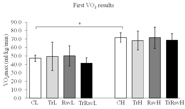
Figure 7: Maximal oxygen uptake results at the beginning of the study ... 47
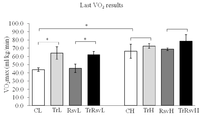
Figure 8: Maximal oxygen uptake results at the end of the study ... 48
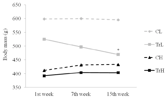
Figure 9: Changing of the body mass at control and trained groups ... 49
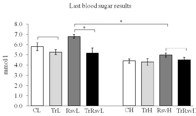
Figure 10: Blood sugar results at the end of the training period ... 50
Figure 11: Results of the rotarod test by the end of the 15 weeks ... 51
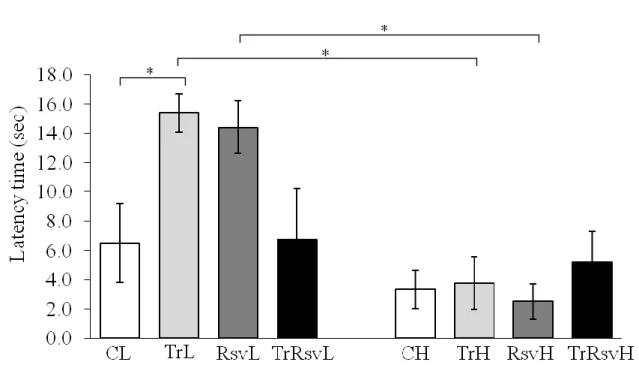
Figure 12: Latency times of the animal groups during an open field test ... 52
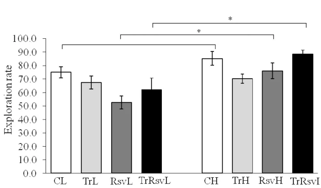
Figure 13: Exploration rate of the animal groups during an open field test ... 53
Figure 14: Results of the new object recognition test ... 54
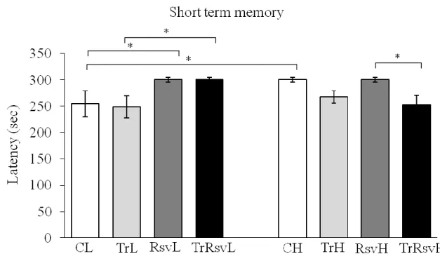
Figure 15: Short term memory was measured 24 hours after the learning period ... 56
Figure 16: Long term memory was measured 10 days after the learning period ... 57
Figure 17: BrdU shows the new cells in the hippocampus... 58
Figure 18: BrdU+NeuN co-localization in the gyrus dentatus ... 59
Figure 19: Sirt1 mRNA levels ... 60
Figure 20: Sirt3 mRNA levels ... 61
Figure 21: Sirt4 mRNA levels ... 62
Figure 22: Sirt6 mRNA levels ... 63
Figure 23: SIRT1 relative density in the brain tissue ... 64
Figure 24: SIRT1 activity (in activity units where the results were divided with the protein amounts) ... 65
Figure 25: Acetylated lysine relative density ... 66
Figure 26: Carbonylated proteins relative density ... 67
Figure 27: NAMPT relative density ... 68
Figure 28: Creb mRNA levels ... 69
Figure 29: BDNF relative density ... 70
7
Figure 30: Poly (ADP)-ribose relative density ... 71
Figure 31: OGG1 relative density ... 72
Figure 32: Acetylated OGG1 levels from histochemistry densities ... 73
Figure 33: Acetylated OGG1 relative density ... 75
Figure 34: Acetylated OGG1 relative density after siRNA treatment... 76
Figure 35: Sirt1 mRNA levels after silencing in the cells ... 77
Figure 36: SIRT1 can deacetylate OGG1; it attenuates the repair, so apoptosis is avoidable ... 82
Table 1: Summary of SIRT1 deacetylation’s known targets ... 14
Table 2: Antibodies in the western blots ... 39
Table 3: Genes and primers in PCR ... 41
8
1. Introduction
1.1 Sirtuins – A new promise of a longer life
One of the biggest wishes of mankind is that of eternal life. Although it is not possible, humanity is still seeking new ways to elongate life. Great milestones show the way that science has already walked on this path. For example we know more and more about cell differentiation, cell proliferation, apoptosis …etc. every day. These processes are built up like a symphony of molecular mechanisms. In the orchestra one of the most important members is DNA (Deoxyribonucleic acid), surrounded by histones. Among many functions histones protect the genetic information by making it untouchable for harmful molecules. In these reactions the key regulators are the histone deacetylases.
There are four classes of deacetylases in mammals, among them, enzymes in class III are unique ones. They are referred to as silent information regulator two proteins (sirtuins), named after their yeast homologue (sir2). (Afshar & Murnane, 1999) Since their first debut this family of molecules has been quoted many times as promises of a longer life.
1.1.1 General characterization of sirtuins
Sirtuins are NAD+-dependent (Nicotinamide adenine dinucleotide-dependent) enzymes that are phylogenetically conserved from archeobacteria to humans. Sequence alignment studies on the sirtuin proteins show that they contain a conserved catalytic core consisting of approximately 275 amino acid residues and variable N- and C- terminal chains. (Sanders, Jackson, & Marmorstein, 2010) The catalytic core exists in an elongated shape and consists of a large Rossmann fold domain (which is a hallmark of NAD+/NADH-binding proteins), a structurally more diverse but smaller zinc-binding domain and many loops connecting these domains (as shown on Figure 1.).
9
Figure 1: Structure of sirtuins
The catalytic core consists of a large Rossmann fold domain, a smaller zinc-binding domain and many loops connecting these domains. (Moniot, Weyand, & Steegborn, 2012)
The Rossmann fold is the large domain of the catalytic core exhibiting the classical α/β Rossmann fold structure. The Rossmann fold is a protein structural motif found in proteins that bind nucleotids, especially the cofactor NAD+. It has all the characteristics of a NAD+-binding site including a Gly-X-Gly sequence for phosphate binding, a pocket for NAD+ and charged residues for the ribose moiety. An unusual characteristic of this family of enzymes is that the NAD+ binds in an inverted direction relative to most NAD+ dehydrogenases. (Finnin, Donigian, & Pavletich, 2001) The Zn-binding domain is a small domain that results from two insertions in the Rossmann fold represents the most diverse region among sirtuin family members. Two structural modules have been suggested for the domain: a three-stranded antiparallel β-sheet and a
10
variable α-helical region depending on the type of the sirtuin. The structural diversity observed in the small domain may potentially have a role in substrate specificity and protein-protein interactions, thus finally affecting the enzyme function. (Zhao, Harshaw, Chai, & Marmorstein, 2001) At the acetyl-lysine binding site the peptide substrate binds to the cleft between the small and large domains with the acetyl-lysine side chain sliding into a hydrophobic tunnel within the cleft. The peptide backbone of the acetyl- lysine chain forms β-sheet-like interactions flanked by two strands in the enzyme to form a three-stranded antiparallel motif called a β-staple. (Cosgrove, et al., 2006) It has been suggested that NAD+ and peptide may bind co-operatively, considering that the region around the peptide binding site adopts a more closed conformation in the presence and not in the absence of bound NAD+. (Avalos, et al., 2002) The crux of peptide binding to sirtuins is said to be mainly controlled by the insertion of the acetyl- lysine into the conserved hydrophobic tunnel and formation of the β-staple as discussed previously. The substrate specificity of sirtuins is more or less thought to lie outside the highly conserved catalytic core of the enzyme. However, it was observed that some sirtuins could distinguish between substrates that have multiple acetylated lysine- residues, suggesting that the catalytic core could be sufficient for substrate specificity.
(Garske & Denu, 2006) (Borra, Langer, Slama, & Denu, 2004) Until this question is answered theoretically every protein with acetyl-lysine binding site can be substrate of sirtuins.
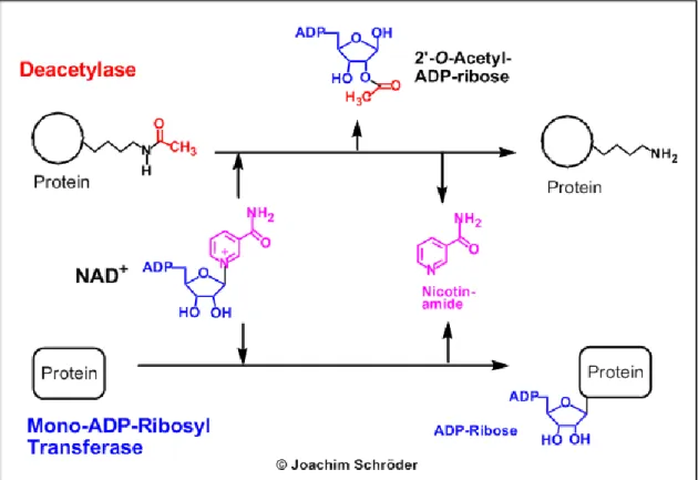
Sirtuins have either deacetylase or mono-ADP-ribosyl transferase activity or both as shown on Figure 2. Both reactions are strictly dependent on NAD+. After NAD+ and the substrate peptide attached to a sirtuin it will cleave the acetyl group and pass it to the NAD+. NAD+ meanwhile will hydrolyse into nicotinamide and 2’-O-acetyl-ADP-ribose.
The dependence of sirtuins on NAD+ links their enzymatic activity directly to the energy status of the cell via the cellular NAD+: NADH ratio, the absolute levels of NAD+, NADH or nicotinamide or a combination of these variables.
11
Figure 2: The main enzymatic activities of sirtuins
Sirtuins have either deacetylase or mono-ADP-ribosyl transferase activities or both.
(Michan & Sinclair, 2007)
Mammals possess seven types of sirtuins. Here I collect the main data in a nutshell about each member, particularly about their function in the nervous system.
1.1.2 SIRT1
This is the best known member among the sirtuin family. It was originally documented to localize in the nucleus, but later studies confirmed that SIRT1 (Sirtuin 1) is able to shift between the nucleus and the cytoplasm. There are target molecules on both sides which can be deacetylated by SIRT1. This was the first sirtuin in which the deacetylation function was proved at first on histone targets, later on several protein targets in the cell. SIRT1 is expressed in every cell of the body in a tissue-specific manner of course. During mouse embryogenesis, SIRT1 is highly expressed in the brain, spinal cord, and dorsal root ganglion, with the peak expression at E4.5. (Salminen
& Kaarniranta, 2012) SIRT1 is also expressed in the adult brain, at high levels in the
12
cortex, hippocampus, cerebellum, and hypothalamus, and at low level in white matter.
Among the various cell types of brain, SIRT1 is predominantly, if not exclusively, expressed in neurons. (Adler, et al., 2007) The only exception is that SIRT1 is found in microglia when co-cultured with neurons. (Schmitz, Mattioli, Buss, & Kracht, 2004) SIRT1 is abundantly expressed in several regions in the hypothalamus of mice, especially in the arcuate, paraventricular, ventro- and dorsomedial nuclei; so calorie restriction increases SIRT1 levels in the hypothalamus, which increases body temperature, food intake, and physical activity. (Ramadori, et al., 2008) (Dietrich, et al., 2010)
SIRT1 is involved in several different mechanisms during the cells’ life, so it has to be precisely regulated on all levels:
- Transcriptional upregulation: The basal level of SIRT1 is regulated by the transcription factor E2F transcription factor 1 (E2F1), through binding to the SIRT1 promoter at a consensus site. Cellular stresses increase the transcriptional activity of E2F1 and upregulate the level of SIRT1. (Wang, et al., 2006) FOXO1 (Forkhead box protein O1) binds to several consensus sites within the SIRT1 promoter and enables its transcription. (Xiong, Salazar, Patrushev, & Alexander, 2011) FOXO3a is another SIRT1 regulator. Starvation in mammal cells activates FOXO3a and consequently augments SIRT1 expression. (Nemoto, Fergusson, & Finkel, 2004)
- Transcriptional downregulation: Hypermethylated in cancer 1 (HIC1) binds SIRT1, forming a transcriptional repression complex through its N-terminus.
This complex directly binds to the SIRT1 promoter, and thereby inhibits SIRT1.
(Chen, et al., 2005) A recent study showed that PPARγ (Peroxisome proliferator-activated receptor γ) binds the promoter of SIRT1 and inhibits its expression. (Han, et al., 2010)
- Post-transcriptional regulation: MicroRNAs are a group of short RNAs with an average length of 22 nucleotides. They cause gene silencing by binding to complementary sequences on their target mRNAs, leading to the degradation of mRNAs. Several microRNAs have been identified that reduce SIRT1 expression, including miR-9, miR-34a, miR-132, miR-181 and miR-199, miR- 217. (Saunders, et al., 2010) (Lee & Kemper, 2010) (Menghini, et al., 2009)
13
Also an RNA-binding protein, HuR associates with the 3′ untranslated region of the Sirt1 mRNA. This interaction leads to increased stability of Sirt1 mRNA, promoting the translation of SIRT1. (Abdelmohsen, K; Pullmann, R Jr, et al., 2007)
- Regulation by other proteins: An active regulator of SIRT1 (AROS) is a recently identified nuclear protein that increases the deacetylating activity of SIRT1.
(Kim, Kho, Kang, & Um, 2007) In contrast, deleted in breast cancer-1 (DBC1), another nuclear protein, functions as a negative regulator. (Chini, Escande, Nin,
& Chini, 2010)
- Post-translational regulation: Sumoylation and desumoylation of SIRT1 can function as a molecular switch to regulate SIRT1 activity in response to cellular stresses. (Zschoernig & Mahlknecht, 2008) It is reported that c-jun N-terminal kinase 1 (JNK1) phosphorylates two serine residues plus Thr530 of SIRT1.
(Nasrin, et al., 2009) The phosphorylation of SIRT1 occurs under oxidative stress and increases the nuclear translocation and enzymatic activity of SIRT1.
The cell cycle checkpoint kinases (CHKs) and dual specificity tyrosine phosphorylation-regulated kinases (DYRKs) are also groups of kinases that phosphorylate SIRT1 to increase its activity. On the other hand a recent study showed that SIRT1 is phosphorylated by mammalian sterile 20-like kinase 1 (MST1) after induced DNA damage, leading to reduced activity of SIRT1.
(Yuan, et al., 2011)
- Metabolic regulation: According to the deacetylation mechanism of SIRT1, it is a reasonable prediction that substrates of SIRT1 will activate it, and products of the reaction will inhibit it. Therefore NAD+ can increase the enzymatic activity of SIRT1, whereas nicotinamide and 2’-O-acetyl-ADP-ribose may inhibit it.
(Neugebauer, Sippl, & Jung, 2008) (Tong & Denu, 2010)
- Pharmacological regulation: Splitomicin and sirtinol, the frequently used SIRT1 inhibitors, are effective both in vitro and in vivo. On the contrary, SIRT1 can be activated by several small-molecule compounds. Most of these activators are polyphenols including resveratrol, quercetin, curcumin, and catechins.
Initially SIRT1 was documented as a histone deacetylase. In nuclei, core histones, including H2, H3 and H4, form ball-like structures for the DNA to wrap around,
14
keeping DNA in a resting status. When acetylated on the lysines, however, histones lose their positive charges and release DNA, allowing DNA unwinding and subsequent gene transcription. In contrast, deacetylation polarizes histones and promotes their binding to DNA, leading to genome-wide but non-specific transcription silencing. (Imai, Armstrong, Kaeberlein, & Guarente, 2000) Since that time it has been demonstrated that SIRT1 has several protein targets both in nucleus and cytoplasm. It is complicated to summarize these interactions, therefore I chose to do it briefly with a help of a table in alphabetical order (Table 1.).
Table 1: Summary of SIRT1 deacetylation’s known targets Target Target’s function SIRT1’s
Effect Literature APE1 (AP
Endonuclease1) base excision repair enzyme ↑ (Yamamori, et al., 2010) eNOS (endothelial
Nitric oxide synthase)
vasodilatator which generates
endothelial nitric oxide ↑ (Mattagajasingh, et al., 2007)
FOXO1 (Forkhead box protein O1)
transcription factor that plays important roles in regulation
of gluconeogenesis and glycogenolysis
↓ (Yang, Hou, Haller, Nicosia, & Bai, 2005)
FOXO3 (Forkhead box protein O3)
upregulates pro-apoptotic factors, but antioxidants as
well
↑ (Wang, Nguyen, Qin,
& Tong, 2007) FOXO4 (Forkhead
box protein O4)
promotes survival against
oxidative stress ↑ (van der Horst, et al., 2004)
HIFs (Hypoxia-
inducible factors) hypoxia-inducible factors ↑ (Dioum, et al., 2009) (Lim, et al., 2010) Ku70 double-stranded DNA break
repair ↑ (Jeong, et al., 2007)
LKB-1 (Liver
kinase B1) activates AMPK ↑ (Zu, et al., 2010)
15 NF-κB (Nuclear
factor kappa-light- chain-enhancer of activated B cells)
upregulates pro-inflammatory
mediators ↓ (Wang, et al., 2009)
NRF2 (Nuclear factor erythroid 2-
related factor 2)
cytoprotective during oxidative stress, carcinogenic
when overactivated
↓
(Kawai, Garduño, Theodore, Yang, &
Arinze, 2011) p300 (Histone
acetyltransferase)
histone acetyltransferase, promoting the transcription of
other transcription factors
↓ (Bouras, et al., 2005)
p53 (Protein 53) upregulates pro-apopototic
molecules (Bcl-2 family) ↓ (Luo, et al., 2001) PARP-1 (Poly
[ADP-ribose]
polymerase 1)
single-stranded DNA break
repair ↓ (Rajamohan, et al.,
2009) PGC-1α
(Peroxisome proliferator- activated receptor gamma coactivator
1-α)
increase mitochondrial
energy metabolism ↑ (Nemoto, Fergusson,
& Finkel, 2005)
PPAR-α (Peroxisome
proliferator- activated receptor
α)
increases anti-inflammatory
mechanisms ↑ (Deplanque, et al.,
2003)
PPAR-γ (Peroxisome
proliferator- activated receptor γ)
downregulates inflammatory
citokines ↓ (Shie, Nivison, Hsu,
& Montine, 2009)
16 RAR-β (Retinoic
acid receptor β)
increases the transcription of
α-secretase ↑
(Donmez G. , Wang, Cohen, & Guarente,
2010) Tau
microtubule-binding protein, promotes the assembly of
microtubules
↓ (Min, et al., 2010)
XPA (Xeroderma pigmentosum, complementation
group A)
nucleotide excision repair
enzyme ↑ (Fan & Luo, 2010)
α-secretase
process soluble APP which helps avoid Alzheimer’s
disease
↑
(Donmez G. , Wang, Cohen, & Guarente,
2010)
1.1.3 SIRT2
This protein is mostly found in the cytoplasm and has both deacetylase and mono-ADP- ribosyl transferase activity. (North, Marshall, Borra, Denu, & Verdin, 2003) New data uncovered a novel role for SIRT2 opening new perspectives for therapeutic intervention in neuroinflammatory disorders. Reduction of SIRT2 in microglia dramatically increased the expression of inflammatory markers, the production of free radicals, and neurotoxicity. Consistent with increased NF-κB-dependent transcription of inflammatory genes, NF-κB was found hyperacetylated in the absence of SIRT2. (Pais, et al., 2013) So SIRT2 has been related to synaptic plasticity, learning and memory that are also in turn related to neuronal motility and migration. Kireev and colleagues showed significantly decreased SIRT2 expressions in the dentate gyrus of old male rats.
Interestingly melatonin or growth hormone treatments have been shown to modulate the pro-antiapoptotic ratio and increase SIRT2’s expression restoring the situation found in young animals. (Kireev, Vara, & Tresguerres, 2013)
17 1.1.4 SIRT3
Among the known sirtuins SIRT3 was identified as a stress responsive deacetylase recently shown to play a role in protecting cells under stress conditions. It has been proven that SIRT3 is a mitochondrial protein with a robust deacetylase activity.
Mitochondria are the major site for the generation of the reactive oxygen radical superoxide, and also where the superoxide is dismuted by mitochondrial MnSOD (Manganese superoxide dismutase). Recent reports show that SIRT3 deacetylates MnSOD at Lys122 and increases its activity, reducing oxidative and radiation stress in mice. Overexpression of SIRT3 protects HEK293 from oxidative stress and prevents age-related cochlear cell death in mice. (Someya, et al., 2010) Overall these observations suggested anti-oxidative and neuroprotective roles of SIRT3. Furthermore, it was found that mitochondrial SIRT3 is increased following PARP-1 mediated NAD+ depletion in neurons, which can be reversed by either inhibition of PARP-1 or exogenous NAD+. (Kim, Lu, & Alano, 2011) The massive amount of ROS produced under this NAD+ depleted condition mediates the increase in mitochondrial SIRT3. By transfecting primary neurons with a SIRT3 overexpressing plasmid or SIRT3 siRNA, it was shown that SIRT3 is required for neuroprotection against excitotoxicity. The ability of calorie restriction to induce SIRT3 expression has been well documented. (Kincaid &
Bossy-Wetzel, 2013) Additional stimulators of the sirtuins have also been proposed, such as resveratrol, a polyphenol found in red wine. Treatment with resveratrol did not affect SIRT3 expression levels. However, a recent report found that a resveratrol derivative, trans-(-)-ε-viniferin, is able to increase SIRT3 expression and provide protection in cell models of Huntington’s disease (HD). Specifically, viniferin treatment of striatal precursor cells overexpressing mutant huntingtin resulted in increased SIRT3 expression, increased the NAD+/NADH ratio, reduced intracellular ROS accumulation, and decreased acetylated MnSOD levels. Thus, SIRT3 is required for viniferin-mediated neuroprotection in HD models. (Fu, et al., 2013)
18 1.1.5 SIRT4
This enzyme also can be found in mitochondria but shows only mono-ADP- ribosyltransferase activity in a NAD+-dependent manner. There are some publications regarding SIRT4’s role in metabolic homeostasis but none of these focus on neural tissues. It has recently been reported that congenital hyperinsulinism/hyperammonemia (HI/HA) syndrome is caused by an activation mutation of glutamate dehydrogenase 1 (GDH1), a mitochondrial enzyme responsible for the reversible interconversion between glutamate and α-ketoglutarate. (Komlos, et al., 2013) GDH1 is allosterically regulated by many factors, and has been shown to be inhibited by SIRT4. SIRT4 is highly expressed in glial cells, specifically astrocytes, in the postnatal brain and in radial glia during embryogenesis. The authors also found that SIRT4 and GDH1 overexpression play antagonistic roles in regulating gliogenesis.
1.1.6 SIRT5
SIRT5 is a mitochondrial sirtuin which can demalonylate and desuccinylate proteins (Peng, et al., 2011) in particular the urea cycle enzyme carbamoyl phosphate synthetase I (CPS I). (Du, et al., 2011) Glorioso and colleagues investigated the transcriptome changes during “normal” human brain aging by microarray analysis in two cohorts and four brain areas, focusing on the overlap of aging and disease pathways, and then tested whether subject molecular brain aging rates were associated with several candidate longevity gene polymorphisms. In support of a genetic modulation or control of this molecular aging-by-disease risk model, they showed that the cross-sectional trajectory of a large component of molecular aging was differentially affected in subjects carrying a common polymorphism in the SIRT5 putative longevity gene (SIRT5prom2), which they also show correlated with reduced SIRT5 expression. Based on these results, they predict that SIRT5-risk allele (C/C) carriers may be at increased risk for mitochondrial- dysfunction related disorders, including Parkinson’s and Huntington’s diseases.
(Glorioso, Oh, Douillard, & Sibille, 2011)
19 1.1.7 SIRT6
According to a study (Schwer, et al., 2010) the highest level of Sirt6 mRNA was detected in the brain, heart and liver and the lowest expression level was observed in skeletal muscle. They also proved that SIRT6 protein is localized in the nucleus of cells.
They could not show any deacetylase activity but they could detect a great mono-ADP- ribosyltransferase activity. Later researchers could provide evidence that SIRT6 deacetylates histone H3K9 and H3K56. (Yang, Zwaans, Eckersdorff, & Lombard, 2009) (Michishita, et al., 2009) Until recently, there was no possibility to study Sirt6 knockout mice, because this deletion was found to be lethal.
In 2010 a group of scientists was able to generate a neural-specific Sirt6 knockout mouse to study the roles of SIRT6 in the central nervous system. (Schwer, et al., 2010) KOSirt6 mice appeared normal at birth, but at 4 weeks of age they were significantly smaller. They also demonstrated that neural-specific Sirt6 deletion, likely through reduced GH and hypothalamic neuropeptide levels – Proopiomelanocortin (POMC), Single-minded homolog 1 (SIM1), and Brain-derived neurotrophic factor (BDNF) -, promotes adult-onset obesity in mice.
1.1.8 SIRT7
This sirtuin is located in the nucleoli, but there were no clue for a long time what kind of enzymatic activity it has. In 2013 Tsai and co-workers evidenced that SIRT7 regulates rDNA transcription and that reduced SIRT7 levels inhibit tumor growth. (Tsai, Greco, & Cristea, 2013) A key feature of cancer cells is uncontrolled proliferation that ultimately overcomes the intrinsic limit of mitotic cycles. However, tumor cells must achieve a critical cell mass before committing to another round of cell division to increase the tumor cell population. Ribosome synthesis is a key process necessary to fulfill the required cell mass. This group of scientists presents the first experimental evidence that SIRT7 interacts with proteins involved in ribosome biogenesis (DNA Polymerase I,III through mammalian target of rapamycin (mTOR)), and that its levels are critical for regulating protein synthesis.
20
More than 2000 publications are dedicated to the therapeutic potential of sirtuins. A main trend is to activate sirtuins via a natural, dietary way. To start a diet like this, it is suggested to consume several types of fruits, vegetables and nuts. These foods contain high amounts of antioxidants. A huge section of antioxidants are polyphenols.
21 1.2 Reservatrol – All good thing come from fruits
Polyphenols are naturally occurring phytochemicals which are present within fruits, vegetables and natural products. These phytoalexins found in the tissues of a widespread range of plants, they characterized by the presence of multiple hydroxyl groups on aromatic rings. They can be divided into two main categories - flavonoids and non flavonoids. There are several subcategories based on the chemical structures as it is nicely demonstrated on the figure of David Vauzour (Figure 3.):
Figure 3: The two main groups of polyphenols
The two main groups of polyphenols are flavonoids and non flavonoids. Within the group of non flavonoids a huge category is stilbenes. In our study we used resveratrol, a stilbene with hydroxyl functional groups associated to the aromatic rings. (Vauzour, 2012)
22
Within the non flavonoids’ group a huge category is stilbenes. Stilbenes possess a 1,2- diarylethenes structure based on the C6-C2-C6 backbone and are usually synthesized in plants in response to infection or injury. Resveratrol (3,5,4'-trihydroxy-stilbene), the main stilbene, can be found in cis or trans configurations. Major dietary sources of resveratrol include grapes, wine and peanuts.
When polyphenols are applied either via intravenous or oral route, the biggest question is if they are capable to overpass the blood-brain barrier or not. Using in vitro models, initial studies have demonstrated that polyphenols permeation through the barrier is dependent on the degree of lipophilicity of each compound, so less polar polyphenols capable of greater brain uptake than the more polar ones. (Youdim, et al., 2003) Studies suggest that polyphenols usually localize in the brain at levels below 1 nmol/g tissue and usually accumulates in a nonregion-specific manner. 14C-labelled grape polyphenols did not show any regional differences in 14C accumulation from anterior to posterior slices of the brain. (Janle, et al., 2010) Although resveratrol accumulates in a low level in the brain there are already publications where its beneficial effects are discussed.
Resveratrol has been reported to be effective in the experimental autoimmune encephalomyelitis with rises in IL-17/IL-10 ratio and with repressed macrophage IL-6 expression. (Imler & Petro, 2009) It was observed to protect PC12 cells against H2O2- mediated oxidative stress (Chen, Jang, Li, & Surh, 2005) and to attenuate cerebral ischemic injury in rat. (Ren, Fan, Chen, Huang, & Yang, 2011) It has been shown that SIRT1 is also activated by resveratrol resulting in cell survival, but it is still under investigation if this activation happens in a direct or an indirect way. Indirect way could happen through a signaling cascade involving cyclic adenosine monophosphate (cAMP), exchange proteins activated by cyclic AMP (Epac1) and 5' AMP-activated protein kinase (AMPK). (Park, et al., 2012)
To sum up the previous facts: sirtuins may have the potential to elongate life. To obtain this it seems to be useful to elevate the level of consumed polyphenols. Although eating antioxidants have several well-documented advantages, for a healthy body everyone should do one more thing: to have some regular exercise!
23 1.3 Exercise – The way to a long and healthy life
If we want to look for the origin of human body and human genetics we need to look further back a few thousand years. Originally we were built up to walk and run tens of miles a day seeking for fruits to collect or animals to hunt. During the past few thousand years life became much easier, or actually too easy. In developed countries people take up a huge protein and sugar surplus day by day. On the other hand we forgot about our in-built locomotion needs. Of course there are honorable exceptions for those who practice some kind of regular exercise. Statistically a cleft is about to open up between these two types of people, so in biology these was a need of a new model which can illustrate the aforementioned differences.
Lauren G. Koch and Steven L. Britton generated a rat model which is close enough to characterize the biological differences between the two extremities. They undertook a large-scale selective breeding program to develop rat lines that would diverge widely for intrinsic aerobic capacity. Six generations of selection produced lines that differed in running capacity by 171%, with most of the change occurring in the high capacity of running line. (Koch & Britton, 2001) Four years later Wisløff et al. characterized the 11th generation of the same model. By this time the low capacity runners (LCR) and high capacity runners (HCR) differed in running capacity by 347%. LCR animals on the other hand started to represent the average person with metabolic syndrome. They even showed all the risk factors linked to metabolic syndrome: weight gain, high blood pressure, reduced endothelial function, hyperinsulinaemia and increased triglyceride concentration in blood. (Wisløff, et al., 2005) In 2008 I had the chance to work with 24 low capacity of running (LCR) and 24 high capacity of running (HCR) male rats from the 22nd generation. Compare to previous works with this animal model I did not focus on cardiac nor skeletal muscle traits. I tried to map the differences between the central nervous system of these animals, especially the brain and hippocampus region.
Several studies reflect that there are huge benefits of regular exercise in the central nervous system. Falone et al. reports that exercise reversed the age-related decline in the level of SIRT1. (Falone, et al., 2012) In their experience hippocampus undergoes significant redox imbalance during the first period of the exercise program, but it seems that this imbalance might have an important role in preparing the cellular environment
24
for the subsequent beneficial modifications. Szabo refers that voluntary exercise may engage proteasome function to benefit the brain after trauma. (Szabo, Ying, Radak, &
Gomez-Pinilla, 2010) Marosi experienced that long-term exercise treatment reduced oxidative stress in the hippocampus of aging rats. Exercise induced an up-regulation of SOD-1 and Glutathione peroxidase (GPx) enzymes, p-AMPK and PGC-1α, that can be related to an improved redox balance in the hippocampus. These results suggest that long-term physical exercise can comprise antioxidant properties and by this way protect neurons against oxidative stress at the early stage of aging. (Marosi, et al., 2012) Radak et al. publicated that exercise can induce neurogenesis via neurotrophic factors, increase capillarization, decrease oxidative damage, and enhance repair of oxidative damage.
Exercise is also effective in attenuating age-associated loss in brain function, which suggests that physical activity-related complex metabolic and redox changes are important for a healthy neural system. (Radak, et al., 2013) In 2005 Adlard proved that increased physical activity decreased the Amyloid beta (Aβ) protein levels in an Alzheimer disease mouse model. Already 1 month of exercise impacted learning and memory according to a Morris water maze task. (Adlard, Perreau, Pop, & Cotman, 2005) In 2010 Radak provided an overview of the positive impacts of exercise on Alzheimer’s disease. According to the review regular physical activity increases the endurance of cells and tissues to oxidative stress, vascularization, energy metabolism, and neurotrophin synthesis, all important in neurogenesis, memory improvement, and brain plasticity. (Radak, et al., 2010)
It is well-proved that exercise has beneficial effects on the central nervous system. On the other hand it is also well-known that exercise generates a huge population of reactive oxygen species due to the increased oxygen consumption. It is still under investigation how training has such beneficial effects despite the ROS.
25
1.4 Reactive oxygen species (ROS) - Enemies within ourselves?
Reactive oxygen species is a general term for molecular oxygen-derived molecules that are reactive species or that are converted easily to reactive species. Many of them are free radicals. There are at least 4 primary sources of free radicals formed endogenously within living organisms:
- peroxisomal oxidation (Aliev, et al., 2008)
- respiratory generation of ATP using oxygen (Beckman & Ames, 1998) - cytochrome P450 enzymes
- cells which use a mixture of oxidants to overcome an infection (Ames, Shigenaga, & Hagen, 1993)
Oxygen-derived free radicals are highly reactive chemical species involved in a variety of disorders. Superoxide anion (O2-), hydroxyl radical (OH-), and hydrogen peroxide (H2O2) are known as reactive oxygen species. As mentioned, they are mostly produced in the mitochondria during the reduction of molecular oxygen to water. Large amount of evidence has shown the important roles of ROS in cell proliferation, homeostasis, intracellular signaling, angiogenesis, and modifications of the extracellular matrix… etc.
On the other hand ROS are described as harmful products and capable of DNA mutations, lipid peroxidation and protein oxidation. All these can lead to inflammation and cell death. (Zhu, Su, Wang, Smith, & Perry, 2007) Fortunately, there are several endogenous antioxidant defense mechanisms, like antioxidant enzymes (catalase, glutathione peroxidase or superoxide dismutase) and non-enzymatic antioxidants (vitamin E, ascorbic acid…). This is important because accumulation of misfolded proteins is a common feature in multiple human diseases, especially in the nervous system. Neurons are particularly sensitive to oxidative stress; therefore the brain is more vulnerable to reactive oxygen species-induced damage due to its high rate of oxygen consumption and high polyunsaturated lipid content. Prevention is very important because regular training increases endurance of cells to oxidative stress, vascularization, energy metabolism and neurotrophin synthesis which can be seen via improved memory and brain plasticity. In connection with Alzheimer disease Dumont and colleagues demonstrated that the overexpression of MnSOD reduced amyloid plaques, improved memory function and protected synapses. (Dumont, et al., 2009) Of course it would be
26
more convenient to look for a therapy which does not require genetic intervention.
Drugs which have antioxidant property do and will have attention. Including but not limited to: statins, alkaloids, catapol and the big family of polyphenols. These molecules have the capacity to chelate metal ions and to directly quench free radical species.
(Perron & Brumaghim, 2009)
Unfortunately antioxidants are not enough to avoid every danger which threats our cells.
Sometimes the amount of free radicals is too high to deal with and molecules of the cells’ get damaged. There have to be mechanisms to repair these damages.
27 1.5 OGG1 – Repair mechanisms during life
DNA damage occurs in daily life and is aggravated following metabolic and oxidative stresses. Accordingly, DNA repair is essential to maintenance of genomic integrity and cellular viability. The severity of DNA damage varies from single base damage to double-stranded DNA breaks. The most common threat which can appear is oxidation.
Reactive oxygen species can originate both from extracellular and intracellular sources.
Among the four bases of DNA guanine has the lowest redox potential, thus it is prone to oxidation resulting 7,8-dihydro-8-oxoguanine (8-oxoG) formation. This lesion is particularly mutagenic because in addition to its ability to form a Watson-Crick pairing with cytosine, 8-oxoG has the ability to form a stable Hoogsteen pair with adenine. This can lead to G:C→T:A transversion after replication. (Kuchino, et al., 1987) Because of the high mutagenic potential during evolution arose a special enzyme to cut out 8-oxoG.
It is 8-oxoguanine DNA glycosylase (OGG) which catalyses the first step of base excision repair in the case of an oxidated guanine.
OGG belongs to the helix-hairpin-helix superfamily of enzymes. OGGs can be divided into three subfamilies: OGG1, OGG2, AGOG. The majority of OGG1 enzymes are found in eukaryotes, OGG2 mostly appears in bacteria and archaea, while AGOG is exclusively found in archaeal organisms. (Robey-Bond, Barrantes-Reynolds, Bond, Wallace, & Bandaru, 2008) Human OGG1 exists in two different splice variants. While hOGG1α can be found in the cytoplasm, nucleus and mitochondria, hOGG1β is only expressed in mitochondria. (Nishioka, et al., 1999) OGG1’s activity can be modified during post-translational changes: OGG1 is phosphorylated in vitro by CDK4 (Cyclin- dependent kinase 4), resulting in a 2.5-fold increase in the 8-oxoG/C incision activity of OGG1. C-Abl tyrosine phosphorylates OGG1 in vitro; however, this phosphorylation event does not affect OGG1 8-oxoG/C incision activity. (Hu, Imam, Hashiguchi, de Souza-Pinto, & Bohr, 2005) On the other hand OGG1 is acetylated on Lys338/Lys341 by p300 which also increased its activity. (Bhakat, Mokkapati, Boldogh, Hazra, &
Mitra, 2006)
The excision activity of OGG1 is quite important because accumulation of 8-oxoG in brain has been implicated in neurodegeneration (Lovell & Markesbery, 2007) (Wang, Markesbery, & Lovell, 2006) (Aguirre, Beal, Matson, & Bogdanov, 2005) It was
28
recently reported that aging results in increased levels of 8-oxoG in the hippocampus, which was associated with decreased level of acetylation of the most powerful repair enzyme of 8-oxoG, OGG1. (Radicella, Dherin, Desmaze, Fox, & Boiteux, 1997) Importance of OGG1’s acetylation is underlined by data showing that exercise increases the acetylated OGG1 levels in muscle of young individuals. (Bori, et al., 2012) Efficient DNA repair has been shown to protect against neurodegeneration and thus amplifies the significance of DNA damage repair in the nervous system. (Liu, et al., 2011)
In contrast to these Stuart et al. proved that OGG1 null mice do not exhibit abnormal phenotype. (Stuart, Bourque, de Souza-Pinto, & Bohr, 2005) These animals accumulated 8-oxoG in mitochondrial DNA 9- to 20-folder higher than wild type, but it does not seem to raise disadvantages. (de Souza-Pinto, et al., 2001) It has even been found that OGG1-deficient mice are resistant to inflammation, implicating involvement of OGG1 in pro-inflammatory signaling. (Touati, et al., 2006) (Mabley, et al., 2005) In 2012 Boldogh et al. published for the first time several lines of evidence that OGG1 is able to bound free 8-oxoG, thus interacting with Ras family GTPases that initiates a signaling cascade. (Boldogh, et al., 2012)
In this context it might be possible that OGG1 is needed to be deactivated sometimes.
To decrease its activity one option is deacetylation, so there might be a connection between OGG1 and SIRT1 deacetylase.
29
2. Objectives of the study
The aim of the study was to test how regular exercise can overcome the health risks which occur at metabolic syndrome. An animal model from the Michigan University was ideal for this purpose. As I mentioned in the “Introduction”, there is a model in which rat lines were developed that diverge widely for their intrinsic aerobic capacity.
This is not the first time when artificial selection was used to investigate such question in exercise. (Swallow, Carter, & Garland, 1998) But as we know this is the first time when researchers selected animals for a very long time (more than ten years passed, which is quite long compare to the life-span of rats) for the final goal to determine the genetic components of aerobic capacity. Of course the 22nd generation, which I worked with, is not enough yet to reach that goal, but ideal to get conclusions about the extremities the two rat types typify. From this point of view observations on this model may reveal mechanisms, which can mean new information after all about us.
The laboratory, where most of the experiments were conducted, has a special interest in addition to sport sciences. Since their discovery, sirtuins have stood in a main focus in many of the investigations. No doubt that sirtuins are a very old and conservative protein family which on the other hand is barely known by modern biology. This experiment was the first in this laboratory when we attempted to get information about sirtuins not only in a descriptive way, but we tried to enlarge their effects by administering a well-known activator: resveratrol.
During my PhD years I had the opportunity to spend some time at the University of Texas. In those days I learnt how to work with cell cultures and I could test my hypothesis on cell culture. According to the observations on rat brain we presumed a connection between sirtuins and OGG1 repair enzyme, so my last hypothesis arose from this topic.
30 Hypotheses:
1. Regular physical activity and resveratrol treatment will enhance the cognitive function of both rat strains.
2. Our aim was to illustrate that the cognitive enhancement was caused via sirtuins and neurotrophic factors in the brain which overall can be seen in neurogenesis.
3. Training and/or resveratrol will compensate the differences which come from the genetic origin of the animals.
4. Sirtuins can deacetylate OGG1 protein and this might moderate its activity.
31
3. Materials and methods
3.1 Origin of the rats
Most of my results are based on the testing of a special type of rats. These Sprague- Dawley rats are artificially selected for intrinsic aerobic endurance running capacity by Lauren G. Koch and Steven L. Britton, who built up a new model to investigate the genetic factors of aerobic endurance. This model building is based on a large scale selective breeding program. Briefly they started the program from 96 male and 96 female rats. Each rat in the founder population was of different parentage. They each were provided food and water ad libitum and placed on a 12:12 hours light-dark cycle.
The protocol for estimation aerobic running capacity required 2 weeks and was started when the rats were 10 weeks old. The first week consisted of introducing each rat to the treadmill (5 minutes, 10 m/min, 15o slope). During the second week, each rat was evaluated for maximal endurance running capacity on five consecutive days. The slope was constant 15o, and the starting speed was 10 m/min. Treadmill velocity was increased by 1 m/min every 2 minutes and each rat was run until exhausted. Using the criterion of single best day, the 13 lowest and 13 highest capacity rats of each sex were selected from the founder population and randomly paired for mating. At 10 weeks of age the offsprings were introduced to the treadmill and subsequently tested for running capacities as described above. The prearranged schedule of matings followed a simple sequence based on assigned family number (1 to 13). I.e.: Founder population - 1x1, 2x2, 3x3… 1st Generation - 1x2, 2x3, 3x4… 2nd Generation - 1x3, 2x4, 3x5… With the use of this technique they could decrease the rate of inbreeding and increased the overall response to selection. (Koch & Britton, 2001) I had the chance to work with 24 low capacity of running (LCR) and 24 high capacity of running (HCR) male rats from the 22nd generation.
32 3.2 Protocols in the animal house
The rats were arrived in September 2008 and were housed 2 per cage. The first week was taken up with adaptation. The animals were provided water and food ad libitum and we kept a 12:12 hours light dark cycle with the light cycle coinciding with daytime.
They were randomly assigned to groups as follows: Control LCR (CL), Trained LCR (TrL), Resveratrol treated LCR (RsvL), Trained and resveratrol treated LCR (TrRsvL), Control HCR (CH), Trained HCR (TrH), Resveratrol treated HCR (RsvH), Trained and resveratrol treated HCR (TrRsvH). All the investigations took 15 weeks and were carried out according to the requirements of the Guiding Principles for Care and Use of Animals in the European Union, approved by the local ethics committee.
3.2.1 Maximal oxygen uptake measurement and training
The first two weeks consisted of teaching the rats how to run on the treadmill. The goal was to run for 10 minutes at a speed 10 m/min on a 5o slope. These days the animals usually slid off the back of the belt so they had to be picked up and moved forward. The failure to run caused the rats to fall onto a 10 x 10 cm electric shock grid that delivered 1.0 mA (3 Hz). Alternatively, at the end of the belt they could be shot with some air.
Finally the rats learned to run on the treadmill. This amount of exposure to treadmill running is likely below that required to produce a significant change in their aerob capacity.
After the learning period each animal’s maximal oxygen uptake was measured with the use of a special rat ergospirometer system (Piston Medical Ltd. Hungary). Briefly the first step was to calibrate the machine and put the rat inside. At the first 10 minutes the machine measures the calm VO2 value. Then we turned on the treadmill inside and started 5 minutes of warm up. From the 15th minute we increased the speed of the treadmill by 5m/min every 3rd minutes. This measurement was kept until: 1: the rat’s VO2 did not change when speed was increased, 2: the rat could not keep the position on the belt of the treadmill, 3: the respiratory quotient (RQ= VCO2/VO2) >1. The VO2 measurement was repeated on every 2nd week and the training was set up according to the VO2 values.
33
The initial parameters at the training were 10 m/min, 30 minutes, on a 5% slope. Then based on the level of VO2 max, the speed corresponding to the 60% VO2 max was determined and used for daily training for 1 hour five times a week.
3.2.2 Drug treatment and corresponding tests
During the 15 weeks of the procedure the animals treated with resveratrol got 100 mg/body mass kg resveratrol solution (made of sterile DW) per os on every 2nd day. The body weight of the animals was measured every week. The blood sugar of the animals was defined once in every month from a drop of blood which was collected from the tail vein. From this drop blood sugar was measured with a quick test.
3.2.3 Balance test
The balance and coordination of the rats was also determined using a rotarod test, in which the rodent is placed on a horizontally oriented, rotating cylinder (rod) suspended above a cage floor, which is low enough not to injure the animal, but high enough to induce avoidance of fall. Rodents naturally try to stay on the rotating cylinder, or rotarod, and avoid falling to the ground. The length of time that a given animal stays on this faster and faster rotating rod is a measure of their balance, coordination, physical condition, and motor-planning. The test measures the functions like balance and coordination of the subjects; especially in testing the effect of experimental drugs.
(Jones & Roberts, 1968) 3.2.4 Behavioral tests
Behavioral tests are meant to measure cognitive ability of rodents. The Novel Object Recognition (NOR) task was used to evaluate cognition, particularly recognition memory, in rodent models. This test is based on the spontaneous tendency of rodents to spend more time exploring a novel object than a familiar one. The choice to explore the novel object reflects the use of learning and recognition memory. The Novel Object Recognition task is conducted in an open field arena with two different kinds of objects.
Both objects are generally consistent in height and volume, but are different in shape and appearance. During habituation, the animals are allowed to explore an empty arena.
34
(It is also called Open field test where the exploration rate can be expressed in numbers because the latency, the grooming, the line-crossing, etc. is counted and the time is measured in every action.) Twenty-four hours after habituation, the animals are exposed to the familiar arena with two identical objects placed at an equal distance. The next day, the rats are allowed to explore the open field in the presence of the familiar object and a novel object to test long-term recognition memory (as shown on Figure 4.). The time spent exploring each object as well as their discrimination index percentage is recorded. This test is useful for assessing impaired cognitive ability in transgenic strains of mice and evaluating novel chemical entities for their effect on cognition.
Figure 4: Novel object recognition test
Y Maze Spontaneous Alternation is a behavioral test to measure the willingness of rodents to explore new environments. Rodents typically prefer to investigate a new arm of the maze rather than return to one that was previously visited. Many parts of the brain, including the hippocampus, septum, basal forebrain, and prefrontal cortex, are involved in this task. Testing occurs in a Y-shaped maze with three opaque plastic arms at a 120° angle from each other (as shown on Figure 5. in our animal house.). After introduction to the center of the maze, the animal is allowed to freely explore the three arms. Over the course of multiple arm entries, the subject should show a tendency to enter a less recently visited arm. The number of arm entries and the number of triads are recorded in order to calculate the percentage of alternation. An entry occurs when all four limbs are within the arm. This test is used to quantify cognitive deficits in transgenic strains of rodents and evaluate novel chemical entities for their effects on cognition.
35
Figure 5: Y maze test
The Passive Avoidance task is a fear-aggravated test used to evaluate learning and memory. In this test, subjects learn to avoid an environment in which an aversive stimulus (such as a foot-shock) was previously delivered. The animals can freely explore the light and dark compartments of the chamber and a mild foot shock is delivered in one side of the compartment. (Figure 6 shows the free exploration before the foot shock in our animal house.) Animals eventually learn to associate certain properties of the chamber with the foot shock. The latency to pass the gate in order to avoid the stimulus is used as an indicator of learning and memory. The Passive Avoidance task is useful for evaluating the effect of novel chemical entities on learning and memory as well as studying the mechanisms involved in cognition. We measured short time (after 24 hours) and long time memory (after 10 days).
http://sbfnl.stanford.edu/cs/bm/lm/
Figure 6: Passive avoidance test
36
In order to detect new cell formation, BrdU was injected into each animal for the last four weeks of the program.
At the end of the experiments the animals were sacrificed two days after the last exercise session to avoid the metabolic effects of the final run. Half of the brain was used for histochemistry. From the other half the hippocampus and the frontal lobe was excised and frozen in liquid nitrogen.
37 3.3 Protocols in the laboratory
3.3.1 Tissue separation
For protein analysis a piece of frontal lobe tissue was separated according to the followings:
The mass of every tissue piece before thawing was measured, and 1 ml of 4oC cold lysis buffer was added.
Lysis buffer contains: 137 mM NaCl (sodium-chloride Sigma-Aldrich #S3014), Tris- HCl pH: 8.0, (prepared and pH adjusted previously from Tris salt (Sigma-Aldrich
#T1503), 1% NP40 (NonidetP-40 Fluka BioChemica #74385), 10% glycerol (Sigma- Aldrich #G5516), 1 mM PMSF (phenylmethylsulfonyl fluoride Sigma-Aldrich #78830), 10 μg/ml aprotinin (Sigma-Aldrich #A6279), 1 μg/ml leupeptin (Sigma-Aldrich
#L8511), 0.5 mM sodium-vanadate (Sigma-Aldrich #590088).
The tissue was smashed in the lysis buffer on ice with a tissue homogenizer. Then the homogenate was shaken on ice for 30 minutes and centrifuged for 15 minutes, 4oC, 15300 RPM (Revolutions per minute, Sigma 2K15 centrifuge, Rotor#: 12148). Finally the supernatant was collected and the pellet was discarded.
The protein concentration of the samples was measured according to the Bradford method with a kit (Bio-Rad DC #500-0002). The Bradford assay is a protein determination method that involves the binding of Coomassie Brilliant Blue G-250 dye to proteins. (Bradford, 1976) The dye exists in three forms: cationic (red), neutral (green), and anionic (blue). Under acidic conditions, the dye is predominantly in the doubly protonated red cationic form (Amax = 470 nm). However, when the dye binds to protein, it is converted to a stable unprotonated blue form (Amax= 595 nm). It is this blue protein-dye form that is detected at 595 nm in the assay using microplate reader. For protein standard bovine serum albumin was used (blank, 1, 2, 4, 8, 15, 20, 25 μg/ml).
The protein concentrations were measured in duplicates. Each standard and unknown sample solution was measured into microplate wells. 1x dye reagent was added to each well, mixed with the pipette and shook gently for 5 minutes. Then samples were
38
measured with the microplate reader (Thermo Scientific) and quantified with the help of Ascent Software. According to the results every sample was diluted to the same concentration.
3.3.2 Western blot
For western blot assays every sample was diluted 1:1 with Laemli buffer. The resolving gels were between 6-15% {10%: 40% distillated water = DW, 33% acrylamide/bis- acrylamide (Sigma-Aldrich #A6050), 25% Tris-HCl pH: 8.8, 10% SDS (sodium dodecyl sulfate Sigma-Aldrich #L3771), 0.1 g/ml ammonium persulfate (Sigma-Aldrich
#A3678), 0.04% N,N,N′,N′-Tetramethylethylenediamine = TEMED (Sigma-Aldrich
#T9281)} and the staching gel was 10% {70% DW, 16.5% acrylamide/bis-acrylamide, 12.5% Tris-HCl pH: 6.8, 10% SDS, 0.1 g/ml ammonium persulfate, 0.1% TEMED}.
Usually 20-50μg protein/well was loaded plus the protein bench mark (Life Technologies # 10748-010). For the electrophoresis a Bio-Rad electrophoresis system was used. The electrophoresis was ready after approx. 1.5 hours, and it needed 1x running buffer {10x Running buffer: 30.3 g Tris powder, 144 g glycine (Sigma-Aldrich
#G8898), 10% SDS + DW fill to 1000ml}. The blotting to the membrane (Millipore, Immobilon PVDF) was made by Bio-Rad Mini blotting system and took 50 minutes with transfer buffer {3.03 g Tris powder, 14.4 g glycine, 100 ml methanol (Molar Chemicals Ltd. #05730) + DW fill to 1000 ml}. After blotting the membranes were blocked between 1-12 hours with 5% nonfat dry milk in 1x TBST {1% 2 M Tris-HCl pH: 7.4, 8.8 g NaCl (Sigma-Aldrich #S7653), 0.1% Tween20 + DW fill to 1000 ml}.
Then the primary antibody was dissolved according to the manufacturers’ protocol into 5% milky TBST or 1% bovine serum albumin (BSA) TBST. The membranes were soaked in the primary antibody solutions between 1-12 hours (See list of the primary antibodies in Table 2.). After the incubation with the primary antibody the membranes were washed in TBST 3 times and soaked into the secondary antibody solution {according to the manufacturers’ protocol the secondary antibody was also solved into 5% milky TBST or 1% BSA TBST}. After incubation with the secondary antibody the membranes were washed in TBST at least 3 times. Labelled protein bands were revealed with the use of Pierce ECL Western Blotting Substrate (Thermo Scientific).
For detection, membranes were exposed to x-ray films. Finally the x-ray films were
39
scanned and the protein densities were quantified using ImageJ (National Institute of Health, USA). On every membrane β-actin was used as internal control.
Table 2: Antibodies in the western blots Antibody Producer & Cataloge
number
Concentration
SIRT1 Abcam, ab53517 1:500
Acetylated Lysine Cell Signaling, #9441 1:1000 Carbonylated proteins
(dinitrophenylhydrazine)
Oxyblot Kit, Millipore
#S7150
1xDNPH (according to the kit’s manual)
NAMPT Abcam, ab37299 1:500
BDNF Santa Cruz, sc-546 1:1000
PAR Calbiochem, #AM80 1:500
OGG1 Abcam, ab204 1:500
Acetylated OGG1 Abcam, ab93670 1:1000
β-Actin Santa Cruz, sc-81178 1:2000
3.3.3 SIRT1 activity assay
Nowadays several SIRT1 activity assay kits are available on the market. During the laboratory measurements there was only the CycLex kit (#CY-1151) which we could use. All the recipes can be found in the kit’s manual. For this measurement a piece of the frontal lobe was homogenized in Lysis buffer, vortexed and kept on ice for 15 minutes. The samples were spinned through a sucrose cushion at 1 300 g for 10 minutes at 4oC. The nuclei pellet was washed in 10 mM Tris HCl pH: 7.5, 10 mM NaCl. Then the pellet was suspended in 100 μl extraction buffer and sonicated for 30 s. After 30 minutes of incubation on ice the samples were centrifuged for 10 minutes at 15 000 RPM. The supernatant’s protein concentration was assassed by Bradford method as described above.
CycLex SIRT1/Sir2 Deacetylase Fluorometric Assay Kit measures the activity of SIRT1 by the basic principle of changing a SIRT1 reaction into the activity of the protease. In order to measure the enzyme activity of SIRT1, which is the NAD+
40
dependent histone deacetylase, this kit is designed so that the activity of NAD+ dependent histone deacetylase can be measured under existence of Trichostatin A, which is the powerful inhibitor of histone deacetylase other than SIRT1. In this kit, fluorophore and quencher are coupled to amino terminal and carboxyl terminal of substrate peptide, respectively, and before reaction of deacetylase, the fluorescence can not be emitted. However, if SIRT1 performs deacetylation, substrate peptide will become cut by the action of protease added simultaneously, quencher will separate from fluorophore, and fluorescence will be emitted. Deacetylase enzyme activity is measured by measuring this fluorescence intensity by a fluorometer (Fluoroskan Ascent Microplate Fluorometer #5210480)
For the assay solutions were mixed into the microplate wells: Assay buffer, Fluoro- Deacetylated Peptide, NAD, TSA, my enzyme sample or recombinant SIRT1 and finally LEP which initiates the reaction. “No enzyme control”, “no NAD control” and positive control (recombinant protein) was used as well. The machine measured the excitation at 340 nm and emission at 440 nm in every 5th minute for 3 hours.
3.3.4 PCR
For the PCR measurements half of the hippocampus was used.
RNA separation was made with RNA NucleoSpin kit (Macherey-Nagel #740955.50) according to the kit’s manual. Shortly the tissue was homogenized in Buffer RA1 with a Teflon homogenizer. Then β-mercaptoethanol was added and vortexed vigorously. The lysate was cleared by filtration through the violet filter via centrifuging it at 11 000 g for 1 minute. Then 70% ethanol was added, mixed well and filtered through the blue filter via centrifugation at 11 000 g for 30 seconds. Membrane Desalting Buffer was added and centrifuged again for 1 minute at 11 000 g. DNase reaction mixture was pipetted onto the filter membrane and incubated for 15 minutes at room temperature. Then the filter membrane was washed once with Buffer RAW2 and twice with Buffer RA3.
Finally the RNA was collected into RNase-free water via centrifugation at 11 000 g for 1 minute.