Case Report
Tremor as an early sign of hereditary spastic paraplegia due to mutations in ALDH18A1
Tibor Kalma´r, Zolta´n Maro´ti, Alı´z Zimmermann, La´szlo´ Sztriha
⇑,1Department of Pediatrics, University of Szeged, Szeged, Hungary Received 6 May 2020; received in revised form 23 July 2020; accepted 23 July 2020
Abstract
Background:TheALDH18A1gene is located at 10q24.1 and encodes delta-1-pyrroline-5-carboxylate synthetase (P5CS), a mito- chondrial bifunctional enzyme that catalyzes the first two steps inde novobiosynthesis of proline, ornithine, citrulline, and arginine.
ALDH18A1-related disorders have been classified into four groups, such as autosomal dominant and recessive hereditary spastic paraplegia (SPG9A and SPG9B, respectively), as well as autosomal dominant and recessive cutis laxa (ADCL3 and ARCL3A, respectively). Neurodegeneration is a characteristic feature of all groups.
Case report:Here, we report a girl with compound heterozygous disease-causing variants (c.-28-2A>G and c.383G>A, p.
Arg128His) in the ALDH18A1gene, revealed by whole exome sequencing. The c.-28-2A>G variant in intron 1, inherited from the mother, is a novel mutation, while the c.383G>A variant in exon 4, inherited from the father, has already been reported.
The patient presented with vigorous infantile tremor preceding progressive spastic paraplegia. Dysmorphic features included elon- gated face, deep-set ears, upturned nose, long philtrum and pointed chin. Intrauterine and postnatal growth retardation, micro- cephaly, global developmental delay and profound intellectual disability were also noticed. Blood fasting ammonia level, plasma proline, ornithine and arginine levels were normal, while citrulline level was slightly decreased. Brain MRI revealed moderate hypo- plasia of the corpus callosum and reduction of white matter volume.
Conclusions:The patient represents SPG9B, a rare form of autosomal recessive hereditary spastic paraplegias. The early onset tremor, preceding lower limb spasticity appears to be a unique early manifestation of neurodegeneration in this case.
Ó2020 The Japanese Society of Child Neurology. Published by Elsevier B.V. This is an open access article under the CC BY-NC-ND license (http://creativecommons.org/licenses/by-nc-nd/4.0/).
Keywords: ALDH18A1-related disorders; Neurodegeneration; Hereditary spastic paraplegia type 9B; Growth retardation; Intellectual disability
1. Introduction
Monoallelic and biallelic mutations in ALDH18A1 (OMIM 138250), located at 10q24.1, can cause neu- rodegeneration in association with various non- neurological features[1–3]. Based on genotypic and phe- notypic features the ALDH18A1-related disorders have been classified into four groups, such as autosomal dom- inant and recessive hereditary spastic paraplegia (SPG9A, OMIM 601162 and SPG9B, OMIM 616586,
https://doi.org/10.1016/j.braindev.2020.07.015
0387-7604/Ó2020 The Japanese Society of Child Neurology. Published by Elsevier B.V.
This is an open access article under the CC BY-NC-ND license (http://creativecommons.org/licenses/by-nc-nd/4.0/).
Abbreviations: P5CS, delta-1-pyrroline-5-carboxylate synthetase;
G5K, glutamate 5-kinase; G5PR, glutamate 5-phosphate reductase;
SPG9A, autosomal dominant hereditary spastic paraplegia; SPG9B, autosomal recessive hereditary spastic paraplegia; ADCL3, autosomal dominant cutis laxa; ARCL3A, autosomal recessive cutis laxa; Gno- mAD, The Genome Aggregation Database
⇑ Corresponding author at: Department of Pediatrics, Division B, University of Szeged, Temesva´ri krt. 35-37, 6726 Szeged, Hungary.
E-mail address:sztriha.laszlo@med.u-szeged.hu(L. Sztriha).
1 ORCID: http://orcid.org/0000-0002-8698-6514.
www.elsevier.com/locate/braindev
respectively), as well as autosomal dominant and reces- sive cutis laxa (ADCL3, OMIM 616603 and ARCL3A, OMIM 219150, respectively)[1–3].ALDH18A1encodes delta-1-pyrroline-5-carboxylate synthetase (P5CS, EC 1.2.1.41 and 2.7.2.11), a mitochondrial bifunctional enzyme that catalyzes the first two steps in the biosyn- thesis of proline, ornithine, citrulline, and arginine from glutamate. It comprises two domains, with different enzymatic activities: an N-terminal glutamate 5-kinase (G5K) domain, responsible for the glutamate phospho- rylation to gamma-glutamyl phosphate, and a C- terminal glutamate 5-phosphate reductase (G5PR) domain, which catalyzes the reduction and conversion to gamma-glutamyl semialdehyde, which is further metabolized to proline and ornithine [1–3]. Two iso- forms of P5CS are generated, differing only by 2 amino acid insert in the G5K domain. The short P5CS isoform has high activity in gut, where it catalyzes an essential step in the arginine biosynthetic pathway. The long iso- form of P5CS is expressed in multiple tissues and is nec- essary for the synthesis of proline from glutamate [4].
Although P5CS expression in the brain is not strong, it has a measurable activity[4].
Each of theALDH18A1-related disorders are rare[1].
We extend the genotypic and phenotypic spectrum of SPG9B by reporting a girl with compound heterozygous ALDH18A1 mutations who had intense tremor in infancy, preceding the development of spastic paraplegia.
2. Case report
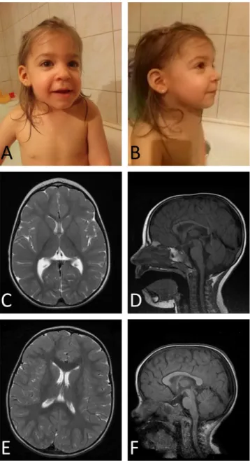
The proband, a girl was born from the first unevent- ful pregnancy to heathy, non-consanguineous Cau- casian parents on the 37th gestational week. Her birth weight was 1950 g (2.2 SD), head circumference 30 cm (2.0 SD) and length 44 cm (1.4 SD). Dysmor- phic features included elongated face, deep-set ears, upturned nose, long philtrum and pointed chin (Fig. 1A, B). She did not have any cutaneous involve- ment. At about 2 months of age fast head and hand tre- mor appeared in the form of rhythmic back-and-forth involuntary movements with low amplitudes both at rest and during action. Fasting did not worsen these move- ments. The tremor became quite vigorous with waxing amplitudes during infancy and gradually waned later.
There was no tremor at rest by the age of 5 years; how- ever, emotional distress, particularly fear still provoked it. She had hypotonia and was unable to sit at age of 10 months, or stand at 12 months of age. She had very short attention span and limited interest in her sur- roundings. Developmental Quotient (DQ) of 45 was found by Brunet-Le´zine test at the age of 2 years.
Fasting ammonia level was normal. Plasma proline, ornithine and arginine levels were also within the normal range, while citrulline level was slightly decreased
(9 mmol/L, normal: 10–50 mmol/L, borderline). The results of other blood tests were normal.
Brain MRI at the age of 2 years revealed reduced vol- ume of white matter and moderate hypoplasia of the corpus callosum (Fig. 1C–F).
At the age of 5.5 years her head circumference was 46 cm (3.5 SD), weight 15 kg, (1.9 SD), and height 98 cm (2.8 SD). By this age, marked spasticity through most of the range of motion (Modified Ashworth scale
Fig. 1. Photos and MR images of the patient. The patient at the age of 2.5 years (A and B). Dysmorphic features can be seen: elongated face, deep-set ears, upturned nose, long philtrum and pointed chin. Parental written permission has been gained to publish the patient’s photos. T2- weigthed axial (C) MR image of the patient shows the paucity of white matter and thinning of the genu of the corpus callosum compared to age-matched control (E). T1 weighted sagittal MR image of the patient (D) demonstrates the hypoplasia of the corpus callosum (D) compared to control (F).
T. Kalma´r et al. / Brain & Development 43 (2021) 144–151 145
2) developed in her lower limbs with brisk deep tendon reflexes. Wide based spastic gait was also observed.
There was no speech and intellectual disability was evi- dent. She had disruptive behavior hindering us from taking formal Intelligence Quotient (IQ) test.
2.1. Genetic analysis
Routine chromosomal analysis by G-banding showed normal 46,XX karyotype. Genomic DNA was extracted from peripheral blood samples with the Puregene kit (Gentra). Array comparative genomic hybridization (aCGH) showed normal genomic copy number (Quanti- tative Genomic Medicine Laboratories, S.L., Barcelona, Spain).
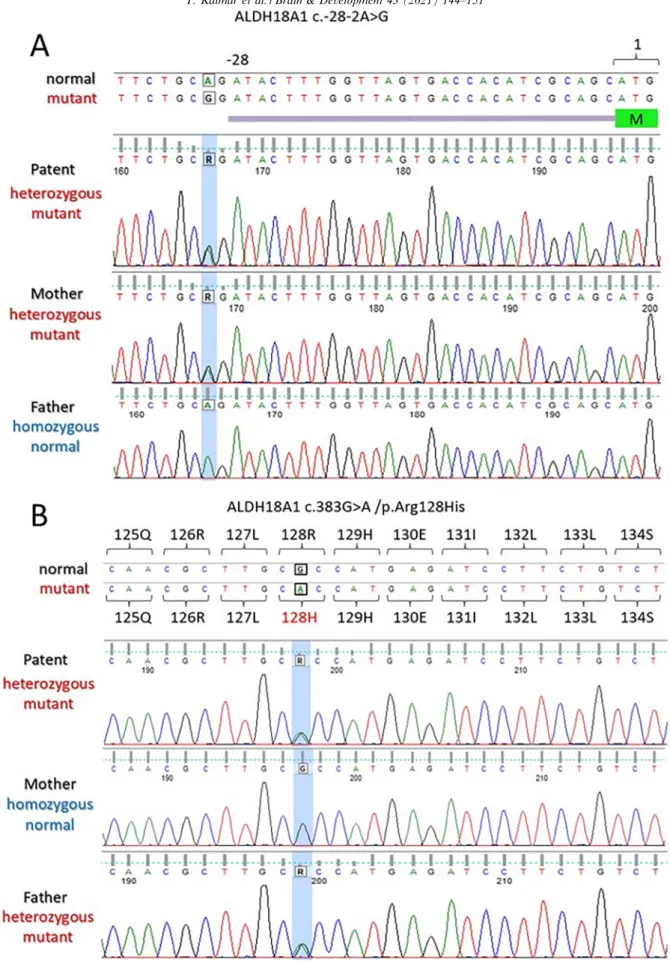
Trio analysis by whole exome sequencing (WES) was performed with CentoXomeÒ Gold at Centogene AG (Rostock, Germany) as described earlier[5]. A heterozy- gous variant in intron 1 (NM_002860.4:c.-28-2A>G) and another heterozygous variant in exon 4 (NM_002860.4:c.383G>A, NP_002851.2:p.Arg128His) of the ALDH18A1 gene were detected (Fig. 2A, B).
The c.-28-2A>G variant is likely pathogenic because it changes the acceptor splice site of intron 1 in the 50- UTR, causing skipping of exon 2 (the start codon is in exon 2). The possible molecular effect of this variant was tested in silico using MutationTaster. It predicted that c.-28-2A>G variant is disease causing (prob:
0.969300883264394); the wild type splice site (tgca|
GATA c.-28) has been lost. This variant is absent in the databases [GnomAD (The Genome Aggregation Database), dbSNP, Exome Variant Server, ClinVar].
However, the p.Arg128His variant has been previously reported in compound heterozygous state as disease- causing for autosomal recessive spastic paraplegia [3].
The c.-28-2A>G variant was also detected in the mother (Fig. 2A) in a heterozygous state, whereas the c.383G>A was detected in the father (Fig. 2B) also in a heterozy- gous state.
3. Discussion
The patient in this report is compound heterozygous for two ALDH18A1 disease-causing mutations. Both mutations affect the G5K domain of the P5CS enzyme.
The phenotypic characteristics, such as intrauterine growth retardation, dysmorphic features, short statue, microcephaly, global developmental delay, cognitive impairment, progressively developing spastic paraplegia and lack of cutaneous manifestations in association with biallelic ALDH18A1 mutations meet the criteria of SPG9B[1–3,6–8].Fifteen patients in 8 families have been described so far with this disorder, including the patient in this report (Table 1). The compound heterozygous mutations in 5 families and homozygous mutations in 3 families distributed randomly in theALDH18A1gene,
affecting both the G5K and G5PR domains (Table 1).
Corpus callosum hypoplasia and thin white matter, found in our patient, have been described in ALDH18A1-related disorders, however they are rare in SPG9B (Table 1). Autopsy findings or neuropathology have never been reported.
The vigorous infantile tremor, the presenting sign in our patient, was unusual and has never been reported so early in SPG9B. Tremor, reported in other cases of SPG9B, started later, at around the 7th and 15th years of age[2,3](Table 1). It can be regarded as a manifesta- tion of neurodegeneration[9]. Tremor has also been seen in patients with ARCL3 due to biallelic ALDH18A1 mutations [10,11]. Tremor can occur in other types of hereditary spastic paraplegias as well, like in SPG4 due to SPASTmutation [12]and SPG11 due to muta- tions in the spatacsingene[13,14].
Neurodegeneration seems to be a common feature of autosomal recessive and autosomal dominant ALDH18A1-related disorders [1]. A unifying view for these disorders has been hypothesized, claiming that the different presentations conform to a disease contin- uum of decreasing severity from the cutis laxa forms ARCL3A and ADCL3 to the motor syndromes SPG9B and SPG9A [1]. Global developmental delay and intel- lectual disability are major manifestations of the central nervous system involvement with equal frequency in both autosomal recessive forms, i.e. in ARCL3A and SPG9B [1]. While hypotonia is a consistent feature in ARCL3A, occasionally followed by pyramidal signs [1,10,15], hypotonia seems to be rare in SPG9B; progres- sive hypertonia, spasticity and pyramidal signs prevail instead [1,3], as in our patient. A transition between these autosomal recessive conditions might be repre- sented by patients reported with biallelic mutations without both cutis laxa and spastic paraplegia [[16], Patient 2 in [17]].
Data has been collected and reviewed by Marco- Marin and coworkers in favor of the view that the sever- ity of the various syndromes inALDH18A-related disor- ders would correspond to higher or lower degrees of loss of P5CS function [1]. Decreased serum P5CS activity was found in a patient with SPG9B due to homozygous p.Ser242Asn mutation in the G5K domain; however, the P5CS protein level and its mitochondrial localization in HeLa cells transfected with the mutant ALDH18A1 plasmids remained unchanged [7]. In another patient with SPG9B due to compound heterozygous mutations in the G5PR domain of ALDH18A1 (p.Arg371Gln and p.Ser497Asn) residual activity of the P5CS was also observed[8]. Indeed, these findings may suggest that the SPG9B phenotype could be associated with residual P5CS activity, while some patients with biallelic null mutations in ALDH18A1 and cutis laxa phenotype exhibited pronounced reduction or total absence of P5CS protein[1,11,18,19].
Fig. 2. Mutations in theALDH18A1gene. (A) A single nucleotide change (boxed) near the 50UTR in the mutant compared to normal (wild type) sequence. The patient inherited this mutation from her mother (R = A/G). (B) A single nucleotide change (boxed) in the exon 4 in the mutant compared to normal (wild type) sequence. The patient inherited this mutation from her father (R = A/G). The mutated positions are highlighted blue in the DNA chromatograms. (For interpretation of the references to colour in this figure legend, the reader is referred to the web version of this article.)
T. Kalma´r et al. / Brain & Development 43 (2021) 144–151 147
Table 1
Reported biallelicALDH18A1variants associated with autosomal recessive spastic paraplegia (SPG9B).
References This study [6] [2] [3]
Number of affected siblings 1 2 1 4
Gender F M F M F F M F
Ethnicity Caucasian Japanese Japanese Caucasian Caucasian Caucasian Caucasian Caucasian
Intron/exon location Intron 1 Exon 4
Exon 2 Exon 4
Exon 2 Exon 4
Exon 3 Exon 14
Exon 4 Exon 15
Exon 4 Exon 15
Exon 4 Exon 15
Exon 4 Exon 15 Nucleotide variation c.-28-2A>G
c.383G>A
c.30C>A c.383G>A (two homozygous
mutations)
c.30C>A c.383G>A (two homozygous
mutations)
c.251G>A c.1741G>A
c.383G>A c.1910T>C
c.383G>A c.1910T>C
c.383G>A c.1910T>C
c.383G>A c.1910T>C
Protein variation ?
p.Arg128His
p.Phe10Leu p.
Arg128His (two homozygous
mutations)
p.Phe10Leu p.Arg128His (two homozygous
mutations)
p.Arg84Gln p.Glu581Lys
p.Arg128His p.Leu637Pro
p.Arg128His p.Leu637Pro
p.Arg128His p.Leu637Pro
p.Arg128His p.Leu637Pro
Protein domain G5K/G5K G5K/G5K G5K/G5K G5K/G5PR G5K/G5PR G5K/G5PR G5K/G5PR G5K/G5PR
Dysmorphic features Yes NA NA NA Yes No Yes Yes
Microcephaly Yes NA NA NA Yes Yes No No
Growth retardation Yes NA NA NA Yes Yes No Yes
Developmental delay Yes Yes Yes Yes Yes Yes Yes Yes
Cognitive impairment
Lower limb spasticity Yes Yes Yes Yes Yes Yes Yes Yes
Ataxia No No No No No No No NA
Cerebellar signs
Tremor (age at onset, years) Yes (<1) No No Yes (15) No No No NA
Epilepsy No No No Yes No No No No
Cutaneous findings No No No No No No No No
Ocular findings No No No No NA NA Probable cataract NA
Brain MRI Corpus callosum hypoplasia Thin white matter
Normal Normal Normal NA NA Thin corpus callosum
Periventricular white matter anomalies Mild cortical atrophy
NA
Plasma amino acids (proline, citrulline, ornithine, arginine)
Normal NA NA Normal NA NA NA NA
(continued on next page)
T.Kalma´retal./Brain&Development43(2021)144–151
Table 1(continued)
Reported biallelicALDH18A1variants associated with autosomal recessive spastic paraplegia (SPG9B).
References [7] [8] [6] [3]
Number of affected siblings 1 2 2 2
Gender F M M M M M M
Ethnicity Chinese Caucasian Caucasian Japanese Japanese Caucasian Caucasian
Intron/exon location Exon 7 Exon 10
Exon 10
Exon 10 Exon 10
Exon 12 Exon 16
Exon 12 Exon 16
Exon 17 Exon 17 Nucleotide variation c.725G>A
(homozygous)
c.1112G>A c.1490G>A
c.1112G>A c.1490G>A
c.1321C>T c.1994G>A
c.1321C>T c.1994G>A
c.2143G>C (homozygous)
c.2143G>C (homozygous) Protein variation p.Ser242Asn
(homozygous)
p.Arg371Gln p.Ser497Asn
p.
Arg371Gln p.Ser497Asn
p.Arg441Ter p.Arg665Gln
p.Arg441Ter p.Arg665Gln
p.Asp715His (homozygous)
p.Asp715His (homozygous)
Protein domain G5K G5PR/G5PR G5PR/
G5PR
G5PR/GSPR G5PR/GSPR GSPR GSPR
Dysmorphic features NA Yes Yes NA NA Yes Yes
Microcephaly NA Yes Yes NA NA Yes Yes
Growth retardation NA Yes Yes NA NA NA NA
Developmental delay No Yes Yes Yes Yes Yes Yes
Cognitive impairment
Lower limb spasticity Yes Yes Yes Yes Yes Yes Yes
Ataxia No No No Yes Yes NA No
Cerebellar signs
Tremor (age at onset, years) No No No No No Yes (7) Yes (7)
Epilepsy No Yes Yes No No No No
Cutaneous findings No No No No No No No
Ocular findings No No No No No NA NA
Brain MRI Normal Increase in the prominence of the cortical sulci
NA Mild cerebellar atrophy
Mild cerebellar atrophy
Normal NA
Plasma amino acids (proline, citrulline, ornithine, arginine)
NA NA NA Normal Normal Normal NA
Abbreviations: M: male, F: female, G5K: glutamate 5-kinase domain, G5PR: gamma-glutamyl phosphate reductase domain, NA: not available.
T.Kalma´retal./Brain&Development43(2021)144–151149
Measurements of blood ammonia and amino acid levels led to inconstant results. Increased blood ammonia and low plasma proline, ornithine, citrulline and arginine were described in both SPG9A and ARCL3A[3,10]. In contrast, the ammonia and amino acid levels were in the normal or low normal range in SPG9B patients regardless of whether the mutation affected the G5K, as in our case, the G5PR, or both domains[2,3,6]. The patient population with ALDH18A1-related disorders is small, hampering the comparison of the sometimes contradictory results gained by various methods in sin- gular cases, or families with different phenotypes.
The pathophysiology of the neurological impairment in ALDH18A-related disorders remains to be clarified.
Reduced cerebral proline and/or creatine synthesis might have a role [1,19], however moonlighting of P5CS protein cannot be ruled out [19,20]. Further research warranted to elucidate the mechanism of neu- rodegeneration in these conditions.
4. Conclusion
We report a girl with a rare form of autosomal reces- sive hereditary spastic paraplegia (SPG9B) due to com- pound heterozygous mutations in theALDH18A1gene.
The c.-28-2A>G variant in intron 1 is a novel mutation;
it was inherited from her mother. The other, c.383G>A variant in exon 4, inherited from her father, has already been published. Vigorous infantile tremor preceding progressive spastic paraplegia was a unique clinical manifestation of the disease. Intrauterine and postnatal growth retardation, dysmorphic features, microcephaly, delayed development and intellectual disability were the other characteristic features of the disorder.
Acknowledgments
The authors are grateful to the patient’s parents for their collaboration.
The authors thank to Professor Eva Morava-Kozicz MD, PhD for the amino acid analysis.
Funding
This study was supported by the GINOP-2.3.2-15-2 grant (TK and ZM) provided by the National Research, Development and Innovation Office (Hungary).
Ethical approval
Written informed parental consent has been obtained.
Written permission has been gained from the parents to publish the patient’s photos in a scientific journal.
The study was approved by the Human Investigation Review Board at Albert Szent-Gyo¨rgyi Clinical Centre, University of Szeged, Hungary.
Conflict of Interest Disclosures
The authors declare no competing interests.
References
[1] Marco-Marin C, Escamilla-Honrubia JM, Lla´cer JL, Seri M, Panza E, Rubio V. D1-Pyrroline-5-carboxylate synthetase defi- ciency: An emergent multifaceted urea cycle-related disorder. J Inherit Metab Dis 2020.https://doi.org/10.1002/jimd 12220.
[2] Steenhof M, Kibaek M, Larsen MJ, Christensen M, Lund AM, Brusgard K, et al. Compound heterozygous mutations in two different domains of ALDH18A1do not affect the amino acid levels in a patient with hereditary spastic paraplegia. Neurogenet 2018;19:145–9.
[3] Coutelier M, Goizet C, Durr A, Habarou F, Morais S, Dionne- Laporte A, et al. Alteration of ornithine metabolism leads to dominant and recessive hereditary spastic paraplegia. Brain 2015;138:2191–205.
[4] Hu CA, Lin WW, Obie C, Valle D. Molecular enzymology of mammalian D1-pyrroline-5-carboxylate synthase. Alternative splice donor utilization generates isoforms with different sensitiv- ity to ornithine inhibition. J Biol Chem 1999;274:6754–62.
[5] Zombor M, Kalma´r T, Nagy N, Bere´nyi M, Telcs B, Maro´ti Z, et al. A novelWDR62missense mutation in microcephaly with abnormal cortical architecture and review of the literature. J Appl Genet 2019;60:151–62.
[6] Koh K, Ishiura H, Beppu M, Shimazaki H, Ichinose Y, Mitsui J, et al. Novel mutations in the ALDH18A1 gene in complicated hereditary spastic paraplegia with cerebellar ataxia and cognitive impairment. J Hum Genet 2018;63:1009–13.
[7] Wei Q, Dong HL, Pan LY, Chen CX, Yan YT, Wang RM, et al.
Clinical features and genetic spectrum in Chinese patients with recessive hereditary spastic paraplegia. Transl Neurodegener 2019;8:19.
[8] Magini P, Marco-Marin C, Escamilla-Honrubia JM, Martinelli D, Dionisi-Vici C, Faravelli F, et al. P5CS expression study in a new family withALDH18A1-associated hereditary spastic para- plegia SPG9. Ann Clin Transl Neurol 2019;6:1533–40.
[9] Torres-Russotto D. Clinical approach to tremor in children.
Parkinsonism Relat Disord 2019;59:111–6.
[10] Baumgartner MR, Rabier D, Nassogne MC, Dufier JL, Padovani JP, Kamoun P, et al. D1-pyrroline-5-carboxylate synthase defi- ciency: neurodegeneration, cataracts and reduced ornithine, citrulline, arginine and proline. Eur J Pediatr 2005;164:31–6.
[11] Fischer B, Callewaert B, Schro¨ter P, Coucke PJ, Schlack C, Ott CE, et al. Severe congenital cutis laxa with cardiovascular manifestations due to homozygous deletions in ALDH18A1.
Mol Genet Metab 2014;112:310–6.
[12] de Bot ST, van den Elzen RTM, Mensenkamp AR, Schelhaas HJ, Willemsen MAAP, Knoers NVAM, et al. Hereditary spastic paraplegia due to SPASTmutations in 151 Dutch patients: new clinical aspects and 27 novel mutations. J Neurol Neurosurg Psychiatry 2010;81:1073–8.
[13] Anheim M, Lagier-Tourenne C, Stevanin G, Fleury M, Durr A, Namer IJ, et al. SPG11 spastic paraplegia. A new cause of juvenile parkinsonism. J Neurol 2009;256:104–8.
[14] Schneider SA, Mummery CJ, Mehrabian M, Houlden H, Bain PG. SPG11 presenting with tremor. Tremor Other Hyperkinet Mov (NY) 2012;2 tre-02-104-666-1.
[15] Wolthuis DFGJ, van Asbeck E, Mohamed M, Gardeitchik T, Lim-Melia ER, Wevers RA, et al. Cutis laxa, fat pads and retinopathy due to ALDH18A1 mutation and review of the literature. Eur J Paediatr Neurol 2014;18:511–5.
[16] Kremer LS, Bader DM, Mertes C, Kopajtich R, Pichler G, Iuso A, et al. Genetic diagnosis of Mendelian disorders via RNA sequencing. Nat Commun 2017;8:15824.
[17] Handley MT, Me´garbane A, Meynert AM, Brown S, Freyer E, Taxlor MS, et al. Loss ofALDH18A1function is associated with a cellular lipid droplet phenotype suggesting a link between autosomal recessive cutis laxa type 3A and Warburg Micro syndrome. Mol Genet Genomic Med 2014;2:319–25.
[18] Skidmore DL, Chitayat D, Morgan T, Hinek A, Fischer B, Dimopoulou A, et al. Further expansion of the phenotypic spectrum associated with mutations inALDH18A1, encodingD1- pyrroline-5-carboxylate synthase [P5CS]. Am J Med Genet Part A 2011;155:1848–56.
[19] Martinelli D, Ha¨berle J, Rubio V, Giunta C, Hausser I, Carrozzo R, et al. Understanding pyrroline-5-carboxylate synthase defi- ciency: clinical, molecular, functional, and expression studies, structure-based analysis, and novel therapy with arginine. J Inherit Metab Dis 2012;35:761–76.
[20] Jeffery C. Protein moonlighting: what is it, and why is it important?. Philos Trans R Soc B 2017;373:20160523.
T. Kalma´r et al. / Brain & Development 43 (2021) 144–151 151