Role of succinate dehydrogenase in pheochromocytomas and paragangliomas
PhD thesis
Nikoletta Katalin Lendvai
Doctoral School of Clinical Medicine Semmelweis University
Supervisor: Attila Patócs MD, Ph.D, M.Sc Official reviewers: Attila Bokor, MD, Ph.D
Judit Berenténé Bene, MD, Ph.D
Head of the Final Examination Committee: Edit Búzás, MD, D.Sc
Members of the Final Examination Committee: Nóra Hosszúfalusi, MD, Ph.D Zsolt Rónai MD, Ph.D
Budapest 2016
1 APPENDIX
Abbreviation
1. INTRODUCTION 6
1.1. Succinate dehydrogenase 6
1.1.1. The function of succinate dehydrogenase and the role of succinate
in cell metabolism 6
1.1.2. The structure of succinate dehydrogenase 8
1.1.3. Chromosomal localization of SDH subunits encoding genes 10
1.2. Pheochromocytoma and paraganglioma 11
1.2.1. Definition, anatomical localisation 11
1.2.2 Clinical features 14
1.2.3. Etiology and genetic background of hereditary pheochromocytomas and paragangliomas 15
1.2.3.1. Familial pheochromocytoma/ paraganglioma syndrome 15
1.2.3.2. Multiple endocrine neoplasia type 2A and 2B 18
1.2.3.3. von Hippel-Lindau syndrome 20
1.2.3.4. Neurofibromatosis type 1 21
1.2.3.5. Recently identified genes causing pheochromocytoma/ paraganglioma 21
1.2.4. Possible genetic modifiers 24
1.2.5. Biochemical characteristics of pheochromocytomas/paragangliomas 24
1.2.6. Diagnosis 26
1.2.6.1. Physical examination and family history 26
1.2.6.2. Biochemical testing 26
1.2.6.3. Imaging studies 28
1.2.6.4. Molecular genetic testing 28
1.2.7. Treatment of pheochromocytoma and paraganglioma 29
2. OBJECTIVES 31
3. METHODS 32
2
3.1. Germline mutation prevalence in Hungarian patients with
pheochromocytoma and/or paraganglioma 32
3.1.1. Patients 32
3.1.2. The first Hungarian case with extra adrenal pheochromocytoma associated with SDHD gene mutation 33
3.1.3. Genetic testing of the RET, VHL, SDHB, SDHC, SDHD, SDHAF2, MAX and TMEM127 genes using Sanger sequencing 34
3.2. The G12S polymorphism of the SDHD gene as a phenotype modifier in patients with MEN2A syndrome 35
3.2.1. Patients 3.2.1.1. Patients with MEN2 syndrome 35
3.2.1.2. Patients with sporadic MTC 36
3.2.1.3. Patients with apparently sporadic PHEO 36
3.2.2. Germline mutation screening of the RET, VHL, SDHB, and SDHD genes 37
3.2.3. Restriction fragment length polymorphism (RFLP) analysis for identification the G12S polymorphism of the SDHD gene 37
3.2.4. Statistical analysis 38
3.3. Biochemical consequences of SDHx mutations, succinate to fumarate ratio in SDHB/D associated paragangliomas 39
3.3.1. Materials 39
3.3.2. Silencing of SDHB in MPC and MTT cells 40
3.3.3. Western blotting 40
3.3.4. Metabolic measurements 41
3.3.5. Statistical analysis 42
4. RESULTS 43
4.1. Germline mutation prevalence in Hungarian patients with pheochromocytoma and/or paraganglioma 43
4.1.1. Genotype-phenotype associations 44
4.1.2. The first Hungarian case with extra adrenal pheochromocytoma associated with SDHD gene mutation 48
3
4.2. The G12S polymorphism of the SDHD gene as a phenotype modifier in
patients with MEN2A syndrome 52
4.3. Biochemical consequences of SDHx mutations, succinate to fumarate ratio in SDHB/D associated paragangliomas 54
4.3.1. Succinate concentration 54
4.3.2. Fumarate concentration 54
4.3.3. Succinate to fumarate ratio 57
4.3.4. Succinate to fumarate ratio in plasma samples 58
4.3.5. Succinate to fumarate ratio in MPC and MPP cells 58
5. DISCUSSION 59
5.1. Germline mutation prevalence in Hungarian patients with pheochromocytoma and/or paraganglioma 59
5.2. The G12S polymorphism of the SDHD gene as a phenotype modifier in patients with MEN2A syndrome 63
5.3. Biochemical consequences of SDHx mutations, succinate to fumarate ratio in SDHB/D associated paragangliomas 65
6. CONCLUSION 67
7. SUMMARY/ÖSSZEFOGLALÁS 69
8. BIBLIOGRAPHY 71
9. BIBLIOGRAPHY OF THE CANDIDATE’S PUBLICATIONS 87
10. ACKNOWLEDGEMENTS 88
4 Abbreviations
ATP adenosine triphosphate CAC Citric acid cycle COV Coefficient of variance
CT Computer Tomography
ETC Electron transport chain FAD Flavine adenine dinucleotide
18F-FDG-PET 2-18F-fluoro-2-deoxy-D-glucose position emission tomography
FH Fumarate hydratase
FMTC Familial Medullary Thyroid Cancer FPGL Familial paraganglioma syndrome
GABA Gamma aminobutyric acid
GC-MS Gas Chromatography-Mass Spectrometry HIF 1α Hypoxia Inducible Factor
H2O2 Hydrogen peroxide
KIF1Bβ Kinesin family member 1B
MDH Malate dehydrogenase
MEN Multiple Endocrine Neoplasia
MIBG Metaiodobenzylguanidine scintigraphy MPC Mouse pheochromocytoma cells MRI Magnetic Resonance Image MTC Medullary Thyroid Cancer
MTT Mouse tumor tissue
NADH Reduced form of nicotinamide adenine dinucleotide
NF Neurofibromatosis
PGL Paraganglioma
PHD Prolyl hydroxylase
PHEO pheochromocytoma
RET Rearranged during transfection
5
RFLP Restriction fragment length polymorphism ROS Reactive oxygen species
SD Standard deviation
SDH Succinate dehydrogenase
SDHA Succinate dehydrogenase subunit A SDHB Succinate dehydrogenase subunit B SDHC Succinate dehydrogenase subunit C SDHD Succinate dehydrogenase subunit D SDHx Succinate dehydrogenase subunits
VHL von Hippel-Lindau
6 1. INTRODUCTION
1.1. Succinate dehydrogenase and function
Succinate dehydrogenase (SDH) described first by Albert Szent-Györgyi in the middle of the 1930’s is part of both the citric acid cycle (CAC) and respiratory electron transfer chain (ETC)/oxidative phosphorylation. 1 SDH catalyzes the oxidation of succinate to fumarate in the mitochondrial matrix and transfers electrons to ubiquinone without pumping protons across the mitochondrial inner membrane. 1
1.1.1. The function of succinate dehydrogenase and the role of succinate
The Krebs cycle consists of chain of chemical reactions in order to generate energy for cells. The whole CAC takes place in the mitochondrial matrix. It uses carbohydrates, fats, amino acids and proteins to oxidase acetyl-coenzyme A (acetyl CoA). The main source of acetyl CoA comes from the glycolysis, but can be derived from fatty acid oxidation as well.
The intermediates of the CAC serve as substrate for biosynthetic pathways.
The CAC result in a total of four molecules of ATP, ten molecules of NADH, and two molecules of FADH2. Electrons from NADH and FADH2 are then transferred to molecular oxygen through the oxidative phosphorylation. 2
Succinate dehydrogenase catalyzes the 7th step of the CAC (tricarboxylic acid cycle or Krebs cycle). It catalyzes the oxidation of succinate to fumarate along with the reduction of ubiquinone (Coenzyme Q) to ubiquinole, by transferring electrons thru FAD-FADH2
(Figure 1).
Succinate dehydrogenase or complex II is also involved in the oxidative phosphorylation (OXPHOS) or electron transport chain, representing the major source of cellular energy.
The OXPHOS takes place in the inner membrane of the mitochondria and consists of four complexes; complex I, II, III and IV. The fifth complex is the ATP synthase, which uses the proton gradient to synthesize 32 to 34 ATP molecules. The flow of electrons from NADH and FADH2 thru the protein complexes is associated with pumping protons to the
7
intermembrane space of the mitochondria, which results in a proton gradient and builds up the transmembrane potential. This is necessary for driving complex V to synthetize ATP.
However, complex II is not coupled with a proton pump and transfers electrons to ubiquinone without contributing to the proton gradient. 3
Figure 1. Citric acetate cycle and oxidative phosphorylation.
Succinate dehydrogenase catalyzes succinate to fumarate oxidation in citric acid cycle and as complex II participates in the electron transfer in the oxidative phosphorylation.
Based on work Osellame LD, Blacker TS DM. Cellular and molecular mechanisms of mitochondrial function. Best Pr Res Clin Endocrinol Metab. 2012;26(6):711–723.
8
The substrate of SDH enzyme, succinate, is a distant product of the α-ketoglutarate dehydrogenase complex. It is involved in several metabolic pathways including a macrophage-specific metabolic pathway generating itaconate 4, it is connected with the metabolism of branched-chain amino acids, heme synthesis, ketone bodies utilization and the GABA shunt. 5,67
Regarding tumorigenesis, succinate is considered as a critical mediator of the hypoxic response, and it has also been suggested that SDH plays an important role in ROS homeostasis of cells producing superoxide and H2O2. 8
Succinate was also involved in posttranslational protein modification called succinylation by this mechanism succinate might be involved in stabilization of certain proteins. In specific tumours it was demonstrated that succinate stabilizes hypoxia inducible factor 1α, a key transcription factor for regulating molecules which are involved in adaptation to hypoxia and in facilitating blood vessel genesis (vascular endothelial growth factor, platelet growth factor etc.). 9
In addition, succinate was discovered to exert its effects outside of cells in para- and autocrine manners, mediated by the expression of at least one plasmalemmal succinate receptor type. 10
Based on these fundamental processes it is not surprising that mutations of genes encoding the subunits of SDH complex have been implicated in the pathogenesis of various diseases including oxidative stress, tumour formation, neurodegeneration, hypoxia or “just simple energy deficiency”.
1.1.2. The structure of SDH
SDH consists of a hydrophilic head that protrudes into the matrix compartment and a hydrophobic tail that is embedded within the IM with a short segment projecting into the soluble intermembrane space. The hydrophilic head consists SDHA (flavoprotein) and SDHB (iron sulphur protein), forming the catalytic core. Here are the binding sites for FAD
9
cofactor and succinate. Three iron-sulphur clusters can be found in the SDHB subunit and these clusters mediate electron transfer to ubiquinone (Figure 2).
The hydrophobic tail consists of SDHC and SDHD subunits. The enzyme complex binds to membrane through these subunits. The structure of the enzyme complex is constructed of six transmembrane helices containing one heme b group and a ubiquinone-binding site. 11 (Figure 2)
Figure 2. Structure of succinate dehydrogenase.
Succinate dehydrogenase [CC-BY-SA-3.0 Steve Cook, based on PDB 1NEK] The ‘top’ of the enzyme pokes into the mitochondrial matrix and oxidises succinate; the ‘bottom’ of the enzyme is dissolved in the lipid of inner mitochondrial membrane, and reduces ubiquinone.
10
1.1.3. Chromosomal localization of genes encoding the SDH subunits
All four subunits of SDH or complex II are encoded by genes located in the nuclear genome. SDHA encoding gene is mapped to the p arm of chromosome 5 at locus 15, SDHB gene is localized on the p arm of chromosome 1 at locus 36. SDHC gene is encoded on the q arm of chromosome 1 at locus 23. SDHD and SDHAF2 genes are encoded on the q arm of chromosome 11 at locus 23.1 and 13, respectively. Number of exons and number of amino acid residues are included in Table 1.
Genetic mutations of these genes are associated with familial paraganglioma syndrome, childhood T-cell acute leukaemia and gastric stromal tumours. 12–16
Table 1. Chromosomal localization of succinate dehydrogenase subunits.
Gene Ensemble ID Chromosomal localization
Number of exons
Number of amino acids
SDHA ENSG00000073578 5p15 15 644
SDHB ENSG00000117118 1p36 8 280
SDHC ENSG00000143252 1q23 6 169
SDHD ENSG00000204370 11q23.1 4 159
SDHAF2 ENSG00000167985 11q13 4 166
SDHA - succinate dehydrogenase subunit A, SDHB - succinate dehydrogenase subunit B, SDHC - succinate dehydrogenase subunit C, SDHD - succinate dehydrogenase subunit D, SDHAF2 – succinate dehydrogenase complex assembly factor 2
11 1.2. Pheochromocytoma and paraganglioma
1.2.1. Definition, anatomical distribution
The term pheochromocytoma means “dusky-colored tumour” and it was historically derived from the color change that occurs when the tumour tissue was immersed in chromate salts. 17 Pheochromocytomas are rare catecholamine-producing tumours arising from neural crest-derived chromaffin cells in the adrenal gland. Chromaffin cells are also found in the sympathetic ganglions; sympathetic extra-adrenal paragangliomas are generally confined to the lower mediastinum, abdomen, and pelvis (e.g. the aortic chemoreceptors and the Zuckerkandl-organ), and are typically hormone secreting. In contrast, parasympathetic paragangliomas are located predominantly on the skull base, neck, and upper mediastinum (e.g. carotid artery/body). 18,1920 (Figure 3)
12
Figure 3. Anatomical distribution of pheochromocytoma and paragangliomas. 21
Based on Lips C, Lentjes E, Höppener J, Luijt R van der, Moll F. Familial paragangliomas.
Hered Cancer Clin Pract. 2006. doi:10.1186/1897-4287-4-4-169.
13
The prevalence of this tumour is estimated between 1:6500 to 1:2500 in the United States 22 but the autopsy reports suggest a prevalence of 1:2000 and suggest that many of these tumours remain undiagnosed. 23,24 An increased frequency is noted in people subjected to chronic hypoxia, living at higher-altitude regions or in the presence of respiratory or heart diseases. 25 The incidence of pheochromocytoma is 2 to 8 per million persons per year. 26 Other tumours, as head-and-neck, abdominal, and pelvic PGL have an incidence of 0.5 per million per year. 27 Paragangliomas in the Zuckerkandl-organ are the most common sympathetic and carotid body tumours are the most common parasympathetic extra adrenal paragangliomas. 28
Pheochromocytoma and paraganglioma can occur at all ages, but have a peek incidence at the 4th and 5th decade, with an almost equal distribution between men and women. 29 30 Pheochromocytoma is present in 0.1% to 1% of patients with hypertension 31,32 and it is present in approximately 5% of patients with incidentally discovered adrenal masses. 33 About 90% of PHEOs are unilateral, but bilateral tumours are seen in higher proportion in syndromic cases. 27
The risk for malignant transformation is greater for extra-adrenal sympathetic paragangliomas than for pheochromocytomas or skull base and neck paragangliomas. 20,25 It is difficult to determine the malignancy of PHEOs and PGLs as only the metastasis to lymph node, bone, liver, or lung confirms malignancy. 19,34 However, pathological criteria, as size, weight, presence of tumour necrosis, a greater than 4% of Ki-67 index and the absence of S100 by immunohistochemistry have been shown to be associated with malignancy. 35
Early diagnosis and resection of the tumour can cure most of the cases. The diagnosis is difficult because clinical features/symptoms can mimic other diseases or can be very unspecific or uncharacteristic.
14 1.2.2. Clinical features
Sympathetic paragangliomas secrete catecholamines, mainly adrenalin and noradrenalin;
parasympathetic paragangliomas are most often (ca. 95%) hormonally silent or have low catecholamine production. Symptoms of PGL/PCC result either from the mass effects or catecholamine hypersecretion. The main symptoms related to hormone hypersecretion are the following:
• Hypertension (paroxysmal or sustained)
• Palpitation and tachycardia
• Sweating attacks
• Headache
• Facial flushing
• Chest and abdominal pain
• Anxiety and panic attacks
• Nausea
• Tremor
• Pallor
• Elevated fasting plasma glucose concentration
The symptoms are usually paroxysmal, but 50-60% of the patients have high blood pressure between the episodes. 36 In some cases the symptoms are more severe and cause cardiovascular and neurological manifestations. 37
Parasympathetic paragangliomas show a slow-growing, painless mass and patients develop symptoms due to pressure of surrounding tissue or nerves: chocking, hoarseness, tickling cough, Horner’s syndrome due to interruption of nervous tissue. 38
15
1.2.3. Etiology/Genetic background and associated hereditary syndromes
Pheochromocytomas and paragangliomas are mainly sporadic tumours, formerly about 10% of all tumours associated with hereditary syndromes, including multiple endocrine neoplasia type 2 (MEN2), von Hippel-Lindau syndrome (VHL) and neurofibromatosis type 1 (NF1). 39 A small percent of PHEO/PGL associated with Carney-triad, Carney-Stratakis syndrome and more rarely with MEN type 1. In the last decade several genes were discovered as genetic causes of pheochromocytoma and paraganglioma, making the prevalence of hereditary PHEO/PGL 30%–35% of all cases.
The common feature of hereditary syndromes is that they show an autosomal dominant inheritance, meaning that the affected individual receives one mutant gene from one of his/her parents. In some cases a de novo mutation in the germline of the patient may occur.
In sporadic (no germline disease-causing mutation) cases somatic, inactivating mutations can also be identified. The most common genes and the associated syndromes are summarized below.
1.2.3.1. Familial paraganglioma syndrome
Familial paraganglioma (FPGL) syndrome is caused by the germline heterozygous mutations of the SDHx genes (SDHB, SDHC, SDHD, encoding subunits B, C and D, respectively) 12,15,40 and the newly identified SDH5 gene. 16 SDHx genes encode subunits of the mitochondrial complex II (succinate dehydrogenase, SDH), an enzyme involved in oxidative phosphorylation and intracellular oxygen sensing and signalling.
Some differences between the mutation types have been observed. Most of the SDHB and SDHC mutations are mutations which result in truncated protein or leading to amino acid change of the iron sulphur clusters in the SDHB, while mutations of the SDHD gene are more likely nonsense mutations and deletions/insertions. The genotype-phenotype associations have been summarized in Table 2.
Biallelic mutation of the SDHA leads to Leigh syndrome, characterized by mitochondrial encephalopathy and optic atrophy. In 2010 the first SDHA gene mutation was described
16
associating with abdominal PGL and head and neck PGL. 41 This latter finding added SDHA to genes representing genetic susceptibility for PHEO/PGL.
SDHB mutations cause hereditary paraganglioma syndrome type 4. SDHB-related PGLs are associated with abdominal, pelvic tumours, and show single presentation in two third of the cases. Mutations in the SDHB gene are associated with a high risk of developing metastases
29 and about 20%–30% of patients already have metastatic disease at the time of the initial diagnosis 42, while mean age at onset is typically 25-30 years. 43 The diagnosis of SDHB- related PHEOs/PGLs is often delayed, most likely because of the less typical catecholamine excess-related clinical presentations compared with other apparently sporadic or hereditary PHEOs/PGLs. This is partially due to the fact that these tumours can have either a biochemically silent phenotype, a low intratumoural catecholamine content, or a purely dopaminergic phenotype. 42,44
SDHC gene mutations cause hereditary paraganglioma syndrome type 3 with autosomal dominant inheritance. Classical clinical presentation for these tumours is solitary head-neck paraganglioma with a low risk of malignancy. The mean age at diagnosis is 38 years. 45
Mutations of the SDHD gene are causing hereditary paraganglioma syndrome type 1. Its clinical presentation shows multiple abdominal and head-neck paragangliomas with an age of onset at 28-31 years. 47, 48 SDHD-related PHEOs/PGLs, especially those derived from the parasympathetic nervous system of the head and neck, are much less aggressive than SDHB-related PGLs. It has to be mentioned that SDHD gene is maternally imprinted, meaning that disease-causing mutations are inherited exclusively from the paternal side.
17
Table 2. Familial paraganglioma syndrome genotype-phenotype association.
Syndrome Chromosomal localisation Gene
Phenotype
PGL1 mutations on chromosome 11
(11q23) SDHD
head and neck, intraabdominal paragangliomas
pheochromocytomas
PGL2 11q13 (SDHAF2, SDH5) paraganglioma
PGL3 1q21-23 (SDHC) paraganglioma
PGL4 1p36 (SDHB) head and neck paragangliomas
pheochromocytomas
PGL - Familial paraganglioma syndrome, SDHAF2 – succinate dehydrogenase complex assembly factor 2, SDHB - succinate dehydrogenase subunit B, SDHC - succinate dehydrogenase subunit C, SDHD - succinate dehydrogenase subunit D
18 1.2.3.2. Multiple endocrine neoplasia type 2A and 2B
The RET (rearranged during transfection) proto-oncogene located on chromosomal region 10q11.2 consists of 21 exons encodes a receptor tyrosine kinase. 48 Germline, gain of function mutations (mainly missense) of the RET proto-oncogene cause multiple endocrine neoplasia type 2 (MEN2), an autosomal dominantly inherited disease with an approximate prevalence of 2.5 per 100.000 in the general population. 48 The gene mutation are found in specific, hot spot regions (exons: 10, 11, 13, 14, 15 and 16) of the RET proto-oncogene. 49 MEN2 has three subtypes: i) MEN2A, characterized by medullary thyroid carcinoma (MTC), pheochromocytoma (PHEO) and primary hyperparathyroidism; ii) MEN2B, which presents with the most aggressive MTC, pheochromocytoma, neuromas and marfanoid phenotype; and iii) familial MTC (FMTC), the mildest form of MTC. (Table 3, Table 4) Pheochromocytoma occurs in approximately 50% of the MEN2 patients. They are bilateral in most of the cases and have a low rate for malignancy. 50,51 Usually, pheochromocytoma is diagnosed at the same time as the medullary thyroid cancer, 52,53 while some more recent studies showed that PHEO was diagnosed after the diagnosis of MTC 54,55, and very rarely PHEO precedes MTC.
Analysis of the RET gene showed that missense mutations in codon 634 in exon 1 were the most frequent mutations (mutation hot-spot) and the TGC634CGC (Cys-Arg) mutation frequently associated with PHEO. 45,53,56
19
Table 3. MEN2 syndromes and associated phenotypes.
MEN Type Clinical
manifestation Genotype
MEN2A
MTC PHEO primer
hyperparathyreosis
mutation s in exon 10 and 11,
codon 634 is a mutation hotspot
MEN2B
MTC (aggressive form)
PHEO
marfanoid habitus mucosal neuromas
Codon 918 in exon16
FMTC MTC (mild form)
Exons: 10,11 or 5, 8, 13-16; codons: 609, 611, 618, 620 or 790, 791, 768, 804
MEN - multiple endocrine neoplasia, MTC - medullary thyroid cancer, PHEO - pheochromocytoma, FMTC - familial medullary thyroid cancer.
Table 4. The effect of RET mutations on the aggressiveness of MTC. 57
Risk of MTC development Mutation in codon 1. Most aggressive, develops in infancy 883, 918, 922 2. Aggressive, develops in childhood 611, 618, 620, 634 3. Less aggressive, develops in older age 609, 768, 790, 791, 804, 891
RET – rearranged during transfection, MTC – medullary thyroid cancer
Based on Brandi ML, Gagel R, Angeli A, Bilezikian J, Beck-Peccoz P, Bordi C, Conte- Devolx B, Falchetti A, Gheri R, Libroia A, Lips C, Lombardi G, Manelli M, Pacini F, Ponder B, Raue F, Skogseid B, Tamburrano G, Thakker R, Thompson N, Tomasetti P, Tonelli F, Wells MS. Guidelines for diagnosis and therapy of MEN type 1 and type 2. J Clin Endocrinol Metab. 2001;86:5658–5671.
20 1.2.3.3. von Hippel-Lindau syndrome (VHL)
The VHL tumour suppressor gene is located on the 3rd chromosome (3p25-26), mutation or deletion of the gene causes the von Hippel-Lindau syndrome (VHL-syndrome). The disease is autosomal dominantly inherited. Pheochromocytoma, retina angioma, cerebral haemangioblastoma, renal carcinoma, cyst of the kidney and pancreas are the main manifestations. In 7-20% of the cases patients develop pheochromocytoma and these tumours are mainly asymptomatic and affect both adrenals. VHL syndrome is classified based on the appearance or lack of pheochromocytoma in subtypes 1, 2A, 2B and 2C (Table 5).
VHL mutations have the highest predominance in paediatric cases and show the highest prevalence for second contra lateral tumours. Therefore, these patients need close follow- up, usually in every 1-3 years after the first diagnosis. 4558
Table 5. Classification of VHL syndrome.
Type Manifestation
Type 1. no PHEO
Type 2. Risk of PHEO
Type 2A Type 2B Type 2C
kidney cancer, low risk of PHEO kidney cancer, high risk of PHEO only PHEO
VHL - von Hippel-Lindau syndrome, PHEO - pheochromocytoma
21 1.2.3.4. Neurofibromatosis 1
Neurofibromatosis type 1 (von Recklinghausen disease) is characterized by skin lesions (café-au-lait spots), growth of tumours along the nerve in the skin (neurofibromas) and nodules in the iris (Lisch-nodules). Patients with NF1 syndrome have an increased risk for developing optic glioma, leukemia and gastrointestinal tumours. The frequency of NF1 is about 1:3000 to 1:4000 and therefore is one of the most frequent autosomal dominant tumour syndrome, however about 50% of the cases result from new mutations.
Pheochromocytoma appears in 1% - 5.7% of all cases, although 50% of the patients with high blood pressure have pheochromocytoma. 59
Inactivating, somatic mutations of NF1 were described in 41% of sporadic PHEO where LOH at the NF1 locus were also described 60, suggesting that lack of function contributes to the pathogenesis of PHEO.
The NF1 gene is located on chromosome 17 (17q11.2), the NF1 protein participates in the regulation of the ras oncogene and its function resembles to tumour suppressor function.
Patients with NF1 syndrome are suggested clinical and laboratory surveillances for PHEO every 3 years. 61
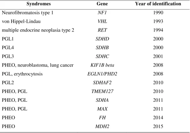
1.2.3.5. Recently identified, novel genes causing PHEO/PGL
TMEM127 (2q11) was identified in 2010 as a new pheochromocytoma susceptibility gene.
62 The encoded protein is a transmembrane protein which is involved in the mammalian target of rapamycin signalling pathway. 46 The TMEM127 associated syndrome is autosomal dominantly inherited. Clinical presentation is characterised by a presence of a mainly unilateral PHEO in patients with no prior family history, however a recent study showed that bilateral, extra-adrenal PGL cases were also described. 63,64
Pathogenic MAX (myc-associated factor X) (14q23) gene variants were shown to predispose to PHEO/PGL. 65 MAX is a transcription factor involved in cellular
22
proliferation, differentiation and apoptosis. It seems that the phenotype associated with MAX mutation shows the appearance of early onset (age <30 years), bilateral PHEO. 66 KIF1B was reported in a single case to be responsible for PHEO. This gene is frequently implicated in inherited and sporadic neural crest tumours such as neuroblastomas. 20,67 EGLN1 (formerly known as PHD2), IDH1, HIF2A and FH genes have been reported to be associated with hereditary PGL/PHEO, but their clinical significance is still unclear (Table 6). 68,69
23
Table 6. Hereditary syndromes and associated genes with year of identification.
Syndromes Gene Year of identification
Neurofibromatosis type 1 NF1 1990
von Hippel-Lindau VHL 1993
multiple endocrine neoplasia type 2 RET 1994
PGL1 SDHD 2000
PGL4 SDHB 2000
PGL3 SDHC 2001
PHEO, neuroblastoma, lung cancer KIF1B beta 2008
PGL, erythrocytosis EGLN1/PHD2 2008
PGL2 SDHAF2 2010
PHEO, PGL TMEM127 2010
PHEO, PGL SDHA 2011
PHEO, PGL MAX 2011
PHEO FH 2014
PHEO MDH2 2015
PHEO – pheochromocytoma, PGL – paraganglioma, NF1 - neurofibromatosis type 1, VHL-von Hippel-Lindau syndrome, RET – rearranged during transfection, SDHAF2 – succinate dehydrogenase complex assembly factor 2, SDHA - succinate dehydrogenase subunit A, SDHB - succinate dehydrogenase subunit B, SDHC - succinate dehydrogenase subunit C, SDHD - succinate dehydrogenase subunit D, KIF1B – kinesin family member 1B, EGLN1 – prolyl hydroxylase domain-containing protein 2, TMEM127 –
transmembrane protein 127 , MAX – myc-associated factor X, FH – fumarate hydratase, MDH2 – malate dehydrogenase.
24 1.2.4. Possible genetic modifiers
Germline gain of function mutations of the RET proto-oncogene cause MEN2. Several important genotype–phenotype associations have been determined; the most commonly affected codon, the codon 634 (nearly 85% of MEN2A cases), frequently associates with PHEO and hyperparathyroidism, whereas mutations of codons 609, 611, 618, and 620 (accounting for 10–15% of MEN2A) usually associate with the milder form of MEN2. 70
53,56
However, the phenotypic heterogeneity observed even in members of the same family suggests that other factors, for example genetic modifiers, may influence the clinical manifestation of the disease. 71–74 As summarized earlier, mutations of SDHx genes are causing PHEO/PGL syndromes. 75 Because both MTC and PHEO/PGL arise from neural crest-derived precursor cells it may be hypothesised that same genetic factors may be involved in both tumour types. Accumulation of amino-acid coding polymorphisms (S163P in SDHB, G12S, and H50R in SDHD) have been found among patients with MTC, especially in those with familial tumours. 76 In addition, these rare genetic variants have been identified in patients with Cowden-like syndrome 77 and the H50R polymorphism has been described in six members of a family with non-RET-associated C-cell hyperplasia and hypercalcitoninemia. 78 These previous data may suggest a possible connection between SDHx polymorphisms and familial MTC and/or C-cell hyperplasia/hypercalcitoninemia.
1.2.5. Biochemical characteristics
The diagnosis of SDHB-related PHEOs/PGLs is often delayed, most likely because of the less typical catecholamine excess-related clinical presentations compared with other apparently sporadic or hereditary PHEOs/PGLs. This is partially due to the fact that these tumours can have either a biochemically silent phenotype, a low intratumoural catecholamine content, or a purely dopaminergic phenotype. 42,44 In contrast, SDHD-related
25
PHEOs/PGLs, especially those derived from the parasympathetic nervous system of the head and neck, are much less aggressive. The presence of SDH mutations impairs oxidative phosphorylation and the Krebs cycle, resulting in metabolic abnormalities, including succinate accumulation. 79
The Warburg effect
The Warburg effect is the 7th hallmark of most cancer types, along with persistent growth signals, evasion of apoptosis, angiogenesis, insensitivity to anti-growth signals, unlimited replication potential, invasion and metastasis. 80
In 1956 Dr. Otto Warburg described the effect that tumour cells show a high glucose uptake in the presence of oxygen accompanied by lactic acid production, aerobic glycolysis. 81 Dr. Warburg’s suspect that functional defect in the mitochondria causes impaired respiration 82 has been proved in the past years. The increasing knowledge of the Warburg effect lead us to new treatment 83 and diagnostic approaches, e.g. 2-18F-fluoro-2- deoxy-D-glucose position emission tomography. The aerobic glycolysis yields only 2 ATP molecules, but/and the tumour cells show an increased glycolysis.
Mutations in SDH genes lead to loss of function in SDH enzyme, which then lead to succinate accumulation. Succinate inhibits prolyl hydroxylases (PHD), which has a role to modify and degrade hypoxia inducible factor 1α (HIF1α). Increasing the levels of HIF1α triggers tumourigenesis 848579.
Presently, the ultimate diagnosis of these tumours is based on immunohistochemistry to detect the presence or absence of the SDHB protein or genetic testing for an SDH mutation or deletion 86,87. Although next-generation sequencing methods will significantly reduce the costs of such testing, currently this genetic testing is still costly and therefore limited or even unavailable in many countries. Neither method can be used e.g., to predict therapeutic responses of these tumours, their resistance to various therapies, for follow-up after a therapy is completed, or to assess their progression over time.
26 1.2.6. Diagnosis
The diagnosis of hereditary pheochromocytomas and paragangliomas is complex, meaning physical examination, family history, biochemical and molecular genetic testing and imaging studies.
Why is it important to suspect PHEO or PGL?
The consequences of catecholamine hypersecretion can lead to serious lesions or even death. As 25-30% of the sporadic cases are caused by a germline mutation, family screening can help to diagnose and treat the tumour earlier.
Malignancy is defined by existence of metastases; and patients with mutation in the SDHB gene have a high risk for metastasis.
1.2.6.1. Physical examination and family history
A detailed family history and personal medical history is very important in cases with PHEO/PGL. Personal medical history should cover the following: symptoms of catecholamine excess, paroxysmal symptoms that may be triggered and enlarging masses.
1.2.6.2. Biochemical testing
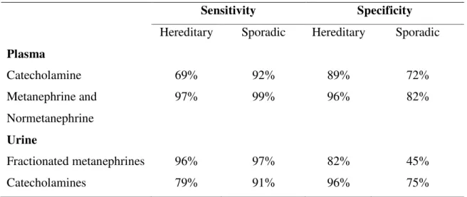
In the diagnosis of PHEO and PGL we can use the biochemical characteristics of the tumour, by measuring the levels of the secreted hormones and their major metabolites.
Plasma free and fractionated metanephrines are the first test to perform when PHEO/PGL is suspected. (Table 7)
27
Table 7. Diagnositic sensitivity of plasma and urinary catecholamines and their metabolites in hereditary and sporadic pheochromocytoma.
Sensitivity Specificity
Hereditary Sporadic Hereditary Sporadic Plasma
Catecholamine 69% 92% 89% 72%
Metanephrine and Normetanephrine
97% 99% 96% 82%
Urine
Fractionated metanephrines 96% 97% 82% 45%
Catecholamines 79% 91% 96% 75%
Based on Lenders JW, Pacak K, Walther MM, et al. Biochemical diagnosis of pheochromocytoma: which test is best? JAMA. 2002;287:1427–1434.
Urine metanephrines have superiority over free catecholamines and vanillylmandelic acid (the end product of catecholamine metabolism). However the diagnostic sensitivity of plasma free metanephrine and normetanephrine are superior 88 and their diagnostic accuracy has been confirmed. Unfortunately, determination of plasma metanephrines and normetanephrin or plasma free catecholamines are not routinely available in many countries.
Liquid chromatography followed by mass spectrometric or electrochemical detection methods are suggested to use for measuring metanephrines and catecholamines.
Patients/specimens should be referred to specialist centres.
Blood sampling for plasma metanephrines should be done after 30 minutes of supine rest.
89 This is usually hard to carry out at clinical centres but doing blood sampling without supine position will lead to an increase in false-positive results. In these cases it is recommended to measure urine fractionated metanephrines. However, false-positive results have a 19-21% rate in plasma free and urine fractionated metanephrines. 90
28
In 50% of the patients with pheochromocytoma, both normetanephrine and metanephrine are elevated; by at least 3 fold or more above the upper cut-off.
The clonidine test is recommended to distinguish the false-positive cases from the true- positive ones.
Some medications can interfere with these measurements or with the catecholamine disposition, not to mention physiological stress with severe conditions.
All positive cases should be followed up, and sometimes second, confirmatory determinations are required.
1.2.6.3. Imaging studies
Imaging studies are recommended to locate the tumour in patients with positive biochemical tests. However, only imaging studies can identify/locate the tumour in patients with biochemically negative results.
CT is the first choice imaging modality for the thorax, abdomen and pelvis, its sensitivity is 88-100%. 91 Tumours greater than 5mm can already be detected by CT.
MRI has a better sensitivity in patients with extra-adrenal, recurrent and metastatic tumours and head and neck paragangliomas. 91–93 MRI is preferred in children, pregnant women and has a sensitivity of almost 100%. Both methods are used for tumour staging as well. 91,94
123I-metaiodobenzylguanidine (123I-MIBG) scintigraphy is a functional imaging modality in metastatic cases and is used when radiotherapy with 131I-MIBG is planned.
In SDHx-related tumours 2-18F-fluoro-2-deoxy-D-glucose positron emission tomography (18F-FDG-PET) has superiority to other imaging techniques. 94
1.2.6.4. Molecular genetic testing
Due to the large number of genes responsible for the development of PHEO/PGL the genetic testing remains a diagnostic challenge. Since 1990, 14 different susceptibility genes have been reported. Both laboratory workload and cost of testing of all genes are still significant despite of the lower price of molecular biological reagents. Phenotype oriented
29
guidelines allow us some priorization in the order of genes tested but after a negative result the remaining genes should also be examined. Therefore, it would be ideal that after exclusion of some syndrome-associated genes based on the obvious phenotype features (i.e.
because of typical manifestation the NF1 gene is rarely tested) all of the remaining genes would be tested at the same time. Recent technical improvements in sequencing technology, the next generation sequencing (NGS) platforms - allow us to use whole exome or targeted resequencing of all these genes. 95 The usefulness of NGS has been demonstrated not only in resequencing of already known genes, but also in discoveries of novel genes associated with PHEO/PGL. Confirmation of results and a negative NGS result does not exclude the possibility of mutations especially the presence of large deletions.
Therefore, the gold standard methodology for identification of pathogenic mutation is the PCR amplification of the coding region of target genes followed by Sanger sequencing. For large deletion analysis multiple ligation probe amplification (MLPA) should be also performed. In addition, the Endocrine Society clinical practice guideline recommend the use of a clinical feature-driven diagnostic algorithm to establish the priorities for specific genetic testing in PHEO/PGL patients with suspected germline mutations delivered within the framework of health-care. 91,96
1.2.7. Treatment of pheochromocytoma and paraganglioma
Surgery is the definitive treatment of PHEO and PGL, if the tumour location allows resection. To prevent cardiovascular complications patients with hormonally functional PHEO/PGL should receive preoperative blockade, the first choice should be α-adrenergic receptor blockers. Calcium channel blockers are the most common add on drugs and β- adrenergic receptor blockers are used in co-administration to control tachycardia. These latter two drugs are not recommended to us in single medication. α-adrenergic receptor blocker treatment should be administered at least 7 days before surgery. 97
For surgery, recommendations suggest minimally invasive techniques. Laparoscopic adrenalectomy is the first choice, but invasive tumours or tumours with size over 6 cm are
30
recommended for open resection. Paragangliomas are suggest for open resection, although in some cases (e.g.: small, non-invasive, location) laparoscopic resection can be done.
After surgery patient personalized follow up is necessary depending on the genetic results.
In syndromic cases, where often bilateral tumours develop, a minimal invasive tumourectomy (adrenal sparing surgery) with left adrenal cortex tissue is advised. 98
31 2. OBJECTIVES
During my PhD training I aimed to collect and to summarize the evidence of the role of SDHx variants in the pathogenesis of PHEO/PGL. My specific aims were:
2.1. to evaluate the role of Mutations of SDHx genes in Hungarian patients with PHEO/PGL
• to determine the prevalence of germline mutations in the SDHx, SDHAF2, MAX and TMEM127 genes in Hungarian patients with apparently sporadic PHEO/PGL.
to describe the detailed phenotype of the first Hungarian case with SDHD gene mutation.
• to collect and to report the genotype-phenotype association in patients with PHEO/PG.
• to identify novel mutations among Hungarian patients with Pheo/PGL
2.2. to test whether polymorphisms of SDHx genes are phenotype modifiers in patients with MEN2A syndrome, therefore I aimed to
• to determine the prevalence of SDHx polymorphisms in patients with RET mutations (MEN2 patietns), in patients with sporadic medullary thyroid cancer (MTC), sporadic PHEO, healthy subjects
2.3. to identify the metabolic consequences of SDHx mutations/deletions in tumour tissues and cell lines. In order to fulfil this aim I aimed:
• to measure the levels of the two Krebs cycle metabolites, succinate and fumarate, in tumour tissue and in human plasma samples obtained from patients with Pheo/PGL
• to determine the succinate to fumarate ratio in mouse pheochromocytoma (MPC) and mouse tumour tissue (MTT) cells
• to test whether this difference propose the implementation of succinate/fumarate measurements in clinical diagnosis.
32 3. METHODS
3.1. Germline mutation prevalence in Hungarian patients with pheochromocytoma and/or paraganglioma
3.1.1. Patients
Our database containing the clinical and laboratory data of 129 patients diagnosed and followed up at the 2nd Department of Medicine, Faculty of Medicine, Semmelweis University with clinical diagnosis of PHEO/PGL between 1998 and 2014 was reviewed in order to select cases for comprehensive genetic testing. All patients underwent genetic counselling and written informed consent was obtained before genetic analysis.
Of these patients, the clinical diagnosis was confirmed by pathological examination of the surgically removed tumour tissues in 92 cases. Mutation screening of the RET and VHL genes identified 4 RET mutation carriers and 4 patients with germline VHL mutations. 99–101 In two cases the specific phenotype features indicated neurofibromatosis type 1. These patients were excluded from this current analysis and SDHB, SDHC, SDHD, SDHAF2, MAX and TMEM127 mutation analysis was performed in 82 cases. The main demographic and pathological data are summarized in Table 8.
33
Table 8. Main genotype-phenotype associations in Hungarian patients with PHEO/PGL.
PHEO – pheochromocytoma, PGL – paraganglioma, SDHD – Succinate dehydrogenase subunit D, SDHB - Succinate dehydrogenase subunit B, TMEM127 – transmembrane protein 127
3.1.2. The first Hungarian case with extra-adrenal pheochromocytoma associated with SDHD gene mutation
In 2002 it became possible to analyse SDHx gene mutations at the 2nd Department of Medicine, Semmelweis University. In the beginning of my PhD work I identified the first disease causing mutation of the SDHD gene in a patient with extra-adrenal pheochromocytoma. This cases represents the first genetically confirmed case of hereditary paraganglioma/pheochromocytoma syndrome due to disease-causing mutation of the SDHD gene in Hungary. With this first case we have the opportunity to demonstrate maternal imprinting and to overview the pathomechanism in hereditary syndrome caused by SDH gene mutation.
Cause of PHEO/PGL Age (years) Malignant/ recurrent Bilateral or multiple locations Genetic cause (n=11) 34,6 (19-51) 3/11 (27.2 %) 8/11 (72.7%)
SDHD (n=1) 32 0 1/1 (100 %)
SDHB (n=7) 31.4 (19-38) 3/7 (42.8%) 5/7 (71.4 %)
TMEM (n=3) 40 (22-51) 0/3 (0 %) 2/3 (66 %)
No genetic cause (n=71) 40,4 (13-78) 12/71 (16.9 %) 3/71 (4.2%) Total (n=82)
38,8 (13-78) 15/82 (18.3%) 11/82 (13.4%)
34
3.1.3. Genetic testing of the RET, VHL, SDHB, SDHC, SDHD, SDHAF2, MAX and TMEM127 Genes Using Sanger Sequencing
After genetic counselling and obtaining informed consent of all 82 patients, underwent genetic testing for the SDHB, SDHC, SDHD, SDHAF2, MAX and TMEM127 using conventional methods including PCR followed by Sanger sequencing. 99–101 Genomic DNA was extracted from peripheral blood using commercially available DNA extraction kits (DNA isolation from mammalian blood, Roche, or DNA isolation kit from blood, Qiagen LTD). Bidirectional DNA sequencing of all these genes and large deletion analysis of the SDHB, SDHC and SDHD genes were performed using multiplex ligation probe amplification. 101
35
3.2. The G12S polymorphism of the SDHD gene as a phenotype modifier in patients with MEN2A syndrome
3.2.1. Patients
Written informed consent was obtained from all patients and family members who participated in the study. Patients underwent a complete clinical examination, laboratory testing, including serum basal calcitonin measurement [hCalcitonin IRMA kit (Diagnostic Systems Laboratories, Inc., Budapest, Hungary), reference range: male, < 15 pg/ml, female,
< 10 pg/ml until December 2007; and Liaison (Diasorin SPA, Stillwater, MN, USA), reference value: male, < 18.9 pg/ml and female, < 6 pg/ml after January 2008], plasma parathyroid hormone (Elecsys; Roche Diagnostics, Basel, Switzerland), urinary catecholamine metabolites (high pressure liquid chromatography with electrochemical detection), and imaging studies, including cervical ultrasonography, thoracal and abdominal computed tomography (CT), and whole-body metaiodobenzylguanidine scintigraphy (MIBG).
3.2.1.1. Patients with MEN2 syndrome
In total, 77 patients with germline RET proto-oncogene mutations who were members of 21 unrelated families with MEN2 syndrome were identified by genetic screening at our centre.
Of the 77 patients, 55 had MEN2A (mean age at diagnosis: 33.4±17 years; range: 7–76 years), three had MEN2B (mean age at diagnosis: 15.6±5 years; range: 10–20 years), and 19 had FMTC (mean age at diagnosis: 23.7±16.8 years; range: 2–57 years). The presence of PHEO and MTC were confirmed by histological examination of surgically removed tumours. Total thyroidectomy was performed in all patients with germline RET mutation in the symptomatic group and was also offered to all individuals from the asymptomatic group.
36 3.2.1.2. Patients with sporadic MTC
The study included 47 unrelated patients with histologically confirmed MTC evaluated consecutively at the 2nd Department of Medicine, Faculty of Medicine, Semmelweis University between 1998 and 2010. There were 15 men (age, mean ± SD, 44.7±13.3; range:
28–82 years) and 32 women (age, mean ± SD, 47.7±12,3; range: 23–76 years).
Preoperative evaluation included medical history, physical examination, thyroid and abdominal ultrasonography, CT or magnetic resonance imaging (MRI), MIBG- scintigraphy, routine biochemical testing, serum calcitonin measurements, and mutation analysis of exons 10–14 of the RET gene.
3.2.1.3. Patients with apparently sporadic PHEO
The study included 48 unrelated patients with histologically confirmed sporadic adrenal pheochromocytomas evaluated consecutively at the 2nd Department of Medicine, Faculty of Medicine, Semmelweis University between 1998 and 2010. There were 16 men (age, mean
± SD, 36±14; range: 13–66 years) and 32 women (age, mean ± S.D, 42±14; range: 19–64 years). Pre-operative evaluation included medical history, physical examination, abdominal ultrasonography, CT or MRI, MIBG-scintigraphy, routine biochemical testing, and 24 h urinary catecholamine metabolite determination. The mutation analysis of RET exons 10–
14 and the entire VHL, SDHB, and SDHD genes revealed no disease-causing mutations.
Patients with confirmed VHL (five patients), SDHB (one patient), or SDHD (one patient) mutations were excluded from the study. Five patients were initially thought to have sporadic pheochromocytoma, but were later identified as having a disease-causing RET mutation and were included in the study as RET mutation carriers. MTC, either by elevated serum calcitonin or by postoperative histology, had been diagnosed in all of these patients.
Genetic counselling and genetic screening for all first-degree relatives have been offered.
37
3.2.2. Germline mutation screening of the RET, VHL, SDHB, and SDHD genes
Genomic DNA was isolated from peripheral blood using the Roche DNA Isolation Kit (Roche Diagnostics GmbH, Mannheim, Germany) and QIAamp DNA Blood Mini Kit (Qiagen Inc., Valencia, CA, USA) in accordance with the manufacturers’ instructions. RET proto-oncogene mutations were detected by direct sequencing as previously reported 99102. Mutation analysis of the VHL, SDHB, and SDHD genes in cases of apparently sporadic PHEO were performed by direct sequencing of the entire coding region of the VHL, SDHB, and SDHD genes, as previously reported 99,101 , and large deletion analysis of the VHL, SDHB, SDHC, and SDHD genes performed using multiplex ligation probe amplification.
101
3.2.3. Restriction fragment length polymorphism (RFLP) analysis for identification the G12S polymorphism of the SDHD gene
The nucleotide change of G to A, which corresponds to the G12S polymorphism, results in the preservation of the BanI restriction cleavage site. Therefore, digestion with the BanI restriction enzyme (New England BioLabs Inc., Ipswich, MA, USA) for 90 min at 37°C was performed after polymerase chain reaction (PCR) amplification of exon 1 of the SDHD gene for genotyping of RET mutation carriers, sporadic MTC patients, and 100 controls (Figure 4). Samples from patients with positive results were examined by direct DNA sequencing. The results obtained with both methods were the same in all cases.
38 Figure 4. Gel electrophoresis and chromatograms.
Panel A: Gel electrophoresis of PCR fragments after digestion with BanI for identification of the G12S polymorphism of the SDHD gene by RFLP (L = DNA ladder; C+ = positive control; heterozygote for G12S; C- = negative control; G12 normal, P1–P5 = patients.
Panel B: Chromatograms of exon 1 of the SDHD gene showing the wild type and the heterozygote form of the G12S (GGT12AGT) polymorphism.
3.2.4. Statistical analysis
Baseline characteristics were compared using the chi-squared test or Fisher’s exact test for qualitative variables, and Student’s t test or Mann-Whitney’s U test for quantitative variables. The statistical package SPSS 15.0 (SPSS Inc., Chicago, IL, USA) was used and p<0.05 was considered statistically significant.
39
3.3. Biochemical consequences of SDHx mutations, succinate to fumarate ratio in SDHB/D associated paragangliomas
3.3.1. Materials
3.3.1.1. Human PHEOs/PGLs
PHEO/PGL tissue samples were collected at the National Institutes of Health (NIH) under clinical protocol 00-CH-0093, approved by the Institutional Review Board of the Eunice Kennedy Shriver National Institute of Child Health and Human Development (NICHD).
Tissue samples were frozen in liquid nitrogen shortly after surgical removal of a tumour.
All patients underwent genetic testing for known PHEO/PGL susceptibility genes except SDHAF2 and neurofibromatosis 1 (NF1); the diagnosis of the latter was based on clinical grounds.
In the present study, we included four groups of tumours: SDHB (10 PGLs), SDHD (5 PGLs), apparently sporadic (6 PHEOs, 4 PGLs), and NF1 (2 PHEOs). NF1-related PHEOs were included because of the genetic background of the mouse PHEO (MPC) and mouse tumour tissue (MTT) cells used in the in vitro experiments. A detailed summary of clinical and patient characteristics is described in Table 1.
3.3.1.2. Plasma samples
Patient blood samples were collected at the NIH under clinical protocol (00-CH-0093), approved by the Institutional Review Board of the NICHD. Blood samples were centrifuged at 3500 rpm at 4°C for 20 minutes, and the plasma was stored at - 80°C until further processing. In the present study we selected three samples for plasma measurements from each group (SDHB, SDHD, and apparently sporadic PHEOs/PGLs).
40 3.3.1.3. MPC and MTT cells
The MPC and MTT cell lines were used as described previously. 103,104 MTT cells are known to be more aggressive than MPC cells and show aggressiveness similar to human disease. 104 MPC and MTT cells were maintained at 21%O2, 5%CO2, 37°C in DMEM (4.5g/L D-glucose, L-glutamine, 110 mg/L sodium pyruvate; Life Technologies Corporation) supplemented with 10% fetal bovine serum (Gibco), 5%heat-inactivated horse serum (Gibco), and Anti-Anti 100_ (Penicillin/Streptomycin, Amphotericin B; Gibco). The medium was changed every 2 to 3 days and cells were passaged when 80%–90%
confluence was reached. 103
3.3.2. Silencing of SDHB in MPC and MTT cells
Early passages of MPC and MTT cells were transduced with lentiviral particles carrying either shRNA targeted against mSDHB or control shRNA (Thermo Fisher Scientific Inc).
The cells were transduced at multiplicity of infection = 1 and maintained according to the manufacturer’s instructions. Medium containing 1 µg/mL puromycin was used to select positive cells.
For the metabolic analysis we seeded 1.5 x 10⁶ cells on a 6-cm dish. After 24 hours, cells were harvested in 1.5 mL PBS and snap-frozen in liquid nitrogen.
3.3.3. Western blotting
To evaluate the degree of SDHB silencing in MPC and MTT cells, Western blot analysis was performed. On 35-mm dishes, 1.0 x 10⁶ cells were plated. The following day, they were lysed and the protein concentration was determined using the Micro BCA Protein Assay Kit (Thermo Fisher Scientific). Thirty micrograms of total protein per well was loaded into a Criterion TGX
41
Precast Gel, 4%–12% (Bio-Rad Laboratories) and transferred to an Immobilon-P membrane (EMD Millipore Corporation). The membrane was blocked in 5% nonfat dry milk in 0.1% Tween in PBS for 1 hour. It was incubated with anti-SDHB antibody (Sigma- Aldrich Co) for 1 hour. β-Actin (Cell Signaling Technology Inc.) was used as a loading control. Proteins were detected using SuperSignal West Pico Chemiluminescent Substrate (Thermo Fisher Scientific), and blots were exposed to High Performance Chemiluminescence film (GE Healthcare) and analyzed with Image J 1.42q software (NIH).
3.3.4. Metabolic measurements
Procedures for the determination of succinate and fumarate have been described elsewhere
105 and are briefly related here. The organic acids were analyzed as their tertiary butyl dimethylsilyl ether derivatives using gas chromatography-mass spectrometry (GC-MS) in the electron impact mode and quantified using the 13C-labeled internal standards for each analyte. The N-methyl-N-(tert-butylmethylsilyl) trifluoroacetamide with 1% tert- butyldimethylchlorosilane reagent was purchased from Pierce Chemical Co, and the 13C- labeled organic acids were procured from Sigma Chemical Co. Samples for GC-MS analysis were prepared by perchloric acid extraction as previously described. 105 The 13C- labeled internal standards were added in two-fold excess of the concentrations of the individual analytes in the tumour tissue to the neutralized PCA extracts. Extracts (5.0 µL) were evaporated under a stream of nitrogen to dryness and were immediately reacted with 5 µL of the sylilating reagent in 15 µL of acetonitrile in 1.5 mL screw capped glass vials and heated to 60°C for 5 minutes. Samples were analyzed on an Agilent 5973 quadrupole GC- MS (Agilent). One microliter of the sample solution was injected onto a 250-µm x 30 m capillary DB-1 (Agilent) column in the splitless injection mode. The mass spectrometer was operated in the electron impact mode (70 eV) and the quadrupole mass analyzer scanned for ions, which corresponded to a loss of 15 mass units (–CH3) from the molecular
42
ion and the base peak of each analyte and its corresponding 13C-labeled internal standard using selected ion monitoring.
3.3.5. Statistical analysis
Data are expressed as means ± SD with coefficient of variation (COV). Student’s t test was applied to determine the significance between the groups, with a P value of less than .05 considered significant. Grubbs’ test was performed using GraphPad to determine whether there were any outliers among the values. The NF1 PHEOs were not statistically analyzed due to the small sample size.
43 4. RESULTS
4.1. Germline mutation prevalence in Hungarian patients with PHEO and/or PGL
Eleven patients were identified to carry mutation in one of the PHEO/PGL associated genes. Together with our previous data demonstrating mutations in RET (n=4) and VHL (n=4) genes, the prevalence of germline disease-causing mutations in Hungarian patients with apparently sporadic, non-syndromic PHEO/PGL was 21.1% (19/90; 11 of 82 cases, 4 RET and 4 VHL mutation carriers). For mutation detection bilateral involvement and multiple tumours had the most positive predictive value. The prevalence of bilateral tumours was significantly higher in mutation carriers than in genetically negative cases (8 of 11, 72.8% vs. 3 of 71, 2.1%; p<0.001).
The mutation spectrum observed in our patients was heterogeneous, the most frequent mutations were detected in the SDHB gene (7 different of which 4 were novel mutations), Three patients had TMEM127 mutations (two novel) and one had mutation in the SDHD gene (Table 9). The chromatograms of all novel mutations identified are presented in Figure 5. All novel SDHB mutation have been submitted to TCA Mutation Database and the new TMEM127 mutations to dbSNP database (http://chromium.lovd.nl /LOVD2/SDH/variants.php?select_db=SDHB&action= view&view=0000838, http://chromium.lovd.nl/LOVD2/SDH/variants.php?select_db=SDHB&action=view& view
=0000839; http://chromium.lovd.nl/LOVD2/SDH/variants.php?select_ db=SDHB
&action=view&view=0000840; http://chromium.lovd.nl/LOVD2/SDH/ variants.php?
select_db=SDHB& action= view&view=0000841).
No mutations in SDHC, SDHAF2, and MAX were identified in our patients.
44 4.1.1. Genotype-phenotype associations
Comparison of the main demographic and clinical data of the genetically positive and negative cases indicated that genetically positive patients were younger, their PHEO/PGL was more frequently malignant, and 72% of cases had bilateral or multiple tumours (Table 8). As expected the malignancy was the highest (3 out of 7 cases) in patients with SDHB mutations. Two patients with mutations SDHB:c758G>A -Cys253Tyr- and the novel SDHB: c.586T>G -Cys196Gly- were lost because of metastatic disease by the age of 35 years. In these patients multiple metastases in bone and liver were observed. In the third case with malignant PGL the novel SDHB: c728G>A Cys243Tyr mutation was identified.
In this patient an intraabdominal PGL with multiple bone metastases was diagnosed.
Another important finding was that the SDHB associated tumours were mainly intraabdominal PGLs (6 out of the 7 cases). In one case with the novel SDHB c607G>T Gly203Stop mutation pheochromocytoma and renal cell carcinoma with oncocytic feature was detected at age of 19 years. The solid architecture, cytoplasmic inclusions of flocculent material and intratumoural mast cells as the main characteristics for SDHB associated renal cell carcinomas could be identified (Figure 6).
Head-neck PGLs were detected in a patient harbouring the SDHB: c286+1G/A mutation, and in a patient with SDHD c.147-148 insA frameshift mutation. In the latter case an intraabdominal PGL was also removed. After 4-8 years follow-up no malignancy was observed in these cases.
TMEM127 mutations were detected in three patients. Two of them had PHEO (one bilateral) while in the third patient with the novel mutation (TMEM127: c467T>A, - Leu155Stop) PHEO and PGL of the head-neck region was also observed. These tumours showed no malignancy. The youngest patient harbouring TMEM127 associated tumour was 22 years old.
45
Table 9. Phenotype characteristics of Hungarian patients with PHEO/PGL.
Case Age Manifestation Gene/Mutation
1 33 Paraganglioma (intrabdominal+head/neck, malignant)
SDHB:c.758G>A Cys253Tyr 2 32 Paraganglioma (intrabdominal+head/neck,
malignant)
SDHB: c.586T>G Cys196Gly * 3 31 Paraganglioma (intrabdominal+head/neck) SDHB: c.586T>C
Cys196Arg*
4 38 Paraganglioma intraabdominal SDHB: c649C>T Arg217Cys 5 19 Pheochromocytoma + renal cell carcinoma SDHB: c.607G>T
Gly203Stop*
6 37 Paraganglioma (head/neck) SDHB: c.286+1G/A,
7 30 Paraganglioma (intraabdominal multiple, malignant)
SDHB: c.728G>A Cys243Tyr*
8 32 Paraganglioma (intrabdominal+head/neck) SDHD: c.147-148 insA 9 51 Pheochromocytoma (bilateral)
Paraganglioma (intraabdominal and head/neck)
TMEM127: c.464T>A Leu155Stop*
10 22 Pheochromocytoma unilateral TMEM127: c419G>A Cys140Tyr
11 47 Pheochromocytoma bilateral TMEM127: c.572delC
*: mutations marked are novel mutations; SDHB (ENSG00000117118); TMEM127 (ENST00000258439). All novel SDHB mutation have been submitted to TCA Mutation Database and the new TMEM127 mutations to dbSNP database.
(http://chromium.lovd.nl/LOVD2/SDH/variants.php?select_db=SDHB&action=view&view
=0000838,
http://chromium.lovd.nl/LOVD2/SDH/variants.php?select_db=SDHB&action=view&view
=0000839,
http://chromium.lovd.nl/LOVD2/SDH/variants.php?select_db=SDHB&action=view&view
=0000840
http://chromium.lovd.nl/LOVD2/SDH/variants.php?select_db=SDHB&action=view&view
=0000841)
SDHB – succinate dehydrogenase subunit B, SDHD – succinate dehydrogenase subunit D, TMEM127 – transmembrane protein 127
46
Figure 5. Results of Sanger sequencing and chromatograms of novel germline variants identified in 6 patients with PHEO/PGL.
SDHB - succinate dehydrogenase subunit B, TMEM127 – transmembrane protein 127, T – thymine, C – cytosine, G – guanine, A – adenine,
47
Figure 6. Immunohistochemical labeling of tumours associated with novel SDHB mutations. Both PGLs and renal cell carcinoma with oncocytic feature associated with SDHB mutations showed no SDHB immunohistochemical staining.
Panel A: Intraabdominal PGL associated with SDHB: c.586 T > G (Cys196Gly), positive control: adrenocortical cells; Panel B: paraganglioma associated with the SDHB: c.728G >
A (Cys243Tyr) mutation, positive control: endothelial cells; Panel C: Renal cell cancer associated with the SDHB: c.607G > T (Gly203Stop) mutation. Panel D: Entrapped non- neoplastic renal tubules showed positive immunohistochemical labeling for SDHB.
PGL – paraganglioma, SDHB – succinate dehydrogenase subunit B