research article
Clinical value of whole-body magnetic resonance imaging in health screening of general adult population
David Laszlo Tarnoki1,2,* Adam Domonkos Tarnoki1,2,* Antje Richter1, Kinga Karlinger2, Viktor Berczi2, Dirk Pickuth1
1 Department of Diagnostic and Interventional Radiology, Caritasklinikum Saarbrücken St. Theresia, Academic Teaching Hospital of Saarland University, Saarbrücken, Germany
2 Department of Radiology and Oncotherapy, Semmelweis University School of Medicine, Budapest, Hungary
Radiol Oncol 2015; 49(1): 10-16.
Received 12 December 2013 Accepted 18 June 2014
Correspondence to: David Laszlo Tarnoki, M.D., Ph.D., Department of Radiology and Oncotherapy, Semmelweis University, Budapest, Hungary, 78/a Ulloi street, Budapest 1082, Hungary. E-mail: tarnoki4@gmail.com
Disclosure: No potential conflicts of interest were disclosed.
* David Laszlo Tarnoki and Adam Domonkos Tarnoki contributed equally to this work.
Background. Whole-body magnetic resonance imaging (WB-MRI) and angiography (WB-MRA) has become in- creasingly popular in population-based research. We evaluated retrospectively the frequency of potentially relevant incidental findings throughout the body.
Materials and methods. 22 highly health-conscious managers (18 men, mean age 47±9 years) underwent WB- MRI and WB-MRA between March 2012 and September 2013 on a Discovery MR750w wide bore 3 Tesla device (GE Healthcare) using T1 weighted, short tau inversion recovery (STIR) and diffusion weighted imaging (DWI) acquisitions according to a standardized protocol.
Results. A suspicious (pararectal) malignancy was detected in one patient which was confirmed by an endorectal sonography. Incidental findings were described in 20 subjects, including hydrocele (11 patients), benign bony lesion (7 patients) and non-specific lymph nodes (5 patients). Further investigations were recommended in 68% (ultrasound:
36%, computed tomography: 28%, mammography: 9%, additional MRI: 9%). WB-MRA were negative in 16 subjects.
Vascular normal variations were reported in 23%, and a 40% left proximal common carotid artery stenosis were de- scribed in one subject.
Conclusions. WB-MRI and MRA lead to the detection of clinically relevant diseases and unexpected findings in a cohort of healthy adults that require further imaging or surveillance in 68%. WB-MR imaging may play a paramount role in health screening, especially in the future generation of (epi)genetic based screening of malignant and athero- sclerotic disorders. Our study is the first which involved a highly selected patient group using a high field 3-T wide bore magnet system with T1, STIR, MRA and whole-body DWI acquisitions as well.
Key words: angiography; incidentaloma; atherosclerosis; high field magnet; diffusion weighted imaging
Introduction
Whole-body magnetic resonance imaging (WB- MRI) has become increasingly popular in the recent decade due to its high soft tissue spatial resolution, multiplanarity, lack of ionising radiation, low inci- dence of nephrotoxicity caused by contrast agents, as well as high sensitivity and specificity in the de-
tection of vascular and malignant diseases.1,2 Due to financial reasons and limited availability, WB- MRI enables an early diagnosis mainly in defined groups of subjects who do not show symptoms yet.
The most common diseases in elderly population include malignant tumours and cardiovascular diseases.3 WB-MRI is capable of detecting a wide range of malignant diseases; such as bronchial car-
cinoma, hepatic malignancies, renal carcinoma, colonic cancer, lymphoma, and also rare malignan- cies such as bone or soft tissue tumours.4
With wider availability of WB-MRI, increased number of incidental findings of potential clinical relevance (36%) have been reported.5 Only a lim- ited number of studies on screening with 1.5-T WB- MRI is available in the literature, often restricted to a single organ system.5 However, the prevalence of incidental findings has not yet been described on a 3-Tesla (T) wide bore device. Furthermore, no screening studies including both whole-body diffusion weighted imaging (DWI) and WB-MRA have been performed.
Our aim was to retrospectively analyze the fre- quencies of potentially relevant incidental findings throughout the body, especially in view of those that require further medical evaluation.
Materials and methods
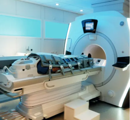
We carried out a retrospective analysis of healthy adults (mainly managers, lawyers, accountants, chief executive officers, company directors) with ex- treme health consciousness who underwent whole- body MRI and MRA at the Institute of Diagnostic and Interventional Radiology, Caritasklinikum Saarbrücken St. Theresia, Germany between March 2012 and September 2013. Subjects were referred to WB-MRI scan by a family doctor, their company or were self referrals. The healthy adults completed a comprehensive questionnaire including their cur- rent symptoms, previous clinical findings, opera- tions, and risk factors. MR studies were acquired on a Discovery MR750w 3 Tesla wide bore device (General Electric Healthcare, GE, Milwaukee, USA; 70 cm wide bore magnet) using T1 weighted (fast Spin Echo technique with a slice thickness of 5 mm), short tau inversion recovery (STIR), and DWI sequences according to a standardized pro- tocol (Figure 1, 2). Depending upon the height of the patient, 6 or 7 slabs were acquired in a slab- by-slab-technique with no continuous table move- ment. The WB-MRI protocol was identical for all participants and included a plain WB-MRI and de- tailed examination of head, neck, chest, abdomen, pelvis, spine and extremities. A rolling platform with extended field of view allowed whole-body examinations with a table range of more than 200 cm, several dozen simultaneous receiver channels, and multiple plugs for attaching several RF coils concurrently enabling the individual to be covered with coils from “head to toe”. The high number of
FIGURE 1. Whole-body MRI set-up of coils (head, chest, abdomen and extremity coils) in a wide bore magnet device.
FIGURE 2. A-C Whole- body MRI acquired on a dedicated whole- body MRI system and matrix coils in asymptomatic, smoker male 46-year-old patient, coronal (A) T1, (B) STIR and (C) DWI images.
A B
C
coils allowed for parallel imaging which speeded up the data acquisition.
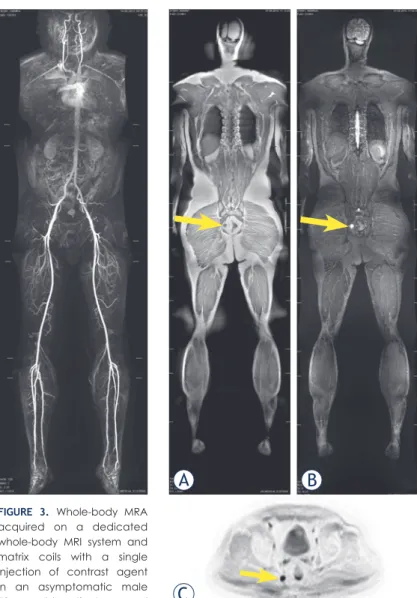
MR angiography (using 3D technique) was per- formed by the administration of 0.45 ml/kg body weight gadolinium contrast agent (0.5 mmol/ml gadoterate meglumine, Dotarem, Guerbet, Roissy, France), and was automatically injected at a flow rate of 1.2 ml/s in the first and 0.6 ml/s in the second phase (Figure 3). The patient was placed in the su- pine position, and the phases were acquired using respiratory gating.
Findings and anatomical variants were docu- mented in a standardized reading protocol. Picture archiving and communication system (AGFA IMPAX and KIS-RIS ORBIS, AGFA Healthcare, Mortsel, Belgium) were used for image storing.
In order to ensure the best possible quality and to minimize the inter-reader variability, three readers reported the whole set of images: first-line reading was performed by a resident in radiology (2-4 years’
experience), followed by two senior radiologists in- cluding the head of the department. The three read- ers evaluated the studies independently from each other. Then the results were discussed in consensus.
Finally, opinion, diagnoses/differential diagnoses and recommendation were evaluated together. The evaluation of images were carried out retrospective- ly by reviewing the reports and images of the pa- tients. Findings were classified as normal, insignifi- cant (abnormalities without well-defined diagnostic and therapeutic consequences according to existing guidelines and best practice recommendations), potentially significant (abnormalities potentially needing further medical evaluation or follow-up), and significant (findings that require further medi- cal evaluation and immediate referral). Statistical analysis was performed by Microsoft Excel. The in- vestigators followed the Helsinki Declarations and the European Council Convention on Protection of Human Rights in Bio-Medicine.
Results
Subject characteristics
Twenty-two healthy adults (18 men, age 47±9 years, mean±standard deviation) who underwent whole- body MRI and MRA imaging between March 2012 and September 2013 were included in the study. The WB-MR scans were analyzed retrospectively. The mean body mass index of the subjects was 25.2 kg/
m2. Fourteen subjects were completely asymptomat- ic. Nine subjects had a history of allergy (e.g., drug, animals, pollens). Nineteen subjects were never smokers, two reported a previous smoking history and one was active smoker. Subjects reported never, occasional, and regular sport activity in 5%, 10%
and 85%, respectively. Four-fifth of the subjects had current symptoms, previous symptoms/surgeries.
WB-MRI findings
A suspicious (pararectal) malignancy was detected in one patient (Figure 4). Two patients had nega- tive MR reports, whereas incidental findings were
FIGURE 3. Whole-body MRA acquired on a dedicated whole-body MRI system and matrix coils with a single injection of contrast agent in an asymptomatic male 52-year-old patient, coronal
reconstruction, pasted image. FIGURE 4. A-C Whole-body MR image of a 52-year- old male patient with history of sleep disorder, night sweats, hypercholesterinaemia and previous smoking. Note the right pararectal 16x14 mm mass (arrows) on the coronal (A) T1 (hypointense signal) and (B) STIR images (hyperintense signal); (C) diffusion weighted (DWI) axial image indicating a restricted diffusion. Inverted DWI image is similar to a PET image.
A B
C
described in 20 subjects. The findings are shown in Figure 5. Hydrocele was the most common inci- dental finding (11 patients; 11 of 18 men), followed by a benign bony lesion in 7 patients. Incidental findings would have needed diagnostic workup at an urologist (17 lesions), rheumatologist (15 le- sions), internist (13 lesions), otorhinolaryngologist (6 lesions), pulmonologist (6 lesions), surgeon (5 le- sions), gynecologist (4 lesions), and dermatologist (1 lesion). Further investigations were recommend- ed in 68% of subjects including eight sonographies (2/3 abdominal), five chest computed tomogra- phies (CT), one pelvic CT, two mammographies and two additional MRIs. In case of the suspicious pararectal malignancy, biopsy was recommended.
The patient had an endorectal sonography which confirmed the presence of a highly suspicious mass, probably a lymph node. A rectoscopy/colo- noscopy was planned, however the patient moved to another city and the further diagnostic/thera- peutic workup is unknown yet.
WB-MRA findings
WB-MRA was negative in 16 subjects. Vascular normal variations (e.g. irregular caliber of the ver- tebral artery, polar renal artery, stronger posterior communicant artery) were reported in five sub- jects, and a non-significant stenosis was described in one subject (Figure 6). A further subject had a possible right subclavian stenosis which might be confounded by motion artifact.
Discussion
To the best of our knowledge, this is the first study to investigate the clinical value of whole-body MRI and MRA in a highly selected group of extremely health conscious general adult population on a 3-T basis using a wide bore magnet including WB-DWI acquisitions. We demonstrated a potentially ma- lignant lesion detection rate of 4.5% in our cohort and a high number of incidental findings (91%) re- quiring further radiological investigations in 68%
of individuals. WB-MRA demonstrated normal vascular variations in 23% of subjects and a non- significant left proximal common carotid artery stenosis in 4.5%.
The patient, who had a suspicious (pararectal) malignant lesion underwent a pelvis CT, which found a 16 x 14 mm large mass with central dis- crete hypodensity in the right pararectal fat tissue and a thickened mesorectal fascia. Tumor markers
FIGURE 5. Prevalence of WB-MRI findings in all patients (%).
FIGURE 6. Coronal reconstruction of whole-body MRA in a 52-year-old male patient with history of appendectomy and previous smoking. Note the 2 mm long, approximately 40%
concentric stenosis of the left common carotid artery 3 cm distal to the aortic arch (arrow). Please note the left vertebral hypoplasia, kinking of the midcervical portion of the right internal carotid artery and the coiling of the distal cervical portion of the left internal cerebral artery.
were in the normal range, and colonoscopy report- ed sigma polyps without dysplasia (no suspicous colon carcinoma). The patient did not show up in the follow-ups later.
Only few studies performed on a lower mag- netic field (usually 1.5 T) have reported WB-MRI screening results in the past years.4,5 Gohde et al. re- ported lower rates of clinically significant inciden- tal findings compared to our results in general (e.g.
peripheral arterial stenoses in 2%, significant inci- dental findings 5–9%), and demonstrated only one malignancy (a renal cell carcinoma).6 Studies per- formed on ‘healthy’ employees reported only few unknown vascular pathologies, usually below 5%
(e.g. silent myocardial infarction, cerebral infarc- tions, significant carotid/renal artery stenoses).7-9 Some population-based studies indicated similar prevalence, for example the Uppsala PIVUS study (performed in 306 70-year-old men) demonstrated significant carotid/renal artery stenoses in 1.5- 1.8%, and abdominal aortic aneurysms in 2%.10 A study of 2536 healthy young men using axial brain MRI reported brain incidental findings with fre- quency of 0.47-1.7%.11 We suspect that the higher prevalence of incidental findings in our study can be referred to the higher magnetic field and the technical novelties in MRI, such as extended body coverage, a rolling platform with extended large field of view and a high number of simultaneous signal receiver channels, and multiple plugs for attaching several RF coils concurrently – allowing
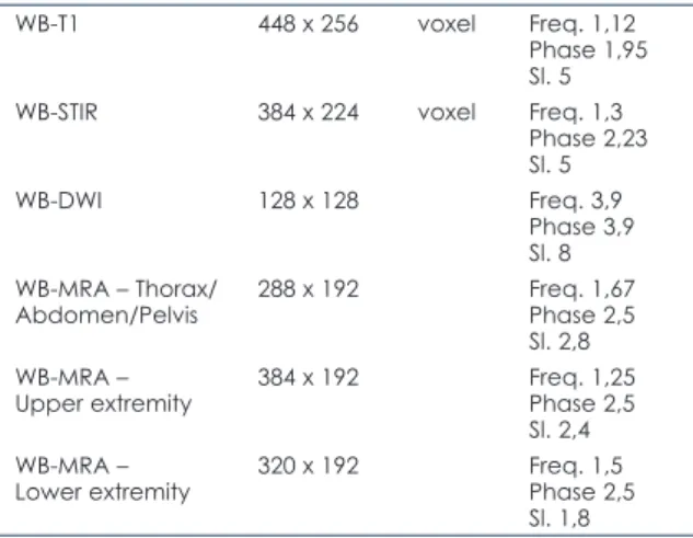
“parallel imaging”.1,12 In our study, homogeneity of the main magnetic field B0 of 3 T bore magnet with the largest bore diameter available on the market, higher signal to noise ratio, better image resolution, shorter acquisition time, and more pa- tient comfort can further increase and facilitate the specificity and patient compliance in contrast to the 1.5 T MRI systems. Spatial resolution data of our MR acquisitions are shown in Table 1.
In our cohort, hydrocele was the most common incidental finding (61% of men). The cause is un- known, however, it might be a marker of physical trauma, infection, or tumor as well. There are ap- proximately 26 million cases of hydrocele world- wide.13 Hydrocele is more common on the right side and it is usually bilateral in elderly patients.14
The second most common incidental finding was the benign bony lesion in 32% of the indi- viduals, most commonly bone islands (these le- sions appeared to be benign but were unspecific, maybe just inhomogeneous bone marrow). These small (majority of lesions measure from 0.1 to 2.0 cm in greatest diameter), asymptomatic lesions can
be found in most parts of the skeleton with a pref- erence for the pelvis, femur, ribs and other long bones (usually at the ends of tubular bones).15 Most of these bone islands do not require treatment after the diagnosis is established.
Due to atherosclerosis as the number one in morbidity and mortality in developed countries and its high prevalence, there is an increasing need to detect the most threatening manifestations of vascular disease well in advance.16 WB-MRA is a promising technique providing the depiction of the arterial system from “head to toe” (except the coro- naries) in less than 45 minutes, visualizing macro- scopic changes in the arterial system and poten- tial organ damage (e.g. cerebral microangiopathy, stroke, myocardial infarction) with high accuracy of up to 95% concerning relevant stenosis.1,17 In our sample, WB-MRA was negative in 73%, and vas- cular normal variations and a non-significant ste- nosis were described. Our findings are comparable with studies performed in asymptomatic patients for cardiovascular diseases where the prevalence for vascular (< 3 T) MR findings were relatively low.4,9,10 However, in high-risk groups, screening studies have revealed many previously unknown vascular pathologies, which in part had been over- seen by common clinical examinations and tests.4 Our most commonly reported vascular normal var- iation was the asymmetry of the vertebral arteries (VAs). The asymmetry of the VAs might be due to hypoplasia which is very common and can be iden- tified on MRI, its prevalence is unknown.18 A Swiss study reported that vertebral artery hypoplasia is more common on the right side.19 Hypertension or
TABLE 1. Spatial resolution data of our study acquisitions
WB-T1 448 x 256 voxel Freq. 1,12
Phase 1,95 Sl. 5
WB-STIR 384 x 224 voxel Freq. 1,3
Phase 2,23 Sl. 5
WB-DWI 128 x 128 Freq. 3,9
Phase 3,9 Sl. 8 WB-MRA – Thorax/
Abdomen/Pelvis 288 x 192 Freq. 1,67 Phase 2,5 Sl. 2,8 WB-MRA –
Upper extremity 384 x 192 Freq. 1,25 Phase 2,5 Sl. 2,4 WB-MRA –
Lower extremity 320 x 192 Freq. 1,5 Phase 2,5 Sl. 1,8
WB = Whole-body; STIR = Short TI Inversion Recovery; DWI = diffusion weighted imaging; MRA = magnetic resonance angiography, Freq.:
frequency, Sl.: slice thickness in mm
hyperlipidemia are hypothesized to be in the back- ground; furthermore, vertebral artery hypoplasia may contribute to a higher risk for posterior circu- lation stroke.19
WB-MRI can be performed by the application of various sequences. T1-weighted images after contrast agent application can depict lesions in pa- renchymal organs and bone and soft tissues due to higher spatial resolution.1,20 STIR can visualize vertebral metastases and bone marrow infiltration with high sensitivity.21 Due to these benefits, these sequences were also acquired in our study in ac- cordance with the literature. However, to the best of our knowledge, DWI has never been applied in a WB-MRI screening study. Advantage of adding a DWI sequence to this study has the depiction of areas of restricted diffusion which allows the better visualization of areas of high cellularity, i.e. malig- nancies.
The ideal screening technique must be both sen- sitive and specific, widely available, cost-effective, reader independent, and without harmful side ef- fects. The diagnostic test must be standardized im- plying a low number of false results.1 In addition, criteria for a screening programme (either Wilson and Jungner, or adapted WHO) should be met. MR is likely to meet these criteria especially in diseases which are ideal for screening, including colorec- tal cancer and cardiovascular disease, as demon- strated with examples in our study.1,22,23 MR angi- ography has also been shown to be equivalently effective in demonstrating vascular abnormalities compared to invasive techniques.1,24 In our study, further investigations were recommended in case of 15 subjects (68%), mainly abdominal sonogra- phies and chest CTs. The incidental findings were mainly related to the fields of urology, rheumatol- ogy and internal medicine in almost two-thirds of the cases. The corresponding screening costs are also determined by these indirect costs related to these subsequent and follow-up tests beyond the direct costs of the screening test itself.1
The strengths of our study include the highly selected patient group, the advantages of high field 3-T wide bore magnet system and the use of whole-body DWI acquisitions. Second, the investi- gated healthy adults represent a small proportion of the population at risk, and the prevalence of ma- lignancies and cardiovascular (atherosclerotic) le- sions are likely to occur in the higher-risk group with lower socioeconomic status. Therefore, these are individuals who are likely to undergo WB-MRI scans in the near future due to financial reasons, and our findings are highly relevant in this context.
However, the present study also has some limita- tions.
The major limitation of our study is the relative- ly low number of subjects, although it is compara- ble to other previous investigations.9 In addition, follow-up analysis of disclosed potentially relevant incidental findings is still incomplete.
In conclusion, our data suggest that 3 T wide bore WB-MRI, DWI and MRA of high diagnostic accuracy lead to the detection of clinically relevant diseases and many incidental findings in a cohort of healthy adults that require further imaging or surveillance in two-third of subjects. Furthermore, research involving large numbers of patients is required to determine the potential benefit or bur- den of communicating incidental findings to study volunteers. Our research was the first one which involved this highly selected patient group, using a high field 3 T wide bore magnet system with T1, STIR, whole-body DWI and MRA acquisitions. Our study was the first which added a DWI sequence to the WB-MRI screening protocol which might help the depiction of areas of malignancies.
Acknowledgments
BBraun Medical Ltd. provided travel support for authors ADT and DLT.
References
1. Ladd SC, Ladd ME. Perspectives for preventive screening with total body MRI. Eur Radiol 2007; 17: 2889-97.
2. Fenchel M, Requardt M, Tomaschko K, Kramer U, Stauder NI, Naegele T, et al. Whole-body MR angiography using a novel 32-receiving-channel magnetic resonance (MR) system with surface coil technology: first clinical experience. J Magn Reson Imaging 2005; 21: 596-603.
3. Sans S, Kesteloot H, Kromhout D. The burden of cardiovascular diseases mortality in Europe. Task Force of the European Society of Cardiology on Cardiovascular Mortality and Morbidity Statistics in Europe. Eur Heart J 1997; 18: 1231-48.
4. Ladd SC. Whole-body MRI as a screening tool? Eur J Radiol 2009; 70: 452- 62.
5. Hegenscheid K, Seipel R, Schmidt CO, Völzke H, Kühn JP, Biffar R, et al.
Potentially relevant incidental findings on research whole-body MRI in the general adult population: frequencies and management. Eur Radiol 2013;
23: 816-26.
6. Gohde SC, Goyen M, Forsting M, Debatin JF. Prevention without radiation - a strategy for comprehensive early detection using magnetic resonance tomography. Radiologe 2002; 42: 622-9.
7. Baumgart D, Egelhof T. Preventive whole-body screening encompassing modern imaging using magnetic resonance tomography. Herz 2007; 32:
387-94.
8. Goehde SC, Hunold P, Vogt FM, Ajaj W, Goyen M, Herborn CU, et al. Full-body cardiovascular and tumor MRI for early detection of disease: feasibility and initial experience in 298 subjects. AJR Am J Roentgenol 2005; 184: 598-611.
9. Kramer H, Schoenberg SO, Nikolaou K, Huber A, Struwe A, Winnik E, et al.
Cardiovascular screening with parallel imaging techniques and a whole- body MR imaging. Radiology 2005; 236: 300-10.
10. Hansen T, Wikström J, Johansson LO, Lind L, Ahlström H. The prevalence and quantification of atherosclerosis in an elderly population assessed by whole- body magnetic resonance angiography. Arterioscler Thromb Vasc Biol 2007;
27: 649-54.
11. Weber F, Knopf H. Incidental findings in magnetic resonance imaging of the brains of healthy young men. J Neurol Sci 2006; 240: 81-4.
12. Quick HH, Vogt FM, Maderwald S, Herborn CU, Bosk S, Göhde S, et al. High spatial resolution wholebody MR angiography featuring parallel imaging:
initial experience. Rofo 2004; 176: 163-9.
13. Michael E, Bundy DA, Grenfell BT. Re-assessing the global prevalence and distribution of lymphatic filariasis. Parasitology 1996; 112: 409-28.
14. Okorie CO, Pisters LL, Liu P. Longstanding hydrocele in adult Black Africans:
Is preoperative scrotal ultrasound justified? Niger Med J 2011; 52: 173-6.
15. Onitsuka H. Roentgenologic aspects of bone islands. Radiology 1977; 123:
607-12.
16. Diehm C, Kareem S, Lawall H. Epidemiology of peripheral arterial disease.
Vasa 2004; 33: 183-9.
17. Fenchel M, Scheule AM, Stauder NI, Kramer U, Tomaschko K, Nägele T, et al. Atherosclerotic disease: whole-body cardiovascular imaging with MR system with 32 receiver channels and total-body surface coil technology—
initial clinical results. Radiology 2006; 238: 280-91.
18. Lovrencic-Huzjan A, Demarin V, Rundek T, Vukovic V. Role of vertebral artery hypoplasia in migraine. Cephalagia 1998; 18: 684-6.
19. Peterson C, Phillips L, Linden A, Hsu W. Vertebral artery hypoplasia: preva- lence and reliability of identifying and grading its severity on magnetic reso- nance imaging scans. J Manipulative Physiol Ther 2010; 33: 207-11.
20. Lauenstein TC, Goehde SC, Herborn CU, Treder W, Ruehm SG, Debatin JF, et al. Three-dimensional volumetric interpolated breath-hold MR imaging for whole-body tumor staging in less than 15 minutes: a feasibility study. AJR Am J Roentgenol 2002; 179: 445-9.
21. Eustace S, Tello R, DeCarvalho V, Carey J, Wroblicka JT, Melhem ER, et al.
A comparison of whole-body turboSTIR MR imaging and planar 99mTc- methylene diphosphonate scintigraphy in the examination of patients with suspected skeletal metastases. AJR Am J Roentgenol 1997; 169: 1655-61.
22. Villavicencio RT, Rex DX. Colonic adenomas: prevalence and incidence rates, growth rates, and miss rates at colonoscopy. Semin Gastrointest Dis 2000;
11: 185-93.
23. Chambless L, Keil U, Dobson A, Mähönen M, Kuulasmaa K, Rajakangas AM, et al. Population versus clinical view of case fatality from acute coronary heart disease—results from the WHO Monica project 1985–1990.
Circulation 1997; 96: 3849-59.
24. Goyen M, Quick HH, Debatin JF, Ladd ME, Barkhausen J, Herborn CU, et al.
Whole body 3D MR angiography using a rolling table platform: initial clinical experience. Radiology 2002; 224: 270-7.