Phytochemistry 190 (2021) 112851
Available online 1 July 2021
0031-9422/© 2021 The Author(s). Published by Elsevier Ltd. This is an open access article under the CC BY license (http://creativecommons.org/licenses/by/4.0/).
The grass root endophytic fungus Flavomyces fulophazii: An abundant source of tetramic acid and chlorinated azaphilone derivatives
P ´ eter J ´ anos Berek-Nagy
a,b, Gerg ˝ o T ´ oth
c, Szilvia B osze ˝
b,d, Lilla Borb ´ ala Horv ´ ath
b,d, Andr ´ as Darcsi
e, S ´ andor Csíkos
a,b, D ´ aniel G. Knapp
a, G ´ abor M. Kov ´ acs
a, Imre Boldizs ´ ar
a,*aDepartment of Plant Anatomy, Institute of Biology, Eotv¨ ¨os Lor´and University, P´azm´any P´eter s´et´any 1/C, Budapest, 1117, Hungary
bNational Public Health Center, Albert Fl´ori´an út 2-6, Budapest, 1097, Hungary
cDepartment of Pharmaceutical Chemistry, Semmelweis University, H˝ogyes Endre u. 9, Budapest, 1092, Hungary
dResearch Group of Peptide Chemistry, E¨otv¨os Lor´and University, E¨otv¨os Lor´and Research Network (ELKH), P´azm´any P´eter s´etany 1/A, Budapest, 1117, Hungary ´
eNational Institute of Pharmacy and Nutrition, Zrínyi u. 3, Budapest, 1051, Hungary
A R T I C L E I N F O Keywords:
Flavomyces fulophazii Periconiaceae Endophytic fungus Phylogeny Vermelhotin Azaphilones Antiproliferative
A B S T R A C T
Fungal endophytes are remarkable sources of biologically active metabolites of ecological and pharmacological significance. In this study, fungal isolates producing yellow pigments and originating from grass roots, were identified as the recently described grass root colonizing dark septate endophyte (DSE), Flavomyces fulophazii (Periconiaceae, Pleosporales). While analyzing the metabolite composition of 17 isolates of this fungus, 11 previously undescribed compounds, including four tetramic acids (dihydroxyvermelhotin, hydroxyvermelhotin, methoxyvermelhotin, oxovermelhotin), and seven chlorinated azaphilones (flavochlorines A–G), together with the known tetramic acid vermelhotin, were tentatively identified by high performance liquid chromatography (HPLC)-tandem mass spectrometry (MS/MS). Among them, flavochlorine A, flavochlorine G, hydrox- yvermelhotin and vermelhotin could be isolated by preparative HPLC, thus their structures were also confirmed by nuclear magnetic resonance (NMR) spectroscopy. Vermelhotin was found to be the main compound, reaching its maximum level of 5.5 mg/g in the in vitro cultures of a selected F. fulophazii isolate. A significant amount of vermelhotin was isolated by preparative HPLC from these cultures (4.8 mg from 1.0 g lyophilized culture), confirming the practical utility of F. fulophazii in high-yield vermelhotin production. The main compounds of this endophyte expressed no activity in standardized plant bioassays (i.e., in the Lactuca sativa seed germination and Lemna minor growth tests). An antiproliferative study of the isolated compounds confirmed moderate activity of vermelhotin against a panel of twelve cancer cell lines, with IC50 ranges of 10.1–37.0 μM, without inhibiting the non-cancer Vero cells, suggesting its selectivity towards cancers.
1. Introduction
Endophytic fungi live inside of healthy plant organs (e.g., roots, stems, leaves), causing no visible symptoms. They are ecologically important and common species: all plants are considered to be potential hosts of various fungal endophytes (Saikkonen et al., 1998; Rodriguez et al., 2009; Porras-Alfaro and Bayman, 2011; Sieber and Grünig, 2013).
The root colonizing fungal endophytes, also called dark septate endo- phytes (DSE) (Rodriguez et al., 2009; Sieber and Grünig, 2013), are relatively frequent worldwide in grassland ecosystems, colonizing healthy roots of different grass species (Mandyam and Jumpponen, 2005; Knapp et al., 2012, 2019; Porras-Alfaro et al., 2008). A plethora of
root endophytes belong to the order Pleosporales, which is one of the most common ascomycetous orders comprising root-associated species in grassland ecosystems (Zhang et al., 2012; Jumpponen et al., 2017).
Secondary metabolites of several pleosporalean fungi have been studied to date (e.g., Yamada et al., 2007; Zhang et al., 2015; Kellogg and Raja 2017; Macia-Vicente et al., 2018); however, metabolite production of ´ numerous pleosporalean fungi is unknown.
Our previous studies, focusing on root endophytic fungal commu- nities of Hungarian and Mongolian grasslands (Knapp et al., 2012, 2015, 2019), confirmed the presence of a DSE fungus, which has recently been described as Flavomyces fulophazii and classified into the family Peri- coniaceae of the order Pleosporales (Knapp et al., 2015; Tanaka et al.,
* Corresponding author.
E-mail address: boldizsar.imre@ttk.elte.hu (I. Boldizs´ar).
Contents lists available at ScienceDirect
Phytochemistry
journal homepage: www.elsevier.com/locate/phytochem
https://doi.org/10.1016/j.phytochem.2021.112851 Received 27 May 2021; Accepted 17 June 2021
2015). When the species was formally described, the “flavo” referred to the remarkable yellow pigments of this DSE, secreted into the culture media (Knapp et al., 2015). Later we isolated eight further DSE strains from Hungarian grasslands, producing also yellow pigments.
Endophytic fungi are abundant sources of biologically active me- tabolites with potential ecological role (Schulz et al., 2002; Hardoim et al., 2015; Maci´a-Vicente et al., 2018). They can protect host plants against pathogenic fungi (Li and Strobel, 2001), parasitic nematodes (Schwarz et al., 2004) and herbivorous insects (Schardl et al., 2007), and can even affect the growth of the plants (Choi et al., 2004; Berthelot et al., 2016) and their seedlings (Rivero-Cruz et al., 2003; García-M´en- dez et al., 2016; Wang et al., 2020). The pharmacological significance (e.
g., antiproliferative properties) of the endophytic metabolites is also extensively studied (Zhang et al., 2006).
The aims of our research were (i) to identify the yellow pigment producing DSE isolates collected from grasslands, (ii) to determine the metabolites of different F. fulophazii isolates grown in vitro, (iii) to analyze tandem mass spectrometry profiles of compounds in order to determine characteristic fragment ions and ion transitions suitable for identification, (iv) to compare metabolite compositions of the different fungal isolates, (v) to isolate compounds, (vi) to study their effect on plants using the lettuce seed germination and Lemna-growth bioassays, and (vii) to evaluate their antiproliferative potential against a panel of 12 human cancer cell lines, cytotoxic effect on non-cancer Vero cells.
2. Results and discussion
2.1. Identification of fungal isolates and their metabolites
In this study, ten Hungarian and seven Mongolian isolates secreting yellow pigments, were investigated (Knapp et al., 2015, 2019) (Table 1).
Besides nine previously identified F. fulophazii strains, we carried out molecular taxonomic identification by sequencing the internal tran- scribed spacer (ITS) region, the universal fungal barcode marker (Schoch et al., 2012) in case of eight further isolates (Table 1). The ITS sequence of all the 17 isolates comprised in the present study were identical thus, confirming their identity as F. fulophazii. The relative frequent isolation of F. fulophazii shows that this DSE is a common fungus in grassland ecosystems. Our previous phylogenetic analyses demonstrated the position of F. fulophazii in the family Periconiaceae in the suborder Massarineae of Pleosporales (Knapp et al., 2015) and albeit the results of analyses of ITS as solely locus should cautiously be considered, our results were in concordance with previous findings
(Fig. 1). At least 12 families belong to Massarineae where Massarinaceae and Periconiaceae are unambiguously in sister position representing two well-supported taxa (Tanaka et al., 2015).
Isolates of F. fulophazii were analyzed by high performance liquid chromatography (HPLC) coupled to ultraviolet (UV) and high resolution tandem mass spectrometry (HR-MS/MS) detections for their metabolite identification. The culture extracts contained a main compound (Fig. 2, compound 5), which can be identified using the molecular formula C12H11O3N (Table 2) and UV spectrum (Supplementary Fig. S1) iden- tical to those of a known tetramic acid-type fungal pigment, vermelhotin (Hosoe et al., 2006). To confirm the structure of compound 5 as ver- melhotin, its isolated sample (a red solid) was analyzed also by NMR spectroscopy (Table 3, Supplementary Fig. S2). Our NMR data deter- mined in chloroform-d were comparable to those reported recently (Leyte-Lugo et al., 2012), confirming compound 5 as an equilibrium mixture of E- and Z-vermelhotin with an E:Z ratio of 4:3. The double set of 1H NMR signals characteristic of the E:Z equilibrium mixture could also be detected in DMSO d6 (Supplementary Figs. S2A, S2B). However, the 1H NMR spectrum in methanol-d4 (Supplementary Fig. S2C) shows broad peaks affected by the E:Z interconversions. The presence of these broad peaks may be the result of faster interconversions between the isomers in the protic solvent methanol than in the aprotic solvents chloroform and DMSO.
To date, four fungal isolates have been reported to produce ver- melhotin, of which two represent unknown species (Hosoe et al., 2006;
Kasettrathat et al., 2008; Leyte-Lugo et al., 2012; Kuhnert et al., 2014).
One of the known sources is Hypoxylon lechatii isolated from ascospores of fruiting body collected from wood of an unknown plant in French Guiana (Kuhnert et al., 2014). The other known source is the strain CRI247-01, which was referred as “unidentified pleosporalean fungus” or “Ascomycete sp.” (Kasettrathat et al., 2008). Based on our molecular taxonomic analysis of the ITS sequence of the strain CRI247-01 grouped into the Periconia clade, we might suspect that this fungus is conspecific with Periconia echinochloae (Fig. 1). Thus, F. fulophazii is not the only species producing vermelhotin in Periconiaceae family, suggesting that this pharmacologically important compound may have chemotaxo- nomic significance in this lineage. Significant cytotoxic, antiplasmodial (Kasettrathat et al., 2008), antimycobacterial (Ganihigama et al., 2015), calmodulin inhibitory (Leyte-Lugo et al., 2012) and anti-inflammatory (Pansanit et al., 2013) activities of vermelhotin were confirmed, high- lighting on the need to make it available for performing further bio- logical tests.
Four minor compounds of the extracts exhibit comparable UV Table 1
Details of Flavomyces fulophazii isolates included in this study.
Isolate No. in this study Other strain/isolate/culture names Collection area Collection date Host plant ITS GenBank accession No. Publication
HF-1 flavo_01 Füloph¨ ´aza, Hungary April 2014 Festuca vaginata MW438310 This study
HF-2 flavo_04 Füloph¨ ´aza, Hungary April 2014 Festuca vaginata MW438311 This study
HF-3 flavo_05 Füloph¨ ´aza, Hungary April 2014 Festuca vaginata MW438312 This study
HF-4 flavo_06 Füloph¨ ´aza, Hungary April 2014 Festuca vaginata MW438313 This study
HF-5 flavo_08 Füloph¨ ´aza, Hungary April 2014 Festuca vaginata MW438314 This study
HF-6 flavo_09 Füloph¨ ´aza, Hungary April 2014 Festuca vaginata MW438315 This study
HF-7 flavo_11 Füloph¨ ´aza, Hungary April 2014 Festuca vaginata MW438316 This study
HF-8 flavo_13 Füloph¨ ´aza, Hungary April 2014 Festuca vaginata MW438317 This study
HF-9 DSE8/143 =CBS 135664 Füloph¨ ´aza, Hungary July 2005 Festuca vaginata KP184000a Knapp et al. (2015) HF-10 DSE8/S =CBS 135761 (T) Füloph¨ ´aza, Hungary July 2012 Festuca vaginata KP184001b Knapp et al. (2015)
MF-1 MF03 Nalaikh, Mongolia October 2016 Stipa krylovii MN537657 Knapp et al. (2019)
MF-2 MF04 Nalaikh, Mongolia October 2016 Stipa krylovii MN537658 Knapp et al. (2019)
MF-3 MF05 Nalaikh, Mongolia October 2016 Stipa krylovii MN537659 Knapp et al. (2019)
MF-4 MF06 Nalaikh, Mongolia October 2016 Stipa krylovii MN537660 Knapp et al. (2019)
MF-5 MF07 Nalaikh, Mongolia October 2016 Stipa krylovii MN537661 Knapp et al. (2019)
MF-6 MF08 Nalaikh, Mongolia October 2016 Stipa krylovii MN537662 Knapp et al. (2019)
MF-7 MF09 =DSE8309 Nalaikh, Mongolia October 2016 Stipa krylovii MN537663c Knapp et al. (2019)
aSequences of further DNA loci of this strain are available: LSU (partial 28S large subunit of the nrRNA gene): KP184039; SSU (partial 18S small subunit of the nrRNA gene): KP184081; ACT (partial actin gene): KP184116; CAL (partial calmodulin gene): KP184159.
b Sequences of further DNA loci of this strain are available: LSU: KP184040; SSU: KP184082; ACT: KP184118; CAL: KP184158.
cSequences of further DNA loci of this strain are available: LSU: MN515261; TEF (translation elongation factor 1-α): MN535259. (T): ex-type culture.
spectra with absorption maxima between 410 nm and 416 nm (Sup- plementary Fig. S1, compounds 1–4). The molecular formulas of com- pounds 2 and 4 (C12H13O4N and C13H15O4N), assigned by HR-MS (Table 2), reveal a formal excess of a H2O molecule in compound 2 and that of a CH3OH molecule in compound 4 relative to vermelhotin (C12H11O3N). Furthermore, by mass fragmentation, neutral losses of water and methanol were observed from the protonated molecular ions of compounds 2 and 4, respectively, resulting in the formation of the ion m/z 218 consistent with the protonated vermelhotin (Supplementary Fig. S3, Table S1). Based on these MS data, compounds 2 and 4 can be considered as new natural vermelhotin derivatives, denominated hydroxyvermelhotin and methoxyvermelhotin (systematic names in Supplementary material). The mass fragment spectra of their protonated molecular ions (m/z 236.09, hydroxyvermelhotin and m/z 250.11, methoxyvermelhotin) equally contain a fragment ion m/z 192.07 (Supplementary Fig. S3, Table S1). Since this ion might be formed by the
loss of substituted terminal ethyl moieties of hydroxyvermelhotin and methoxyvermelhotin [C12–C13 ethyl with hydroxyl (Fig. 2C) or methoxyl groups (Fig. 2E)], the C-12 position of the hydroxyl and methoxyl groups in these molecules was tentatively confirmed. Among them hydroxyvermelhotin could be isolated in pure form as a red solid.
Hydroxyvermelhotin was analyzed by NMR in DMSO d6 and methanol- d4 (note: this compound could not be analyzed in chloroform due to its poor solubility) (Table 4, Supplementary Fig. S4). The NMR spectra of the isolated hydroxyvermelhotin and vermelhotin in these solvents are similar; however, there are specific differences (Tables 3 and 4, Sup- plementary Figs. S2, S4). Based on their NMR spectra in methanol-d4, instead of the olefinic protons in the side chain (protons on C-11 and C- 12) of vermelhotin, hydroxyvermelhotin contains an sp3 methylene (Table 4, δC: 41.4; δH: 2.85m, 2H). Hydroxyvermelhotin is also present in an equilibrium mixture of E:Z isomers (4:3, E:Z ratio) as the double set of
1H NMR signals was detected in DMSO d6 (Table 4, Supplementary Fig. 1. Maximum Likelihood (ML) phylogenetic tree of ITS sequences of representative species in Periconiaceae and Massarinaceae in the suborder Massarineae (Pleosporales). Highlighted sections indicate affiliations to families. Flavomyces fulophazii isolates and the vermelhotin producing CRI247-01 strain (see Kasettrathat et al., 2008) are shown in bold. ML bootstrap support values (≥70) are shown at branches. GenBank accession numbers of the sequences and strain numbers are shown before and after the species names, respectively. Three representative species of the family Lentitheciaceae served as multiple outgroups (highlighted with blue). The scale bar indicates 0.05 expected changes per site per branch. . (For interpretation of the references to color in this figure legend, the reader is referred to the Web version of this article.)
Fig. S4B). The circular dichroism (CD) spectrum of hydroxyvermelhotin, showing zero signal, confirmed this compound to be a racemic mixture.
Based on the molecular formulas of compounds 1 and 3 (C12H13O5N and
C12H11O4N, assigned by HR-MS, Table 2) and on their fragmentation behavior (Supplementary Fig. S3, Table S1) similar to hydrox- yvermelhotin and methoxyvermelhotin, these compounds were Fig. 2.A HPLC separation of the extract prepared from Flavomyces fulophazii culture sample HF-3A [full chromatogram A was recorded using UV detection (λ =280 nm), and trace chromatograms (B, C, D, E, F) were obtained by MS detection, monitoring the extracted ion current for m/z 252.1 (B), m/z 236.1 (C), m/z 234.1 (D), m/z 250.1 (E) and m/z 218.1 (F), corresponding to tetramic acids] and HR-MS spectra (B′, C′, D′, E′, F′) of tetramic acids 1 (dihydroxyvermelhotin), 2 (hydroxyvermelhotin), 3 (oxovermelhotin), 4 (methoxyvermelhotin) and 5 (vermelhotin), respectively, along with their chemical structures.
Table 2
High resolution mass-spectral (positive ion mode) data for compounds detected in Flavomyces fulophazii culture extracts.
Compound Formula Detected ion Detected formula Calculated m/z Found m/z diff (ppm)
No.a Name
1 10,11-dihydroxy-vermelhotin C12H13O5N [M+H]+ C12H14O5N 252.0866 252.0865 −0.472
[M+Na]+ C12H13O5NNa 274.0686 274.0684 −0.853
2 11-hydroxy-vermelhotin C12H13O4N [M+H]+ C12H14O4N 236.0917 236.0915 −0.908
[M+Na]+ C12H13O4NNa 258.0737 258.0735 −0.500
3 11-oxo-vermelhotin C12H11O4N [M+H]+ C12H12O4N 234.0761 234.0758 −1.129
[M+Na]+ C12H11O4NNa 256.0580 256.0577 −1.363
4 11-methoxy-vermelhotin C13H15O4N [M+H]+ C13H16O4N 250.1074 250.1072 −0.698
[M+Na]+ C13H15O4NNa 272.0893 272.0890 −1.136
5 vermelhotin C12H11O3N [M+H]+ C12H12O3N 218.0812 218.0809 −1.145
[M+Na]+ C12H11O3NNa 240.0631 240.0628 −1.351
6a flavochlorine E C16H18O5NCl [M+H]+ C16H19O5NCl35 340.0946 340.0944 −0.785
[M +H+2]+ C16H19O5NCl37 342.0917 342.0912 −1.335
6b flavochlorine F C16H18O5NCl [M+H]+ C16H19O5NCl35 340.0946 340.0944 −0.696
[M +H+2]+ C16H19O5NCl37 342.0917 342.0913 −1.160
7 flavochlorine A C15H18O3NCl [M+H]+ C15H19O3NCl35 296.1048 296.1045 −1.039
[M +H+2]+ C15H19O3NCl37 298.1018 298.1016 −0.763
8 flavochlorine B C13H14O2NCl [M+H]+ C13H15O2NCl35 252.0786 252.0783 −1.162
[M +H+2]+ C13H15O2NCl37 254.0756 254.0753 −1.310
9 flavochlorine C C16H20O3NCl [M+H]+ C16H21O3NCl35 310.1204 310.1202 −0.863
[M +H+2]+ C16H21O3NCl37 312.1175 312.1171 −1.178
10 flavochlorine G C18H22O4NCl [M+H]+ C18H23O4NCl35 352.1310 352.1307 −0.859
[M +H+2]+ C18H23O4NCl37 354.1281 354.1276 −1.418
11 flavochlorine D C13H13O3Cl [M+H]+ C13H14O3Cl35 253.0626 253.0622 −1.417
[M +H+2]+ C13H14O3Cl37 255.0596 255.0593 −1.444 aNumbers of compounds correspond to those in Figs. 2 and 3.
tentatively identified as further new natural vermelhotin derivatives, denominated dihydroxyvermelhotin and oxovermelhotin, respectively (systematic names in Supplementary material).
The intensive ratio 3:1 of isotope peaks [M +H]:[M +H +2] in the HR-MS spectra of compounds 6a, 6b, 7–11 supports the presence of one chlorine atom in their structures (Fig. 3). Among them compounds 7 and 10 could be isolated as yellow amorphous solids. The molecular ion peak of compound 7 in its HR-MS spectrum corresponds to the molec- ular formula C15H18O3NCl (Table 2). The 1H NMR spectrum of this compound in methanol-d4 indicated the presence of two aliphatic methyl groups at δ 1.11 (t, J =7.3 Hz) and δ 3.96 (s), among them the latter was assumed to be an O-methyl group (Table 5, Supplementary Fig. S5). Altogether four methylene protons (δ 1.81, 2.95, 3.91 and 4.43) were observed. Based on COSY correlation, the two methylene protons with higher chemical shifts (δ 3.91 and 4.43) are in vicinal positions.
Additionally, the molecule contains a propyl side chain (δ 1.11 (CH3), δ 1.81 (CH2) and δ 2.95 (CH2)). Three unsaturated proton singulets (δH: 8.79, 7.51 and 6.44) and six quaternary carbons (δC: 174.8, 157.9, 147.5, 146.9, 107.1, 106.6) indicate a highly conjugated, planar struc- ture. Moreover, signals of one chlorine bearing carbon atom at δ 106.8, one carbonyl at δ 174.8, one aromatic O-methyl at δ 157.8 and three sp2 carbons at δ 105.7, 114.02 and 140.8, together with a literature survey (McMullin et al., 2013) indicated a chlorinated, and nitrogenated aza- philone structure (Table 5, Supplementary Fig. S5).
Azaphilones consisting of a pyranoquinone core could react with primary amines via a Schiff base formation, resulting in the exchange of the pyran oxygen for nitrogen (Gao et al., 2013). Thus, azaphilones may also present as their nitrogenated counterparts. As a further chemical characteristic of azaphilones, a large-scale presence of their chlorinated analogues was determined. Azaphilones identified so far exhibit signif- icant cytotoxic and antibacterial activities (Gao et al., 2013).
Comprehensive one- and two-dimensional NMR analyses (COSY, HSQC and HMBC, Supplementary Fig. S5) confirmed the structure of compound 7 and designated the annelation at position 4a and 8a. The HMBC correlations from H-12 to C-8 and from H-4 to C-9 confirmed the positions of the O-methyl group and the propyl side chain at the C-8 and C-3, respectively. The HMBC correlation between H-1 and C-1’ indicated that the nitrogen occupies position 2 and bears a hydroxyl ethyl group.
Mass fragment spectra of the protonated molecular ion m/z 296.10 of compound 7 (Supplementary Table S2, Fig. S6) confirmed (i) the consecutive losses of –CH3, –CH2-CH3 and –CH2-CH2-CH3 radicals generated from the propyl side chain, after detachment of the hydroxyl ethyl group from the heterocyclic nitrogen (ions [M +H-a]+, [M +H- a-b]+, [M +H-a-c]+, [M + H-a-d]+in Fig. 4 and Supplementary Table S2), (ii) the formation of the bicyclic chlorinated backbone as a relative stable fragment ion ([g +H]+in Fig. 4 and Supplementary Table S2) and (iii) the neutral loss of a formyl chloride from the bicyclic chlorinated backbone (ion [g + H–CHClO]+ in the Supplementary Table 3
NMR spectroscopic data of E and Z diastereoisomers of vermelhotin (compound 5) in chloroform-d, DMSOd6 and methanol-d4.
No.a chloroform-d DMSOd6 methanol-d4b
δH (E-) δC (E-) δH (Z-) δC (Z-) δH (E-) δC (E-) δH (Z-) δC (Z-) δH δC
1 (N) 5.62, 1H, brs – 5.50, 1H, brs – 7.45 brs, 1H – 7.65 brs, 1H – – –
2 – 171.4 – 172.4 – 170.9 – 170.0 – 175.5
3 – 98.1 – 97.9 – 97.8 – 97.7 – 98.7
4 192.5 194.6 – 192.4 – 194.8 – 195.8
5 3.79, s, 2H 50.4 3.82, s, 2H 50.9 3.58, s, 2H 49.9 3.63, s, 2H 50.4 3.74, s, 2H 54.8
6 – 165.4 – 166.7 – 163.9 – 165.3 – 162.3
7 8.17, d (9.2), 1H 116.0 8.18, d (9.2), 1H 116.5 8.03, d (9.0), 1H 115.3 8.00, d (9.0), 1H 115.1 8.10, br, 1H 116.5 8 7.40, dd (9.2, 7.0),
1H 141.5 7.41, dd (9.2, 7.0),
1H 142.0 7.63, dd (9.0, 7.2),
1H 142.1 7.65, dd (9.0, 7.2),
1H 142.8 7.67 dd (9.4, 7.2),
1H 144.7
9 6.29, d (7.0), 1H 107.5 6.28, d (7.0), 1H 107.5 6.63, d (7.2), 1H 108.1 6.63 d, (7.2), 1H 108.0 6.60 d (7.2), 1H 109.9
10 – 158.7 – 158.8 – 157.3 – 157.4 – 160.8
11 6.17, dq (15.3, 1.5),
1H 122.1 6.16, dq (15.3, 1.5),
1H 122.1 6.38, dq (15.5, 1.5),
1H 122.7 6.38, dq (15.5, 1.5),
1H 122.8 6.35 dq (15.5, 1.5),
1H 123.5
12 7.42, dq (15.3, 7.1),
1H 138.6 7.39, dq (15.3, 7.1),
1H 139.3 7.18, dq (15.5, 7.0),
1H 136.1 7.17 dq (15.5, 7.0),
1H 136.5 7.35, br, 1H 139.5
13 2.01, dd (7.1, 1.5),
3H 19.0 1.99, dd (7.1, 1.5),
3H 18.9 1.97, dd (7.0, 1.5),
3H 18.3 1.95, dd (7.0, 1.5),
3H 18.4 1.99 dd (7.2, 1.5),
3H 18.8
aCorresponding numbered molecular structure is found in Fig. 2.
b Diastereoisomers E and Z vermelhotin could not be separately detected in methanol-d4.
Table 4
NMR spectroscopic data of E and Z diastereoisomers of 11-hydroxyvermelhotin (compound 2) in DMSOd6 and methanol-d4.
No.a DMSOd6 methanol-d4b
δH (E-) δC (E-) δH (Z-) δC (Z-) δH δC
1 (NH) 7.50, brs, 1H 7.60, brs, 1H – –
2 170.4 169.7 – 175.9
3 97.9 97.7 – 98.9
4 192.4 193.9 194.5
5 3.53, s, 2H 51.3 3.59, s, 2H 51.8 3.80, s, 2H 54.9
6 163.5 164.8 – 164.4
7 8.07, d, (9.4), 1H 115.1 8.05 d, (9.4), 1H 114.9 8.17, br, 1H 116.0
8 7.62, dd (9.4, 7.2),1H 142.0 7.65, dd (9.4, 7.2), 1H 142.5 7.67, dd (9.2, 7.0), 1H 144.7
9 6.64 d (7.2), 1H 109.0 6.62 d (7.2), 1H 108.7 6.68, d (7.0), 1H 110.1
10 – 157.6 – 157.5 – 160.9
11 2.74 m, 2H 40.4 2.69, m, 2H 39.9 2.85,m, 2H 41.4
12 4.19, m, 1H 61.3 4.14, m, 1H 60.7 4.29, m, 1H 61.7
13 1.14, d (6.0), 3H 21.9 1.13, d (6.0), 3H 21.8 1.25 d (6.0), 3H 22.4
aCorresponding numbered molecular structure is found in Fig. 2.
b Diastereoisomers E and Z hydroxyvermelhotin could not be separately detected in methanol-d4.
Table S2). Based on these results, a new natural chlorinated, and nitrogenated azaphilone derivative was identified, denominated flavo- chlorine A (systematic name in Supplementary material).
Mass fragment spectra, generated from the protonated molecular ions of the other chlorinated metabolites (6a, 6b, 8–11), show ions corresponding to the [g +H]+and [g +H–CHClO]+fragments of flavochlorine A (Fig. 4, Supplementary Tables S2, S3, and Fig. S6).
Consequently, all these chlorinated compounds were presumed to contain the same base skeleton (indicated by symbol g in Fig. 4).
Structures of the side chains, attached to the C-3 and N-2 positions of compounds 8, 9 and 11, could be also identified by interpreting their fragment ion spectra (Supplementary Table S2). Accordingly, com- pounds 8, 9 and 11 were tentatively identified as further new natural
azaphilone derivatives, denominated flavochlorine B, flavochlorine C and flavochlorine D, respectively (Fig. 3) (systematic names in Supple- mentary material). Flavochlorins A, B and C belong to the group of nitrogenated azaphilones, and accordingly they do not react with amines. Since non nitrogenated azaphilones like flavochlorine D can react with amino acids (Gao et al., 2013), the difference between the molecular formulas of compound 10 (C18H22O4NCl) and flavochlorine D (C13H13O3Cl) (Table 2) can be interpreted by a valine or a valine isomer incorporation into the flavochlorine D molecule, leading to the forma- tion of compound 10. The NMR spectra of this isolated compound (Table 5, Supplementary Fig. S7) confirmed the presence of norvaline, a structural isomer of valine, because a –CH2-CH2-CH-COOH-(CH3) moi- ety was determined on the N atom (Figs. 3 and 4). Mass fragmentation mechanism of this protonated compound was comparable to that of flavochlorine A (as detailed above), revealing the detachment of the moiety –CH2-CH2-CH-COOH-(CH3) from the N atom (Supplementary Tables S2 and S3, ion with m/z 252.08) and the consecutive losses of –CH3, –CH2-CH3 and –CH2-CH2-CH3 from the propyl side chain (Sup- plementary Tables S2 and S3, ions with m/z 237.06, m/z 223.04, m/z 209.02). According to these spectral data, compound 10 was determined to be a new nitrogenated azaphilone derivative, denominated flavo- chlorine G (systematic name in Supplementary material). The configu- ration of the chiral carbon C-3’ in flavochlorine G could not be determined, because isolated flavochlorine G contained undefined im- purities interfering with exact structure determination. Taking into consideration the reactivity of not nitrogenated azaphilones towards amino acids, the difference between the molecular formulas of com- pound 6a (C16H18O5NCl) and flavochlorine D (C13H13O3Cl) (Table 2), suggests a serine incorporation into the flavochlorine D molecule, leading to the formation of compound 6a. Characteristic fragment ions of the protonated compound 6a, such as m/z 310.08, m/z 292.07, m/z 278.09 and m/z 252.08, formed by the loss of CH2O, CH4O2
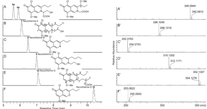
(CH2O + H2O), H2CO3 (CO2+H2O) and C3H4O3 (hydroxypropanoic acid moiety, marked with symbol a in Fig. 4), support the presence of serine in this molecule (Supplementary Table S3, Fig. 3). Based on these data, along with other mass fragmentation properties reported in the Fig. 3.Extracted ion chromatograms (A–F) for m/z 340.1 (A), m/z 296.1 (B), m/z 252.1 (C), m/z 310.1 (D), m/z 352.1 (E), and m/z 253.1 (F) corresponding to azaphilones, and HR-MS spectra (A′–F′) of azaphilones 6a (flavochlorine E), 7 (flavochlorine A), 8 (flavochlorine B), 9 (flavochlorine C), 10 (flavochlorine G) and 11 (flavochlorine D), respectively, along with their chemical structures (note: HR-MS spectrum of 6b (flavochlorine F) comparable with that of compound 6a, was not depicted). Chromatograms and spectra were obtained from a HPLC separation of the extract prepared from Flavomyces fulophazii culture sample HF-3A.
Table 5
NMR data of flavochlorine A (compound 7) and flavochlorine G (compound 10) in methanol-d4.
No.a flavochlorine A flavochlorine G
δH δC δH δC
1 8.795, s, 1H 140.8 8.80, s, 1H 141.3
3 – 147.5 – 145.9
4 7.51, s, 1H 114.0 7.49, s, 1H 115.2
4a – 146.9 146.9
5 – 106.8 106.7
6 – 174.8 174.6
7 6.44, s, 1H 106.6 6.45, s, 1H 106.0
8 – 157.9 – 158.4
8a – 107.1 – 107.8
9 2.95, t, (7.9), 2H 35.3 2.90, t (7.6), 2H 33.62
10 1.80, m, 2H 22.8 1.80, m, 2H 21.95
11 1.11, t, (7.4), 3H 13.6 1.10, t, (6.9), 3H 12.90 1′ 4.43, t, (5.0), 2H 57.5 4.32, t (7,3), 1H 54.49
2′ 3.91, t, (5.0), 2H 61.6 2.24, m, 2H 34.22
3′ – – 3.25, m, 2H 48.21
4′ – – 1.31, d, (7.0), 3H 20.97
5′ – 176.3
O–Me 3.96, s, 3H 56.4 3.95, s, 3H 55.46
aCorresponding numbered molecular structures are found in Fig. 3.
Supplementary Table S3, compound 6a was tentatively identified as a new natural serine analogue of flavochlorine D, denominated flavo- chlorine E (systematic name in Supplementary material). The molecular formula of compound 6b (C16H18O5NCl, Table 2) was identical to that of the serine derivate flavochlorine E, revealing compound 6b to be a constitutional isomer of flavochlorine E. An evaluation of their mass fragmentation processes suggests, that the hydroxyl methyl group in the serine motif of flavochlorine E may be present at the propyl side chain of compound 6b as a methyl ether, connected to the C-9 carbon (Fig. 3, Supplementary Table S3). Thus, compound 6b was tentatively identified as the seventh new natural azaphilone derivative of the fungus F. fulophazii, denominated flavochlorine F (systematic name in Supple- mentary material).
2.2. Amounts of compounds in the in vitro cultures of F. fulophazii Amounts of all identified compounds were determined by HPLC-MS in the lyophilized cultures of ten Hungarian and seven Mongolian F. fulophazii isolates grown in three replicates (Supplementary Table S4, Fig. S8). Among tetramic acids and azaphilones, vermelhotin and fla- vochlorine A were found to be the main compounds, respectively, in all samples. The highest amounts of vermelhotin were determined in cul- tures of the Hungarian and Mongolian isolates HF-3 (5.3 mg/g) and MF- 7 (5.5 mg/g), respectively (data are averages, calculated from contents of three replicate cultures).
In order to isolate vermelhotin by preparative HPLC, three-three lyophilized cultures of the isolate HF-3 and those of MF-7 were pooled and extracted (six cultures, in total). Since the total weight of these pooled cultures was 1.09 g, 5.9 mg vermelhotin could be isolated as the calculated maximum yield (CMY). The preparative HPLC isolation could be regarded to be effective, according to a comparison of the CMY of vermelhotin (5.9 mg) with the amount of vermelhotin isolated from the lyophilized cultures (4.8 mg), thus suggesting the practical utility of F. fulophazii in high-yield vermelhotin production. Accumulation of the vermelhotin derivatives (dihydroxyvermelhotin, hydroxyvermelhotin,
methoxyvermelhotin and oxovermelhotin) showed a close correlation with that of vermelhotin, resulting in their highest amounts also in the cultures of isolates HF-3 and MF-7 (Supplementary Table S4). Among these vermelhotin derivatives, hydroxyvermelhotin was determined to be the most abundant compound, reaching its maximum levels of 1.1 mg/g and 1.0 mg/g in the cultures of isolates HF-3 and MF-7, respectively (average values, calculated from contents of three parallel cultures). Accordingly, in addition to vermelhotin, hydroxyvermelhotin could also be isolated from the pooled cultures of isolates HF-3 and MF-7 by preparative HPLC.
A relative high-level accumulation of the azaphilone flavochlorine A was detected in cultures of isolates MF-3 (MF-3B, 4.2 mg/g), HF-1 (HF- 1A, 3.1 mg/g) and HF-9 (HF–9B, 2.2 mg/g). However, flavochlorine A levels were at least one order of magnitude smaller in the corresponding parallel cultures of these isolates. Consequently, in order to isolate fla- vochlorine A, cultures of different isolates (detailed above), containing the highest levels of this compound, were pooled and extracted. Accu- mulation of azaphilones B–G, occurring as minor compounds relative to flavochlorine A, showed a close correlation with the accumulation of flavochlorine A.
We found the metabolite production of F. fulophazii highly variable, resulting in differences in the amounts of compounds among the isolates and also among the parallel cultures of one isolate.
2.3. Activity of isolated compounds on the seed germination of Lactuca sativa and on the growth of Lemna minor plants
Metabolites of endophytic fungi can protect the host plants against various classes of pests (like pathogenic fungi, parasitic nematodes, herbivorous insects, etc.) thus, indirectly promoting plant growth (Li and Strobel, 2001; Schwarz et al., 2004; Schardl et al., 2007). Some volatile organic compounds were also confirmed as enhancers of plant development, affecting directly the plants (Berthelot et al., 2016).
However, several endophytic metabolites have been shown to be phytotoxic, inhibiting growth of the plants (Choi et al., 2004) and their Fig. 4.Characteristic MS fragmentation of azaphilone compounds 6a, 7–11 (A) and 6b flavochlorine F (B) along with their backbone specific fragment ion structure (C). Corresponding fragment ions generated from protonated molecular ions of these azaphilones by various collision induced dissociation energies, are detailed in the Supplementary Tables S2 and S3.
seedlings (Rivero-Cruz et al., 2003; García-M´endez et al., 2016; Wang et al., 2020).
To test the effects of our isolated metabolites on plants, two stan- dardized assays, i.e., the Lactuca sativa seed germination- and the Lemna minor growth test, both having international recommendations, were performed (Priac et al., 2017; Vanhoutte et al., 2017). Neither the seed germination nor the reproduction and leaf development were affected by the metabolites of F. fulophazii, suggesting their no direct effects on plants (Supplementary Figs. S9 and S10).
2.4. Antiproliferative activity of isolated compounds
Antiproliferative activity of the isolated pure compounds vermel- hotin, hydroxyvermelhotin and flavochlorine A was tested in vitro against 12 human cancer cell lines and the non-cancer Vero cells (Table 6). A significant in vitro antiproliferative activity of vermelhotin was demonstrated against a panel of cancer cell lines including HepG2 (human hepatocellular liver carcinoma) and HL-60 (human promyelo- cytic leukemia) (Kasettrathat et al., 2008). Our study verified the inhibitory effect of vermelhotin on the proliferation of HepG2 and HL-60 cells, with IC50 values of 10.1 μM and 9.4 μM (correspond to 2.19 μg/mL and 2.03 μg/mL), respectively, as these values are consistent with those reported recently (2.50 μg/mL for HepG2 and 1.60 μg/mL for HL-60) (Kasettrathat et al., 2008). For the first time, our results confirmed significant moderate antiproliferative activity of ver- melhotin against the further ten cancer cell lines included in our experiment, with IC50 ranges of 12.1–37.0 μM, without inhibiting the non-cancer Vero cells, suggesting its selectivity towards cancer cells.
Regarding the novel compounds, neither the vermelhotin-related hydroxyvermelhotin nor the azaphilone flavochlorine A were found to be effective antiproliferative compounds in our study (Table 6).
Comparing the structures of vermelhotin and hydroxyvermelhotin, the non-activity of hydroxyvermelhotin can be explained by its higher po- larity that may hinder the penetration into the cancer cells.
3. Conclusions
Our frequently occurring, grass colonizing root endophytic fungus,
Flavomyces fulohazii was proved to be a novel source of secondary me- tabolites, accumulating four new tetramic acids, seven new chlorinated azaphilones and the already known but rarely occurring, valuable tet- ramic acid, vermelhotin. The mass fragmentation of these compounds, determined by HR-MS/MS, resulted in the formation of structure- specific diagnostic ions, such as ions at m/z 181 and m/z 180 corre- sponding to the protonated bicyclic chlorinated backbone of azaphilones and their nitrogenated counterparts, respectively. Comparing the metabolite composition of different fungal isolates, optimum sources for the isolation of selected compounds could be defined, allowing the isolation of vermelhotin, hydroxyvermelhotin and flavochlorine A as pure compounds. Among them, vermelhotin showed selective anti- proliferative activity against 12 cancer cell lines compared to the normal Vero cells, suggesting its potential use in cancer therapy. However, seed germination and plant growth were not affected by these compounds.
4. Experimental
4.1. Fungal cultures and molecular identification
Isolates of the endophytic fungus F. fulophazii (Pleosporales), collected from different Hungarian (ten isolates, indicated as HF-1–HF- 10) and Mongolian (seven isolates, indicated as MF-1–MF-7) locations (Knapp et al, 2015, 2019), were used in this experiment (Table 1, Sup- plementary Fig. S8). For molecular phylogenetic identification, total DNA was extracted from the isolates HF-1–HF-8 with the NucleoSpin Plant II DNA isolation kit (Macherey-Nagel, Düren, Germany) following the manufacturer’s instructions. The nuc rDNA internal transcribed spacer (ITS) region was amplified with the primer pair ITS1F (Gardes and Bruns 1993) and ITS4 (White et al., 1990) using DreamTaq poly- merase (Thermo Fisher Scientific, Vilnius, Lithuania). Sequencing was carried out with the primers used for amplification by LGC (Berlin, Germany). Sequences were assembled in Staden Package (Staden et al., 2000) and deposited in GenBank under accession numbers MW438310–MW438317 (Table 1). To analyze the phylogenetic position of F. fulophazii in the suborder Massarineae, representative sequences of the families Periconiaceae and the neighboring Massarinaceae were involved in the analysis. To study the phylogenetic position of the pleosporalean strain CRI247-01, the other known source of vermelhotin (Kasettrathat et al., 2008), its nuc rDNA ITS sequence was also involved in the dataset. In the phylogenetic analysis, three representative species of the family Lentitheciaceae, Darksidea alpha (CBS 135650), Keissleriella breviasca (CBS 139691) and Lentithecium clioninum (CBS 139694) served as multiple outgroups. The sequences from the NCBI GenBank were combined and aligned using the online version of MAFFT 7 (Katoh and Standley 2013) and the E–INS–i method. Maximum likelihood (ML) phylogenetic analysis was carried out with the RAXMLGUI 1.3 imple- mentation (Silvestro and Michalak 2012; Stamatakis 2014). A GTR +G nucleotide substitution model was used for nucleotide partitions with ML estimation of base frequencies. ML bootstrap (BS) analysis with 1000 replicates was used to test the support of the branches. The phylogenetic tree (Fig. 1) was visualized and edited in MEGA 7 (Kumar et al., 2016).
4.2. Preparation of F. fulophazii culture extracts for analysis and isolation
Each isolate was grown in three replicates (labeled as A, B, C) in Petri dishes (60 mm ×15 mm) on Potato dextrose agar medium (VWR, Hungary) at room temperature in dark for 30 days. The voucher speci- mens of all isolates are available in the Department of Plant Anatomy, E¨otv¨os Lor´and University, Budapest, Hungary and two isolates were also deposited in the CBS collection (as indicated in Table 1). Complete in vitro cultures containing culture medium with the fungal mycelium grown on it were lyophilized and pulverized. The NMR solvents chlo- roform-d, dimethyl sulfoxide-d6 (DMSO d6) and methanol-d4 were purchased from Sigma-Aldrich, Hungary. The other materials and Table 6
Antiproliferative activity of vermelhotin, hydroxyvermelhotin and flavochlorine A isolated from Flavomyces fulophazii culture extracts.
Cell line Antiproliferative activity (IC50, μM) vermelhotin hydroxy-
vermelhotin flavochlorine
A referencea
Dau Sal
A2058 12.1 >100 67.9 0.31
HepG2 10.1 >100 >100 1.22 5.83
A431 19.9 >100 >100 0.7
U87 28.6 >100 >100 0.42 0.8
EBC-1 20.0 >100 >100 1.2
SH-SY5Y 12.9 >100 >100 0.7
HT-29 31.4 >100 >100 0.24
HL-60 9.4 >100 >100 0.025
MonoMac-
6 17.1 >100 >100 0.6 2.23
LCLC-
103H 37.0 >100 >100 8.6
HEK-293 21.6 >100 >100 no
data
H838 22.1 >100 >100 10.2
VERO >100 >100 >100 no
data 1–5 literature data, obtained from Lajko et al. (2018); Kiss et al. (2019); Baranyai ´ et al. (2017); Tripodi et al. (2020); Orb´an et al. (2011), respectively.
aDaunomycin (Dau, against all cells) and 5-chloro-2-hydroxy-N-[4-(trifluoro- methyl)phenyl]benzamide (Sal, against the HepG2, U87 and MonoMac-6 cells) were used as positive control.
reagents applied in the analysis and isolation of fungal metabolites, such as acetonitrile, distilled water, formic acid, methanol (Reanal, Hungary) were all of analytical reagent grade of the highest purity available.
Extracts for analysis: Aliquots of the powdered cultures (10.0 mg) were extracted with 5 mL of methanol at 60 ◦C, via a reflux condenser, for 30 min. The insoluble, centrifuged material was subsequently re- extracted in the same way. The supernatants were combined and these combined solutions were dried by a rotary vacuum evaporator at 40 ◦C.
Before analysis, these dried extracts were dissolved in methanol.
Extracts for isolation: Procedure was the same as described above, except for the amounts of lyophilized and pulverized cultures. To isolate vermelhotin, hydroxyvermelhotin and flavochlorine G, total amount of six unified cultures (HF-3A, HF–3B, HF–3C, MF-7A, MF-7B, MF-7C) were extracted while to isolate flavochlorine A and flavochlorine G three unified cultures (MF-3B, HF-1A, HF–9B) were extracted. Com- pounds of these extracts were separated by preparative HPLC.
4.2.1. 11-Hydroxyvermelhotin (compound 2)
Red, amorphous solid; UV (MeOH) λmax (log ε) 276 (3.60), 425 (3.42) nm; for 1H NMR and 13C NMR data, see Table 4; for HR-Orbitrap-MS data, see Table 2.
4.2.2. Flavochlorine A (compound 7)
Yellow, amorphous solid; UV (MeOH) λmax (log ε) 287 (3.91), 386 (3.56) nm; for 1H NMR and 13C NMR data, see Table 5; for HR-Orbitrap- MS data, see Table 2.
4.3. General experimental procedures
Analytical HPLC hyphenated with UV and high-resolution Orbitrap mass spectrometry detections: A Dionex Ultimate 3000 UHPLC system (3000RS diode array detector (DAD), TCC-3000RS column thermostat, HPG- 3400RS pump, SRD-3400 solvent rack degasser, WPS-3000TRS auto- sampler), hyphenated with a Orbitrap Q Exactive Focus Mass Spec- trometer equipped with electrospray ionization (ESI) (Thermo Fischer Scientific, Waltham, MA, USA) was used for chromatographic separation and high resolution mass spectral analysis. The HPLC separations were performed on a Kinetex C18 column (75 × 3 mm; 2.6 μm) (Phe- nomenex, USA). Eluents: eluent A, 0.1% v/v formic acid, eluent B, acetonitrile:0.1% v/v formic acid (80:20, v/v). Linear gradient: 0.0 min, 10% B; 12.0 min, 70% B; flow rate: 0.3 mL/min; column temperature:
25 ◦C; injected volume: 1.0–5.0 μL. The ESI source was operated in positive ionization mode and operation parameters were optimized automatically using the built-in software. The working parameters were as follows: spray voltage, 3500 V (+); capillary temperature 256 ◦C;
sheath-, auxiliary- and spare-gases (N2): 47.50, 11.25 and 2.25 arbitrary units, respectively. The resolution of the full scan was of 70,000 and the scanning range was between 120 and 600 m/z units. MS/MS scans were acquired at a resolution of 35,000, using collision energy of 10, 15, 20, 30, 45 and 75 eV. DAD spectra were recorded between 250 and 600 nm.
Preparative HPLC: A Pharmacia LKB HPLC (Uppsala, Sweden) system (2248 pumps, VWM 2141 UV detector) was connected to a preparative HPLC column: Gemini, 5 μm, C6-Phenyl, 100 ×21.2 mm (Phenom- enex, USA). The eluents were the same as described above. Linear gradient: 0.0 min, 10% B; 20.0 min, 70% B; flow rate: 5.0 mL/min;
column temperature: ambient; injected volume: 500 μL.
Nuclear magnetic resonance (NMR) spectroscopy: NMR spectra of the isolated compounds were recorded in methanol-d4 and chloroform- d and DMSO d6 at 25 ◦C on a Varian DDR spectrometer (599.9 MHz for
1H and 150.9 MHz for 13C) equipped with a dual 5 mm inverse detection gradient (IDPFG) probe-head. Standard pulse sequences and parameters were used to obtain 1D 1H, various 2D [1H–1H] COSY, [1H–13C] HSQC and [1H–13C] HMBC spectra. Chemical shifts were referenced relative to the appropriate solvent resonances.
Circular dichroism (CD) spectroscopy: The CD spectrum of isolated
hydroxyvermelhotin was recorded in methanol on a Jasco J720 Spec- tropolarimeter (Jasco INC, Tokyo, Japan). The spectra were accumu- lated three times with a bandwidth of 1 nm and a scanning step of 0.2 nm at a scan speed of 50 nm/min.
4.4. Compound quantification by HPLC-MS
In order to quantify compounds by HPLC-MS, extracted ion chro- matograms (EICs) of their molecular ions were recovered from the total ion current chromatograms, and an external standard method was applied by using EICs. Linear regression analysis of the isolated ver- melhotin, hydroxyvermelhotin and flavochlorine A, was performed in the range of 0.10–16.0 ng of their injected amounts, resulting in appropriate R2 values (0.998, 0.999 and 0.998 for vermelhotin, hydroxyvermelhotin and flavochlorine A, respectively). Amounts of tetramic acids (dihydroxyvermelhotin, oxovermelhotin and methox- yvermelhotin) and flavochlorines B–G were calculated using the cali- bration curve for isolated hydroxyvermelhotin and flavochlorine A, respectively.
4.5. In vitro activity tests of isolated compounds on the seed germination of Lactuca sativa and on the growth of Lemna minor plants
4.5.1. Lactuca sativa seed bioassay
Lactuca sativa L. var. capitata ‘Attrakci´o’ (R´edei Kertimag, Hungary) seeds were washed twice with DW and were placed into Petri dishes (90 ×15 mm) containing filter paper (ø 75 mm, VWR, Hungary) (5 seeds in all Petri dishes). Dilution series of the isolated compounds were made in the concentration range of 1.0–100 μM by DW. The seeds were treated with 1.9 mL amounts of these solutions for 5 days at room temperature on natural light (the control was treated with DW). The length of the root and the hypocotyl was then measured by the software ImageJ (NIH, USA) using the screened pictures of the seedlings.
4.5.2. Lemna minor bioassay
Lemna minor L. (clone 9441) was cultured during stock cultivation in a light chamber (16 h light exposure) at 24 ◦C on a liquid medium (Appenroth et al., 1996). The medium was changed every 14 days. Two weeks prior to the tests, fronds were transferred into Steinberg medium to acclimate and the medium was changed after 7 days (Naumann et al., 2007). Dilution series of the isolated compounds were made in the concentration range of 1.0–100 μM by Steinberg medium. The fronds were treated with 1.9 mL amounts of these solutions in 24-well plates for 7 days (in a light chamber, 16 h light exposure) at 24 ◦C (the control was grown on Steinberg medium). Fronds were then counted and the total leaf area in each well was measured by the software ImageJ using the screened pictures of the plates (Schneider et al., 2012).
Statistical analyses of the Lactuca sativa and Lemna minor bioassays were conducted by Prism v.8.0.1 (GraphPad, USA). The normality of the datasets was tested by Shapiro-Wilk test. In case of normality, one-way ANOVA was performed. When data did not exhibit normality, Kruskal- Wallis test was carried out. To compare the treatments with the con- trol, we used Dunett and Dunn post hoc tests, respectively at α =0.05.
4.6. Determination of in vitro cytostatic effect of isolated compounds A2058 (Human skin, melanoma), HepG2 (Human liver, hepatocel- lular carcinoma), HT-29 (Human colon, colorectal adenocarcinoma), HL-60 (Human peripheral blood, acute promyelocytic leukemia) and MonoMac-6 (Human peripheral blood, acute monocytic leukemia) cells were cultured in RPMI-1640 medium (Lonza) containing 10% FBS (Gibco, Thermo Fisher Scientific, Waltham, MA, USA) 1% Penicillin/
Streptomycin (from 10,000 units penicillin and 10 mg streptomycin/
mL, Gibco) and 2 mM L-glutamine (Lonza). A431 (Human skin, epidermoid carcinoma), U87 (Human brain, glioblastoma), EBC-1 (Human lung, squamous cell carcinoma, bronchi), SH-SY5Y (sub-line