Edited by:
Leo Van Overbeek, Wageningen University & Research, Netherlands Reviewed by:
Macit Ilkit, Çukurova University, Turkey Jason Sahl, Northern Arizona University, United States Anne D. Van Diepeningen, Fungal Biodiversity Centre (KNAW), Netherlands
*Correspondence:
Tamás Papp pappt@bio.u-szeged.hu
Specialty section:
This article was submitted to Plant Microbe Interactions, a section of the journal Frontiers in Microbiology Received:09 February 2018 Accepted:03 May 2018 Published:23 May 2018 Citation:
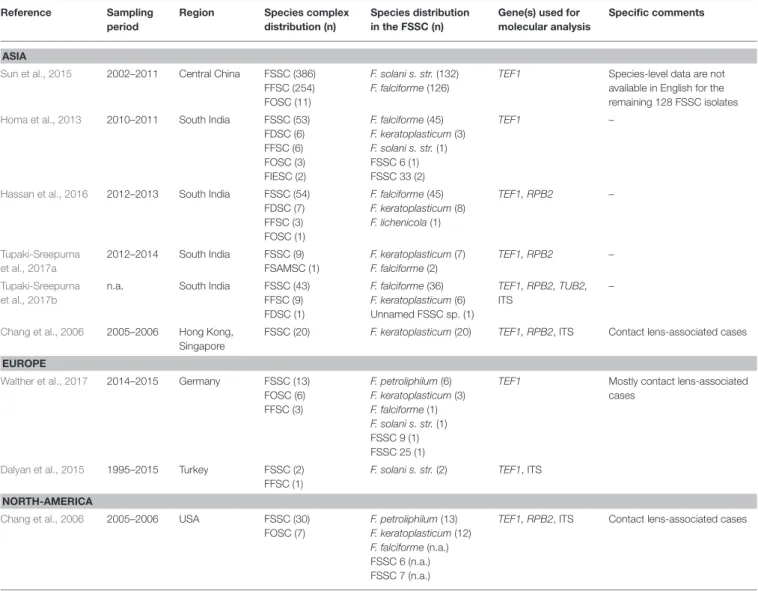
Homa M, Galgóczy L, Manikandan P, Narendran V, Sinka R, Csernetics Á, Vágvölgyi C, Kredics L and Papp T (2018) South Indian Isolates of the Fusarium solani Species Complex From Clinical and Environmental Samples: Identification, Antifungal Susceptibilities, and Virulence.
Front. Microbiol. 9:1052.
doi: 10.3389/fmicb.2018.01052
South Indian Isolates of the
Fusarium solani Species Complex From Clinical and Environmental Samples: Identification, Antifungal Susceptibilities, and Virulence
Mónika Homa1,2, László Galgóczy2,3, Palanisamy Manikandan4,5,6,
Venkatapathy Narendran4, Rita Sinka7, Árpád Csernetics2, Csaba Vágvölgyi2, László Kredics2and Tamás Papp1,2*
1MTA-SZTE “Lendület” Fungal Pathogenicity Mechanisms Research Group, Szeged, Hungary,2Department of Microbiology, Faculty of Science and Informatics, University of Szeged, Szeged, Hungary,3Division of Molecular Biology, Biocenter, Medical University of Innsbruck, Innsbruck, Austria,4Aravind Eye Hospital and Postgraduate Institute of Ophthalmology, Coimbatore, India,5Department of Medical Laboratory Sciences, College of Applied Medical Sciences, Majmaah University, Majmaah, Saudi Arabia,6Greenlink Analytical and Research Laboratory India Private Limited, Coimbatore, India,
7Department of Genetics, Faculty of Science and Informatics, University of Szeged, Szeged, Hungary
Members of theFusarium solanispecies complex (FSSC) are the most frequently isolated fusaria from soil. Moreover, this complex solely affects more than 100 plant genera, and is also one of the major opportunistic human pathogenic filamentous fungi, being responsible for approximately two-third of fusariosis cases. Mycotic keratitis due to Fusarium species is among the leading causes of visual impairment and blindness in South India, but its management is still challenging due to the poor susceptibility of the isolates to conventional antifungal drugs. Aims of the present study were to isolate South Indian clinical and environmental FSSC strains and identify them to species level, to determine the actual trends in their susceptibilities to antifungal therapeutic drugs and to compare the virulence of clinical and environmental FSSC members.
Based on the partial sequences of the translation elongation factor 1α gene, the majority of the isolates—both from keratomycosis and environment—were confirmed as F. falciforme, followed by F. keratoplasticum and F. solani sensu stricto. In vitro antifungal susceptibilities to commonly used azole, allylamine and polyene antifungals were determined by the CLSI M38-A2 broth microdilution method. The first generation triazoles, fluconazole and itraconazole proved to be ineffective against all isolates tested.
This phenomenon has already been described before, as fusaria are intrinsically resistant to them. However, our results indicated that despite the intensive agricultural use of azole compounds, fusaria have not developed resistance against the imidazole class of antifungals. In order to compare the virulence of different FSSC species from clinical and environmental sources, aDrosophila melanogastermodel was used. MyD88 mutant flies having impaired immune responses were highly susceptible to all the examined fusaria. In wild-type flies, oneF. falciformeand twoF. keratoplasticumstrains also reduced the survival significantly. Pathogenicity seemed to be independent from the origin of the isolates.
Keywords: keratomycosis,Fusarium solanispecies complex,F. falciforme, molecular identification, antifungal susceptibility,Drosophila melanogaster, virulence
INTRODUCTION
The genus Fusarium is a large group of hyaline filamentous fungi firstly described byLink (1809). According to the recent literature, it comprises approximately 200–300 species belonging to 20–22 species complexes (O’Donnell et al., 2013, 2015; Al- Hatmi et al., 2016). Fusaria are common soil saprophytes;
however, they are also known as phytopathogens (Coleman, 2016). Two Fusarium species were recently included in the list of the top ten plant pathogenic fungi with both economic and scientific importance (Dean et al., 2012). The members of this genus may also interact with plants as endophytic root colonizers (Bacon and Yates, 2006); furthermore, they may be responsible for a wide range of human infections in either immunocompetent or immunocompromised patients (Garnica and Nucci, 2013). In accordance with the current species complex descriptions, at least ten of them have been reported to have human pathogenic representatives (Al-Hatmi et al., 2016). Last but not least, plumbing systems are also proven environmental reservoirs of human-pathogenicFusariumspecies (Short et al., 2011).
Taxonomy of the genusFusariumis changing intensely since 2011, when the era of the dual nomenclature ended (Hawksworth et al., 2011) and a comprehensive phylogenetic study of the genus discovered that the traditionally knownFusariumis not monophyletic (Gräfenhan et al., 2011). Based on these results, Gräfenhan et al. (2011)proposed to restrict the nameFusarium to the Gibberellaclade and at the same time to reallocate the medically important Fusarium solani species complex (FSSC) andFusarium dimerumspecies complex (FDSC) to other genera.
After the release of this study,Lombard et al. (2015)were the first who suggested to useNeocosmospora solaniinstead ofF. solani, andNeocosmospora falciformisinstead ofF. falciforme. However, in this study we would like to follow a previously published proposal ofGeiser et al. (2013)by keeping the historical concept ofFusariumand use the names well-known in medical mycology.
In South India, a frequent scenario of fungal keratitis (keratomycosis) is that agricultural workers are infected after a corneal injury caused by plant or soil materials during their regular activities (Dóczi et al., 2004; Homa et al., 2013). Based on the recent reports,Fusariumspecies—and among them the members of the FSSC—are the most frequently isolated causative agents of fungal keratitis in this region (Chakrabarti and Singh, 2011; Homa et al., 2013; Hassan et al., 2016). The FSSC comprises at least 60 haplotypes, out of which 22 have been reported to have clinical associations (van Diepeningen et al., 2014; Al- Hatmi et al., 2016) with poor susceptibility to commonly used antifungal drugs (Azor et al., 2007). As consequence of the narrow range of therapeutic options, the treatment ofFusarium keratitis is extremely challenging and the lack of a prompt and effective therapy often results in corneal opacification or complete blindness (Shukla et al., 2008). Therefore, the rapid identification of the causative agent and the determination of its antifungal susceptibility are essential to choose the best therapeutic option. Presumably, the intensive agricultural and clinical (mis)use of antifungal compounds have also influenced the current susceptibility profile of the genus (Al-Hatmi et al.,
2016). Thus, besides clinical studies, it is also crucial to evaluate the development of antifungal resistance among environmental strains to follow up the impact of fungicides used in the field.
Among FSSC species, F. falciformewas the most prevalent species isolated from human mycotic keratitis in South India (Homa et al., 2013; Hassan et al., 2016; Tupaki-Sreepurna et al., 2017a,b). However, it is unclear what lies in the background of its dominance: its environmental frequency or its high virulence. FSSC is reported to be more virulent than other species complexes of the genus (Mayayo et al., 1999); however, the virulence of different FSSC species has not been compared before.
To answer the questions above, virulence studies are inevitable.
The objectives of the present study were (I) to isolate FSSC strains from keratomycosis patients, agricultural source and natural environments in South India; (II) to identify the strains at the species-level using molecular methods;
(III) to determine their in vitro susceptibilities to commonly used antifungal agents; (IV) to compare the species diversity and the antifungal susceptibility profiles of the clinical and environmental isolates; (V) to compare the virulence of different clinical and environmental FSSC members; and (VI) to present and discuss the clinical details of the investigated keratomycosis cases.
MATERIALS AND METHODS
Patients Specimens and Fusarium Isolates
A total of 22 Fusarium isolates derived from patients with keratomycoses attending the Aravind Eye Hospital and Postgraduate Institute of Ophthalmology (Coimbatore, Tamilnadu, India) along with 20 environmental FSSC isolates from the same region were investigated (Table 1). Corneal scrapings were performed by an ophthalmologist under strict aseptic conditions, on each base of the corneal ulcer using a Kimura’s spatula after instillation of 4% preservative-free lidocaine. Materials obtained from scraping the leading edge and the base of the ulcers were inoculated directly onto 5%
sheep blood agar, chocolate agar, potato dextrose agar (PDA) and into brain heart infusion broth without gentamicin sulfate (Himedia Laboratories, India). Sheep blood agar and chocolate agar plates were incubated at 37◦C, while PDA plates and brain heart infusion bottles were incubated at 27◦C for 3 weeks.
To isolate fusaria from environmental sources, soil and plant parts (i.e., root and stem) were collected from gardens, parks, yards and agricultural fields in the surrounding regions of Coimbatore in November 2012. One gram of each collected soil sample was suspended in 10 ml sterile distilled water. The stock solutions were diluted 10 and 100 times and spread over Rose Bengal-Chloramphenicol agar (Himedia Laboratories, India) plates. The collected plant parts were pre-washed in sterile distilled water, surface-sterilized in 75% ethanol for 5 min and in 95% ethanol for 5 min, then rinsed in sterile distilled water for three times to remove ethanol residues. The sterilized parts were cut into small pieces, placed on Rose Bengal-Chloramphenicol agar plates and incubated at 25◦C for 72 h. All fungal colonies from Rose Bengal-Chloramphenicol agar were subcultured into PDA plates using the cross-streak method. ThenFusarium-like
TABLE1|MolecularidentificationandantifungalsusceptibilitiesofFusariumstrainsisolatedfromkeratitiscasesandenvironmentalsamplesinSouthIndia. SZMCNo.Yearof isolationOriginStateSpeciesbased onTEF1AccessionNo. ofTEF1MIC(µg/ml) AMBCLTECNFLCITCKTCNTMTRB CLINICALISOLATES SZMC114382005GobichettypalayamTamilnaduF.falciformeHE647902282>64>32321664 SZMC114392005ErodeTamilnaduF.falciformeHE6479034162>64>3216864 SZMC114412005GobichettypalayamTamilnaduF.falciformeHE6479068162>64>323284 SZMC114072005CoimbatoreTamilnaduF.falciformeHE6479090.2581>64>32884 SZMC114082005PalladamTamilnaduF.falciformeHE6479100.581>64>3216816 SZMC114422005CoimbatoreTamilnaduF.falciformeHE6479114162>64>3216832 SZMC114432004PalladamTamilnaduF.falciformeHE6479144161>64>328816 SZMC114112005KangeyamTamilnaduF.falciformeHE647915482>64>3232832 SZMC114122005KangeyamTamilnaduF.falciformeHE6479162162>64>3232832 SZMC114142004PalghatKeralaF.keratoplasticumHE647919281>64>328816 SZMC114472005HarurTamilnaduF.falciformeHE647924141>64>323280.5 SZMC114482005CoimbatoreTamilnaduF.falciformeHE647927484>64>3232832 SZMC114492004PalghatKeralaF.falciformeHE6479288162>64>32161632 SZMC114192005CoimbatoreTamilnaduF.falciformeHE647929488>64>3264832 SZMC114252005KrishnagiriTamilnaduF.falciformeHE6479374161>64>3216816 SZMC114542005TirupurTamilnaduF.falciformeHE647944421>64>32880.5 SZMC114552005TiruchengoduTamilnaduF.falciformeHE647945484>64>32641616 SZMC114562005KangeyamTamilnaduF.falciformeHE6479460.580.532>32284 SZMC114572005TirupurTamilnaduF.falciformeHE647948484>64>3232864 SZMC114312005ErodeTamilnaduF.falciformeHE647949484>64>3264832 SZMC114322005TirupurTamilnaduF.falciformeHE647950182>64>328816 SZMC114582005OttanchatramTamilnaduF.falciformeHE647951248>64>32844 MICrange(µg/ml)0.25–82–160.5–832–>64>322–644–160.5–64 MIC50(µg/ml)482>64>3216832 GMMIC(µg/ml)3.28.82.1>64>3218.18.519.0 ENVIRONMENTALISOLATES SZMC213292012Soil,flowergardenTamilnaduF.falciformeMG2724214168>64>32161664 SZMC213302012Soil,flowergardenTamilnaduF.keratoplasticumMG2724224168>64>3264832 SZMC213312012Soil,outdoor flowerpotTamilnaduF.falciformeMG2724232162>64>326484 SZMC213322012Soil,outdoor flowerpotTamilnaduF.falciformeMG272424488>64>32641632 SZMC213332012Soil,outdoor flowerpot
TamilnaduF.falciformeMG272425488>64>32161632 SZMC213342012Soil,outdoor flowerpotTamilnaduF.falciformeMG272426488>64>3264864 (Continued)
TABLE1|Continued SZMCNo.Yearof isolationOriginStateSpeciesbased onTEF1AccessionNo. ofTEF1MIC(µg/ml) AMBCLTECNFLCITCKTCNTMTRB SZMC213352012Soil,bananatreeTamilnaduF.falciformeMG2724274164>64>3232832 SZMC213362012Soil,bananatreeTamilnaduF.falciformeMG2724284164>64>3232832 SZMC213372012Soil,bananatreeTamilnaduF.falciformeMG2724292162>64>326484 SZMC213382012Soil,gardenTamilnaduF.falciformeMG2724301816>64>3264816 SZMC213392012Soil,yardTamilnaduF.falciformeMG272431484>64>32641632 SZMC213402012Soil,yardTamilnaduF.falciformeMG272432484>64>32641632 SZMC213422012Soil,yardTamilnaduF.falciformeMG272433481>64>321688 SZMC213432012Soil,parkTamilnaduF.falciformeMG272434482>64>3216816 SZMC213442012Soil,parkTamilnaduF.falciformeMG2724352168>64>3232816 SZMC213452012Soil,maizefieldTamilnaduF.falciformeMG272436482>64>328816 SZMC213462012Soil,sorghumfieldTamilnaduF.falciformeMG272437282>64>321688 SZMC213482012Soil,unknown cultivatedfieldTamilnaduF.solanis.str.MG2724382816>64>32884 SZMC213502012Sorghum,rootTamilnaduF.falciformeMG272439282>64>324816 SZMC213512012Tomato,rootTamilnaduF.falciformeMG2724404161>64>322816 MICrange(µg/ml)1–48–161–16>64>322–648–164–64 MIC50(µg/ml)484>64>3232816 GMMIC(µg/ml)3.010.64.0>64>3224.39.517.8 AMB,amphotericinB;CLT,clotrimazole;ECN,econazole;FLC,fluconazole;GM,geometricmean;ITC,itraconazole;KTC,ketoconazole;MIC,minimuminhibitoryconcentration;MIC50,MICfor50%ofthetestedisolates;NTM,natamycin; SZMC,SzegedMicrobiologicalCollection,UniversityofSzeged,Szeged,Hungary;TEF1,translationelongationfactor1α;TRB,terbinafine.
colonies were purified and identified by macro- and microscopic characteristics. From both clinical and environmental samples, the purified fungal colonies were sub-cultured and stored on PDA plates at 4◦C until further investigations.
Molecular Identification
Isolates suspected to be Fusarium sp. based on their macromorphological characteristics and microscopic features were further subjected to molecular identification. All isolates were grown in Potato Dextrose Broth (Sigma-Aldrich, USA) at 25◦C in a shaker (New Brunswick Scientific Co., Inc., USA) at 220 rpm for 5 days, and subsequently genomic DNA was extracted with the MasterPure Yeast DNA Purification Kit (Epicentre Biotechnologies, USA) in accordance with manufacturer’s instructions. FSSC isolates were selected using the FSSC-specific PCR as described by He et al. (2011) and confirmed with an EcoRI digestion-based PCR-RFLP method (Homa et al., 2013). For the species-level identification of FSSC- positive isolates, the 5′portions of translation elongation factor 1α(TEF1) coding region and introns were amplified (O’Donnell et al., 1998). After Sanger sequencing (LGC Genomics GmbH, Germany) theTEF1sequences were deposited in the GenBank (https://www.ncbi.nlm.nih.gov/nucleotide/) under the accession numbers listed in Table 1 and used as BLAST (Altschul et al., 1990) queries against the Fusarium MLST database (http://www.westerdijkinstitute.nl/fusarium/) (O’Donnell et al., 2010).
All the confirmed isolates were deposited in the Szeged Microbiological Collection (SZMC; http://szmc.hu/; http://www.
wfcc.info/ccinfo/collection/by_id/987) under the strain numbers listed inTable 1.
Phylogenetic Analysis
Besides the FSSC strains isolated from clinical and environmental sources, two clinical members of theF. dimerumspecies complex (SZMC 11496 and SZMC 11540) were involved in the analysis as an outgroup. The sequences were aligned by Muscle v3.8.31 (Edgar, 2004) and manually refined in BioEdit v7.1.3.0 (Hall, 1999). Substitution models for the final alignment were selected by the AICc function in jModelTest 2.1.10 (Posada, 2008).
Trees were inferred by Maximum Likelihood (ML) and Bayesian MCMC approaches. ML bootstrapping was performed in PhyML 3.0 under the TrN+G model of sequence evolution, using the nearest-neighbor interchange branch swapping algorithm and 1000 replicates of non-parametric bootstrap analysis (Guindon and Gascuel, 2003). ML bootstrap values>69% were considered as significant support (Soltis and Soltis, 2003). Bayesian MCMC analyses were run in MrBayes 3.1.2 (Huelsenbeck and Ronquist, 2001). One cold and three incrementally heated chains were run in two replicates sampling every 100th generation. Chain length was set to 10,000,000 generations and a burn-in value of 100 000 generations was chosen using the Tracer 1.4 software (Rambaut and Drummond, 2007). Post-burn-in trees were summarized in a 50% majority rule consensus tree in MrBayes. Posterior probabilities >0.94 were considered as significant.
Antifungal Susceptibility Tests
Antifungal susceptibility tests were performed as described in the Clinical and Laboratory Standards Institute (CLSI) M38- A2 broth microdilution method (Clinical Laboratory Standards Institute, 2008). Pharma grade powders of amphotericin B (AMB), clotrimazole (CLT), econazole (ECN), fluconazole (FLC), itraconazole (ITC), ketoconazole (KTC), terbinafine (TRB) (Sigma-Aldrich, USA), and commercially available natamycin (NTM) eye drops (Lalitha et al., 2008a) (Natamet, 5% suspension, Sun Pharmaceutical Ind. Ltd., India) were included in the tests.
Conidial suspensions were prepared in 0.85% saline solution from 5-day-old cultures grown on PDA plates and diluted in RPMI-1640 medium (Sigma-Aldrich, USA) adjusting the final inoculum density to 104 CFU/ml. Fungal growth was evaluated after incubation for 48 h at 35◦C without shaking.
Minimal inhibitory concentration (MIC) was determined as the lowest concentration of an antifungal agent that inhibited completely the growth of the tested isolates compared to the drug-free control medium. For FLC and KTC, the MICs were defined as the lowest concentrations of the drugs that cause approximately 50% reduction in growth. MIC50was determined as the MIC inhibiting the growth of 50% of all the tested isolates.
Aspergillus flavusATCC 204304 andCandida kruseiATCC 6258 were included as quality control strains. Each experiment was performed in triplicates.
Survival Experiments in Drosophila melanogaster
To examine the background ofF. falciformedominance in South Indian human keratomycosis cases, the virulence of six FSSC isolates, i.e.,F. falciformeSZMC 11407, SZMC 11408, and SZMC 21332,F. keratoplasticum SZMC 11414 and SZMC 21330, and F. solani s. str.SZMC 21348 was examined inD. melanogaster.
Conidial suspensions were prepared with sterile phosphate buffer saline (PBS; 137 mM NaCl, 2.7 mM KCl, 10 mM Na2HPO4, 2 mM KH2PO4, pH 7.4) from 5-day-old cultures grown on PDA plates at 35◦C. The final inoculum densities were adjusted to 1× 10⁷conidia/ml with PBS.
Drosophilastocks were raised and kept following the infection on standard cornmeal agar medium at 25◦C. The Oregon R strain, originally obtained from the Bloomington stock center, was used as the wild type throughout the experiments.
MyD88c03881 flies having impaired immune responses were described previously (Tauszig-Delamasure et al., 2002). Infection was performed by dipping a thin needle in a suspension of fungal conidia (107conidia/ml) or PBS for the uninfected control, and subsequently the thorax of the anesthetized fly was pricked. Flies were counted at different points of time to monitor survival. Flies were moved into fresh vials every other day. Each experiment was performed with approximately 60 flies for each genotype.
The results shown inFigure 1are representative of at least three independent experiments.
Statistical Analyses
All statistical analyses were performed in SigmaPlot (version 14.0). Two samplet-test was used to reveal significant differences between the antifungal susceptibility profiles of clinical and
FIGURE 1 |Survival rates of wild-type (Oregon) and MyD88c03881 mutant flies infected with clinicalF. falciformeSZMC 11407 and SZMC 11408(A,B), clinical F. keratoplasticumSZMC 11414(C), environmentalF. keratoplasticumSZMC 21330(D), environmentalF. falciformeSZMC 21332(E), and environmentalF. solani s.
str.SZMC 21348(F)strains. The control groups were injected with sterile PBS.
environmental isolates. Fisher’s exact test was applied to compare the species composition of clinical and environmental isolates.
Kaplan-Meier survival curves were generated in order to present the results of the survival experiments in D. melanogaster.
The Log-Rank statistic was used to decide whether there is a statistically significant difference between the curves. To identify the group—or groups—of flies that differ from the others, the Holm-Sidak multiple comparison procedure was applied (Glantz, 2012). Significance level was set atp<0.05.
Ethics Statement
Due to its observational nature, no formal ethics approval was required for this study. In order to protect the patients’
anonymity, identifying information were not included in the manuscript.
RESULTS
Clinical Characteristics of Patients With Fusarium Keratitis
Clinical data are available for all cases but one (Strain No.
SZMC 11425) (Table 2). Out of the 22 keratitis isolates, 20 were isolated from patients residing in Tamilnadu, while the rest were from Kerala. Majority of the infections (n=13) were registered between June and August. None of the patients had any underlying conditions. Half of the patients reported trauma as a predisposing factor, while 10 patients could not recall any injury prior to the infection. The severity of the ulcer was recorded as mild in six, moderate in seven and severe in eight cases. The most common therapeutic approach was the combined topical application of NTM, ITC, and ECN eye drops (n=16), which were supplemented with systemic KTC in 12 cases. Surgical intervention (therapeutic penetrating keratoplasty, TPK) was needed in four severe cases, where the topical and systemic drugs could not improve the patients’ condition. The registered final outcomes were as follows: 16 patients were healed completely; the therapy failed in five cases and one patient was lost to follow-up.
Identification of the Fusarium Isolates Based on Molecular Markers
BLAST searches with the partialTEF1sequences revealed that most of the isolates derived from both human keratomycoses and the environment belong to F. falciforme (n = 41; FSSC 3 + 4, O’Donnell et al., 2008). A clinical (SZMC 11414) and an environmental (SZMC 21330) isolate were identified as F. keratoplasticum (FSSC 2, Short et al., 2013), while another isolate from soil (SZMC 21348) was confirmed asF. solani s. str.
(FSSC 5,Schroers et al., 2016;Table 1). Statistically significant association between the investigated FSSC species and their source was not detected. In order to examine the phylogenetic distribution of the isolates, a phylogenetic reconstruction was also performed using the above-mentionedTEF1locus, which confirmed the BLAST-based identifications (Figure 2).
Antifungal Susceptibilities
Table 1 summarizes the MIC values of the eight investigated antifungal agents. Clinical and environmental strains showed
similar susceptibilities. However, environmental isolates proved to be significantly (p=0.01) less susceptible to ECN than the clinical FSSC isolates. In all other cases, statistically significant differences were not detected between these two populations. The lowest MICs were recorded for AMB and ECN (0.25–16µg/ml).
MIC values of CLT and NTM were between 2 and 16µg/ml, while the activities of TRB and KTC varied in the MIC ranges of 0.5–64 and 2–64µg/ml, respectively. ITC and FLC proved to be ineffective in the tested concentration ranges.
Virulence
In the case of wild type Oregon flies, the clinicalF. falciforme strains SZMC 11407 and SZMC 11408 and the environmental FSSC 5 strain SZMC 21348 proved to be avirulent; the survival rates 6 days post infection (dpi) were 79±21%, 76±6%, and 61± 15% (Figures 1A,B,F). At the same time, infection with theF. keratoplasticumstrains SZMC 11414 and SZMC 21330 and the environmentalF. falciformestrain SZMC 21332 resulted in a significant reduction in the survival rate compared to the control group, the 6 dpi survival rates were 33±18%, 46±12%, and 46
±23% (Figures 1C–E), respectively. All six tested strains reduced the 6 dpi survival rates of the MyD88-mutant MyD88c03881 flies to 4–15% (Figures 1A–F).
DISCUSSION
The population of tropical/subtropical countries such as India is more prone to eye infections, especially to fungal keratitis caused byFusariumspp. generally due to the climatic conditions.
Regular monitoring of the disease is essential for its effective management (Lalitha et al., 2008b; Kredics et al., 2015).
As it is shown in Table 2, we found the highest incidence of FSSC keratitis cases in July. Previously, Lin et al. (2012) also observed an uneven distribution ofFusariumkeratitis cases throughout the year in South India with a major peak of registered cases in July. This peak was associated with the windy season in June-July, when dust particles are presumed to be the main causes of ocular trauma (Lin et al., 2012). This theory is reinforced by the clinical records summarized in Table 2, where dust was mentioned as a predisposing factor for the infection only in June and July. A minor peak of keratitis cases in January—which was detected byLin et al. (2012) and was attributed to the intensive agricultural activities of the harvest season resulting in elevated concentrations of conidia in the air and frequent ocular injuries due to soil or plant debris particles—
was not observed in our study. Interestingly,Lin et al. (2012)also observed that environmental humidity (dry and wet season) was not a significant factor in the seasonal patterns of fungal keratitis.
We observed some major variations especially in the risk factors and treatment ofFusariumkeratitis cases when compared our data with the study ofWalther et al. (2017)from Germany.
Based on the clinical details, trauma was the most frequently recorded predisposing factor for Fusarium keratitis in India (Bharathi et al., 2007; Tupaki-Sreepurna et al., 2017a), whereas keratomycoses were rare in temperate climates and more commonly associated with the use of soft contact lenses (Keay et al., 2011; Walther et al., 2017). As shown in Table 2, most