Contents lists available atScienceDirect
Biochemical Pharmacology
journal homepage:www.elsevier.com/locate/biochempharm
Volatile anaesthetics inhibit the thermosensitive nociceptor ion channel transient receptor potential melastatin 3 (TRPM3)
Balázs Kelemen
a,b,2, Erika Lisztes
a,3, Anita Vladár
a,b, Martin Hanyicska
a,b, János Almássy
a, Attila Oláh
a,4, Attila Gábor Szöllősi
c,5, Zsófia Pénzes
b,c, János Posta
d, Thomas Voets
e,f,6, Tamás Bíró
c,g,7,1, Balázs István Tóth
a,1,8,⁎aLaboratory of Cellular and Molecular Physiology, Department of Physiology, Faculty of Medicine, University of Debrecen, Debrecen, Hungary
bDoctoral School of Molecular Medicine, University of Debrecen, Debrecen, Hungary
cDepartment of Immunology, Faculty of Medicine, University of Debrecen, Debrecen, Hungary
dLaboratory of Toxicology, Department of Forensic Medicine, Faculty of Medicine, University of Debrecen, Debrecen, Hungary
eLaboratory of Ion Channel Research, VIB-KU Leuven Center for Brain & Disease Research, Leuven, Belgium
fDepartment of Cellular and Molecular Medicine and TRP Research Platform Leuven (TRPLe), KU Leuven, Leuven, Belgium
gHungarian Center of Excellence for Molecular Medicine, Szeged, Hungary
A R T I C L E I N F O Keywords:
Volatile anaesthetics TRP ion channels TRPM3 Nociception Thermosensation
A B S T R A C T
Background:Volatile anaesthetics (VAs) are the most widely used compounds to induce reversible loss of con- sciousness and maintain general anaesthesia during surgical interventions. Although the mechanism of their action is not yet fully understood, it is generally believed, that VAs depress central nervous system functions mainly through modulation of ion channels in the neuronal membrane, including 2-pore-domain K+ channels, GABA and NMDA receptors. Recent research also reported their action on nociceptive and thermosensitive TRP channels expressed in the peripheral nervous system, including TRPV1, TRPA1, and TRPM8. Here, we in- vestigated the effect of VAs on TRPM3, a less characterized member of the thermosensitive TRP channels playing a central role in noxious heat sensation.
Methods:We investigated the effect of VAs on the activity of recombinant and native TRPM3, by monitoring changes in the intracellular Ca2+concentration and measuring TRPM3-mediated transmembrane currents.
Results:All the investigated VAs (chloroform, halothane, isoflurane, sevoflurane) inhibited both the agonist- induced (pregnenolone sulfate, CIM0216) and heat-activated Ca2+signals and transmembrane currents in a concentration dependent way in HEK293T cells overexpressing recombinant TRPM3. Among the tested VAs, halothane was the most potent blocker (IC50= 0.52 ± 0.05 mM). We also investigated the effect of VAs on native TRPM3 channels expressed in sensory neurons of the dorsal root ganglia. While VAs activated certain sensory neurons independently of TRPM3, they strongly and reversibly inhibited the agonist-induced TRPM3 activity.
Conclusions:These data provide a better insight into the molecular mechanism beyond the analgesic effect of VAs and propose novel strategies to attenuate TRPM3 dependent nociception.
https://doi.org/10.1016/j.bcp.2020.113826
Received 25 November 2019; Accepted 22 January 2020
⁎Corresponding author.
E-mail address:toth.istvan@med.unideb.hu(B.I. Tóth).
1TB and BIT contributed equally to the work as senior authors.
20000-0002-8994-0132.
30000-0002-8517-6536.
40000-0003-4122-5639.
50000-0001-6046-8236.
60000-0001-5526-5821.
70000-0002-3770-6221.
80000-0002-4103-4333.
Biochemical Pharmacology 174 (2020) 113826
Available online 25 January 2020
0006-2952/ © 2020 The Author(s). Published by Elsevier Inc. This is an open access article under the CC BY license (http://creativecommons.org/licenses/BY/4.0/).
T
1. Introduction
Volatile anaesthetics (VAs) are the most commonly used compounds to maintain general anaesthesia during operation both in human ther- apeutic interventions and in animal experiments [1]. Although the exact mechanisms, whereby VAs cause a reversible loss of conscious- ness are not yet fully understood, it is generally accepted that they suppress the activity of the central nervous system by specifically tar- geting cellular proteins, including several ion channels, among others.
A few of these ion channels, especially GABAAreceptors and various K+ channels, are considered to play a central role in the general depression of the central nervous system functions resulting in reversible loss of the consciousness initiated by VAs[2–4]. However, VAs can also influence several other ion channels, including voltage-gated Na+, K+and Ca2+
channels [5,6]. Moreover, in recent years, their action on sensory transient receptor potential (TRP) channels has also been reported.
TRP channels form a heterogeneous and multifunctional group in the voltage gated-like superfamily of ion channels. The 6-transmem- brane domain containing TRP proteins form non-specific, mostly Ca2+
permeable cationic channels, and are functional as homo- or hetero- tetramers [7–9]. Most TRP channels are considered to function as polymodal “cellular sensors” sensitive to diverse changes in the phy- sico-chemical environment (e.g. temperature, pH, osmolarity, ionic concentrations, endogenous mediators, external chemical irritants, etc.) [10,11]and increasing body of evidence indicates their emerging roles in various diseases [12,13]. In primary sensory neurons, the most widely studied sensory TRPs are the thermosensitive TRPV1, TRPA1 and TRPM8 channels. Their versatile role in diverse (patho)physiolo- gical sensory processes, including thermosensation, itch, pain (together with different forms of hyperalgesia and allodynia), and inflammation has inspired a plethora of research studies [14–21]. This in turn has urged several pharmaceutical companies to find effective tools tar- geting these thermosensitive TRP channels as potential novel drugs to manage several, mostly pain related, clinical syndromes[22]. Recently, VAs were also reported to activate TRPA1[23,24], to sensitize TRPV1 [25]and also to modulate TRPM8[26], and these results may explain some adverse effects related to general anaesthesia.
TRPM3 was recently introduced as a novel thermosensitive noci- ceptor TRP channel expressed by a large subset of primary sensory neurons of the dorsal root and trigeminal ganglia (DRG and TG).
TRPM3 has an essential role in acute heat pain sensation[27], it con- tributes to the development of inflammatory heat hyperalgesia and transmits chemical pain sensation evoked by its endogenous steroid ligand pregnenolone sulfate (PregS)[28]. The stimulation of TRPM3 by certain ligands e.g. CIM0216, or special ligand combinations, e.g. co- application of PregS and the antifungal agent clotrimazole revealed an alternative ion permeation pathway beyond the main pore, which re- sulted in a strong inward current at negative membrane potentials. This unique current displays the general characteristics of the so-called omega currents reported earlier in mutant K+and Na+channels, and may have a high (patho)physiological relevance exacerbating TRPM3 related pain sensation[29–31]. The above results, together with the fact that modulation of TRPM3 activity, in contrast with TRPV1, did not induce any change in the core body temperature of experimental ani- mals [28], introduce TRPM3 as an appealing drug target for novel therapeutic interventions in pain related syndromes. However, in order to exploit the putative therapeutic potential of TRPM3 as a potential drug target, a better understanding of its regulation and pharmacolo- gical interactions are of greatest importance.
In this study, we investigated the effect of VAs on recombinant and native TRPM3 ion channels and found that they potently inhibit TRPM3 function. These results further enhance our knowledge about VAs’
mechanism of action and may contribute to the development of novel analgesics targeting TRPM3.
2. Materials and methods
2.1. Cell culturing and isolation of sensory neurons
Native HEK293T cells, and HEK293T cells stably overexpressing the mouse TRPM3α2 variant (HEK-M3 cells) were cultured as described before[28]. In brief, cells were cultured in DMEM medium (Invitrogen, Paisley, UK), supplemented with 10% foetal bovine serum, (In- vitrogen), 50 U/ml penicillin, 50 μg/ml streptomycin (both from ThermoFisher, Waltham, MA, USA), 10 mM Glutamax, Non-Essential- Amino-Acids and 200 μg/ml Hygromycin (all from Invitrogen) at 37 °C.
Sensory neurons of dorsal root ganglia (DRGs) were obtained from adult (8–12 week old) C57BL6 mice, as described before[28]. Briefly, mice were euthanized by CO2, DRGs were isolated and digested with collagenase and dispase (both from Invitrogen). Suspension of sensory neurons was seeded on poly-L-lysine HBr (Sigma Aldrich, St.Louis, MO, USA) coated glass bottom culture dishes (MatTek, Ashland, MA, USA) and cultured in Neurobasal medium supplemented with 2% B-27 sup- plement (both from Invitrogen), 2 mM L-glutamine, 100 µg/ml peni- cillin/streptomycin (both from ThermoFisher), and 100 ng/ml ß-NGF (Pepro Tech EC, Ltd., London, UK) at 37 °C in 5% CO2containing at- mosphere. Neurons were used for experiments within 24 to 36 h fol- lowing isolation. We aimed at obtaining high number of sensory neu- rons yielding n ≥ 50 neurons in each experimental group for highly reliable statistical analysis. Therefore, respecting the 3R principles, we established multiple primary sensory neuron cultures from an animal and used them in independent measurements. Cultures were randomly recruited into the different experimental groups, and cultures from 3 individual animals were recruited into each group. Altogether, 12 an- imals were used for the study. All experimental procedures and animal husbandry were conducted following the European Parliament and the Council Directive (2010/63/EU) and national legislation.
2.2. Fluorescent Ca2+measurements
Fluorescent measurement of cytoplasmic Ca2+ concentration in HEK cells and in individual DRG neurons were performed according to our previously optimized protocols: HEK-M3 cells were seeded in Poly- L-Lysine HBr (Sigma-Aldrich) coated 96-well/clear-bottom plates (Greiner Bio-One, Frickenhausen, Germany) at a density of 100,000 cells per well in normal cell culture medium and incubated overnight.
Next day, cells were loaded with 2 μM Fura-2-AM (Invitrogen) at 37 °C for 30 min, and washed three times with Ca2+-buffer (150 mM NaCl, 5 mM KCl, 1 mM MgCl2x6H2O, 2 mM CaCl2x2H2O, 10 mM glucose xH2O, 10 mM HEPES, pH 7.4 (all from Sigma-Aldrich)). The plates were then placed into a FlexStation 3 fluorescent microplate reader (Molecular Devices, Sunnyvale, CA, USA) and cytoplasmic Ca2+con- centration (reflected by the ratio of fluorescence measured at λEX1: 340 nm, λEX2: 380 nm, λEM: 516 nm (F340/F380)) was monitored during application of compounds in various concentrations. During the mea- surements, cells in a given well were exposed to only one given con- centration of the agents. Measurements were carried out at ambient temperature.
To determine the VAs’ effect on the temperature-evoked activation of TRPM3, we used a Fluo-4 based assay and a QPCR system. HEK-M3 and non-transfected HEK293T cells were loaded with 2 µM Fluo-4-AM (Invitrogen) for 30 min, then they were trypsinised and re-suspended in Ca2+- buffer in the presence or absence of VAs and transferred into PCR tubes with optical transparent cover (200.000 cells/tube, 200 μL).
Fluorescence was measured with a Stratagene Mx3005P QPCR instru- ment (Agilent Technologies Santa Clara, CA, USA) using an appropriate filter set while the well temperature was increased from 25 °C to 46 °C in steps of 3 °C.
To measure cytoplasmic Ca2+ concentration in individual DRG neurons, we used a microscope based calcium imaging system. On the day after the isolation, DRG neurons were loaded with 2 µM Fluo-4-AM
(Invitrogen) dissolved in normal Ca2+-buffer, then placed on the stage of a Zeiss LSM 5 Live confocal fluorescent microscope (Carl Zeiss AG, Oberkochen, Germany) and Fluo-4 loaded cells were captured with constant settings in every 1 s (λEX: 488 nm, λEM: 516 nm). During the measurements, cells were continuously perfused with Ca2+-buffer and different compounds were applied via the perfusion. Data were pre- sented as F1/F0, where F0is the average fluorescence of the baseline (before the first compound application) and F1is the actual fluores- cence. Experiments were performed at room temperature (21–22 °C).
2.3. Electrophysiology
HEK-M3 cells were seeded to 12 mm glass coverslips previously coated with poly-L-lysine HBr (Sigma-Aldrich) and whole cell patch clamp measurements were carried out by using an Axopatch 1.D am- plifier and Clampex 10.2 software (Molecular Devices). Pipettes with final resistances of 2–5 MΩ were fabricated and filled with intracellular solution containing 100 mM aspartic acid, 45 mM CsCl, 1.144 mM MgCl2, 10 mM HEPES, and 10 mM EGTA (all from Sigma-Aldrich). pH was adjusted to 7.2 using CsOH (VWR, Radnor, PE, USA). Experiments were performed in a bath solution composed of 150 mM NaCl, 1 mM MgCl2, and 10 mM HEPES buffered to pH 7.4 (NaOH) (all from Sigma- Aldrich). To record TRPM3 mediated currents, the holding potential was 0 mV and cells were ramped every 2 s from −150 to +150 mV over the course of 200 ms. Recorded data were analysed and plotted using Origin 9.0 software (OriginLab Corporation, Northampton, MA, USA).
2.4. Preparation of working solutions of VAs
10 mM stock solutions of VAs were prepared in extracellular solu- tion by a rigorous overnight stirring in air-tight closed vials. From these stock solutions, fresh dilutions of the final working solutions were prepared and used for measurements within 45 min. If needed, new working solutions were diluted in every 30 min.
2.5. Gas chromatography/mass spectrometry (GC/MS)
The stability of the stock solutions of VAs in an open, freely venti- lating system was checked by GC/MS. Stock solutions prepared as de- scribed above were kept in an open vial at room temperature for 45 min simulating the conditions in an open perfusion system used for patch clamp and Ca2+measurements. During this incubation time, samples were taken at different time points and subjected for analysis im- mediately. The active agents in the samples were identified by a GC/MS method using an Agilent 7980B-5977A instrument (Agilent Technologies, Santa Clara, CA, USA) at the Toxicology Laboratory of the Institute of Forensic Medicine of the University of Debrecen. The injection volume was 0.2 µL, and the injector temperature was 250 °C.
The applied capillary column was a J&W DB-35MS UI, 30 m × 0,25 mm × 0,25 µm. The detection parameters were the fol- lowings: Sampling: split; split ratio: 20:1. Oven temperature program:
65 °C (2 min. hold time), heating: 50 °C/min to 230 °C; Interface:
280 °C; MS source: 230 °C; Ionization: EI; Detection mode: SCAN. The
‘area under curve’ of the specific peaks were determined in arbitrary units and normalized to that of obtained from the sample taken at the beginning (0 min) of the incubation.
2.6. Chemicals
Chloroform and halothane were obtained from Sigma Aldrich, iso- flurane (sold as Vetflurane) was from Virbac (Carros, France) and se- voflurane from AbbVie Inc. (North Chicago, IL, USA). Pregnenolone sulphate (PregS), allyl isothiocyanate (AITC), capsaicin, thymol, CIM0216 were obtained from Tocris Bioscience (Bristol, UK).
Isosakuranetin was obtained from Carl Roth (Karlsruhe, Germany).
Working solutions of VAs were prepared as described above. All other drugs were prepared as stock solutions in dimethyl sulfoxide (DMSO), and then diluted into extracellular solutions to reach the desired final concentration. Concentration of the vehicle (DMSO) in the final working solutions was 0.1%.
2.7. Curve fitting
Logistic dose-response curves were fitted using the equation y = A2 + (A1 − A2)/(1 + (x/x0)^p) where the calculated parameters are: A1: initial value (ymin), A2: final value (ymax), x0: center (EC50) and p is the calculated power. Fittings were carried out and parameters were calculated using Origin 9.0 (OriginLab Corporation).
2.8. Data and statistical analysis
Electrophysiological data were analysed using ClampFit 10.0 soft- ware (Axon Instruments, Foster City, CA, USA). IBM SPSS Statistics 23.0 (IBM Corporation. Armonk, NY, USA) and Origin 9.0 (OriginLab Corporation) were used for statistical analysis and data display. When testing antagonistic effect of isosakuranetin, agonist induced Ca2+
signals in the presence and absence of isosakuranetin were compared pairwise by two-tailed Student’s t-test for independent samples. To control unwanted variances of measured currents between HEK-M3 cells, TRPM3 currents measured in the presence of VAs were normal- ized, and compared to the agonist induced current (considered as 100%) in the same cell and two-tailed Student’st-test for one sample was used for statistical evaluation. In case of Ca2+signals recorded on sensory neurons, if it is not mentioned otherwise, signal amplitudes were normalized to the 1st agonist evoked signal, considered as 100%, to control variances experienced between individual neurons. Then, data were subjected for statistical analysis using one-way ANOVA with Dunnett post-hoc test to compare the effect of VAs to vehicle control. In every case, P < 0.05 was regarded as showing significant differences between group means. All data are presented as mean ± SD.
3. Results
3.1. VAs formed stabile solutions
In our study, we investigated the effect of VAs with various chemical structures (Fig. 1a) on TRPM3 ion channel functions. Due to their vo- latile and lipophilic nature, we assumed that it might be challenging to prepare a stabile aqueous solution of VAs suitable for measurements, although they reportedly can be solved > 10 mM in water at 25 °C[32].
Therefore, we aimed at testing the stability of the stock solutions of VAs applied in an open, freely ventilating system mimicking the conditions in our perfusion and liquid handling systems used in the experiments.
For this, we performed GC/MS measurements on 10 mM stock solutions of the VAs, which were prepared by rigorous overnight stirring in an airtight vial, as described in the Materials and methods. The con- centration of the stock solutions in open vials did not change drama- tically during 45 min (Fig. 1b). Therefore, working solutions were freshly prepared from 10 mM stock solutions stored in airtight vials and used for measurements within 30 min. After 30 min, new working so- lutions were prepared.
3.2. VAs inhibited chemical agonist induced activation of recombinant TRPM3
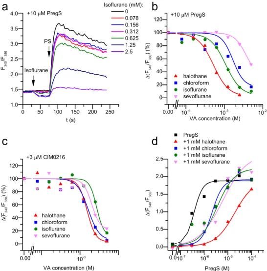
First, we investigated the effect of VAs on recombinant TRPM3 by carrying out Ca2+measurements on HEK-M3 cells. During the mea- surements, we applied various concentrations of VAs followed by the application of TRPM3 agonists PregS or CIM0216 in the continuous presence of VAs, as shown inFig. 2a. This experimental design enabled us to investigate both potential activating and inhibiting effect of VAs.
We found that none of the investigated VAs activated the recombinant TRPM3, whereas each of them inhibited the TRPM3-mediated Ca2+
signals evoked by the endogenous TRPM3 agonist PregS and by the more potent synthetic TRPM3 agonist CIM0216[30]in a dose depen- dent manner (Fig. 2a-c). The IC50 values for halothane, chloroform, isoflurane, and sevoflurane were (mean ± SEM) 0.52 ± 0.05 mM, 1.67 ± 0.03 mM, 1.09 ± 1.49 mM and, 3.83 ± 2.39 mM , respec- tively, when TRPM3 was activated by 10 µM PregS (Fig. 2b), while the IC50 values were 1.52 ± 0.05 mM, 1.64 ± 0.25 mM, 2.71 ± 0.88 mM and, 2.0 ± 0.28 mM when activated by 3 µM CIM0216, respectively (Fig. 2c). Moreover, the EC50value of PregS was shifted from 0.37 ± 11 µM to 19.33 ± 4.24 µM (~51× fold), 2.13 ± 0.35 µM (~6× fold), 3.49 ± 1.3 µM (~8× fold), and 6.14 ± 6.39 µM (~15× fold) by 1 mM halothane, chloroform, iso- flurane, and sevoflurane, respectively (Fig. 2d).
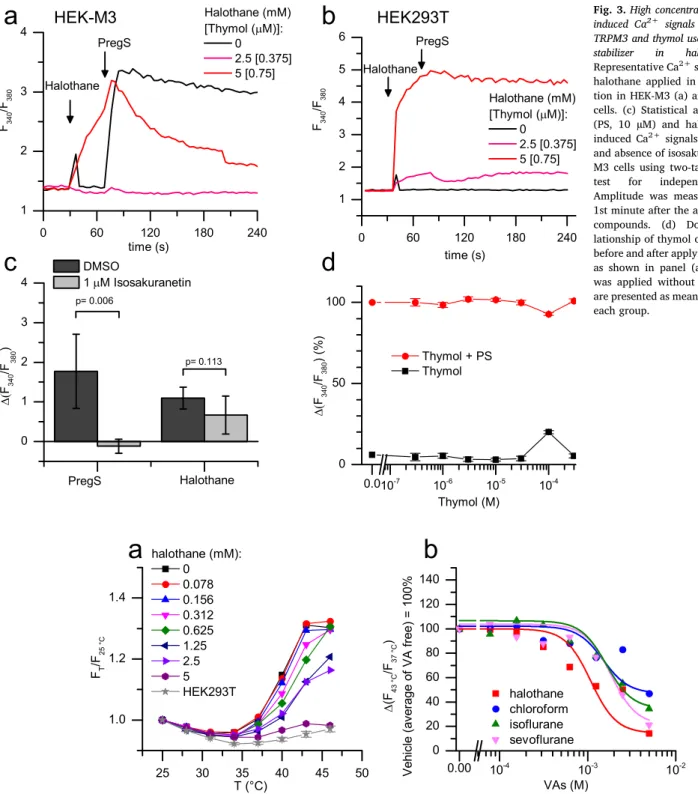
Although halothane was found to be the most potent inhibitor of TRPM3 among the tested VAs, it also evoked robust Ca2+ transients when applied alone at high concentrations (≥5 mM) (Fig. 3a). This stimulatory effect of halothane was found to be independent of TRPM3, because (i) it was equally observed in native HEK293T cells and HEK- M3 cells (Fig. 3a-b) and (ii) the halothane-induced Ca2+signals were not inhibited by the TRPM3 antagonist isosakuranetin (Fig. 3c). It is important to mention that the commercially available halothane ap- plied in the current study also contained ca. 150 ppm thymol, as sta- bilizer resulting in ca. 0.75 µM thymol in the most concentrated (5 mM) halothane working solution tested. Since thymol was reported to be an activator of TRPV3 [33,34] and TRPA1 [35], other thermosensitive members of TRP family, we also investigated the effect of thymol on TRPM3, to clarify its potential contribution to the effect of halothane.
Our results indicated that thymol applied at concentrations up to 300 µM did not affect TRPM3 functions: it neither activated HEK-M3 cells nor inhibited their activation evoked by PregS (Fig. 3d).
3.3. VAs inhibited the heat-evoked activation of TRPM3
Next, we tested whether the VAs influence activation of TRPM3 by thermal stimuli, in addition to their effect on ligand-induced activation of the channel. HEK-M3 cells were challenged by precise temperature steps using a Quantitative real-time PCR instrument, and the in- tracellular Ca2+concentration was monitored in the presence of VAs at various concentrations. Our results revealed that VAs also inhibited warming-induced activation of TRPM3: the obtained IC50 values of halothane, chloroform, isoflurane, and sevoflurane were 1.08 ± 0.58 mM, 1.50 ± 0.63 mM, 1.64 ± 0.59 mM, and 1.83 ± 0.63 mM, respectively, when cells were heated from 37 to 43 °C (Fig. 4).
3.4. VAs inhibited TRPM3 mediated transmembrane currents
In whole-cell patch-clamp measurements, PregS induced an out- wardly rectifying whole-cell current in HEK-M3 cells (Fig. 5a-b). This TRPM3-mediated current was partially inhibited by 1 mM chloroform, halothane and isoflurane, and almost fully abolished when these VAs were applied at 5 mM. Sevoflurane was less effective: when applied at 1 mM, it only minimally inhibited the PregS-induced TRPM3 current, whereas at 5 mM it evoked a marked but incomplete inhibition (Fig. 5c). The effects of the VAs were fast and reversible (Fig. 5a).
The PregS-induced current is conducted by the canonical pore of the TRPM3, but CIM0216 also induces the opening of an additional per- meation pathway through the voltage-sensing domain of TRPM3 re- sulting in the appearance of a marked inward current at negative membrane potentials [29–31]. We found that VAs also inhibited the opening of the alternative ion permeation pathway, when activated by CIM0216 (Fig. 5d-f).
3.5. VAs inhibited native TRPM3 in sensory neurons of mouse dorsal root ganglia
To study the effect of VAs on native TRPM3, we investigated sensory neurons isolated from mouse DRGs. We used Ca2+imaging to probe PregS responses in the presence and absence of VAs (Fig. 6a). We considered the PregS-responsive (PregS+) neurons as TRPM3 expres- sing (TRPM3+) neurons. As suggested in previous studies, our results also confirmed that some sensory neurons were directly stimulated by VAs: 15.3%, 21%, 5.1%, and 1,8% of the DRG neurons were activated by 1 mM halothane, chloroform, isoflurane and sevoflurane, respec- tively (Fig. 6b). The distribution of the neurons activated by VAs was similar in the TRPM3+ and TRPM3- population and, importantly, the large majority of TRPM3+ neurons was not activated by VAs (VA- neurons) (Fig. 6b). These results clearly argue for the conclusion that VAs activated a subset of sensory neurons independently of TRPM3. To investigate the VAs’ potential inhibitory effect on native TRPM3, we compared the PregS evoked Ca2+signals in the presence and absence of VAs. We analysed only the TRPM3+ but VA- sensory neurons to eliminate the influence of the TRPM3-independent Ca2+signals evoked by VAs on PregS-induced responses. In our experiments, we applied repeated 2-minute-long pulses of PregS in the presence or absence of VAs, as shown inFig. 6a. Only those cells that responded to a depo- larizing pulse evoked by 25 mM KCl at the end of the measurement were considered as sensory neurons. We found that all of the in- vestigated VAs applied at 1 mM decreased the PregS-induced, TRPM3- mediated Ca2+transients. The inhibitory effect of VAs was reversible within 4 min after application (Fig. 6c).
Since activation of TRP channels results in depolarization of sensory neurons, Ca2+signals evoked by activating TRP channels may be fur- ther amplified by the consequent activation of voltage-gated Ca2+
channels. Previous studies revealed that VAs inhibit some voltage-de- pendent Ca2+channels, as well[36]. Therefore, we considered, that the inhibition of voltage-gated Ca2+ channels might contribute to the Fig. 1.The structure and stability of the investigated volatile anaesthetics (VAs).(a)
Chemical structure of VAs investigated in this study: halothane (a halogenated alkene), chloroform (trichloromethane), isoflurane (a halogenated methyl-ethyl ether) and sevoflurane (fluorinated methyl-isopropyl ether). (b) The stability of the most concentrated 10 mM stock solutions in open, freely ventilating vials was checked by GC/MS in the given timeframe as described in the “Materials and methods” section (n = 3 at each point).
apparent inhibition of signals induced by PregS or agonists of other TRP channels. To challenge the role of voltage-gated Ca2+ channels, we tested whether or not VAs influence Ca2+responses evoked by acti- vating other native thermosensitive TRP channels expressed on sensory neurons. During these experiments, we slightly modified the previous protocol as illustrated inFig. 6d. Because the effect of capsaicin and AITC is poorly reversible, we applied isoflurane before and together with the 1st agonist application, and the effect was compared to the 2nd agonist application after the wash out of the VAs. If the inhibitory effect of VAs experienced in our previous experiments were partially or totally mediated by some other voltage-gated Ca2+channels, then VAs should also inhibit Ca2+transients evoked by activating other TRP channels that cause depolarization of sensory neurons. Our results refuted this theory. The amplitudes of the Ca2+transients activated by the TRPV1- specific agonist capsaicin and the TRPA1 agonist AITC did not decrease in the presence of isoflurane, whereas, the PregS-evoked Ca2+ tran- sients were again significantly decreased (Fig. 6d-e). These results clearly argue for the conclusion that VAs inhibit PregS-induced re- sponses via TRPM3, without influencing voltage gated Ca2+channels.
4. Discussion
Activation of several members of the superfamily of the voltage- gated ion channels[37]is influenced by VAs, and a number of these channels, in particular the hyperpolarization activated and cyclic nu- cleotide gated channel 1 (HCN1)[38], shaker-related delayed rectifier K+channels (Kv1)[39]and two-pore-domain K+channels (K2P) have been implicated in the induction of general anaesthesia. Voltage gated
Na+and Ca2+channels can also be inhibited by VAs[36,40]. More- over, recent studies reported the effect of VAs on thermosensitive TRP channels, as well. The cold- and menthol-activated TRPM8, after an initial activation, was inhibited by VAs. Likewise TRPC5[41], another cold-sensitive family member, was inhibited by halothane and chloro- form[42]. The warmth sensor TRPM2 was not influenced by halothane or chloroform[41], whereas the noxious heat sensor TRPV1 was sen- sitized[25], or, if applied at higher concentration, even activated by VAs[43]. Moreover, irritant VAs, isoflurane and desflurane directly activated TRPA1, a general target of several irritant chemicals, whereas the non-irritating halothane and sevoflurane did not induce TRPA1 activation[24]. These results can explain some adverse effects often associated with general anaesthesia induced by certain VAs. Indeed, irritant VAs evoke mechanical hyperalgesia and bronchoconstriction, impaired respiratory pattern, augmented laryngeal C-fiber activity and stimulate tracheal CGRP release mainly mediated by TRPA1 [24,44–46]. In our current study, we investigated the effect of VAs on TRPM3, a less characterized thermo-nociceptive TRP channel, which together with TRPV1 and TRPA1 play a crucial role in acute heat pain sensation[27,28].
In contrast to the other two heat-pain sensors TRPV1 and TRPA1, TRPM3 was found to be neither sensitized nor activated by any of the investigated VAs. In contrast, activation of TRPM3 by both chemical ligands and heat was markedly inhibited by the investigated VAs.
Among those, halothane was found to be the most potent, inhibiting PregS-evoked TRPM3 activity with an IC50of approximately 0.5 mM, equivalent to ca. 2-times the minimal alveolar concentration (2 MAC) that induces anaesthesia in different species[2,47]. Although the IC50
Fig. 2.Identification of different VAs as in- hibitors of the recombinant TRPM3. (a) Averaged Ca2+responses (n = 5) on HEK-M3 cells evoked by 10 µM PregS (PS) in the pre- sence of various concentrations of isoflurane, as indicated. Compounds were added at the indicated time points. (b) Concentration de- pendent inhibitory effect of VAs on the re- combinant TRPM3 activated by 10 µM PregS.
The measurements were carried out as shown in panel (a), VAs were applied 30 s in advance to the agonist. (c) Concentration dependent inhibitory effect of VAs on the recombinant TRPM3 activated by CIM0216. The measure- ments were carried out as in panel (a) but 3 µM CIM0216 was applied instead of PregS.
(d) Dose-response relationship of PregS in the presence and absence of various VAs, as in- dicated in the figure. The measurements were carried out as in panel (a), but PregS was applied at various concentration and VAs were applied in 1 mM. Data are presented as mean ± SD, n = 5 in each group.
of chloroform was slightly higher (ca. 1.67 mM), this value also cor- responded to approximately 1.5 MAC[5]. Other VAs were less effective in clinically relevant concentrations: the IC50 value for isoflurane (≈1.1 mM) is equivalent to about 3 MAC, whereas the IC50of sevo- flurane approached 10 MAC against PregS-evoked activation. Applied at 1 mM, each VA shifted the PregS activation curve of TRPM3 toward higher concentrations. Again, halothane was found to be the most ef- fective, as it increased the EC50of PregS approximately 50-fold. Im- portantly, at 1 mM, all investigated VAs inhibited the activation of native TRPM3 in sensory neurons of mouse DRGs, as well. Moreover,
VAs inhibited not only the activity of TRPM3 induced by PregS, but they also inhibited the effect of the synthetic agonist CIM0216 and the heat-induced TRPM3 responses with very similar potencies.
The sensitivity of TRPM3 toward some VAs seems to be slightly lower than sensitivity of ion channels generally believed to mediate anaesthesia. Clinically relevant concentrations (≈1 MAC) of volatile anaesthetics activate several members of the K2P channel family known to conduct background K+ currents, which crucially contribute to the negative membrane potential[3]. For example, the EC50of halothane and sevoflurane that induce TASK-1 mediated K+ currents were Fig. 3.High concentration of halothane induced Ca2+ signals independently of TRPM3 and thymol used in 150 ppm as stabilizer in halothane. (a-b) Representative Ca2+signals evoked by halothane applied in high concentra- tion in HEK-M3 (a) and HEK293T (b) cells. (c) Statistical analysis of PregS (PS, 10 µM) and halothane (5 mM) induced Ca2+signals in the presence and absence of isosakuranetin in HEK- M3 cells using two-tailed Student’st- test for independent samples.
Amplitude was measured within the 1st minute after the application of the compounds. (d) Dose-response re- lationship of thymol on HEK-M3 cells, before and after applying 10 µM PregS, as shown in panel (a-b), but thymol was applied without halothane. Date are presented as mean ± SD, n = 5 in each group.
Fig. 4.VAs inhibited thermal activation of TRPM3.(a) Changes in intracellular Ca2+concentration of HEK-M3 cells upon elevating temperature in the presence of various concentrations of halothane as indicated in the figure. Native HEK293T cells were used as negative control. (b) Dose dependent inhibitory effect of VAs on the recombinant TRPM3 activated by temperature shift from 37 to 43 °C. The measurements were carried out as in panel (a). Data are presented as mean ± SD, n = 5 in each group.
0.23 mM and 0.29 mM (near to 1 MAC), respectively[48]. Moreover, NMDA receptor mediated currents were also effectively inhibited by isoflurane and sevoflurane with reported IC50values between 0.25 and 1.3 MAC and ca. 1.25 MAC, respectively[49,50]. The EC50of isoflurane and sevoflurane potentiating GABA induced activity of GABAA re- ceptors was found also around 1 MAC (0.29 mM and 0.33 mM, re- spectively) [49,51]. However, the potency of halothane inhibiting NMDA receptor mediated postsynaptic excitatory currents (IC50= 0.57 mM, equivalent with ca. 2 MAC)[52]and potentiating
GABA induced GABAAcurrents (EC50 = 0.67 mM, ca. 2 MAC)[53]
were very close to the value we found for TRPM3.
Although we analysed the α2 variant and native channels from mice, we strongly believe that our findings can be easily extrapolated to native human channels which is not characterized functionally. Until today, the best characterized variant is the mTRPM3α2 [54]which functionally seem to be hardly distinguishable from the native mouse and the recombinant human TRPM3: each of these variants form Ca2+- permeable cation channels [55–57], are activated by PregS [58,59], Fig. 5.Reversible inhibition of whole-cell TRPM3 cur- rents by various VAs.(a) Representative time courses illustrating the effect of PregS and chloroform on TRPM3 mediated transmembrane currents at −80 and +120 mV. Currents were measured during a voltage ramp from −150 to +150 mV applied at every 2 s. (b) I/V relationship of the transmembrane currents at different time points as indicated in panel (a), illustrating the concentration dependent inhibi- tion of PregS induced currents by chloroform. (c) Statistical analysis of VAs’ effect on PregS induced transmembrane currents (n = 5 in each group, ex- cept in case of isoflurane, where n = 6). The mea- surements were carried out as shown in panel (a). (d) Representative time courses illustrating the effect of CIM0216 and halothane on TRPM3 mediated trans- membrane currents at −80 and +120 mV. A voltage ramp from −150 to + 150 mV was applied at every 2 s. (e) I/V relationship of the transmembrane cur- rents at different time points as indicated in panel (d), illustrating the concentration dependent inhibi- tion of CIM0216 induced currents by halothane. (f) Statistical analysis of VAs’ effect on CIM0216 induced transmembrane currents (n = 6, except in case of chloroform, where n = 5). The measurements were carried out as in panel (d). Effect of VAs was com- pared to the agonist (PregS or CIM0216) induced current before applying the VAs (one samplet-test, control agonist induced current is considered as 100%). Box plots represent the range between 25 and 75 percentile values, horizontal lines mark the median values, square symbols mark the means and whiskers represent SD.
and undergo very similar regulation by phosphatidylinositol 4,5-bi- sphosphate [60,61] and by the βγ subunit of G protein coupled re- ceptors [62–65]. Heat sensitivity is also a shared feature of the re- combinant (mTRPM3α2) and native TRPM3 channels [27,28].
Importantly, both mouse and human TRPM3 were effectively blocked by diclophenac and primidone [59,66]. These close functional and pharmacological similarities suggest that our findings on mouse TRPM3 can be generalized to native human channels.
While our results indicate that VAs inhibit homotetrameric TRPM3,
potential heteromerization between different TRP channel subunits can influence the functional characteristic of the tetramer channels, as de- scribed for example for the thermosensitive members of the TRPV fa- mily[67,68]. However, available data do not suggest significant par- ticipation of TRPM3 in forming heteromeric TRP channels. Although the closest relative TRPM1 was shown to be able to form hetero- multimeric channels with TRPM3, TRPM4 was found not to interact with TRPM3 [69]. Considering that expression of TRPM1 is mainly restricted to melanocytes and retinal bipolar cells[70,71], and it is not Fig. 6.VAs inhibited TRPM3 dependent Ca2+
transients in somatosensory neurons isolated from mouse DRGs.(a) Representative traces showing typical changes of intracellular Ca2+concentra- tion in DRG neurons from wild-type mice in re- sponse to 20 µM PregS, 1 mM halothane, and their combination as indicated in the panel.
25 mM KCl was used as positive control to de- polarize the neuronal cell membrane. (b) Percentage of DRG neurons responding to PregS (PS+) and various VAs applied in 1 mM (VA+).
The measurements were carried out as in panel (a). (c) Statistical analysis of VAs’ effect (1 mM each) on PregS induced Ca2+transients in PregS +/VA− sensory neurons. Values are given as percentage of the first PregS-induced Ca2+
transient without VAs. Dots represents in- dividual neurons, and horizontal lines indicate mean values. Effect of VAs was compared to the vehicle treated control group by ANOVA and Dunnett’s post-hoc test. (d) Representative traces showing typical changes in intracellular Ca2+
concentration in DRG neurons in response to PregS (multiple traces), AITC, and capsaicin in the presence and absence of 1 mM isoflurane, as indicated in the figure. (e) Statistical analysis on the amplitude of PregS, AITC and capsaicin in- duced Ca2+in the presence and absence of 1 mM isoflurane. Measurements were carried out as in panel (d). Effect of isoflurane was compared to transients evoked by isoflurane free agonist as 100% by one-samplet-test. Dots represents in- dividual neurons, and horizontal lines indicate mean values.
expressed in mouse DRGs[72], we do not assume significant hetero- merization which would influence the responsiveness of TRPM3 to VAs.
However, PregS and VAs induced responses may be influenced by the presence of alternative targets expressed by sensory neurons. Indeed, although ca. half of the sensory neurons isolated from wild type DRG are activated by PregS and considered as TRPM3+ neurons, about 10%
of the neurons obtained from TRPM3−/− mice are also activated by PregS [27,28] indicating the presence of TRPM3 independent me- chanisms. This has to be considered when interpreting our experiments carried out on sensory neurons: a low percentage of the PregS induced responses is probably independent of TRPM3. However the exact nature of additional target(s) is mainly elusive. Although sensory neurons of the DRG express both NMDA and GABAAreceptors, their role is ques- tionable in PregS induced responses without their primary ligands.
Although an emerging body of evidence supports the direct action of VAs on TRP channels, we have only a limited knowledge about the underlying mechanisms and the potential binding sites. Studies on TRPA1 and TRPV1 highlighted the role of the pore domain in forming the binding pocket for VAs[43,73], although molecular dynamics si- mulation suggested multiple binding sites on TRPV1[74]. Our experi- ments did not directly address the mechanism of action of VAs on TRPM3, but we observed in our electrophysiological measurements that VAs inhibited not only the canonical pore currents but also the alter- native pore currents induced by CIM0216 at negative membrane po- tential[30]. These currents are mediated by the opening of an alter- native permeation pathway established by the voltage sensing domain of the channel[29,31,75]. The finding that both the canonical and the alternative pore mediated currents are blocked by VAs indicates that VAs do not act as classical pore blockers, but rather inhibit channel gating via a conformational change affecting activation by various mechanisms.
Our results not only reveal an additional ion channel affected by VAs, but also extend our knowledge about the pharmacological inter- actions of TRPM3 that potentially modulate sensory functions mediated by this channel. TRP channels are promising targets pursued by several pharmaceutical companies for the development of novel drugs to manage several, mostly pain related, clinical syndromes [22,76,77].
Despite significant efforts, several TRP-targeting drugs, in particular the first generation of antagonists targeting TRPV1, failed in clinical trials due to undesirable side effects such as hyperthermia and impaired noxious heat sensation [22,78,79]. From this point of view, TRPM3 may be a safer target, since its activation by PregS, in contrast to cap- saicin, a potent and selective activator of TRPV1[80], did not affect core body temperature[28]. To date, only a few blockers of TRPM3 have been characterised, and in animal models some of these were indeed found to inhibit TRPM3-mediated pain, including the flavanone derivative isosakuranetin[81]or the antiepileptic drug primidone[66].
Our results introduce VAs as a new class of TRPM3 inhibitors. A po- tential advantage of VAs is that they are well established, approved compounds and large amount of data available about their safety in human applications. However, their slightly lower affinity to TRPM3 than to several other targets, e.g. to GABAAand K2P channels, can limit their usage in clinical applications as TRPM3 targeting blockers. In spite of concerns about specificity of VAs, our data provide important new information contributing to the characterization of TRPM3 as potential pharmacological target of novel analgesics. The knowledge about the general mechanism of action of VAs may help identify targetable re- gions and basic biophysical and molecular interactions which can be utilized in future studies applying rational drug design approach. In the other hand, characterizing VAs, as a new class of chemicals inhibiting TRPM3 may also advance the field of pharmacochemistry of TRP channels.
In conclusion, we characterised TRPM3 on sensory neurons as a potential target of VAs. These findings may contribute to the better understanding of the analgesic effect of VAs, and may aid in the further development of TRPM3 modulators as novel analgesics.
5. Authors’ contributions
Balázs Kelemen, Thomas Voets, Tamás Bíró and Balázs István Tóth conceptualized the project and designed the study. Balázs Kelemen, Erika Lisztes, Anita Vladár, Martin Hanyicska, János Almássy, Attila Oláh, Attila Gábor Szöllősi, Zsófia Pénzes, János Posta, Balázs István Tóth conducted the research presented in the manuscript. Balázs Kelemen, János Almássy, Attila Oláh, Attila Gábor Szöllősi, János Posta, Thomas Voets, Tamás Bíró, and Balázs István Tóth performed formal analysis of the results. All authors contributed significantly to the writing, editing and review of the final version of the manuscript.
Tamás Bíró and Balázs István Tóth were responsible for funding ac- quisition.
CRediT authorship contribution statement
Balázs Kelemen:Conceptualization, Investigation, Formal analysis, Writing - original draft, Writing - review & editing. Erika Lisztes:
Investigation, Writing - original draft. Anita Vladár: Investigation, Writing - original draft.Martin Hanyicska: Investigation, Writing - original draft.János Almássy:Investigation, Formal analysis, Writing - original draft.Attila Oláh: Investigation, Formal analysis, Writing - original draft. Attila Gábor Szöllősi:Investigation, Formal analysis, Writing - original draft.Zsófia Pénzes:Investigation, Formal analysis, Writing - original draft.János Posta:Investigation, Formal analysis, Writing - original draft. Thomas Voets: Conceptualization, Formal analysis, Writing - original draft, Writing - review & editing.Tamás Bíró:Funding acquisition, Formal analysis, Writing - original draft, Writing - review & editing. Balázs István Tóth: Conceptualization, Funding acquisition, Supervision, Project administration, Writing - original draft, Writing - review & editing.
Declaration of Competing Interest
TB and AO provide consultancy services to Phytecs Inc. (TB) and Botanix Pharmaceuticals Ltd. (AO). TV is co‐ inventor on patents en- titled “Treatment of pain” derived from WO2012149614, and his lab has received research funding for pain-related research from industrial parties. Botanix Pharmaceuticals Ltd, Phytecs Inc., and the founding sponsors had no role in conceiving the study, designing the experi- ments, writing of the manuscript, or in the decision to publish it. Other authors declare no conflict of interest.
Acknowledgements
The presented work was supported by research grants of the National Research, Development and Innovation Office (K_120187, PD_121360, PD_128034, FK_125053, FK_125055, GINOP-2.3.2-15- 2016-00050). The project has received funding from the EU’s Horizon 2020 research and innovation program under grant agreement No.
739593. The work of AO, AGSz, MH and BIT was supported by the New National Excellence Program of the Ministry for Innovation and Technology (ÚNKP-19-4-DE-287, ÚNKP-19-4-DE-142, ÚNKP-19-3-I-DE- 140, and ÚNKP-19-4-DE-285). The work of AO, AGSz, JA, and BIT was supported by the János Bolyai Research Scholarship of the Hungarian Academy of Sciences and JA was supported by the Lajos Szodoray Research Scholarship of the Faculty of Medicine, University of Debrecen.
References
[1] J.G. Bovill, Inhalation anaesthesia: from diethyl ether to xenon, Handb. Exp.
Pharmacol. (2008) 121–142,https://doi.org/10.1007/978-3-540-74806-9_6.
[2] N.P. Franks, General anaesthesia: from molecular targets to neuronal pathways of sleep and arousal, Nat. Rev. Neurosci. 9 (2008) 370–386,https://doi.org/10.1038/
nrn2372.
[3] Y. Li, J. Xu, Y. Xu, X.-Y. Zhao, Y. Liu, J. Wang, G.-M. Wang, Y.-T. Lv, Q.-Y. Tang,
Z. Zhang, Regulatory effect of general anesthetics on activity of potassium channels, Neurosci. Bull. 34 (2018) 887–900,https://doi.org/10.1007/s12264-018-0239-1.
[4] P.S. Garcia, S.E. Kolesky, A. Jenkins, General anesthetic actions on GABA(A) re- ceptors, Curr. Neuropharmacol. 8 (2010) 2–9,https://doi.org/10.2174/
157015910790909502.
[5] N.P. Franks, W.R. Lieb, Molecular and cellular mechanisms of general anaesthesia, Nature 367 (1994) 607–614,https://doi.org/10.1038/367607a0.
[6] C. Nau, Voltage-gated ion channels, Handb. Exp. Pharmacol. (2008) 85–92,https://
doi.org/10.1007/978-3-540-74806-9_4.
[7] B. Nilius, T. Voets, TRP channels: a TR(I)P through a world of multifunctional ca- tion channels, Pflugers Arch. 451 (2005) 1–10,https://doi.org/10.1007/s00424- 005-1462-y.
[8] S.F. Pedersen, G. Owsianik, B. Nilius, TRP channels: an overview, Cell Calcium 38 (2005) 233–252,https://doi.org/10.1016/j.ceca.2005.06.028.
[9] B. Nilius, G. Owsianik, The transient receptor potential family of ion channels, Genome Biol. 12 (2011) 218,https://doi.org/10.1186/gb-2011-12-3-218.
[10] D.E. Clapham, TRP channels as cellular sensors, Nature 426 (2003) 517–524, https://doi.org/10.1038/nature02196.
[11] T. Voets, K. Talavera, G. Owsianik, B. Nilius, Sensing with TRP channels, Nat.
Chem. Biol. 1 (2005) 85–92,https://doi.org/10.1038/nchembio0705-85.
[12] B. Nilius, G. Owsianik, T. Voets, J.A. Peters, Transient receptor potential cation channels in disease, Physiol. Rev. 87 (2007) 165–217,https://doi.org/10.1152/
physrev.00021.2006.
[13] B.I. Tóth, B. Nilius, Transient Receptor Potential Dysfunctions in Hereditary Diseases: TRP Channelopathies and Beyond, in: TRP Channels as Therapeutic Targets, Academic Press, Boston, 2015: pp. 13–33.http://www.sciencedirect.com/
science/article/pii/B9780124200241000023(accessed September 1, 2016).
[14] K. Talavera, B. Nilius, T. Voets, Neuronal TRP channels: thermometers, pathfinders and life-savers, Trends Neurosci. 31 (2008) 287–295,https://doi.org/10.1016/j.
tins.2008.03.002.
[15] J. Vriens, B. Nilius, T. Voets, Peripheral thermosensation in mammals, Nat. Rev.
Neurosci. 15 (2014) 573–589,https://doi.org/10.1038/nrn3784.
[16] D.N. Cortright, A. Szallasi, TRP channels and pain, Curr. Pharm. Des. 15 (2009) 1736–1749.
[17] D. Julius, TRP channels and pain, Annu. Rev. Cell Dev. Biol. 29 (2013) 355–384, https://doi.org/10.1146/annurev-cellbio-101011-155833.
[18] B.I. Tóth, A. Szallasi, T. Bíró, Transient receptor potential channels and itch: how deep should we scratch? Handb. Exp. Pharmacol. 226 (2015) 89–133,https://doi.
org/10.1007/978-3-662-44605-8_6.
[19] C. Moore, R. Gupta, S.-E. Jordt, Y. Chen, W.B. Liedtke, Regulation of pain and Itch by TRP channels, Neurosci. Bull. 34 (2018) 120–142,https://doi.org/10.1007/
s12264-017-0200-8.
[20] X. Dong, X. Dong, Peripheral and central mechanisms of itch, Neuron 98 (2018) 482–494,https://doi.org/10.1016/j.neuron.2018.03.023.
[21] N.A. Veldhuis, D.P. Poole, M. Grace, P. McIntyre, N.W. Bunnett, The G protein- coupled receptor-transient receptor potential channel axis: molecular insights for targeting disorders of sensation and inflammation, Pharmacol. Rev. 67 (2015) 36–73,https://doi.org/10.1124/pr.114.009555.
[22] M.M. Moran, M.A. McAlexander, T. Bíró, A. Szallasi, Transient receptor potential channels as therapeutic targets, Nat. Rev. Drug Discov. 10 (2011) 601–620,https://
doi.org/10.1038/nrd3456.
[23] J.A. Matta, P.M. Cornett, R.L. Miyares, K. Abe, N. Sahibzada, G.P. Ahern, General anesthetics activate a nociceptive ion channel to enhance pain and inflammation, PNAS 105 (2008) 8784–8789,https://doi.org/10.1073/pnas.0711038105.
[24] H. Eilers, F. Cattaruzza, R. Nassini, S. Materazzi, E. Andre, C. Chu, G.S. Cottrell, M. Schumacher, P. Geppetti, N.W. Bunnett, Pungent general anesthetics activate transient receptor potential-A1 to produce hyperalgesia and neurogenic broncho- constriction, Anesthesiology 112 (2010) 1452–1463,https://doi.org/10.1097/
ALN.0b013e3181d94e00.
[25] P.M. Cornett, J.A. Matta, G.P. Ahern, General anesthetics sensitize the capsaicin receptor transient receptor potential V1, Mol. Pharmacol. 74 (2008) 1261–1268, https://doi.org/10.1124/mol.108.049684.
[26] F. Vanden Abeele, A. Kondratskyi, C. Dubois, G. Shapovalov, D. Gkika, J. Busserolles, Y. Shuba, R. Skryma, N. Prevarskaya, Complex modulation of the cold receptor TRPM8 by volatile anaesthetics and its role in complications of gen- eral anaesthesia, J. Cell Sci. 126 (2013) 4479–4489,https://doi.org/10.1242/jcs.
131631.
[27] I. Vandewauw, K. De Clercq, M. Mulier, K. Held, S. Pinto, N. Van Ranst, A. Segal, T. Voet, R. Vennekens, K. Zimmermann, J. Vriens, T. Voets, A TRP channel trio mediates acute noxious heat sensing, Nature 555 (2018) 662–666,https://doi.org/
10.1038/nature26137.
[28] J. Vriens, G. Owsianik, T. Hofmann, S.E. Philipp, J. Stab, X. Chen, M. Benoit, F. Xue, A. Janssens, S. Kerselaers, J. Oberwinkler, R. Vennekens, T. Gudermann, B. Nilius, T. Voets, TRPM3 is a nociceptor channel involved in the detection of noxious heat, Neuron 70 (2011) 482–494,https://doi.org/10.1016/j.neuron.2011.02.051.
[29] J. Vriens, K. Held, A. Janssens, B.I. Tóth, S. Kerselaers, B. Nilius, R. Vennekens, T. Voets, Opening of an alternative ion permeation pathway in a nociceptor TRP channel, Nat. Chem. Biol. 10 (2014) 188–195,https://doi.org/10.1038/nchembio.
1428.
[30] K. Held, T. Kichko, K. De Clercq, H. Klaassen, R. Van Bree, J.-C. Vanherck, A. Marchand, P.W. Reeh, P. Chaltin, T. Voets, J. Vriens, Activation of TRPM3 by a potent synthetic ligand reveals a role in peptide release, PNAS 112 (2015) E1363–E1372,https://doi.org/10.1073/pnas.1419845112.
[31] K. Held, F. Gruss, V.D. Aloi, A. Janssens, C. Ulens, T. Voets, J. Vriens, Mutations in the voltage-sensing domain affect the alternative ion permeation pathway in the TRPM3 channel, J. Physiol. (Lond.) 596 (2018) 2413–2432,https://doi.org/10.
1113/JP274124.
[32] T. Seto, T. Mashimo, I. Yoshiya, M. Kanashiro, Y. Taniguchi, The solubility of vo- latile anaesthetics in water at 25.0 degrees C using 19F NMR spectroscopy, J.
Pharm. Biomed. Anal. 10 (1992) 1–7.
[33] H. Xu, M. Delling, J.C. Jun, D.E. Clapham, Oregano, thyme and clove-derived fla- vors and skin sensitizers activate specific TRP channels, Nat. Neurosci. 9 (2006) 628–635,https://doi.org/10.1038/nn1692.
[34] A.K. Vogt-Eisele, K. Weber, M.A. Sherkheli, G. Vielhaber, J. Panten, G. Gisselmann, H. Hatt, Monoterpenoid agonists of TRPV3, Br. J. Pharmacol. 151 (2007) 530–540, https://doi.org/10.1038/sj.bjp.0707245.
[35] S.P. Lee, M.T. Buber, Q. Yang, R. Cerne, R.Y. Cortés, D.G. Sprous, R.W. Bryant, Thymol and related alkyl phenols activate the hTRPA1 channel, Br. J. Pharmacol.
153 (2008) 1739–1749,https://doi.org/10.1038/bjp.2008.85.
[36] N.P. Franks, W.R. Lieb, Selective actions of volatile general anaesthetics at mole- cular and cellular levels, Br. J. Anaesth. 71 (1993) 65–76,https://doi.org/10.1093/
bja/71.1.65.
[37] F.H. Yu, V. Yarov-Yarovoy, G.A. Gutman, W.A. Catterall, Overview of molecular relationships in the voltage-gated ion channel superfamily, Pharmacol. Rev. 57 (2005) 387–395,https://doi.org/10.1124/pr.57.4.13.
[38] P.A. Goldstein, HCN1 channels as targets for volatile anesthetics: coming to the fore, Anesth. Analg. 121 (2015) 594–596,https://doi.org/10.1213/ANE.
0000000000000871.
[39] M.I. Lioudyno, A.M. Birch, B.S. Tanaka, Y. Sokolov, A.L. Goldin, K.G. Chandy, J.E. Hall, M.T. Alkire, Shaker-related potassium channels in the central medial nucleus of the thalamus are important molecular targets for arousal suppression by volatile general anesthetics, J. Neurosci. 33 (2013) 16310–16322,https://doi.org/
10.1523/JNEUROSCI.0344-13.2013.
[40] H.C. Hemmings, Sodium channels and the synaptic mechanisms of inhaled anaes- thetics, Br. J. Anaesth. 103 (2009) 61–69,https://doi.org/10.1093/bja/aep144.
[41] Y.M. Bahnasi, H.M. Wright, C.J. Milligan, A.M. Dedman, F. Zeng, P.M. Hopkins, A.N. Bateson, D.J. Beech, Modulation of TRPC5 cation channels by halothane, chloroform and propofol, Br. J. Pharmacol. 153 (2008) 1505–1512,https://doi.
org/10.1038/sj.bjp.0707689.
[42] K. Zimmermann, J.K. Lennerz, A. Hein, A.S. Link, J.S. Kaczmarek, M. Delling, S. Uysal, J.D. Pfeifer, A. Riccio, D.E. Clapham, Transient receptor potential cation channel, subfamily C, member 5 (TRPC5) is a cold-transducer in the peripheral nervous system, PNAS 108 (2011) 18114–18119,https://doi.org/10.1073/pnas.
1115387108.
[43] C. Kimball, J. Luo, S. Yin, H. Hu, A. Dhaka, The pore loop domain of TRPV1 is required for its activation by the volatile anesthetics chloroform and isoflurane, Mol. Pharmacol. 88 (2015) 131–138,https://doi.org/10.1124/mol.115.098277.
[44] F. Li, C.J. Guo, C.-C. Huang, G. Yu, S.M. Brown, S. Xu, Q. Liu, Transient receptor potential A1 activation prolongs isoflurane induction latency and impairs re- spiratory function in mice, Anesthesiology 122 (2015) 768–775,https://doi.org/
10.1097/ALN.0000000000000607.
[45] T. Mutoh, Y. Taki, H. Tsubone, Desflurane but not sevoflurane augments laryngeal C-fiber inputs to nucleus tractus solitarii neurons by activating transient receptor potential-A1, Life Sci. 92 (2013) 821–828,https://doi.org/10.1016/j.lfs.2013.02.
[46] T.I. Kichko, F. Niedermirtl, A. Leffler, P.W. Reeh, Irritant volatile anesthetics induce015.
neurogenic inflammation through TRPA1 and TRPV1 channels in the isolated mouse trachea, Anesth. Analg. 120 (2015) 467–471,https://doi.org/10.1213/ANE.
0000000000000568.
[47] N.P. Franks, W.R. Lieb, Temperature dependence of the potency of volatile general anesthetics: implications for in vitro experiments, Anesthesiology 84 (1996) 716–720,https://doi.org/10.1097/00000542-199603000-00027.
[48] J.E. Sirois, Q. Lei, E.M. Talley, C. Lynch, D.A. Bayliss, The TASK-1 two-pore domain K+ channel is a molecular substrate for neuronal effects of inhalation anesthetics, J. Neurosci. 20 (2000) 6347–6354.
[49] Z. Ming, D.J. Knapp, R.A. Mueller, G.R. Breese, H.E. Criswell, Differential mod- ulation of GABA- and NMDA-gated currents by ethanol and isoflurane in cultured rat cerebral cortical neurons, Brain Res. 920 (2001) 117–124,https://doi.org/10.
1016/s0006-8993(01)03044-x.
[50] M.W. Hollmann, H.T. Liu, C.W. Hoenemann, W.H. Liu, M.E. Durieux, Modulation of NMDA receptor function by ketamine and magnesium. Part II: interactions with volatile anesthetics, Anesth. Analg. 92 (2001) 1182–1191,https://doi.org/10.
1097/00000539-200105000-00020.
[51] K. Nishikawa, N.L. Harrison, The actions of sevoflurane and desflurane on the gamma-aminobutyric acid receptor type A: effects of TM2 mutations in the alpha and beta subunits, Anesthesiology 99 (2003) 678–684,https://doi.org/10.1097/
00000542-200309000-00024.
[52] M. Perouansky, E.D. Kirson, Y. Yaari, Mechanism of action of volatile anesthetics:
effects of halothane on glutamate receptors in vitro, Toxicol. Lett. 100–101 (1998) 65–69,https://doi.org/10.1016/s0378-4274(98)00166-0.
[53] L.H. Lin, L.L. Chen, J.A. Zirrolli, R.A. Harris, General anesthetics potentiate gamma- aminobutyric acid actions on gamma-aminobutyric acidA receptors expressed by Xenopus oocytes: lack of involvement of intracellular calcium, J. Pharmacol. Exp.
Ther. 263 (1992) 569–578.
[54] J. Oberwinkler, S.E. Philipp, TRPM3, Handb. Exp. Pharmacol. 222 (2014) 427–459, https://doi.org/10.1007/978-3-642-54215-2_17.
[55] C. Grimm, R. Kraft, S. Sauerbruch, G. Schultz, C. Harteneck, Molecular and func- tional characterization of the melastatin-related cation channel TRPM3, J. Biol.
Chem. 278 (2003) 21493–21501,https://doi.org/10.1074/jbc.M300945200.
[56] N. Lee, J. Chen, L. Sun, S. Wu, K.R. Gray, A. Rich, M. Huang, J.-H. Lin, J.N. Feder, E.B. Janovitz, P.C. Levesque, M.A. Blanar, Expression and characterization of human transient receptor potential melastatin 3 (hTRPM3), J. Biol. Chem. 278
(2003) 20890–20897,https://doi.org/10.1074/jbc.M211232200.
[57] J. Oberwinkler, A. Lis, K.M. Giehl, V. Flockerzi, S.E. Philipp, Alternative splicing switches the divalent cation selectivity of TRPM3 channels, J. Biol. Chem. 280 (2005) 22540–22548,https://doi.org/10.1074/jbc.M503092200.
[58] T.F.J. Wagner, S. Loch, S. Lambert, I. Straub, S. Mannebach, I. Mathar, M. Düfer, A. Lis, V. Flockerzi, S.E. Philipp, J. Oberwinkler, Transient receptor potential M3 channels are ionotropic steroid receptors in pancreatic beta cells, Nat. Cell Biol. 10 (2008) 1421–1430,https://doi.org/10.1038/ncb1801.
[59] H. Suzuki, E. Sasaki, A. Nakagawa, Y. Muraki, N. Hatano, K. Muraki, Diclofenac, a nonsteroidal anti-inflammatory drug, is an antagonist of human TRPM3 isoforms, Pharmacol. Res. Perspect. 4 (2016) e00232, ,https://doi.org/10.1002/prp2.232.
[60] B.I. Tóth, M. Konrad, D. Ghosh, F. Mohr, C.R. Halaszovich, M.G. Leitner, J. Vriens, J. Oberwinkler, T. Voets, Regulation of the transient receptor potential channel TRPM3 by phosphoinositides, J. Gen. Physiol. 146 (2015) 51–63,https://doi.org/
10.1085/jgp.201411339.
[61] D. Badheka, I. Borbiro, T. Rohacs, Transient receptor potential melastatin 3 is a phosphoinositide-dependent ion channel, J. Gen. Physiol. 146 (2015) 65–77, https://doi.org/10.1085/jgp.201411336.
[62] D. Badheka, Y. Yudin, I. Borbiro, C.M. Hartle, A. Yazici, T. Mirshahi, T. Rohacs, Inhibition of transient receptor potential melastatin 3 ion channels by G-protein βγ subunits, Elife 6 (2017),https://doi.org/10.7554/eLife. 26147.
[63] T. Quallo, O. Alkhatib, C. Gentry, D.A. Andersson, S. Bevan, G protein βγ subunits inhibit TRPM3 ion channels in sensory neurons, Elife 6 (2017),https://doi.org/10.
7554/eLife. 26138.
[64] O. Alkhatib, R. da Costa, C. Gentry, T. Quallo, S. Bevan, D.A. Andersson, Promiscuous G-protein-coupled receptor inhibition of transient receptor potential melastatin 3 Ion channels by Gβγ subunits, J. Neurosci. 39 (2019) 7840–7852, https://doi.org/10.1523/JNEUROSCI.0882-19.2019.
[65] S. Dembla, M. Behrendt, F. Mohr, C. Goecke, J. Sondermann, F.M. Schneider, M. Schmidt, J. Stab, R. Enzeroth, M.G. Leitner, P. Nuñez-Badinez, J. Schwenk, B. Nürnberg, A. Cohen, S.E. Philipp, W. Greffrath, M. Bünemann, D. Oliver, E. Zakharian, M. Schmidt, J. Oberwinkler, Anti-nociceptive action of peripheral mu- opioid receptors by G-beta-gamma protein-mediated inhibition of TRPM3 channels, Elife 6 (2017),https://doi.org/10.7554/eLife. 26280.
[66] U. Krügel, I. Straub, H. Beckmann, M. Schaefer, Primidone inhibits TRPM3 and attenuates thermal nociception in vivo, Pain 158 (2017) 856–867,https://doi.org/
10.1097/j.pain.0000000000000846.
[67] W. Cheng, C. Sun, J. Zheng, Heteromerization of TRP channel subunits: extending functional diversity, Protein Cell 1 (2010) 802–810,https://doi.org/10.1007/
s13238-010-0108-9.
[68] M. Schaefer, Homo- and heteromeric assembly of TRP channel subunits, Pflugers Arch. 451 (2005) 35–42,https://doi.org/10.1007/s00424-005-1467-6.
[69] J. Frühwald, J. Camacho Londoño, S. Dembla, S. Mannebach, A. Lis, A. Drews, U. Wissenbach, J. Oberwinkler, S.E. Philipp, Alternative splicing of a protein do- main indispensable for function of transient receptor potential melastatin 3 (TRPM3) ion channels, J. Biol. Chem. 287 (2012) 36663–36672,https://doi.org/
10.1074/jbc.M112.396663.
[70] S. Irie, T. Furukawa, TRPM1, Handb. Exp. Pharmacol. 222 (2014) 387–402,https://
doi.org/10.1007/978-3-642-54215-2_15.
[71] E. Oancea, J. Vriens, S. Brauchi, J. Jun, I. Splawski, D.E. Clapham, TRPM1 forms ion channels associated with melanin content in melanocytes, Sci. Signal. 2 (2009) ra21,https://doi.org/10.1126/scisignal.2000146.
[72] I. Vandewauw, G. Owsianik, T. Voets, Systematic and quantitative mRNA expres- sion analysis of TRP channel genes at the single trigeminal and dorsal root ganglion level in mouse, BMC Neurosci. 14 (2013) 21,https://doi.org/10.1186/1471-2202- 14-21.
[73] H.T. Ton, T.X. Phan, A.M. Abramyan, L. Shi, G.P. Ahern, Identification of a putative binding site critical for general anesthetic activation of TRPA1, PNAS 114 (2017) 3762–3767,https://doi.org/10.1073/pnas.1618144114.
[74] C. Jorgensen, C. Domene, Location and character of volatile general anesthetics binding sites in the transmembrane domain of TRPV1, Mol. Pharm. 15 (2018) 3920–3930,https://doi.org/10.1021/acs.molpharmaceut.8b00381.
[75] K. Held, T. Voets, J. Vriens, Signature and pathophysiology of non-canonical pores in voltage-dependent cation channels, Rev. Physiol. Biochem. Pharmacol. 170 (2016) 67–99,https://doi.org/10.1007/112_2015_5003.
[76] J. Sousa-Valente, A.P. Andreou, L. Urban, I. Nagy, Transient receptor potential ion channels in primary sensory neurons as targets for novel analgesics, Br. J.
Pharmacol. 171 (2014) 2508–2527,https://doi.org/10.1111/bph.12532.
[77] B. Nilius, A. Szallasi, Transient receptor potential channels as drug targets: from the science of basic research to the art of medicine, Pharmacol. Rev. 66 (2014) 676–814,https://doi.org/10.1124/pr.113.008268.
[78] J.-D. Brederson, P.R. Kym, A. Szallasi, Targeting TRP channels for pain relief, Eur. J.
Pharmacol. 716 (2013) 61–76,https://doi.org/10.1016/j.ejphar.2013.03.003.
[79] M.M. Moran, TRP channels as potential drug targets, Annu. Rev. Pharmacol.
Toxicol. 58 (2018) 309–330,https://doi.org/10.1146/annurev-pharmtox-010617- 052832.
[80] M.J. Caterina, M.A. Schumacher, M. Tominaga, T.A. Rosen, J.D. Levine, D. Julius, The capsaicin receptor: a heat-activated ion channel in the pain pathway, Nature 389 (1997) 816–824,https://doi.org/10.1038/39807.
[81] I. Straub, U. Krügel, F. Mohr, J. Teichert, O. Rizun, M. Konrad, J. Oberwinkler, M. Schaefer, Flavanones that selectively inhibit TRPM3 attenuate thermal noci- ception in vivo, Mol. Pharmacol. 84 (2013) 736–750,https://doi.org/10.1124/mol.
113.086843.
![proteinstoPEinthedouble-membranephagophore,whichisanimportantmolecularstepintheautophagicprocess.Thephagophoreengulfscargotargetedfordegradation toreceivemoreattention.Allformsofautophagy,namely,macro-andmicroautophagy ].Whilethefirst Atg geneswerediscover](data:image/gif;base64,R0lGODlhAQABAIAAAP///wAAACH5BAEAAAAALAAAAAABAAEAAAICRAEAOw==)