Peri-Operative Changes of Immune Sensitivity in Patients Undergoing Cardiac Surgery with Cardiopulmonary Bypass or Interventional
Cardiology Procedures.
PhD thesis Gabor Erdős, M.D.
Doctoral School of Clinical Medicine, Semmelweis University
PhD Program Director: Prof. Barna Vásárhelyi, Ph.D.
Supervisor: Dr. István Kocsis, Ph.D.
Official reviewers: Dr. Andrea Székely, Ph.D.
Dr. Rita Padányi, Ph.D.
Head of the Final
Examination Committee: Dr. Szabolcs Várbíró, Ph.D.
Members of the Final
Examination Committee: Prof. Dr. Katalin Darvas, Ph.D, D.Sc.
Dr. Petronella Hupuczi, Ph.D.
Budapest, 2015
DOI:10.14753/SE.2016.1872
2 TABLE OF CONTENTS
Chapter page
List of abbreviations 5
1. Introduction 9
1.1. The systemic inflammatory response syndrome (SIRS) 9
1.1.1. General aspects 9
1.1.2. Pathophysiology and characteristics 10
1.1.3. Definition and the role of inflammatory markers 11
1.2. SIRS in cardiac surgery 12
1.3. SIRS in interventional cardiology procedures 14
2. Objectives 15
3. Methods 18
3.1. Patients` selection 18
3.2. Cardiac surgery and extracorporeal perfusion techniques 21
3.3. Interventional cardiology procedures 26
3.4. Blood sampling for inflammatory markers 29
3.4.1. CD62L shedding assay 30
3.4.2. HLA-DR expression 30
3.4.3. ELISA 33
3.4.3.1. IL-6 33
3.4.3.2. IL-8 34
3.4.3.3. IL-10 35
3.4.3.4. sCD62L 35
3.4.3.5. sTLR-2 36
3
Chapter page
3.4.3.6. ADAM 17 (TACE) 36
3.4.3.7. CRP 36
3.4.4. Determination of myocardial injury 37
3.5. Statistical analyses 38
3.5.1. Study 1 38
3.5.2. Study 2 and Study 3 38
3.5.3. General considerations 38
4. Results 39
4.1. Assessment of immune sensitivity in cardiopulmonary bypass
procedures using CD62L shedding assay. 39
4.1.1. Patients 39
4.1.2. CD62L shedding assay 39
4.1.2.1. CD62L shedding between the different perfusion techniques
and cardiac surgery procedures 44
4.1.3. HLA-DR expression 46
4.1.4. Association of HLA-DR expression data with CD62L
shedding data 48
4.1.5. IL-8 plasma level 48
4.1.6. Plasma levels of the soluble factors 48
4.2. Investigation of inflammatory response to treatment of AS
and investigation of inflammatory response to surgical treatment of AS. 52
4.2.1. Patients 52
4.2.2. HLA-DR expression 56
4.2.3. Cytokine release 56
4.2.3.1. IL-6 56
4.2.3.2. IL-8 57
DOI:10.14753/SE.2016.1872
4
Chapter page
4.2.3.3. IL-10 57
4.2.4. sCD62L levels 58
4.2.5. White blood cell concentration 58
4.2.6. CRP 59
5. Discussion 62
5.1. The utility of CD62L shedding assay towards established
inflammatory markers 62
5.2. The impact of treatment modality on the inflammatory response 64
6. Conclusions 67
7. Summary 68
8. Összefoglalás 69
9. References 70
10. List of own publications 84
11. Acknowledgements 90
5 LIST OF ABBREVIATIONS
ACCP - American College of Chest Physicians ACT - Activated clotting time
ADAM 17 - ADAM metalloproteinase domain 17 AKI - Acute kidney injury
ALI - Acute lung injury ANOVA - Analysis of variance AS - Aortic valve stenosis BIS - Bispectral index BSA - Body surface area
°C - Celsius
CABG - Coronary artery bypass grafting CAD - Coronary artery disease
CA - Carbohydrate antigen CD - Cluster of differentiation CE - Conformité européenne
CECC - Conventional extracorporeal circulation cm2 - Square centimeter
COPD - Chronic obstructive pulmonary disease CRP - C-reactive protein
DOI:10.14753/SE.2016.1872
6 ECC - Extracorporeal circulation ECG - Electrocardiogram
EDTA - Ethylene diamine tetra acetic acid EEG - Electroencephalography
ELISA - Enzyme-linked immunosorbent assay
ES - EuroSCORE
FACS - Fluorescence-activated cell sorting
g - Gram
h - Hour
ICU - Intensive care unit HLA-DR - Human leukocyte antigen
hrs - Hours
IBW - Ideal body weight IMC - Intermediate care unit
IL - Interleukin
IU - International unit
Kg - Kilogram
L - Liter
LTA - Lipoteichoic acid m2 - Square meter
7
mg - Milligram
mL - Milliliter
MECC - Minimized extracorporeal circulation
min - Minutes
µL - Microliter
mmHg - Millimeters of mercury
nm - Nanometer
NO - Nitric oxide
NOS - Nitric oxide synthase ns - Non-significant
NT-proBNP - Amino terminal B-type natriuretic peptide OD - Optical density
OPCAB - Off-pump coronary artery bypass grafting
pg. - Picogram
RVP - Rapid ventricular pacing
s - Seconds
SAVR - Surgical aortic valve replacement sCD62L - Soluble L-selectin
SIRS - Systemic inflammatory response syndrome SCCM - Society of Critical Care Medicine
DOI:10.14753/SE.2016.1872
8 STS - Society of Thoracic Surgery
TACE - Tumor necrosis factor-α-converting enzyme
TA-TAVI - Transapical transcatheter aortic valve implantation
TA-TAVR - Transapical transcatheter aortic valve replacement (synonymously used for
TA-TAVI)
TAVI - Transcatheter aortic valve implantation
TF-TAVI - Transfemoral transcatheter aortic valve implantation
TF-TAVR - Transfemoral transcatheter aortic valve replacement (synonymously used
for TF-TAVI)
TLR - Toll-like receptor
sTLR - Soluble toll-like receptor TNF-α - Tumor necrosis factor alpha VS - Valve surgery
WBC - White blood cell
9 1. INTRODUCTION
1.1. The systemic inflammatory response syndrome
1.1.1. General aspects
The systemic inflammatory response syndrome (SIRS) is a serious, potentially life- threatening clinical condition that can be defined as a pro-inflammatory state that involves the entire human organism.[1] The prevalence of SIRS is high, and it affects one-third of all hospitalized patients and 50 - 80 % of critically ill patients treated in the intensive care unit (ICU).[2]
In contrast to sepsis, which is the result of a confirmed infectious process, SIRS occurs without a proven source of infection and usually occurs in association with a non-infectious insult or a tissue injury (e.g., autoimmune disorder, pancreatitis, vasculitis, thrombo- embolism, burns). Although differing in origin,[3] SIRS and sepsis involve common patho- physiologic pathways and therapeutic strategies. Moreover, there is a continuum between the entities: SIRS may evolve to sepsis, which has a high risk of further developing to severe sepsis and septic shock. Accordingly, prognosis and patient outcome are poorer with the increasing severity of the inflammatory processes and associated organ dysfunction or failure.[2, 4, 5]
Despite of the advances in intensive care therapy and the use of new-generation antimicrobial-therapeutics, lethality due to SIRS and sepsis remained unchanged between 20 - 30 %, in septic shock even between 70 - 80 %, in the last two decades.[6, 7]
Thus, the occurrence of SIRS still represents a relevant socio-economic and financial factor that should be addressed early in patient selection and allocation to a specific treatment strategy, especially in elderly patients.[8]
DOI:10.14753/SE.2016.1872
10 1.1.2. Pathophysiology and characteristics
An injury to the human body triggers an almost uniform response of the innate and humoral immune system.[9, 10] Within minutes following tissue damage, the production of pro-inflammatory cytokines increases, thus forming a complex signaling system together with molecules involved in neuroendocrine, hemostatic and metabolic processes. The net effect of the inflammatory response is the restoration of fluid and cardiovascular homeo- stasis and the promotion of wound healing.[11] After uncomplicated operations or once healing is established, the pro-inflammatory state becomes attenuated, and anti- inflammatory components predominate until the non-inflammatory condition is restored in the normal physiologic state.
SIRS and sepsis can be viewed as pro-inflammatory responses that include an exaggerated production of pro-inflammatory mediators. The associated cellular and microvascular injury leads to devastating consequences for cell integrity, energy supply and oxygen delivery, which results in the alteration of enzyme function, increased apoptosis and mitochondrial dysfunction. The concomitant activation of the coagulation cascade also leads to diffuse microvascular thrombosis and associated ischemic (multi)organ damage, which determine patient outcome.
Paradoxically, the human organism rapidly suffers from an increased susceptibility to infection in SIRS, although a heightened non-specific immunity is present.[10] As a con- sequence, a parallel state of inflammation and immunoparesis results. At this stage, invading microorganisms cannot be combated effectively due to the consumption and subsequent imbalance of immune components.[10] In elderly patients in particular, the ability of the immune system to address an injury is markedly diminished, which puts this patient population at a high risk for SIRS and sepsis.[12, 13] Due to factors known as inflammageing and immunosenescence, both the baseline inflammatory state and the inflammatory response through non-specific (innate) and specific (adaptive) pathways are suppressed and thereby contribute to an early immunoparesis with a more severe inflammatory course.[14-20]
11
1.1.3. Definition and the role of inflammatory markers
As endorsed by the American College of Chest Physicians (ACCP) and the Society of Critical Care Medicine (SCCM),[21, 22] the diagnosis of SIRS is confirmed when at least two of the following conditions are recognized:
Body temperature less than 36 °C or greater than 38 °C
Heart rate greater than 90 beats / minute
Respiratory rate greater than 20 breaths / minute or an arterial PaCO2 less than 32 mmHg
Abnormal white blood cell count less than 4 cells per µL or greater than 12 cells per µL or 10 % bands.
Considering these conditions, clinicians are able to identify with high sensitivity patients who have SIRS both early and in real time to initiate further diagnostic measures and accurate therapy.[3]
In contrast, this classification is less appropriate for detecting the impact of different therapeutic strategies (e.g., gold standard surgery vs. minimally invasive intervention) on the extent of the inflammatory cascade activated. Because the response to the infection, rather than the infection itself, seems to be the major determinant of patient outcome,[2]
investigating the inflammatory markers, especially those that reflect critical nodes on the inflammatory pathways, is advisable.[2, 10]
The modern diagnostic and laboratory methods (e.g., enzyme-linked immunosorbent assay, fluorescence-activated cell sorting) allow for the determination of nearly all of the factors involved in inflammatory processes. Because different components of the pro- and anti- inflammatory cascade peak heterogeneously after a procedure, an objective assessment of the injury on the immune system is possible when multiple inflammatory markers are sampled at different peri-procedural time points.[3, 9, 23, 24]
DOI:10.14753/SE.2016.1872
12 1.2. SIRS in cardiac surgery
In cardiac surgery, both preexisting disorders (e.g., impaired left ventricular function, diabetes) and surgical factors such as sternotomy, pericardectomy or cardiac ischemia can cause a pathological inflammatory response and concomitant organ dysfunction, which manifests as coagulopathy, cardiac or respiratory insufficiency, or cognitive dysfunction.[25-28]. In addition to the surgical trauma, the usage of extracorporeal circulation (ECC) technique is considered to be a further cause of abnormalities in the inflammatory response and related SIRS development.[29-33] The contact of blood with air and the foreign surfaces that are included in the ECC circuit (e.g., tubings, oxygenator) triggers a complex cascade of reactions that involve the inflammatory, coagulation and complement systems.[34-36] Leukocytes, platelets, and endothelial cells are indirectly activated by humoral mechanisms or receive a direct stimulation via lipopolysaccharides, ischemic-reperfusion injury or mechanic stress during ECC runs. The complex interaction ultimately results in a pro-coagulatory and vasodilatatory state. Together with the pro- inflammatory state that occurs at this time, this interaction leads to severe disturbances of the microcirculation and a concomitant organ dysfunction, bleeding (due to hyperfibrinolysis) and to a vasoplegic shock with high lethality as a further manifestation of SIRS. In addition to the above-mentioned, different organs are directly affected during and after periods of ECC. Even after an uncomplicated ECC course, post-operative pulmonary complications such as acute lung injury (ALI) can occur because the lungs are not ventilated during ECC and risk being exposed to an ischemic-reperfusion injury with consecutive increased permeability, interstitial edema and atelectasis with an increased shunt fraction and pulmonary vascular resistance. The risk and severity of pulmonary injury has been directly linked to ECC and mortality.[37-39]
Additionally, during this procedure, the heart suffers from ischemia, a complication that cannot be entirely avoided despite the application of cardioplegia. After cardioplegic cardiac arrest, leukocytes migrate into the myocardium and produce endothelial lesions and cell edema in the process. The cardial reperfusion injury aggravates the local inflammatory response, which often manifests as myocardial stunning, ischemia, dysfunction and β-
13
adrenergic desensitization and leads to difficulties in weaning the patient from the cardio- pulmonary bypass.[40, 41]
The ECC-induced inflammatory response plays a pivotal role in the pathogenesis of neurologic injury.[42] Focal cerebral deficits, seizures and, in up to 69 % of the patients, cognitive dysfunction and disability can occur, which contribute to increased mortality.[43]
A main factor in the emergence of neurologic complications following ECC seems to be related to the alteration of nitric oxide (NO) homeostasis. NO is formed via the up- regulation of neuronal NO synthase (nNOS) and is a potent neurotoxin that causes cerebral vasodilatation and the associated cerebral perfusion deficit. [44, 45]
Post-operative renal and hepatic ischemia-reperfusion injury, which is induced by the inflammatory response during ECC, further contributes to organ dysfunction after cardiac surgery and increases both the length of ICU stays and patient mortality.[46, 47] Finally, ECC-associated immunosuppression, which results from anti-inflammatory cytokine production and the impairment of cell-mediated immunity, contributes to an altered inflammatory response and adverse outcome after cardiac surgery.[48, 49]
In conclusion, the incidence of SIRS remains a serious complication after cardiac surgery with ECC. Once activated, a complex cascade of humoral and cell-mediated reactions is initiated, which is self-limited in the majority of cases. Nevertheless, the coincidence of potential confounders (e.g., age, pre-operative health condition, intra-operative complications, transfusion, anesthetic technique, perfusion technique) may act unfavorably (“multiple hit scenario”), which can lead to a dysbalance of pro- and anti-inflammatory pathways and the subsequent development of SIRS and multiple organ dysfunction syndrome (MODS), with the associated poor outcomes. Future therapeutic strategies, including technical innovations, which allow for cardiac surgery using modified variants of ECC, or even without the use of ECC, may represent a promising alternative to modulate the peri-operative inflammatory response, especially considering those patients who might be expected to have an increased risk for SIRS.
DOI:10.14753/SE.2016.1872
14
1.3. SIRS in interventional cardiology procedures
Interventional and invasive cardiology procedures include treatments such as trans- catheter aortic valve implantation (TAVI) via the transfemoral (TF-TAVI) or the trans- apical route (TA-TAVI). Since its introduction into clinical practice in 2007, more than 9000 high-risk patients with severe aortic valve stenosis (AS) who are not suitable for surgical aortic valve replacement (SAVR) undergo TAVI in Europe every year.[50]
Although in TAVI extracorporeal circulation is entirely avoided, SIRS remains a common phenomenon.[51] According to previous reports, SIRS occurred in TAVI in 40 - 60 % of affected individuals and was independently associated with acute kidney injury (AKI), worse short-term outcome and mortality.[52-56]
In contrast to the mechanisms that are known to contribute to SIRS in cardiac surgery, SIRS pathogenesis and triggering factors have not been completely identified in TAVI.
Currently, intra-procedural low cardiac output states, such as periods with rapid ventricular pacing (RVP) for aortic valve implantation in the native aortic annulus, are considered partially accountable for the induction of ischemic injury in the organs and, consequently, for inducing SIRS.[56-58]
As expected, the inflammatory response is present in TAVI procedure, though to a lesser extent than is observed in cardiac surgery with ECC.[53] In TF-TAVI procedures, an even more significant reduction of SIRS can be anticipated.[59] In TF-TAVI, in contrast to TA- TAVI, no thoracotomy and no myocardial incision are performed. Instead, the aortic bio- prosthesis is delivered by protrusion of the delivery system via the descending aorta in a retrograde fashion following the puncture of the femoral artery. Thus, the myocardium, as a potential source of pro-inflammatory cytokines, is less injured when this technique is implemented.[60]
At present, several new (less invasive) TAVI devices, aortic bioprostheses and prosthesis deployment techniques are on the horizon. Therefore, it is likely that SIRS as currently detected in TAVI procedures will be diminished to an even greater extent in the near future.
15 2. OBJECTIVES
In preceding studies to the PhD thesis a) patients undergoing TF-TAVI and TA- TAVI were examined for procedural complications and predictors for adverse events in a prospective observational manner and b) the clinical outcomes of TAVI patients were evaluated in comparison with medical treatment and SAVR by using data from the institutional prospective registry.[61, 62] In both studies and despite the high procedural success rates, the all-cause mortality rates were high in the selected high-risk patient population with severe AS allocated to TAVI or SAVR (22.6 and 22.4 %, respectively).
Because the contribution of treatment modality (surgical vs. interventional) on the patients’
peri-procedural course, especially in terms of inflammatory response and immuno- modulation, was not evident at that time, subsequent studies were performed to investigate these issues in detail. It was hoped that these results may contribute to finding a perennial pattern of immune response that is characteristic for each procedure. Such information would help predict more precisely the extent of the immunomodulation that occurs during procedures, which would allow cardiac surgeons and interventional cardiologists to select the most appropriate treatment strategy for an individual patient.
The aim of the PhD thesis was to provide a detailed insight into the inflammatory response and the changes in immune sensitivity that occur in the peri-operative period during different cardiac surgery and interventional cardiology procedures. In particular, this project intends to investigate the influence of two different extracorporeal circulation techniques and non-pharmacological treatment strategies on the peri-procedural immune response in patients with coronary or valvular heart disease to better understand the impact of these strategies on the patients’ peri-operative course and procedural outcome.
Data from two prospective observational studies (Study 1 and Study 2) and from one prospective randomized controlled trial (Study 3) were then obtained. All studies were conducted as single-center investigations in a tertiary referral hospital in which cardiac surgery and interventional cardiology procedures are performed on a daily basis.
DOI:10.14753/SE.2016.1872
16
The aim of Study 1 (Assessment of the immune sensitivity in cardiopulmonary bypass procedures using CD62L shedding assay) was to investigate changes in the perioperative immune sensitivity in patients who were scheduled to undergo coronary artery bypass graft surgery (CABG), cardiac valve surgery (VS) or a combination of both by using the conventional extracorporeal circulation (CECC) technique. The main focus was on CECC- induced immunomodulation, which was determined by the quantification of a broad network of molecules that are involved in peri-operative immune reactions (i.e., interleukins, human leukocyte antigen expression, plasma concentration of soluble factors as soluble L-selectin or soluble toll-like receptor). In addition and compared with the established methods that are commonly used to describe immune response, the functional status of the immune system was determined by using a novel laboratory method to assess the responsiveness of inflammatory effector cells directly. This method, referred to as the CD62L shedding assay, quantifies the cleavage of the adhesion molecule L-selectin (CD62L) from the surface of neutrophils (membrane-bound part of CD62L molecules) after ex-vivo microbial stimulation with toll-like receptor (TLR) ligands, thereby providing information on granulocyte and monocyte sensitivity.
The functional changes in immunity have not been quantified using the CD62L shedding assay under these clinical circumstances. The CD62L shedding assay may provide a novel and more detailed view of the immunological processes that are induced by the use of extracorporeal circulation.
The aim of Study 2 (Investigation of inflammatory response to treatment of AS) was to investigate patients with severe symptomatic AS for an inflammatory response during the peri-operative course. The main focus lay in the comparison of cardiac surgical and interventional cardiology modalities in the treatment of aortic stenosis. Whereas SAVR using CECC represents the current gold standard, over the last decade, TAVI has become widely available for selected high-risk patients who have multiple co-morbidities and allows for the treatment of AS without the use of any extracorporeal circulation support.
Few previous studies have investigated the difference in the extent of the inflammatory response in SAVR and TAVI. Our results may contribute to an improved
17
understanding of immunological processes and the inflammatory response in patients with AS, which, due to the increased age and frailty of affected individuals, can be completely different compared with other cardiac surgical or interventional cardiology patients.
Study 3 (Investigation of inflammatory response to surgical treatment of AS) investigated, in a randomized controlled fashion, the inflammatory response occurred in patients who were treated solely surgically for AS. The main focus lay on the comparison of two different modalities of extracorporeal circulation: the CECC and its less invasive variant, the minimized extracorporeal circulation (MECC) technique. This issue has also been the subject of previous studies investigating patients undergoing coronary artery bypass graft surgery. The technical composition of the MECC system differs, however, for CABG and SAVR. Moreover, the pathological mechanisms that are involved in coronary artery disease (CAD) and AS also differ.[63-65] Whereas inflammation predominates in CAD, the progression of AS is driven primarily by osteoblast processes that lead to calcification. It is therefore likely that patients who are treated for AS exhibit a milder immune response despite being treated with a similar extracorporeal circulation technique.
The general study hypothesis stated that significant peri-operative immune changes are detectable during different cardiac surgery and interventional cardiology procedures and that both the established measurement of molecules involved in the inflammatory cascade and the novel CD62L shedding assay would produce complementary results.
Further, the most severe impairment of the immune system was expected in patients undergoing cardiac surgery using CECC, followed by patients treated with MECC. In contrast, only minimal changes in peri-operative immune sensitivity were anticipated in patients treated for AS with the TAVI technique, primarily due to the avoidance of extracorporeal circulation and related surgical trauma. Altogether, a correlation was expected between the extent of inflammatory response and the peri-operative course.
DOI:10.14753/SE.2016.1872
18 3. METHODS
3.1. Patients` selection
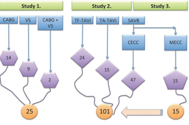
We studied the peri-operative changes of immune sensitivity in different patient populations. In the population planned to undergo cardiac surgery, patients selected for CABG, VS or for a combined cardiac surgical procedure (CABG + VS) treated on CECC or MECC were eligible to be included in the studies. In the population planned to undergo interventional cardiology procedure, patients with severe AS, defined as an aortic valve area <1 cm2 and an aortic valve mean pressure gradient >40 mmHg, allocated either to SAVR using CECC or TAVI using the transapical or the transfemoral access route were eligible.
Figure 1. illustrates patients` distribution between the studies.
In the study periods all elective patients were screened for study purposes. General inclusion criteria were: age ≥18 years and written informed consent. The exclusion criteria included local or systemic infection apparent on pre-treatment clinical investigation or in routine laboratory tests, chronic intake of immunosuppressive drugs, steroids or antibiotics, active liver or renal disease including renal failure requiring hemodialysis. Likewise, emergency treatment or patients’ participation in another trial was an exclusion criterion.
Table 1. summarizes the main determinants of each study.
All procedures and subsequent clinical or laboratory investigations were performed as a single-center prospective observational study in a tertiary referral hospital. Patients`
recruitment was made in a consecutive fashion according to the order of allocation to the cardiac surgical or interventional cardiology program. In patients presenting with AS, the appropriate treatment modality (surgical vs. interventional) was selected by the Institutional Heart Team consisting of cardiac surgeons and interventional cardiologists. TAVI was considered appropriate in respective patients with an age >80 years and/or a logistic EuroSCORE (ES) calculated risk of mortality ≥15 %, a Society of Thoracic Surgeons
19
(STS) score risk of mortality ≥10 %, or in those with an age >70 years with a predicted or prohibitive risk of morbidity or/and mortality for the surgical aortic valve replacement.
Source: own design
Figure 1. Patients` distribution between the studies. The figure shows patients` allocation to different cardiac surgical and interventional cardiology therapies (blue square), the overall number of individuals in each treatment group (purple shapes) and in each study (orange circles).
All patients gave written informed consent for cardiac surgery with extracorporeal circulation, interventional cardiology procedure, the related anesthesia technique and to the use of anonymized data for research purposes. Ethical approval for all study-relevant investigations was provided by the local Ethics Committee.
DOI:10.14753/SE.2016.1872
20 Table 1. Main determinants of the studies.
Study 1 Study 2 Study 3
Modality
prospective observational trial
randomized controlled trial Patient population CABG, VS,
CABG + VS
VS (AS) and TAVI
VS (AS)
Inclusion criteria ≥18 yrs., IC
Exclusion criteria infection, intake of immunosuppressant or antibiotics, liver disease, renal disease, emergency treatment, participation in another study Primary aims
(comparators)
CECC-induced immunomodulation (established markers vs. CD62L assay)
Inflammatory response in patients with AS
(SAVR vs. TAVI)
Inflammatory response in patients with AS
(CECC vs. MECC) Determined
inflammatory markers
CD62L shedding and
IL-8, HLA-DR, sCD62L, sTLR-2, ADAM17
IL-6, Il-8, IL-10 HLA-DR sCD62L CRP, WBC Sampling time
points
Pre-operatively
End of surgery
48 hrs postop.
Pre-operatively
4, 24 and 48 hrs postop.
Legend: CABG indicates coronary artery bypass graft surgery; VS, cardiac valve surgery;
AS, aortic valve stenosis; TAVI, transcatheter aortic valve implantation; yrs., years; IC, informed consent; CECC, conventional extracorporeal circulation; MECC, minimized extracorporeal circulation; SAVR, surgical aortic valve replacement; CD62L, L-selectin;
IL, interleukin; HLA, human leukocyte antigen; s, soluble; TLR, toll-like receptor; ADAM, ADAM metalloproteinase domain 17; CRP, C-reactive protein; WBC, white blood cell.
21
3.2. Cardiac surgery and extracorporeal perfusion techniques1
All single cardiac valve surgery (i.e., aortic valve replacement, mitral valve replacement or reconstruction) and combined cardiac surgical procedures were performed using CECC. Coronary artery bypass graft surgery was solely performed using MECC.
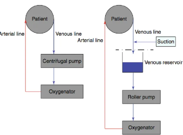
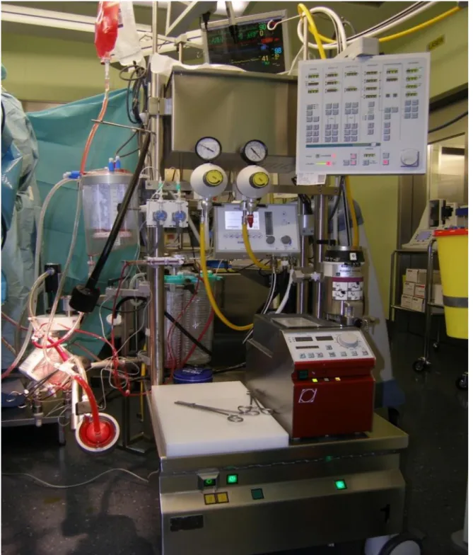
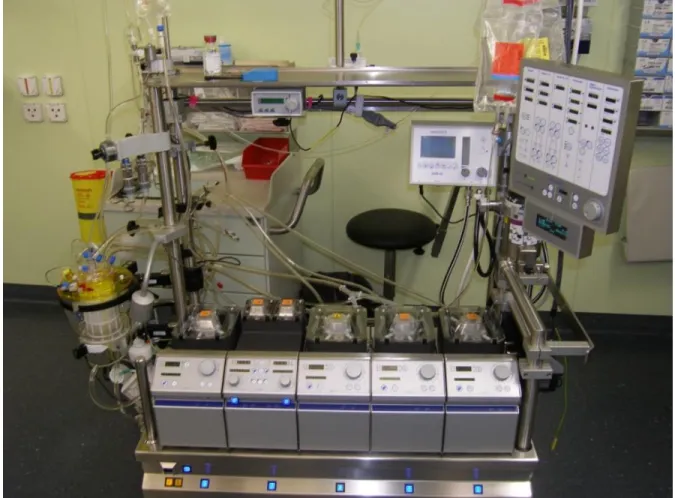
Figures 2-4. illustrate the schematic composition and the real life setup of the different extracorporeal systems as used in the studies.
The MECC system (Jostra AG, Hirrlingen, Germany) used in this study consisted of a centrifugal pump (Rotaflow, Maquet, Gossau, Switzerland), a microporous capillary membrane oxygenator (Quadrox; Maquet, Gossau, Switzerland, Capiox FX25; Terumo, Tokyo, Japan or Affinity Fusion, Medtronic, MN, USA), an anesthesia vapor (isoflurane), a pulmonary artery vent line and a suction device, which controlled blood aspiration by an optoelectrical sensor (SmartSuction, Cardiosmart, Muri, Switzerland). In contrast to MECC, CECC included a roller pump (Maquet, Rastatt, Germany), a hardshell venous reservoir, conventional cardiotomy suction, and a left ventricular vent line inserted via the right upper pulmonary vein. MECC was primed with 600 mL of Ringer’s solution and 5.000 IU of heparin, whereas CECC was primed with 500 mL of hydroxyethyl starch (130/0.4), 1,000 mL of Ringer’s solution, 100 mL of 20 % mannitol and 10.000 IU of heparin. None of the extracorporeal circuits used a heparin coating or neutrophil filter.
Figure 2. illustrates the different composition of the CECC and the MECC system.
Continuous anesthesia monitoring consisted of ECG, pulse oximetry, capnography, invasive arterial and central venous pressure, transesophageal echocardiography and processed EEG (BISTM, Covidien, Medtronic, MN, USA).
1 except for the figures, the text is quoted from Ref. 85
DOI:10.14753/SE.2016.1872
22
General anesthesia induction was performed with midazolam, etomidate and sufentanil, followed by isoflurane and sufentanil maintenance. Cardiac surgery was performed through a median sternotomy. After heparinization (MECC, 400 IU/kg ideal body weight (IBW);
CECC, 500 IU/kg IBW), extracorporeal circulation was established, with arterial inflow through the ascending aorta and venous drainage through a two-stage cannula secured in the right atrium. An activated clotting time (ACTkaolin) of >480 s (ACT Plus, Medtronic, MN, USA) was targeted in both groups and monitored every 20 min.
All patients received a bolus of tranexamic acid (30 mg/kg IBW) followed by a continuous infusion (15 mg/kg IBW/h) until sternal closure. Extracorporeal circuit flow rates were set to 2.0 L/min/m2 body surface area (BSA) in MECC, and 2.4 L/min/m2 BSA in CECC. Core temperature was allowed to drift to a nadir of 34 °C. Myocardial protection was applied with a single dose of 100 mL of crystalloid cardioplegia (Cardioplexol, Bichsel Laboratory, Interlaken, Switzerland) followed by high-potassium cold blood cardioplegia, with repetition if any electrical or mechanical activity was observed. Following separation from extracorporeal bypass, heparin was neutralized with protamine sulfate in a ratio of 1:1 with regard to the initial bolus.
23
Source: Department of Cardiothoracic Surgery, University Hospital Bern, Switzerland
Figure 2. Schematic configuration of the MECC (left picture) and the CECC (right picture) system. Note that in the MECC system consists of a different pump (centrifugal pump vs.
roller pump) and does not have a venous reservoir. Likewise, suctioning of blood from the surgical field (optoelectrical device vs. conventional suction device) and the blood venting technique (pulmonary artery vent vs. pulmonary vein vent) are divergent.
DOI:10.14753/SE.2016.1872
24
Source: Department of Cardiothoracic Surgery, University Hospital Bern, Switzerland
Figure 3. MECC setup as used in the study.
25
Source: Department of Cardiothoracic Surgery, University Hospital Bern, Switzerland
Figure 4. CECC setup as used in the study.
DOI:10.14753/SE.2016.1872
26 3.3. Interventional cardiology procedures
TA-TAVI and TF-TAVI were performed according to the strategy described previously in detail.[66, 67] There was no pre-specified recommendation regarding access route selection (transfemoral vs. transapical), which was chosen according to the decision of the interventionist and the local expertise.
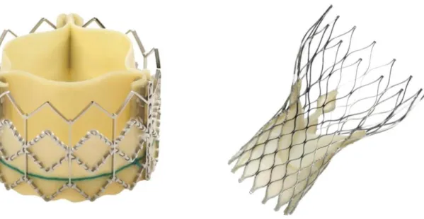
In all TA-TAVI cases the balloon-expandable Edwards Sapien XT aortic bioprosthesis (Edwards Lifesciences, CA, USA) was selected. In TF-AVI procedures depending on the aortic valve and annular calcific load and distribution, either the Edwards Sapien XT aortic bioprosthesis or the self-expanding CoreValve Revalving System (Medtronic, MN, USA) was chosen.
Figure 5. displays the two bioprostheses used in the studies.
TA-TAVI included echocardiography-guided anterolateral mini-thoracotomy, access site preparation, insertion of the apical introducer and balloon aortic valvuloplasty of the calcified native aortic valve during rapid ventricular pacing followed by angiography- guided delivery of the TA-TAVI device. The self-expanding CoreValve was deployed after puncture of the femoral artery and introduction of the bioprosthesis in the aortic annulus through the descending aorta.
Figure 6. displays the different access routes indicating bioprosthesis positioning into the aortic annulus.
All TA-TAVI procedures were performed in general endotracheal anesthesia using total intravenous anesthesia (TIVA) with propofol 2 % (Disporivan™, AstraZeneca AG, Zug, Switzerland) combined with remifentanil (Ultiva™, GalaxoSmithKline AG, München- buchsee, Switzerland). Continuous anesthesia monitoring consisted of ECG, pulse oximetry, capnography, invasive arterial and central venous pressure, transesophageal echocardiography and processed EEG (BISTM, Covidien, Medtronic, MN, USA).
27
Source: Edwards LifeSciences. www.edwards.com
Figure 5. Transcatheter aortic valve implantation using the Edwards Sapien XT aortic bioprosthesis (transfemoral and transapical cases, left picture) and the CoreValve Revalving System (only transfemoral cases, right picture). Note the divergent form and implantation procedure of the bioprostheses.
DOI:10.14753/SE.2016.1872
28
Source: Edwards LifeSciences. www.edwards.com
Figure 6. Transcatheter aortic valve implantation using the transfemoral (left picture) and the transapical (right picture) access route. Note that in TF-TAVI no myocardial puncture and incision occurs.
Post-operatively, patients were either extubated in the angiography suite, or remained intubated until circulatory and respiratory stabilization. All TA-TAVI and all intubated TF- TAVI patients were transferred to the intensive care unit, whereas extubated TF-TAVI patients transferred to the intermediate care unit (IMC).
29
3.4. Blood sampling and determination of inflammatory response markers1
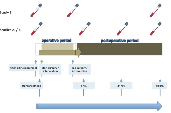
In all cases, blood was collected in three different tubes (2x EDTA tubes with a filling volume of 2.7 mL and 1x heparinized tube with a filling volume of 4.7 mL), which were immediately transferred to the in-house laboratory. The first sample (baseline value) was drawn in all studies from the patient`s arterial line prior to the application of anesthetic drugs. The subsequent samples were drawn from the central venous line after 4 (Study 2 and Study 3), 24 (Study 2 and Study 3) and 48 hrs (Studies 1, 2 and 3) post-operatively.
Figure 7. displays the blood sampling strategy in the studies.
Source: own design
Figure 7. Frequency of blood sampling in each study. The figure illustrates the blood sampling time points for inflammatory markers in relation to the peri-procedural processes.
DOI:10.14753/SE.2016.1872
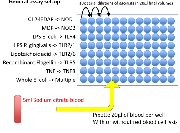
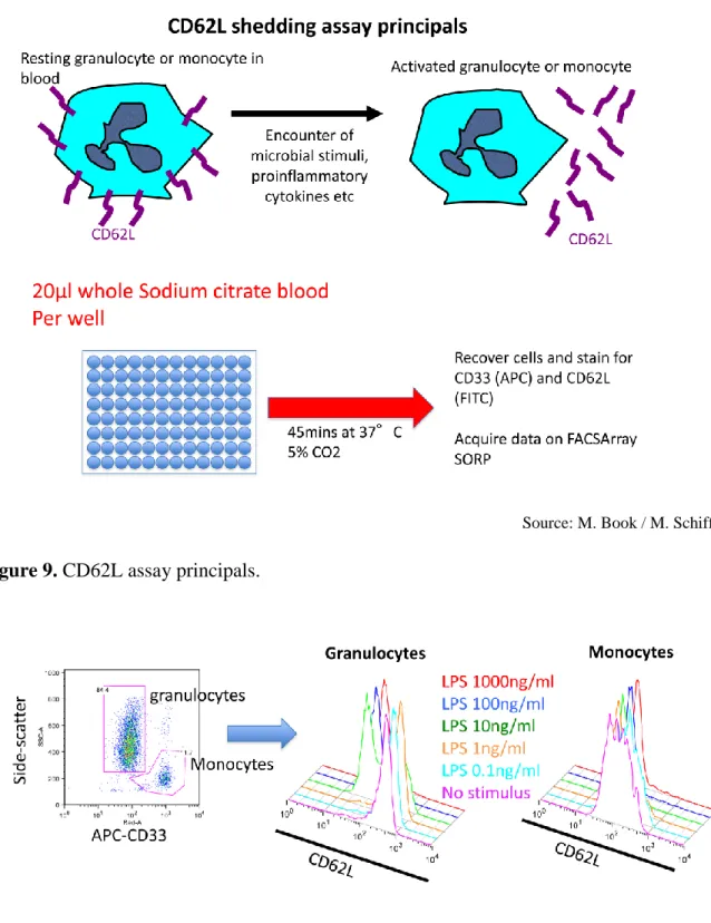
30 3.4.1. CD62L shedding assay
Twenty-five ml citrate whole blood was stimulated with 25 ml of six 10-fold dose- titrations starting at 10 mg/ml (LTA, Invivogen, San Diego, USA), 20 ng/ml (TNF, R&D).
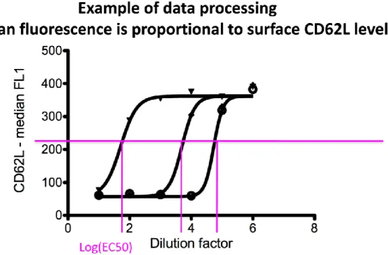
After 45 minutes incubation at 37 °C / 5 % CO2, cells were washed in PBS/BSA 1% and stained with 25 ml PBS/BSA 1% containing APC-anti-human CD33 (allowing identification of both granulocytes and monocytes in combination with side-scatter measurement, and remaining stable during stimulation) and FITC-anti-human CD62L antibodies (Biolegend, San Diego, USA) for 15 minutes. Red blood cells were lysed in 200 ml FACS Lysis solution (BD Biosciences, San Diego, USA) and acquired on a FACSArray SORP (BD Biosciences, San Diego, USA). Blood monocytes and granulocytes were distinguished on the basis of CD33 expression and side scatter using FlowJo Software (Tree Star Inc., Ashland, USA). Median FITC fluorescence intensity is computed for each sample’s granulocytes and monocytes and plotted against dilution factor to enable parallel analysis of multiple agonists. Four-parameter curves are fitted using non-linear regression and LogEC50 values were extracted, corresponding to the dilution factor giving 50%
granulocyte or monocyte CD62L-shedding. The corresponding ligand concentration was calculated and plotted for each ligand.
Figure 8. – 11. displays the operational steps of CD62L assay.
3.4.2. HLA-DR expression
Fifty microliters of heparinized blood were stained with 20 µL of Quantibrite™
anti-human-HLA-DR PE (phycoerythrin)/anti-monocyte PerCP (peridinin chlorophyll protein)-Cy5.5 (anti-human-CD14 and anti-human-CD64) (BD Biosciences) at room temperature in the dark for 25 min. Red blood cells were subsequently lysed with FACS lysis solution (BD Biosciences) for 5-10 min and washed twice with phosphate-buffered saline solution (Sigma-Aldrich) and fixed with 400 µL of 4 % paraformaldehyde. One unstained sample was treated in the same manner. The fluorescence intensity of the samples
31
was measured on an LSR II (BD Biosciences) using the software FACSDiva v6.1.3 (BD Biosciences) as duplicates. A total of 500-1,000 monocyte events were recorded. For quantification of the signal, Quantibrite™ PE beads (BD Biosciences) were acquired with each measurement. The FACS data were analyzed using FlowJo v7.5 (Treestar) with gating for CD14- and CD64-positive cells (monocytes). The HLA-DR PE channel was calibrated using the data from the PE beads, which allows fluorescence intensity to be correlated with the mean number of PE molecules per cell. The results were recorded as the median of the calibrated PE channel fluorescence intensity of each sample. The mean and standard deviation were calculated for the duplicate samples, and an analysis of variance (ANOVA) was performed using GraphPad Prism v5.04 (GraphPad Software).
Source: M. Book / M. Schiff
Figure 8. General CD62L assay setup.
DOI:10.14753/SE.2016.1872
32
Source: M. Book / M. Schiff
Figure 9. CD62L assay principals.
Source: M. Book / M. Schiff
Figure 10. Identification of granulocytes and monocytes.
33
Source: M. Book / M. Schiff
Figure 11. Data processing to determine logEC50.
3.4.3. ELISA
Plasma from each time point was separated from 5 mL of EDTA-whole blood by centrifugation at 3,000 g for 5 min and stored at -80 °C.
3.4.3.1. IL-6
Interleukin-6 levels were measured by a sandwich ELISA kit (eBioScience), and all samples were diluted 1:2 using the reagents provided by the kit. The standards ranged from
DOI:10.14753/SE.2016.1872
34
1.56 to 100 pg/mL. The provided 96-well plate was treated according to the manufacturer’s instructions, and all standards, controls and samples were loaded in duplicate. Optical density was measured using an eL800 microplate reader (Biotek Instruments) set to record at 450 and 630 nm. Blank values and OD values at 630 nm were subtracted from all OD 450 nm values, and the IL-6 concentration of all samples was determined by a 4-parameter curve fit of the standards (Gen5 v.1.09 software, Biotek Instruments). Samples that fell out of the standard curve range were repeated at a higher dilution. According to the manufacturer, the sensitivity and intra- and inter-assay variations were 0.92 pg/mL, 3.4 % and 5.2 %, respectively. A coefficient of variation of the sample duplicates of below 20 % was considered acceptable.
3.4.3.2. IL-8
Interleukin-8 levels were measured by a sandwich ELISA kit (eBioScience), and all samples were diluted 1:2 using the reagents provided by the kit. The standards ranged from 15.6 pg/ml to 1000 pg/ml. The provided 96-well plate was treated according to the manufacturer’s instructions and all standards, controls and samples were loaded in duplicate. Optical density was measured using an eL800 microplate reader (Biotek Instruments) set to record at 450 and 630 nm. Blank values and OD values at 630 nm were subtracted from all OD 450 nm values and the IL-8 concentration of all samples was determined by a 4-parameter curve fit of the standards (Gen5 v.1.09 software, Biotek Instruments). Samples that fell out of the standard curve range were repeated at a higher dilution. According to the manufacturer, the sensitivity and the intra- and inter-assay variations were 2 pg/ml, 6.3 % and 8.7 %, respectively. A coefficient of variation of the sample duplicates of below 20 % was considered acceptable.
35 3.4.3.3. IL-10
Interleukin-10 levels were measured by a sandwich ELISA kit (eBioScience), and all samples were diluted 1:2 using the reagents provided by the kit. The standards ranged from 3.1 to 200 pg/mL. The provided 96-well plate was treated according to the manufacturer’s instructions, and all standards, controls and samples were loaded in duplicate. Optical density was measured using an eL800 microplate reader (Biotek Instruments) set to record at 450 and 630 nm. Blank values and OD values at 630 nm were subtracted from all OD 450 nm values, and the IL-10 concentration of all samples was determined by a 4-parameter curve fit of the standards (Gen5 v.1.09 software, Biotek Instruments). Samples that fell out of the standard curve range were repeated at a higher dilution. According to the manufacturer, the sensitivity and intra- and inter-assay variations were 1.0 pg/mL, 3.2 % and 5.6 %, respectively. A coefficient of variation of the sample duplicates of below 20 % was considered acceptable.
3.4.3.4. sCD62L
Soluble L-selectin (sCD62L) levels were measured by a sandwich ELISA kit (eBioScience), and all samples were diluted 1:200 using the reagents provided by the kit.
The standards ranged from 0.4 to 25 ng/mL. The provided 96-well plate was treated according to the manufacturer’s instructions, and all standards, controls and samples were loaded in duplicate. Optical density was measured using an eL800 microplate reader (Biotek Instruments) set to record at 450 and 630 nm. Blank values and OD values at 630 nm were subtracted from all OD 450 nm values, and the sCD62L-selectin concentration of all samples was determined by a 4-parameter curve fit of the standards (Gen5 v.1.09 software, Biotek Instruments). Samples that fell out of the standard curve range were repeated at a higher dilution. According to the manufacturer, the sensitivity and intra- and inter-assay variations were 0.198 pg/mL, 3.7 % and 4.2 %, respectively. A coefficient of variation of the sample duplicates of below 20 % was considered acceptable.
DOI:10.14753/SE.2016.1872
36 3.4.3.5. sTLR-2
Soluble toll-like receptor (sTLR)-2 plasma concentrations were detected using a human TLR-2 ELISA kit (Cusabio, Wuhan, China). Samples were diluted 1:3 and measured together with standards and blanks in duplicate. The detection range (standard curve) was 0.312–20 ng/ml, intra- and interassay variation was given as 8 and 10 % CV by the manufacturer.
3.4.3.6. ADAM 17 (TACE)
All samples were measured undiluted and in duplicates using a TACE human ELISA kit (Abcam, Cambridge, UK) according to the manufacturer’s instructions. The detection range (standard curve) was 78.15 – 5000 pg/ml, intra- and interassay variation was given as 10 and 12 % CV by the manufacturer.
3.4.3.7. CRP
C-reactive protein (CRP) levels were measured by an ELISA kit (eBioScience), and all samples were diluted 1:8,000, 1:16,000, 1:32,000, 1:40,000 or 1:64,000 using the reagents provided by the kit. The dilution was determined by several pre-tests. The standards ranged from 0.15 to 10 ng/mL. The provided 96-well plate was treated according to the manufacturer’s instructions, and all standards, controls and samples were loaded in duplicate. Optical density was measured using an eL800 microplate reader (Biotek Instruments) set to record at 450 and 560 nm. Blank values and OD values at 560 nm were subtracted from all OD 450 nm values, and the IL-6 concentration of all samples was determined by a 4-parameter curve fit of the standards (Gen5 v.1.09 software, Biotek Instruments). Samples that fell out of the standard curve range were repeated at a higher dilution. A coefficient of variation of the sample duplicates of below 20 % was considered acceptable.
37 3.4.4. Determination of myocardial injury
Intra-procedural cardiac tissue injury was determinated by the analysis of IL-6 levels and their course during the observation period. As reported by previous investigators, Il-6 levels are the highest in the coronary sinus blood under cardiopulmonary bypass conditions, indicating that cardiac myocytes represent the major source in the release of this pro-inflammatory cytokine when myocardial ischemia occurs.[33, 60, 68]
DOI:10.14753/SE.2016.1872
38 3.5. Statistical analyses
3.5.1. Study 1: Assessment of the immune sensitivity in cardiopulmonary bypass procedures using CD62L shedding assay.
Comparisons between the groups and repeated measurements were tested for normality by the Shapiro-Wilk test. In cases of non-normal distribution, non-parametric testing was performed by Friedman Repeated Measures ANOVA on ranks with the Tukey post-hoc test for all pairwise multiple comparisons. Comparisons between the different tests for immune sensitivity were performed using the Pearson product moment correlation.
3.5.2. Study 2 and Study 3: Investigation of inflammatory response to treatment of AS and investigation of inflammatory response to surgical treatment of AS.
Different treatment strategies for aortic valve stenosis were compared using a general linear model for repeated measures (as in Study 1) followed by Bonferroni correction. The Greenhouse-Geisser correction was applied to detect inter-subject effects.
3.5.3. General considerations
Data are presented as integral number (percentage), mean ± standard deviation or median with interquartile ranges as appropriate. A p-value <0.05 was considered significant. In case of multiple comparisons and where Bonferroni adjustment of the Wilcoxon tests were performed, p values <0.0125 (p =0.05/4) were considered significant.
Statistical analyses were performed with SigmaPlot software version 12.0 (Systat Software Inc., San Jose, CA, USA) in Study 1, and with SPSS v22 (IBM Statistics; USA) in the Studies 2 and 3.
39 4. RESULTS
4.1. Assessment of the immune sensitivity in cardiopulmonary bypass procedures using CD62L shedding assay.
4.1.1. Patients
Overall, 25 consecutive patients completed Study 1. Among these, 14 patients underwent CABG, 9 patients VS (aortic valve replacement (5 patients) and mitral valve replacement (1 patient) or reconstruction (3 patients)) and 2 patients a combined cardiac surgery procedure (CABG with aortic valve replacement or CABG with mitral valve reconstruction). Eleven patients were treated using CECC and fourteen patients using MECC.
Table 2. summarizes the basic clinical and procedural characteristics in the peri-operative period.
4.1.2. CD62L shedding assay
Granulocytes and monocytes were stimulated either with lipoteichoic acid (LTA) from the gram positive bacterium Staphylococcus aureus or with tumor necrosis factor alpha (TNF-α) to assess changes in the sensitivity of inflammatory effector cells.
Granulocyte and monocyte sensitivity to LTA decreased at the end of cardiac surgery but recovered to the baseline after 48 hrs. Decreased immune sensitivity was apparent as a roughly 10-fold increase of the initial LTA concentration was necessary to cause shedding of 50 % of CD62L from the cell surface of granulocytes or monocytes.
DOI:10.14753/SE.2016.1872
40
Table 2. Basic clinical and procedural characteristics in the peri-operative period displayed according to the type of surgery and type of extracorporeal circulation.
Type of surgery All CABG VS CABG + VS p
Number of patients, n 25 14 9 2
Age, yrs. 67 ± 12 71 ± 10 63 ± 12 54 ± 19 ns
Female sex, n 12 (48) 6 (43) 5 (55) 1 (50) ns
Diabetes, n 9 (36) 7 (50) 2 (22) 0 (0) ns
EuroScore (additive) 6 ± 3 5 ± 2 6 ± 4 6 ± 7 ns
Surgery time, min. 230 ± 58 228 ± 41 232 ± 77 235 ± 106 ns Time on ECC, min 83 ± 42 60 ± 14 112 ± 51 105 ± 49 .003
LOS, days 9.7 ± 2.7 9.4 ± 2 10.3 ± 3.8 9 ± 0 ns
In-hospital mortality 0 (0) 0 (0) 0 (0) 0 (0) ns
Type of ECC CECC MECC p
Number of patients, n 11 14
Age, yrs. 61 ± 12 71 ± 10 ns
Female sex, n 6 (54) 6 (43) ns
Diabetes, n 2 (18) 7 (50) ns
EuroScore (additive) 7 ± 4 5 ± 2 ns Surgery time, min. 233 ± 77 228 ± 41 ns Time on ECC, min 211 ± 48 60 ± 14 < .001
LOS, days 10 ± 3 9 ± 2 ns
In-hospital mortality 0 (0) 0 (0) ns
Legend: CABG, coronary artery bypass graft surgery; VS, valve surgery; ECC, extracorporeal circulation; CECC conventional extracorporeal circulation; MECC, minimized extracorporeal circulation; LOS, length of post-operative hospital stay. Values are presented as number (percentage) and as mean ± standard deviation.
41 Figure 12. and Table 3. display the relevant results.
Source: Reference 69.
Figure 12. Granulocyte and monocyte sensitivity to LTA stimulation
Legend. LC50 represents the concentration of LTA which was necessary to cause shedding of 50 % of the membrane-bound CD62L molecules of granulocytes or monocytes. Thus, high LTA concentration values are associated with decreased sensitivity of the affected cells. Black lines = data from an individual patient; orange lines = median values of all patients at a certain blood sampling point with interquartile ranges (= purple lines).
*indicates p <0.05 compared with the previous sampling time point.
DOI:10.14753/SE.2016.1872
42
Table 3. Granulocyte and monocyte concentrations after activation with LTA or TNF-α at different peri-procedural time points.
Before surgery End of surgery 48 hrs p LTA
Granulocytes 1225 [999;2364] 9602 [1242;10000] 1177 [916;2748] .001 Monocytes 191 [112;508] 780 [113;1535] 97 [74;164] .004 TNF-α
Granulocytes 2 [2;9] 20 [2;28] 3 [2;5] .010
Monocytes 1 [0;2] 1 [1;3] 2 [1;3] .057
Legend: LTA indicates lipoteichoic acid; TNF-α, tumor necrosis factor alpha. Values are median with interquartile ranges (q25/q75).
Likewise, TNF-α stimulation of granulocytes showed a decreased sensitivity of these cells at the end of cardiac surgery as revealed by a 10-fold increased TNF-α concentration required to produce 50 % shedding of the membrane-bound fraction of CD62L. In contrast, monocyte sensitivity to TNF-α stimulation was not diminished at the end of surgery and after 48 hrs.
Figure 13. and Table 3. display the relevant results.
43
Source: Reference 69.
Figure 13. Granulocyte and monocyte sensitivity to TNF-α stimulation.
Legend. LC50 represents the concentration of LTA which was necessary to cause shedding of 50 % of the membrane-bound CD62L molecules of granulocytes or monocytes. Thus, high LTA concentration values are associated with decreased sensitivity of the affected cells. Black lines = data from an individual patient; orange lines = median values of all patients at a certain blood sampling point with interquartile ranges (= purple lines).
*indicates p <0.05 compared with the previous sampling time point.
DOI:10.14753/SE.2016.1872
44
4.1.2.1. CD62L shedding between the different perfusion techniques and cardiac surgery procedures
Granulocyte and monocyte stimulation with LTA and TNF-α and associated CD62L shedding did not reveal differences between CECC and MECC or CABG, VS and combined cardiac procedures, respectively.
Table 4. summarizes the changes in LTA and TNF-α concentrations according to the ECC types.
Table 5. summarizes the changes in LTA and TNF-α concentrations according to cardiac surgery procedures.
Table 4. Impact of ECC type on LTA / TNF-α values in the peri-operative period.
CECC MECC p
LTA Before surgery
granulocytes 1770 [966;2564] 1112 [946;2342] ns
monocytes 264 [119;494] 174 [90;575] ns
After surgery
granulocytes 10000 [1242;12951] 6209 [1211;10000] ns monocytes 804 [103;2000] 769 [185;1429] ns 48 hrs granulocytes 1291 [986;2748] 1086 [830;3758] ns
monocytes 111 [74;175] 96 [59;109] ns
TNF-α Before surgery
granulocytes 2 [2;8] 3 [2;12] ns
monocytes 1 [1;2] 1 [0;2] ns
After surgery
granulocytes 20 [2;36] 12 [2;25] ns
monocytes 2 [1;4] 1 [0;4] ns
48 hrs granulocytes 3 [2;5] 4 [1;6] ns
monocytes 2 [1;2] 2 [1;7] ns
Legend: LTA indicates lipoteichoic acid; TNF-α, tumor necrosis factor alpha; CECC, conventional extracorporeal circulation; MECC, minimized extracorporeal circulation.
Values are median with interquartile ranges (q25/q75).
45
Table 5. Impact of different cardiac surgery procedures on LTA / TNF-α values in the peri- operative period.
CABG VS p
LTA Before surgery
granulocytes 1112 [859;2133] 1770 [1087;7319] ns
monocytes 319 [93;634] 191 [117;379] ns
After surgery
granulocytes 10000 [1526;10000] 9205 [1195;22629] ns monocytes 870 [355;1918] 290 [98;1397] ns 48 hrs granulocytes 1086 [850;2973] 1618 [1013;5220] ns
monocytes 96 [63;110] 116 [74;192] ns
TNF-α Before surgery
granulocytes 3 [2;10] 2 [2;9] ns
monocytes 1 [0;2] 1 [0;3] ns
After surgery
granulocytes 4 [2;22] 28 [2;50] ns
monocytes 1 [0;3] 2 [2;4] ns
48 hrs granulocytes 3 [1;5] 3 [2;4] ns
monocytes 2 [1;6] 2 [1;2] ns
Legend: LTA indicates lipoteichoic acid; TNF-α, tumor necrosis factor alpha; CABG coronary artery bypass graft surgery; VS, valve surgery. Values are median with interquartile ranges (q25/q75).
DOI:10.14753/SE.2016.1872
46 4.1.3. HLA-DR expression
Monocytic HLA-DR expression could be determinated in 17 of the 25 patients (68
%). Median density of surface HLA-DR decreased significantly at the end of surgery. After 48 hrs monocyte density did not recover to the baseline, but rested significantly reduced in comparison to the value at the first sampling time point.
Table 6. and Figure 14. display the results.
Table 6. Monocytic HLA-DR density in the peri-operative period.
Before surgery End of surgery 48 hrs p HLA-DR
CD14/CD64 19204
[12740;24268]
9694
[7907;11039]
6969
[4303;11976]
.001
Legend: HLA-DR indicates human leukocyte antigen; CD14/64, cluster of differentiation 14/64 monocytes. Values are median with interquartile ranges (q25/q75).
47
Source: Reference 69.
Figure 14. Monocytic HLA-DR expression
Legend. HLA-DR/cell means HLA-DR molecules per CD14 and CD64 positive monocytes. Black lines = data from an individual patient; orange lines = median values of all patients at a certain blood sampling point with interquartile ranges (= purple lines).
*indicates p <0.05 compared with the first sampling time point.
DOI:10.14753/SE.2016.1872
48
4.1.4. Association of HLA-DR expression data with CD62L shedding data.
Regression analyses revealed no significant association between the functional sensitivity analysis of the immune system (CD62L shedding) or to the monocytic HLA-DR expression values in the peri-operative period:
LTA / TNF: r = 0.10 p = 0.400 LTA / HLA-DR: r = -0.21 p = 0.078 TNF / HLA-DR: r = -0.13 p = 0.280
4.1.5. IL-8 plasma level
IL-8 plasma levels were determined in 18 of 25 patients (72 %) due to limited sample volume. IL-8 levels significantly increased at the end of surgery as compared to the first sampling point. After 48 hrs IL-8 decreased in comparison to the sampling point at the end of surgery, however, the pre-operative value was not reached. The IL-8 concentration after 48 hrs was still significantly increased when compared to the first sampling time point.
Figure 15. shows the peri-operative course of IL-8 plasma levels.
4.1.6. Plasma levels of the soluble factors
sCD62L plasma levels significantly decreased at the end of surgery. After 48 hrs the levels were still significantly lower as at the first sampling time point indicating that sCD62L did not reach pre-operative values after 48 hrs.
Figure 16. shows the peri-operative course of sCD62L.
49
Source: Reference 69.
Figure 15. Peri-operative concentration of IL-8.
Legend. Black lines = data from an individual patient; orange lines = median values of all patients at a certain blood sampling point with interquartile ranges (= purple lines).
*indicates p <0.05 compared with the first sampling time point.
DOI:10.14753/SE.2016.1872
50
sTLR-2 showed a significant increase at the end of surgery. After this peak sTLR-2 levels decreased to a minimum at 48 hrs showing a significant difference to the sampling point at the end of surgery.
ADAM17 levels increased constantly during the peri-operative course, however, a significant difference between two sampling points cannot be detected.
Table 7. displays the peri-operative values of sTLR-2 and ADAM17.
Table 7. sTLR-2 and ADAM17 concentrations in the peri-operative course.
Before surgery End of surgery 48 hrs p sTLR-2 1.4 [1.2;2.4] 2.5 [1.9;3.6] 0.8 [0.6;1.2] .004 ADAM17 436 [227;849] 574 [274;769] 761 [402;1145] .401 Legend: sTLR indicates soluble toll-like receptor; ADAM 17, ADAM metalloproteinase domain 17. Values are median with interquartile ranges (q25/q75).
51
Source: Reference 69.
Figure 16. Peri-operative course of sCD62L.
Legend. Black lines = data from an individual patient; orange lines = median values of all patients at a certain blood sampling point with interquartile ranges (= purple lines).
*indicates p <0.05 compared with the first sampling time point.
DOI:10.14753/SE.2016.1872
52
4.2. Investigation of inflammatory response to treatment of AS and investigation of inflammatory response to surgical treatment of AS.
Study 2 and Study 3 were performed independently with either a prospective observational (Study 2) or a randomized controlled design (Study 3). Nevertheless, both studies investigate the same topic (AS) in the same patient population: in Study 2 cardiac surgical patients are investigated in comparison to interventionally treated patients (SAVR vs.
TAVI) and in Study 3 cardiac surgical patients are investigated relating to different extra- corporeal perfusion techniques (SAVR with CECC vs. SAVR with MECC), respectively.
Moreover, in both studies the same inflammatory markers and the same sampling points were used.
Thus, for a better readability and related understanding of the scientific message, the results of both studies and their interpretation are presented together in the following chapters.
4.2.1. Patients
Overall, 101 patients treated with a different modality (surgical or interventional) for severe symptomatic aortic stenosis were compared. Patients allocation to the different treatment modalities were as follows: TF-TAVI: 24 patients; TA-TAVI: 15 patients; SAVR with CECC: 47 patients and SAVR with MECC: 15 patients (Figure 1.).
Patients` characteristics were significantly different in the four groups. Patients in the MECC group were the youngest, whereas patients in the TF-TAVI group were the oldest (mean age MECC: 65 ± 8yrs.; TF-TAVI 82 ± 4 yrs., p <0.001). The extent of heart failure, as a result of severe aortic stenosis over years, represented by the NYHA classification, was in the majority of surgical patients mild (percentage of NYHA class II in MECC: 60 %;
CECC: 49 % vs. others, p =0.02). In contrast, in the interventional groups the extent of heart failure was severe (percentage of NYHA class III in TF-TAVI: 62 % and TA-TAVI 66 % vs. others, p =0.01). Co-morbidities, as coronary artery disease (CAD), chronic obstructive pulmonary disease (COPD) or a higher procedural risk (redo procedure) were more frequently present in patients selected for an interventional treatment (e.g., percentage
53
of patients with CAD: TF-TAVI: 58 % vs. others, p <0.001). The longest procedural time was present in the CECC group, whereas TF-TAVI was performed the fastest (median procedural time: CECC: 165 min. vs. TF-TAVI 75 min., p <0.001). Intermediate care unit (IMC) or intensive care unit (ICU) stay was roughly double in patients selected for TF- TAVI as in the CECC group (median stay: TF-TAVI 48 hrs vs. CECC: 22 hrs, p <0.001).
Systemic inflammatory syndrome (SIRS), defined according to the current guidelines, was present in 27 % of TA-TAVI patients, whereas other groups were only minimally affected (percent of patients with SIRS: TA-TAVI: 27 % vs. others, p = 0.02).
Table 8. summarizes patients` pre-operative and procedural characteristics.
DOI:10.14753/SE.2016.1872