Chapter 5
Altered Potassium Ion Homeostasis in Hearing Loss
Altered Expression of Connexins and Kir2.1 Potassium Ion Channels in Hearing Loss Patients
Viktoria Szuts, Janos Andras Jarabin, Nikoletta Nagy, Ferenc Otvos, Roland Nagy, Attila Nagy,
Katalin Halasy, Laszlo Rovo, Marta Szell and Jozsef Geza Kiss
Additional information is available at the end of the chapter http://dx.doi.org/10.5772/intechopen.77732
Provisional chapter
© 2016 The Author(s). Licensee InTech. This chapter is distributed under the terms of the Creative Commons Attribution License (http://creativecommons.org/licenses/by/3.0), which permits unrestricted use, distribution, and reproduction in any medium, provided the original work is properly cited.
Altered Potassium Ion Homeostasis in Hearing Loss
Altered Expression of Connexins and Kir2.1 Potassium Ion Channels in Hearing Loss Patients
Viktoria Szuts, Janos Andras Jarabin,
Nikoletta Nagy, Ferenc Otvos, Roland Nagy,
Attila Nagy, Katalin Halasy, Laszlo Rovo, Marta Szell and Jozsef Geza Kiss
Additional information is available at the end of the chapter
Abstract
Connexins, Kv-type ion channels, and pannexins have a dominant role in maintaining the potassium ion homeostasis in the cochlea. The cellular background currents are sustained by Kir2.1 ion channels; however, their involvement in the hearing system is less clear. In this study, the mutations of gap junction proteins beta 2 (GJB2), beta 3 (GJB3) and beta 6 (GJB6) were screened in the white Caucasian population in Hungary using gene mapping and immunofluorescence methods from translated proteins of these genes—connexins on blood cells. Expression of connexins and Kir2.1 ion channels was investigated in the blood cells of deaf patients prior to cochlear implantation, and the results show significantly decreased amounts of connexin26 and connexin43. In addition, the coexpression of Kir2.1 ion channels with synapse-associated 97 proteins was partially impaired. Our investiga- tion revealed a reduced level of Kir2.1 channels in deaf patients indicating a crucial role for the functional Shaker superfamily of K+ channels in the non-diseased hearing system.
Keywords: hearing loss, cochlea, GJB gene mutation, connexin26, connexin43, Kir2.1 ion channels, lymphocytes
1. Introduction
Mutations in connexin26 (Cx26) and connexin30 (Cx30) have frequently been associated with hearing loss and deafness, and some Cx26 mutations have also been found to contribute to
© 2018 The Author(s). Licensee IntechOpen. This chapter is distributed under the terms of the Creative Commons Attribution License (http://creativecommons.org/licenses/by/3.0), which permits unrestricted use, distribution, and reproduction in any medium, provided the original work is properly cited.
over 50% of hearing loss or nonsyndromic deafness cases in different human populations investigated in the last decades [1–6].
Recently, a fast and new non-invasive method has been developed in our laboratory to identify mutated genes for GJB2 leading to hearing loss or deafness [7]. This method has allowed us to collect and store the dried blood spots (DBS) in Guthrie cards for mutation analysis or expressed proteins of genes. The early detection of genetic mutations for inner ear impairment is crucial to provide hearing rehabilitation with an outstanding functional outcome, i.e. to maintain the development of the peripheral and central auditory pathway for unhindered future benefits.
There are a number of modern technologies in the field of cochlear implantation and to fulfill this aim it is crucial to address the audiological and logopedical progress in a timely manner.
Cx26 and connexin43 (Cx43) have a major role in cell-to-cell interactions and in the perme- ability of the channels for diverse ions and small molecules, which maintain the physiological condition of the cochlea [1, 8]. Connexins form hemichannels in the cochlea where the Cx26 channels are the most abundant and are involved in the potassium-recycling pathway of the cochlea. Translated proteins from GJ genes make up channels by head-to-head docking of two connexin hexamers referred to as hemichannels (HCs) or connexons; one from each neighbor- ing cell [9, 10]. Cx26 proteins were localized in the outer membrane of the cochlea.
The position of mutations in the GJB2 gene (Cx26 protein) is different across certain popula- tions. The known mutation in the populations of northern European descent is a base pair deletion at position 35 in the GJB2 gene (known as 35delG; Cx26 protein) while the 235delC mutation, which occurs more frequently in Asian populations, misses a base pair at position 235 (Cx26 protein) [11–13]. Furthermore, patients with a single base pair deletion at posi- tion 167 (167delT) (Cx26 protein) are commonly found in communities of eastern European (Ashkenazi) Jewish ancestry [14, 15].
Different connexins may be important factors in the flawless cell-to-cell interactions that maintain the fast electrogenic mechanisms between the cochlea and neuronal system [16].
Other ion currents are also involved in this process. The voltage-dependent inward rectifier potassium current (IK1) has a role in maintaining the resting membrane potential, contributing to the beginning and the final repolarization in all cells. The IK1 current in different tissues is sustained mostly by Kir2.x (Kir2.1, Kir2.2, and Kir2.3) ion channels [17–23]. Auxiliary sub- units and the synapse-associated protein 97 (SAP97) modulate the function of these channels [24, 25]. IK1 currents have a fundamental role in muscle cells, but their molecular background in the hearing system is still uncertain. We have limited information on the impact of the altered gap-junctions [26] and Kir2.1 in the inner membrane of the cochlea [20, 21, 27], the neural connection and the expressed potassium ion channels on blood cells. The exploration of the type and mechanism of these channels is one of the aims of the present work.
Hereditary hearing diseases are known to be associated with mutations, such as the auto- somal recessive non-syndromic hearing loss (ARNSHL), seizures, sensorineural deafness [5, 28–30], Pendred syndrome (PDS) [5, 29–31], deafness [7, 27–35], and rarely the hearing loss overlaps with Andersen-Tawil syndrome [2, 36, 37]. The Andersen-Tawil syndrome is characterized by mutated KCNJ2 genes expressing malfunctioned Kir2.1 proteins, which leads to functional abnormalities and causing deafness with cardiomyopathy [36–38].
The IK1 current is sustained -by ion channels that consist of heteromeric assemblies of Kir2.1, Kir2.2, and Kir2.3 α-subunits [22, 39]. This current significantly contributes to the maintenance and regulation of the resting membrane potential [17, 40] and strongly affects the final repo- larization of the action potential (AP) [8, 9, 17, 18, 41]. The Kir2.x channels are under the con- trol of the intracellular scaffolding, trafficking and regulatory proteins, which strongly adjust their physiological functions. Kir2.x isoforms colocalize with membrane associated guanylate kinase (MAGUK) proteins [24, 42]. One of these is the SAP97 anchoring protein, which has an important interaction with Kir2.x channel complexes in neuronal cells and myocytes [25, 42, 43]. Various Kir2.x isoforms may cooperate differently with the MAGUK proteins in the com- plexes that traffic Kir2.x ion channels to the plasma membrane (PM), and anchor and stabilize these channels into the sarcolemma of cardiac muscle [22, 25, 43]. The Kir2.1 channels may be exported from the Golgi in a signal-dependent manner and this process may be disrupted by certain diseases [44]. The age-dependent SAP97 expression in the human heart was evaluated for the first time in our laboratory [45], and it seems that the regulation and modulation of these ion channel complexes have a strong effect in the development of different tissue types in diverse hereditary diseases associated with cardiomyopathy and deafness too [36–39, 46].
These genes have demonstrated a gender differences and have been remodeled in cardiomy- opathy [40, 41]. IK1 currents are active during the time course of AP [20, 21, 47, 48], but their molecular background basis is poorly characterized on blood cells.
Our hypothesis states that connexins and Kir2.x ion channels, which are impaired at the tran- scription and translated protein level in deafness, have an essential function in the hearing system. The aim of this study was to monitor the mutations of expressed GJs and KCNJ2 genes in the Hungarian population and to screen the expression of hearing-related genes and proteins involved in the maintenance of the ionic homeostasis in the cochlea. An additional goal was to find an opportunity to obtain cells from the body that present the disrupted ionic status control characteristic of the pathophysiological condition of deafness. For this pur- pose, we used non-invasive methods to find the representative and irrevocable changes in peripheral blood cells of patients. Our group has previously demonstrated that connexin ion channels, especially the Cx26 mutations, are characteristic indicators in patients with hearing loss [7, 48]. In this study we investigated ion channels related to connexins in patients with altered hearing and detected the expression level of Cx26, Cx43 and Kir2.1 channels in the blood cells. Patients have hearing defects and these results highlight to the role of gap junc- tions and hemichannels in K+ removal and recycling in the inner ear, as well as possible roles for nutrient passage in the cochlea [9, 26, 48].
2. Material and methods
2.1. Human patients
Eighty consecutive, non-randomized profoundly hearing-impaired cochlear implant candi- dates were recruited in this study cohort; 45 females and 35 males aged between 0.5 and 72 years (average: 25.8 ± 24.4 years). The control group was made up of 13 subjects with objec- tively measured binaural normal hearing sensitivity; 7 females and 6 males aged between 3
and 59 years (average: 27.4 ± 17.2 years) (Tables 1 and 2). Details of patient demographics, clinical diagnoses, and genetic investigations are presented in Table 1. All patients in the dis- eased group had nonsyndromic sensorineural hearing loss, without any organic abnormali- ties (neither anatomical nor developmental alterations) including overlapping diseases. Prior to cochlear implantation, patients did not receive medication. The investigations conformed to the Declaration of Helsinki. Experimental protocols were authorized by the University of Szeged and National Scientific Research Ethical Review Boards (No. 38/2014). The blood cells were taken and kept in cold (4–6°C) for 2–4 h prior to investigations.
2.2. Objective tests used for the evaluation of hearing sensitivity 2.2.1. Impedance audiometry
2.2.1.1. Tympanometry
A measurement of compliance change in the middle ear apparatus to transmit sounds to the inner ear as air pressure is varied in the external auditory canal [49, 50]. Equipment used:
r36m Clinical Middle Ear Analyzer (Resonance, Gazzaniga, Italy).
2.2.1.2. Acoustic reflex test
Following high intensity acoustic stimulation, a sudden decrease in compliance occurs with an approximate 10 ms delay as a consequence of the contraction of the stapedius muscle.
Equipment used: r36m Clinical Middle Ear Analyzer (Resonance, Gazzaniga, Italy).
Patients Sex Age Diagnosis Mutation
1 F 5.0 ND NO
2 M 39.0 ND NO
3 F 59.0 ND NO
4 F 16.7 ND NO
5 F 57.9 ND NO
6 M 22.0 ND NO
7 M 23.9 ND NO
8 M 22.1 ND NO
9 F 24.0 ND NO
10 M 17.9 ND NO
11 F 17.9 ND NO
12 F 17.7 ND NO
13 M 3.0 ND NO
ND: Non-Diseased; F: female; M: male.
Table 1. Data of non-diseased patients without mutations in GJB2 (Cx26) genes in Hungarian population: non-diseased patients.
Patients Sex Age Diagnosis Mutation
1 M 1.7 Hearing loss
2 M 46.3 Hearing loss
3 M 2.5 Hearing loss GJB2 (heterozygote c.35delG)
4 F 4.4 Hearing loss
5 F 1.1 Hearing loss
6 M 2.8 Hearing loss
7 M 16.1 Hearing loss
8 M 23.5 Hearing loss
9 M 1.4 Hearing loss GJB2 (homozygote c.35delG)
10 F 14.5 Hearing loss
11 F 26.9 Hearing loss
12 F 27.6 Hearing loss GJB2 (homozygote c.35delG)
13 M 14.5 Hearing loss
14 F 1.8 Hearing loss
15 F 65.7 Hearing loss
16 F 64.6 Hearing loss GJB2 (homozygote c.35delG)
17 M 2.1 Hearing loss GJB2 (heterozygote c.35delG)
18 M 70.4 Hearing loss
19 F 70.0 Hearing loss
20 F 41.9 Hearing loss
21 F 1.8 Hearing loss
22 F 71.0 Hearing loss
23 F 2.2 Hearing loss GJB2 (heterozygote missense c.101 T/C
p.Met34Thr (rs35887622, CM077555, CM970679))
24 F 9.9 Hearing loss GJB2 (homozygote c.35delG)
25 M 6.9 Hearing loss GJB2 (heterozygote c.35delG)
26 F 1.9 Hearing loss GJB2 (homozygote c.35delG)
27 M 2.7 Hearing loss GJB2 (homozygote c.35delG)
28 M 1.3 Hearing loss
29 F 2.9 Hearing loss GJB2 (homozygote c.35delG)
30 M 5.4 Hearing loss GJB2 (heterozygote c.35delG)
31 M 69.6 Hearing loss
32 F 11.3 Hearing loss
33 F 54.0 Hearing loss
34 F 69.5 Hearing loss
Patients Sex Age Diagnosis Mutation
35 F 23.7 Hearing loss
36 M 3.1 Hearing loss
37 M 61.6 Hearing loss
38 F 15.4 Hearing loss
39 F 27.7 Hearing loss
40 F 76.9 Hearing loss
41 F 41.7 Hearing loss
42 M 2.0 Hearing loss
43 M 1.2 Hearing loss
44 M 2.6 Hearing loss GJB2 (heterozygote missense c.119c/a
p.Ala40Glu (rs111033296, CM051511)
45 F 48.0 Hearing loss
46 F 59.1 Hearing loss GJB2: c.101 T/C p.Met34Thr rs35887622
homozygote,
GJB2: c.139G/T p.Glu47Ter rs104894398 homozygote
47 M 52.7 Hearing loss GJB2: c.35delG homozygote
48 M 19.7 Hearing loss
49 M 2.6 Hearing loss GJB2 (homozygote c.35delG)
50 M 6.6 Hearing loss
51 M 58.5 Hearing loss
52 M 13.2 Hearing loss
53 F 27.3 Hearing loss
54 F 34.9 Hearing loss
55 F 58.9 Hearing loss
56 F 29.8 Hearing loss
57 F 49.2 Hearing loss
58 F 77.3 Hearing loss
59 M 51.2 Hearing loss
60 M 3.1 Hearing loss GJB2 (homozygote c.35delG)
61 M 52.6 Hearing loss
62 F 40.9 Hearing loss GJB2 (heterozygote c.35delG)
63 F 5.7 Hearing loss
64 M 1.5 Hearing loss GJB2 (homozygote c.35delG)
65 F 2.5 Hearing loss
66 F 2.0 Hearing loss
2.2.2. Otoacoustic emission (OAE)
Spontaneous and evoked sound waves that originate from the inner ear are transmitted through the ossicles and the eardrum into the external auditory meatus can be measured.
Equipment used: Eclipse EP15 (Interacoustics A/S, Middelfart, Denmark [51].
2.2.3. Auditory brain stem responses (ABR)
Auditory brain stem responses are far-field potentials that measure all ranges of the early auditory evoked potentials (AEP) using signal averaging [52, 53]. It consists of a series of seven waves (I-VII.) occurring within about 10 ms from stimulus onset [54]. Applied stimuli: Click and CE-Chirp LS. Equipment used: Eclipse EP15 (Interacoustics A/S, Middelfart, Denmark).
Click stimulus: used to test the critical high frequency region (approximately 2–4 kHz). Chirp stimulus: applied to activate the entire cochlea instantaneously.
2.2.4. Auditory steady state responses (ASSR)
Auditory steady state responses are AEPs that are used to objectively estimate the hearing sensitivity in individuals with normal hearing sensitivity and with various degrees and configurations of sensorineural hearing loss (SNHL) [55]. Equipment used: Eclipse EP15 (Interacoustics A/S, Middelfart, Denmark) [50, 55].
Patients Sex Age Diagnosis Mutation
67 F 64.6 Hearing loss *GJB3 (c.316C/T p.Arg106Cys
(rs147106166) heterozygote)
68 F 2.8 Hearing loss
69 F 7.1 Hearing loss
70 M 23.7 Hearing loss
71 F 66.5 Hearing loss
72 M 68.7 Hearing loss
73 F 5.9 Hearing loss GJB2 (heterozygote c.35delG)
74 M 6.2 Hearing loss
75 F 20.9 Hearing loss
76 M 9.4 Hearing loss GJB2 (heterozygote c.35delG)
77 F 45.2 Hearing loss
78 M 1.4 Hearing loss
79 F 3.4 Hearing loss
80 F 55.8 Hearing loss
F: female; M: male.*Mutation of GJB3 gene.
Table 2. Data of hearing loss patients carrying mutations in GJB2 (Cx26) genes in Hungarian population: patients with hearing loss.
2.3. Genetic investigations
Peripheral blood samples were drawn from the affected patients (n = 80). Genomic DNA was isolated following the instructions of QIAamp DNA Blood Mini Kit (Qiagen, USA). The coding regions and the flanking introns of the GJB2, GJB3 and GJB6 genes were amplified by polymerase chain reaction (PCR) using DreamTaq Green PCR Master Mix (Thermo Scientific, USA). Primer sequences used for the genetic analyses were designed with Primer 3 (http://
bioinfo.ut.ee/primer3/) online software. The quality and quantity of PCR products were mea- sured by electrophoresis on 2% agarose gel (SeaKem LE agarose, Lonza), which were used as templates for sequencing. Direct sequencing of PCR products was performed on a traditional capillary sequencer (ABI Prism 7000) and compared with the wild-type gene sequences using the Ensemble Genome Browser.
2.4. Immunofluorescence
Blood samples were collected in parallel for protein analyses in tubes containing EDTA as anti- coagulant. Blood cells of non-diseased and hearing loss patients were washed with phosphate buffer saline (PBS, pH 7.3) and centrifuged gently. Cells, mostly lymphocytes, were attached to slides, blocked with 2% bovine serum albumin and incubated overnight at 4°C with mouse anti-Cx26 (1:100 dilution, Millipore, USA), or rabbit anti-Cx43 (1:100 dilution, Cell Signaling Technology, USA), or rabbit anti-Kir2.1 antibodies (1:100 dilution, Alomone Ltd., Jerusalem) with mouse anti-SAP97 (1:300 dilution, LifeSpan, USA) [48]. After washing with PBS, the cells were incubated with Alexa 488-labeled anti-rabbit IgG (1:400 dilution; Molecular Probes, USA) plus Cy3-conjugated anti-mouse IgG (1:400 dilution; Jackson Immuno Research Laboratories, Inc., USA) secondary antibodies and counterstained with 2,4 diamino-2-phenylindol (DAPI) (Hoechst 33258) for 10 minutes. Detection was performed using an Olympus FV1000 confocal laser scanning microscope (Olympus Life Science Europa GmbH, Hamburg, Germany). The scale bar was labeled in every figure (n = 2–3 specimens for each antibody staining) from 12 patients and the analysis was performed using Fluoview ver. 4.0 programme.
3. Results
3.1. Examination of patients expecting cochlear implantation
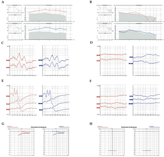
The outer ear was inspected by physical examination and found to be anatomically and func- tionally intact in all subjects prior to any hearing assessment (Figure 1A–H). Hearing sensitiv- ity was subsequently evaluated with objective procedures, applying the crosscheck principle to achieve reliable results, confirmed by multiple analyses [49]. The middle ear is essentially an impedance matching system. Impedance is simply a measure of acceptance or rejection of the energy per unit time. While transmitted through the tympanic membrane and other struc- tures of the middle ear, acoustic signals can be modified, depending on the mass, the elasticity and the resistance of the system that would determine the amount of energy accepted or reflected. The overall configuration of tympanometry and acoustic reflex tests provide greater authority and assurance in the evaluation of middle ear pathologies and conductive hearing
loss. Middle ear analysis was performed prior to any other tests to unquestionably exclude any pathology that might alter subsequent results [49, 50]. Otoacoustic emissions (OAEs) indicate the functional integrity of the outer hair cells in the inner ear [49, 51]. The source that generates retrograde sound transmission is the frequency following cell-vibration of the outer hair cells, representing their active, non-linear characteristics. There is a distinction between spontaneous emissions (SOAE) and evoked emissions (EOAE). Several modes of stimula- tion and registration (i.e. post or pre-stimulatory) are known to provide information about outer hair cell function [49, 50]. Distortion product otoacoustic emission (DPOAE) [49, 51] is one of them. Representative results of a non-diseased subject (Table 1), (Figure 1A, C, E, G) and a hearing-impaired patient (Table 2), (Figure 1B, D, F, H) are presented. All curves are color-coded: red lines represent the right ear while blue lines represent the left ear. Figure 1A and B clearly shows that the active, non-linear vibrations of the outer hair cells to an exter- nal sound stimulus are sensitively recordable with DPOAE exhibiting the normal function of the endocochlear cellular amplification mechanisms (Figure 1A). In hearing-impaired patients, DPOAE can be registered up to a moderate degree of sensorineural hearing loss;
however, the signal tapers off beyond a certain extent (Figure 1B). Neural responses dur- ing an auditory brainstem response test (ABR) are generated subcortically by the auditory nerve and subsequent brain stem fiber tracts and nuclei. Using either click or chirp stimuli is still the gold standard for electro-physiologically assessing the integrity of the auditory nerve and the brain stem pathways as they lead the electrical stimuli towards the cortex [52–55]
(Figure 1C and D). Neuronal junction points in the brainstem as generator relay structures are identified in the form of waves on the electrophysiological recordings in normal subjects (Figure 1C); while these points are not registered in patients with a profound hearing loss (Figure 1D). Using CE Chirp-LS stimuli (Figure 1E and F) while registering ABRs, the syn- chronous activation is achieved in the cochlea, thus a more prominent wave IV-V is observed in a non-diseased patient (Figure 1E). In patients with profound hearing loss, ABRs are miss- ing (Figure 1F). Auditory steady state responses on amplitude-modulated stimuli are highly relevant, since it takes many years to objectively estimate hearing sensitivity in individuals with normal hearing and with various degrees and configurations of SNHL. The objective estimation of hearing sensitivity is performed by auditory steady-state response (ASSR) tests (Figure 1G and H). In a non-diseased subject, normal thresholds were registered on all the measured frequencies (Figure 1G); while a profound hearing loss is demonstrated in a patient with hereditary sensorineural deafness (Figure 1H).
3.2. Mutation of connexin genes in blood cells in patients with hearing loss
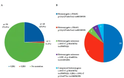
In all patients the most frequently occurring GJ genes in the cochlea were tested for mutations of GJB genes from DNA of blood cells as described above. Figure 2 shows the distribution in patients from Hungary involving 13 non-diseased patients and 80 patients selected for cochlear implantation. The results of the mutation analysis of GJB2, GJB3 and GJB6 genes are shown in Figure 2A. 25.0% of hearing loss patients carried a mutation in the GJB2 gene, and only 1 out of 80 patients (1.2%) had a mutation in the GJB3 gene (Figure 2A). Mutations in GJB6 genes were not detected in this study (Table 1). In the ancestors of the Hungarian Caucasian population, the 35delG mutation in the GJB2 gene appeared most frequently in homozygous state (50%) (Figure 2B) and c.35delG (p.Gly12ValfsTer2 (rs80338939)) in heterozygotes (50%). Among the
heterozygotes, the tests showed 35% mutations of heterozygote c.35delG p.Gly12ValfsTer2 (rs80338939), 5% heterozygote missense c.101T>C p.Met34Thr (rs35887622), 5% heterozygote missense c.119C>A p.Ala40Glu (rs111033296) and 5% of compound heterozygous c.101T>C
Figure 1. Objective clinical measures of the auditory function in patients with hearing loss. Representative results of a non-diseased patient (A, C, E, G) and a patient with impaired hearing (B, D, F, H) are presented. All curves are color-coded.
Red represents the right ear, and blue represents the left ear. A-B: The active, non-linear vibrations of the outer hair cells to external sound stimuli are sensitively recordable with the distortion product otoacoustic emission test (DPOAE) exhibiting the normal function of the endocochlear cellular amplification mechanisms (A). In hearing impaired patients, DPOAE can be registered up to a moderate degree of sensorineural hearing loss. However, the signal tapers off beyond a certain extent (B). C-D: Auditory brainstem response tests (ABR) precisely reveals the functional integrity of the vestibulocochlear nerve leading the electrical stimuli towards the cortex. Neuronal junction points in the brainstem as generator relay structures are identified in the form of waves in the electrophysiological recordings in normal subjects (C); however, they are not present in patients with a profound hearing loss (D). E-F: The objective estimation of hearing sensitivity was performed by auditory steady-state response (ASSR) tests. In a non-diseased subject, normal thresholds were registered on all the measured frequencies (E), while a profound hearing loss is shown in a patient with hereditary sensorineural deafness (F).
G-H: Used while registering ABRs, synchronous activation is achieved in the cochlea, thus a more prominent CE chirp-LS stimuli wave IV-V is observed in a non-diseased subject (G). In patients with profound hearing loss, ABRs are missing (H).
p.Met34Thr (rs35887622), GJB2: c.139G>T p.Glu47Ter (rs104894398). The distribution of these ratios for homozygotes and heterozygotes may change with time, but the rate of this change cannot be estimated at this moment.
3.3. Distribution of connexins and Kir2.1 ion channels with modulators on blood cells
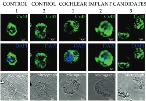
The expression of Cx26 and Cx43 channel proteins was abundant in non-diseased patients (control 1 and 2) but much poorer in blood cells of deaf patients as seen in Figure 3 and variable in hearing loss patients (see 1–3 on Figure 4). In all cells, the nuclei were coun- terstained with DAPI (blue) and scale bars are shown on each image. Thirteen patients with hearing loss were tested for ion channels and compared to controls (with same age and sex) in each experiment. The protein pattern of both Cx26 and Cx43 changed on the surface of blood cells (Figures 3 and 4), appearing as large patches in the control cells and reorganized in smaller groups of connexins in diseased cells. Furthermore, the shape of the area around the nucleus was round in diseased cells and the cytosol was disintegrated. The protein level of Cx26 was variable on the surface of blood cells from patients with hearing loss. We could not identify the GJB2 gene mutation in Patient 1, but Patient 2 and 3 showed mutations with decreased protein level of Cx26. We suppose that Patient 1 has a mutation in another type of gene.
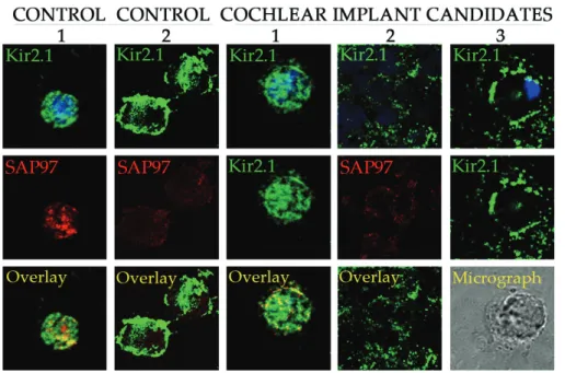
The Kir2.1 ion channels colocalized with SAP97 protein in non-diseased patients, but we determined only partial or no overlayed channel complexes (Figure 5) in cells from cochlear implant candidates. The protein pattern was altered and the levels were decreased in hearing loss patients compared to controls. The SAP97 anchoring protein is redistributed in the blood cells and do not colocalize often with Kir2.1 channels in diseased cells. The levels of Kir2.1
Figure 2. Genetic mutations for GJB genes were tested in hearing loss patients prior to implantation (n = 80). (A) Prevalence of GJB2 and GJB3 gene mutations in subjects with non-syndromic hearing impairment (n = 21/80). (B) Prevalence of different GJB2 mutations in subjects with non-syndromic hearing impairment (n = 20/80).
Figure 4. Alteration of protein levels of connexin43 ion channels in blood cells in patients with deafness. Representative figures from non-diseased and hearing loss patients. Cx43: Connexin43; DAPI: 2,4-diamino-2-phenylindole.
Figure 3. Pattern of connexin26 ion channels on blood cells of patients expecting cochlear implantation. Cx26:
Connexin26, DAPI: 2,4-diamino-2-phenylindole.
protein were varied in each cochlear implant candidates, just as in Patient 1 compared to Patient 2 or 3. The amount of SAP97 declined in the lymphocytes of Patient 2 and 3 with GJB2 gene mutations. The Kir2.1 channels did not perfectly colocalize with SAP97 in the lympho- cytes of Patients 1– 3 with hearing loss (Figure 5).
4. Discussion
The main purpose of this study was to detect the mutations of genes GJB2, GJB3 and GJB6 as well as the translated proteins Cx26, Cx43 and Kir2.1 ion channels from blood cells in cochlear implant candidates. The cochlear implantation was required in hearing impaired patients for based on indication criteria. Hearing sensitivity was evaluated with objective methodolo- gies [56], applying the crosscheck principle. The outer and middle ear anatomy and function proved to be normal in all the recruited subjects of this cohort.
The study provided the following key results: (1) the different GJB mutations were detected in hearing loss in 50% homozygote and 50% heterozygote form for GJB genes in a Hungarian population. (2) Cx43 levels decreased moderately in blood cells. (3) Our investigation revealed a reduced level for Kir2.1 channels as well as for the SAP97 protein. (4) The extent of colocalization of Kir2.1 ion channels with SAP97 anchoring proteins was less marked or partially redistributed in blood cells from patients with hearing loss. (5) Cx26 mutation impacts hearing loss.
Figure 5. The disrupted protein pattern and coexpression of Kir2.1 ion channel with declined SAP97 on blood cells of hearing loss patients. SAP97: Synapse-associated protein 97; DAPI: 2,4-diamino-2-phenylindole.
4.1. Significance of connexins in hearing system and alteration in deafness
These data are consistent with earlier reports on the decrease of GJB2 gene expression in deaf- ness [7, 57], and this study is the first to demonstrate the declined Cx26 protein level in patients carrying these mutations. Recently, more than 100 mutations have been reported, among which the GJB2 gene mutations are the most common cause of nonsyndromic deafness [1]. In this study we demonstrated a similar ratio of GJB2 gene mutations in a Hungarian population in accordance with the earlier findings of Nagy et al. [7]. Additionally, more heterozygote forms were identified in this investigation. The position of joint mutations differs across many popu- lations. This very likely holds true for other genes as well, but GJB2 gene is probably the most examined gene among the genes involved in hearing-related problems. The allelic frequency of three GJB2 gene mutations have been shown to exhibit differences based on ethnicity, which in some cases also translated to geographic regions [11]. The 35delG mutation of the coding exon of GJB2 gene has a frequency of 2–4% in the Caucasian population [58]. Geographically this holds true for Europe, the Middle East and some populations in North America.
The GJB2 gene encodes the Cx26 protein, which assembles to form channels between the cells in the cochlea. These gap junction proteins allow the rapid removal of K+ from the base of hair cells, resulting in the recycling of this ion back to the endolymph to maintain the cochlear homeostasis in humans [59]. Kamiya et al. investigated the expression of Cx26 and Cx30 in the gap junction macromolecular complex [60], where Cx26 is the most abundant channel in the cochlea of mice. They concluded that the disruption of Cx26 was the earliest alteration of the Cx26-dependent gap junction plaque during the embryonic development of mice with connexin-associated deafness. The degradation of the gap junction macromolecu- lar complex leads to loss of hearing function [60] caused by diets [61].
The abnormal or missing gap junctions likely alter the level of potassium ions, which may affect the function and survival of cells that are essential for hearing in the inner ear. The spatial and temporal heteromeric associations of Cx26 and Cx30 proteins in the inner ear have been confirmed earlier [26, 62]. This finding has not been reconfirmed until now. Forge’s results had an essential role to understand the pathophysiologic processes of nonsyndromic deafness caused by mutations in GJB2 genes.
Previously, we have demonstrated that the Cx26 mutation frequently occurs in the Hungarian population [7]. In this study we demonstrated that the ratio of homozygotes and heterozy- gotes is 1.0:1.0 in the mutated GJB2 genes, which suggests that the frequency of heterozygotes is rather high in hearing impaired patients. Further investigations are required to identify additional mutated genes. The combination of heterozygote forms of additional channels may also be involved in the causes leading to deafness in the Caucasian population, because about 73.8% of hearing impaired subjects is not carriers for mutations of the GJB2 gene.
Connexins are 25% more abundant in the sensory epithelial cells of the inner ear than in mam- malian supporting cell types and these cells infrequently contain more than 100,000 channels.
Forge and co-workers have demonstrated [62] that four connexin isotypes, Cx26, Cx30, Cx31 and Cx43, can be found in different mammalian species, in the cochlea and three of them,
Cx26, Cx30 and Cx43, in the vestibular organs. The spatial distribution of connexins in the inner ear has been extensively investigated and it was suggested that these connexin channels are isolated both spatially and functionally. The Cx26 and Cx30 are found in supporting cells of the organ of Corti, in the basal cell region of the stria vascularis, and in type 1 fibrocytes of the spiral ligament, but no other connexin type was detected in this area. Cx31 localizes around the type 2 fibrocytes and below the spiral prominence. Cx30 was not expressed here and the amount of Cx26 expression was low in the above areas [62]. Cx43 was only detected in one region where “tension fibrocytes” line the inner aspect of the otic capsule. It is an important result that Cx26 colocalizes with Cx30 as detected by electron microscopic and immunoprecipitation methods for protein expression.
The majority of gap junction forming proteins in the inner ear potassium-recycling pathway includes Cx26, Cx30 and Cx43. The heteromeric association of Cx26 and Cx30 proteins indi- cates a fine regulation in the connexin composition of the gap junction channels in the inner membrane of the cochlea, and the altered potassium homeostasis might also be related to the redistributed ion channels on blood cells. The heteromeric coexpression of Cx40-Cx43-Cx45 was evaluated by Desplantez et al. [63, 64] in liver cells, and low levels of Cx40/Cx43 coex- pression was detected in heart tissues demonstrating that the Cx40:Cx43 ratio regulates the composition and electrical properties of gap junctions [65–68], which may also be the case in the cochlea.
The age of the 35delG mutation was estimated by Van Lear and co-workers [12]. This muta- tion was tested in ancestors for about 500 generations, indicating an approximate age of 10,000 years in various populations. The mutations in the Hungarian Caucasian population demonstrate a European average.
We saw a high expression of Cx26 and Cx43 proteins in the blood cells of non-diseased patients where the high intensity staining on the surface of cells indicated a high abundance of Cx26.
In the presence of the GJB2 gene mutation, the Cx26 protein level decreased and the pattern of the distribution was disrupted. These results suggest that the expression of GJB2 gene is regulated at the level of transcription and translation in non-diseased patients. Furthermore, it supported the results that the GJB2 gene deficiency is presumably associated with cochlear developmental disorders [66].
Our results indicate that the absence or a reduced presence of Cx26 channels contributes to the development of deafness in humans, which is in support of earlier results [1, 7, 57]. The widely distributed Cx26 and Cx30 and Cx43 proteins have an essential role in the regulation of early ear development [9, 26, 35] and also in the regulation of potassium pathways in the inner ear of adults [26, 62, 67].
4.2. The possible role of Kir2.1 ion channels in the cochlea
Kir2.x ion channels have an important physiological function in transporting potassium ions back into the cells, but the mutations of these ion channels are rarely detected. The role of Kir2.1 ion channels, well-known in Andersen-Tawil syndrome, has only recently been
confirmed for deafness [36, 37, 69], and this suggests the importance of in the auditory system.
The spatial and temporal localization of Kir2.x channels has been mapped in the inner ear by Forge et al. [62], Jagger et al. [26] and its significance has been described by Meredith et al.
[70]. Kir2.1 was characterized as a strong inwardly rectifying ion channel in the vestibular type II hair cells by Zampini et al. [18], Correia et al. [71] in avian and mammalian settings too [47, 69]. An inwardly rectifying potassium channel itself, Kir5.1 plays a vital role in regulating cochlear K+ circulation which is necessary for normal hearing, and it has age-related reduction in expression [71] just like SAP97, but contrary to the Kir2.1 ion channels [45]. The develop- mental study by Ruan et al. [20, 21] carried out in mouse has shown the temporal pattern of Kir2.1 channels in different parts of the hearing system. Kir2.1 has a temporal expression in the hair cells of mouse cochleae that correlates with the functional maturation of the hair cells and the neurons. The stronger signals were generally detected in the apical parts of the cochlea and on row 3 outer hair cells after birth. However, the Kir2.1 ion channels in human adults have been detected mostly in the vestibular system. In all types of cells the mutations of these channels still indicated that the homomer and heteromer Kir2.x channel compositions are necessary in the mammalian inner ear. Lewin and Holt [47], and also Meredith and Rennie [70] identified the functional contribution of Kir2.1 in the IK1 current in utricle hair cells. They demonstrated that Kir2.1 is required for IK1 currents in type II utricle hair cells and that Kir2.1 contributes to the hyperpolarized resting potentials [46] and also to the fast, small amplitude receptor potentials.
The effective functioning of the Kir2.x protein tripartite complex, requires an association of proteins involving SAP97 and calcium calmodulin kinase II (CaMKII) with Kir2.1 ion chan- nels, and Kir2.6 ion channel also has an essential role to deliver the matured Kir2.x protein to the membrane surface [42, 43]. In this study, we showed that the amount of Kir2.1 channel was close to the control in hearing loss patients where we could not detect GJB2 gene muta- tions. The low level of SAP97 anchoring protein decreased drastically in hearing loss patients.
Our results confirmed the colocalization of Kir2.1 ion channel with SAP97 anchoring protein in non-diseased patients but only partial colocalization with disrupted clustering occurred on the surface of blood cells in patients with deafness. This redistribution of the Kir2.1 channel complex, and also the clearly visible reorganization of Cx26 and Cx43 channels, suggests the instability of Kir2.1 and connexin macromolecule complexes in blood cells. This redistribu- tion of Kir2.1 ion channels and Cx26 on the surface of blood cells may lead to a weakened physiological function of Kir2.1 channels and gap junctions, which, in turn, may influence the potassium homeostasis in the sensorineural system [73, 74]. These direct or indirect interac- tions are essential for the normal physiological function of the Kir2.1 complex and inward rectifier currents contributing to normal potassium ion homeostasis [71, 72].
The development of new diagnostic techniques for molecular biology analysis together with deep insights into molecular physiology requires subcellular nanotechnology tools in biomedical research and clinical diagnostics. In this study, we report on hearing impaired patients with a series of hearing screenings using modern equipment, and we have developed a new molecular method to study the critical potassium ion channels contributing to normal cochlear function.
5. Conclusion
Altered expression of Cx26 with Kir2.1 ion channel complex in hearing loss patients pro- foundly determines the potassium-recycling pathway and the alteration of these channels in blood cells in deafness detectable using molecular tools.
The Kir2.x ion channels and connexin channels may have an essential role in the IK1 currents of the cochlea and vestibule during the development process and may serve as diagnostic markers in the early stage of the pathophysiology of hearing loss.
Acknowledgements
This study was supported by grant EFOP-3.6.2-16-2017-00009, which supported MSz, NN, JGK, JAJ, and RN. VSz was supported through the NKFIH-112688 and OTKA K112688 grants.
The authors thank Professor Dr. Gyozo Garab and his group for complementary protein stud- ies, Dr. Ferhan Ayaydin, Zsuzsanna Koszo, Ildiko Kelemen Valkonyne, and Peter Deak for their assistance in confocal microscopy.
Conflict of interest
The authors have nothing to disclose.
Author contributions
MS was the mentor who guided the genetic research study; JAJ and LR tested patients and wrote the manuscript; NN designed and performed the genetic analyses; JGK, AN, RN KH, VSZ carried out protein experiments, analyzed data, and VSZ, FO, JAJ and KH wrote the first draft of the manuscript.
Abbreviation
ABR auditory brain stem responses AEP auditory evoked potentials
AP action potential
ARNSHL autosomal recessive non-syndromic hearing loss
ASSR auditory steady-state responses CaMKII calcium calmodulin kinase II Cx26 connexin26
Cx30 connexin30 Cx31 connexin31 Cx43 connexin43 Cx45 connexin45
DAPI 2,4-diamino-2-phenylindole
DBS dried blood spots
DNA deoxyribonucleic acid
DPOAE distortion product otoacoustic emission EDTA ethylenediaminetetraacetic acid EOAE evoked otoacoustic emissions GJB2 gap junction protein beta 2 GJB3 gap junction protein beta 3 GJB6 gap junction protein beta 6 HC hemichannel
Kv-type voltage-gated potassium channel
Kir2.x potassium inward-rectifier ion channel 2.x MAGUK membrane associated guanylate kinase OAE otoacoustic emission
PCR polymerase chain reaction
PDS Pendred syndrome
PM plasma membrane
SAP97 synapse-associated protein 97 SNHL sensorineural hearing loss SOAE spontaneous otoacoustic emission
Author details
Viktoria Szuts1,2*, Janos Andras Jarabin3, Nikoletta Nagy4, Ferenc Otvos5, Roland Nagy3, Attila Nagy3,6, Katalin Halasy7, Laszlo Rovo3, Marta Szell4,8 and Jozsef Geza Kiss3
*Address all correspondence to: szutsvik@hotmail.com
1 Institute of Plant Biology, Biological Research Centre, Hungarian Academy of Sciences, Szeged, Hungary
2 Department of Food Engineering, Faculty of Engineering, University of Szeged, Szeged, Hungary
3 Department of Oto-Rhino-Laryngology and Head-Neck, Faculty of Medicine, University of Szeged, Szeged, Hungary
4 Department of Medical Genetics, Faculty of Medicine, University of Szeged, Szeged, Hungary
5 Institute of Biochemistry, Biological Research Centre, Hungarian Academy of Sciences, Szeged, Hungary
6 Department of Medical Physics and Informatics, Faculty of Medicine and Faculty of Sciences and Informatics, University of Szeged, Szeged, Hungary
7 Department of Anatomy and Histology, University of Veterinary Medicine, Budapest, Hungary
8 MTA-SZTE Dermatological Research Group, University of Szeged, Szeged, Hungary
References
[1] Kemperman MH, Hoefsloot LH, Cremers CWRJ. Hearing loss and connexin 26. Journal of the Royal Society of Medicine. 2002;95(4):171-177. DOI: 10.1258/jrsm.95.4.171
[2] Zelante L, Gasparini P, Estivill X, Melchionda S, D’Agruma L, Govea N, Milá M, Della Monica M, Lutfi J, Shohat M, Mansfield E, Delgrosso K, Rappaport E, Surrey S, Fortina P. Connexin26 mutations associated with the most common form of non-syndromic neu- rosensory autosomal recessive deafness (DFNB1) in Mediterraneans. Human Molecular Genetics. 1997;6(9):1605-1609. DOI: 10.1093/hmg/6.9.1605
[3] Estivill X, Rabionet R. The Connexin-deafness homepage World Wide Web URL: [http://
www.iro.es/deafness] [Accessed: November 2001]
[4] Marlin S, Garabédian EN, Roger G, Moatti L, Matha N, Lewin P, Petit C, Denoyelle F.
Connexin 26 gene mutations in congenitally deaf children: Pitfalls for genetic counsel- ing. Archives of Otolaryngology – Head & Neck Surgery. 2001;127(8):927-933
[5] Mueller RF, Nehammer A, Middleton A, Houseman M, Taylor GR, Bitner-Glindzciz M, Van Camp G, Parker M, Young ID, Davis A, Newton VE, Lench NJ. Congenital non-syndromal sensorineural hearing impairment due to connexin 26 gene muta- tions—Molecular and audiological findings. International Journal of Pediatric Otorhino- laryngology. 1999;50(1):3-13. DOI: 10.1016/S0165-5876(99)00242-6
[6] Chan DK, Schrijver I, Chang KW. Connexin-26-associated deafness: Phenotypic vari- ability and progression of hearing loss. Genetics in Medicine. 2010;12(3):174-181. DOI:
10.1097/GIM.0b013e3181d0d42b
[7] Nagy AL, Csáki R, Klem J, Rovó L, Tóth F, Tálosi G, Jóri J, Kovács K, Kiss JG. Minimally invasive genetic screen for GJB2 related deafness using dried blood spots. International Journal of Pediatric Otorhinolaryngology. 2010;74(1):75-81. DOI: 10.1016/j.ijporl.
2009.10.021
[8] Pointis G, Gilleron J, Carette D, Segretain D. Physiological and physiopathological aspects of connexins and communicating gap junctions in spermatogenesis. Philosophical Tran- sactions of the Royal Society, B: Biological Sciences. 2010;365(1546):1607-1620. DOI:
10.1098/rstb.2009.0114
[9] Forge A, Jagger DJ, Kelly JJ, Taylor RR. Connexin30-mediated intercellular communica- tion plays an essential role in epithelial repair in the cochlea. Journal of Cell Science.
2013;126(7):1703-1712. DOI: 10.1242/jcs.125476
[10] Fiori MC, Reuss L, Cuello LG, Altenberg GA. Functional analysis and regulation of purified connexin hemichannels. Frontiers in Physiology. 2014:71. DOI: 10.3389/fphys.
2014.00071
[11] Shearer AE, Eppsteiner RW, Booth KT, Ephraim SS, Gurrola J, Simpson A, Black- Ziegelbein EA, Joshi S, Ravi H, Giuffre AC, Happe S, Hildebrand MS, Azaiez H, Bayazit YA, Emin Erdal M, Lopez-Escamez JA, Gazquez I, Tamayo ML, Gelvez NY, Leal GL, Jalas C, Ekstein J, Yang T, Usami S, Kahrizi K, Bazazzadegan N, Najmabadi H, Scheetz TE, Braun TA, Casavant TL, LeProust EM, Smith RJ. Utilizing ethnic-specific differences in minor allele frequency to recategorize reported pathogenic deafness variants. American Journal of Human Genetics. 2014;95(4):445-453. DOI: 10.1016/j.
ajhg.2014.09.001
[12] Van Laer L, Coucke P, Mueller RF, Caethoven G, Flothmann K, Prasad SD, Chamberlin GP, Houseman M, Taylor GR, Van de Heyning CM, Fransen E, Rowland J, Cucci RA, Smith RJH, Van Camp G, Van Camp G. A common founder for the 35delG GJB2 gene mutation in connexin 26 hearing impairment. Journal of Medical Genetics. 2001;38(8):515- 518. DOI: 10.1136/jmg.38.8.515
[13] Mahdieh N, Rabbani B. Statistical study of 35delG mutation of GJB2 gene: A meta- analysis of carrier frequency. International Journal of Audiology. 2009;48(6):363-370.
DOI: 10.1080/14992020802607449
[14] Morell RJ, Kim HJ, Hood LJ, Goforth L, Friderici K, Fisher R, Van Camp G, Berlin CI, Oddoux C, Ostrer H, Keats B, Friedman TB. Mutations in the connexin 26 gene (GJB2)
among Ashkenazi Jews with nonsyndromic recessive deafness [see comments]. The New England Journal of Medicine. 1998;339:1500-1505. DOI: 10.1056/NEJM199811193392103 [15] Lerer I, Sagi M, Malamud E, Levi H, Raas-Rothschild A, Abeliovich D. Contribution of
connexin 26 mutations to nonsyndromic deafness in Ashkenazi patients and the vari- able phenotypic effect of the mutation 167delT. American Journal of Medical Genetics.
2000;95(1):53-56. DOI: 10.1002/1096-8628(20001106)95:1<53::AID-AJMG11>3.0.CO;2-2 [16] Kelley PM, Cohn E, Kimberling WJ. Connexin 26: Required for normal auditory func-
tion. Brain Research Reviews. 2000;32:184-188. DOI: 10.1016/S0165-0173(99)00080-6 [17] Hibino H, Inanobe A, Furutani K, Murakami S, Findlay I, Kurachi Y. Inwardly rectify-
ing potassium channels: Their structure, function, and physiological roles. Physiological Reviews. 2010;90(1):291-366. DOI: 10.1152/physrev.00021.2009
[18] Zampini V, Masetto S, Correia MJ. Elementary properties of Kir2.1, a strong inwardly rectifying K+ channel expressed by pigeon vestibular type II hair cells. Neuroscience.
2008;155(4):1250-1261. DOI: 10.1016/j.neuroscience.2008.06.048
[19] Hofherr A. Selective Golgi export of Kir2.1 controls the stoichiometry of functional Kir2.X channel heteromers. Journal of Cell Science. 2005;118(9):1935-1943. DOI: 10.1242/jcs.02322 [20] Ruan Q, Chen D, Wang Z, Chi F, Yin S, Wang J. Topological and developmental expres- sion gradients of Kir2.1, an inward rectifier K+ channel, in spiral ganglion and cochlear hair cells of mouse inner ear. Developmental Neuroscience. 2008;30(6):374-388. DOI:
10.1159/000164687
[21] Ruan Q, Chen D, Wang Z, Chi F, He J, Wang J, Yin S. Effects of Kir2.1 gene transfection in cochlear hair cells and application of neurotrophic factors on survival and neurite growth of co-cultured cochlear spiral ganglion neurons. Molecular and Cellular Neurosciences.
2010;43(3):326-339. DOI: 10.1016/j.mcn.2009.12.006
[22] Rook MB. Physiologic function of I(K1) requires a combination of Kir2 isoforms. Heart Rhythm. 2007;4(4):497-498. DOI: 10.1016/j.hrthm.2006.12.041
[23] Anumonwo JMB, Lopatin AN. Cardiac strong inward rectifier potassium channels. Journal of Molecular and Cellular Cardiology. 2010;48(1):45-54. DOI: 10.1016/j.yjmcc.2009.08.
013
[24] Leonoudakis D, Conti LR, Radeke CM, McGuire LMM, Vandenberg CA. A multi- protein trafficking complex composed of SAP97, CASK, Veli, and Mint1 is associated with inward rectifier Kir2 potassium channels. The Journal of Biological Chemistry.
2004;279(18):19051-19063. DOI: 10.1074/jbc.M400284200
[25] Vaidyanathan R, Taffet SM, Vikstrom KL, Anumonwo JMB. Regulation of cardiac inward rectifier potassium current (IK1) by synapse-associated protein-97. The Journal of Biolo- gical Chemistry. 2010;285(36):28000-28009. DOI: 10.1074/jbc.M110.110858
[26] Jagger DJ, Forge A. Connexins and gap junctions in the inner ear—It’s not just about K+
recycling. Cell and Tissue Research. 2015;360(3):633-644. DOI: 10.1007/s00441-014-2029-z
[27] Isomoto S, Kondo C, Kurachi Y. Inwardly rectifying potassium channels: Their molecu- lar heterogeneity and function. The Japanese Journal of Physiology. 1997;47(1):11-39.
DOI: 10.2170/jjphysiol.47.11
[28] Kelsell DP, Dunlop J, Stevens HP, Lench NJ, Liang JN, Parry G, Mueller RF, Leigh IM.
Connexin 26 mutations in hereditary non-syndromic sensorineural deafness. Nature.
1997;387:80-83. DOI: 10.1038/387080a0
[29] Abdelhadi O, Iancu D, Stanescu H, Kleta R, Bockenhauer D. EAST syndrome: Clinical, pathophysiological, and genetic aspects of mutations in KCNJ10. Rare Diseases. 2016;
4(1):e1195043. DOI: 10.1080/21675511.2016.1195043
[30] Csanády M, Faragó M, Forster T, Hőgye M, Gy P. Study of the course of inheritance of dilated familial cardiomyopathy. European Heart Journal. 1991;12:191
[31] Denoyelle F, Marlin S, Weil D, Moatti L, Chauvin P, Garabédian EN, Petit C. Clinical features of the prevalent form of childhood deafness, DFNB1, due to a connexin-26 gene defect: Implications for genetic counselling. Lancet. 1999;353:1298-1303. DOI: 10.1016/
S0140-6736(98)11071-1
[32] Abe S, Usami S, Shinkawa H, Kelley PM, Kimberling WJ. Prevalent connexin 26 gene (GJB2) mutations in Japanese. Journal of Medical Genetics. 2000;37:41-43
[33] Marlin S, Garabedian EN, Roger G, Moatti L, Matha N, Lewin P, Petit C, Denoyelle F.
Connexin 26 gene mutations in congenitally deaf children: Pitfalls for genetic counselling.
Archives of Otolaryngology – Head & Neck Surgery. 2001;127:927-933
[34] Martini A, Calzolari F, Sensi A. Genetic syndromes involving hearing. International Journal of Pediatric Otorhinolaryngology. 2009;73(Sp1):S2-S12. DOI: 10.1016/S0165-5876 (09)70002-3
[35] Nickel R. Forge a gap junctions and connexins in the inner ear: Their roles in homeosta- sis and deafness. Current Opinion in Otolaryngology & Head and Neck Surgery. 2008 Oct;16(5):452-457. DOI: 10.1523/JNEUROSCI.1932-14.2014
[36] Plaster NM, Tawil R, Tristani-Firouzi M, Canún S, Bendahhou S, Tsunoda A, Donaldson MR, Iannaccone ST, Brunt E, Barohn R, Clark J, Deymeer F, George AL, Fish FA, Hahn A, Nitu A, Ozdemir C, Serdaroglu P, Subramony SH, Wolfe G, Fu YH, Ptácek LJ. Mutations in Kir2.1 cause the developmental and episodic electrical phenotypes of Andersen’s syn- drome. Cell. 2001;105(4):511-519. DOI: 10.1016/S0092-8674(01)00342-7
[37] Tristani-Firouzi M, Etheridge SP. Kir 2.1 channelopathies: The Andersen-Tawil syndrome.
Pflügers Archiv / European Journal of Physiology. 2010;460(2):289-294. DOI: 10.1007/s00424-0 10-0820-6
[38] Jun AI, McGuirt WT, Hinojosa R, Green GE, Fischel-Ghodsian N, Smith RJ. Temporal bone histopathology in connexin 26-related hearing loss. Laryngoscope. 2000;110:269- 275. DOI: 10.1097/00005537-200002010-00016
[39] Zobel C, Cho HC, Nguyen TT, Pekhletski R, Diaz RJ, Wilson GJ, Backx PH. Molecular dis- section of the inward rectifier potassium current (IK1) in rabbit cardiomyocytes: Evidence for heteromeric co-assembly of Kir2.1 and Kir2.2. The Journal of Physiology. 2003;550(Pt 2):
365-372. DOI: 10.1113/jphysiol.2002.036400
[40] Gaborit N, Varro A, Le Bouter S, Szuts V, Escande D, Nattel S, Demolombe S. Gender- related differences in ion-channel and transporter subunit expression in non-diseased human hearts. Journal of Molecular and Cellular Cardiology. 2010;49:639-646. DOI:
10.1016/j.yjmcc.2010.06.005
[41] Yanni J, Tellez TO, Mackewski JO, Mackiewicz U, Beresewich A, Beresevicz A, Billeter R, Dobrzynski H, Boyett M. Changes of ion channels gene expression underlying heart failure induced sinoatrial node dysfunction. Circular Heart Failure. 2011;4:496-508. DOI:
10.1161/CIRCHEARTFAILURE.110.957647
[42] Mustroph J, Maier SL, Wagner S. CaMKII regulation of cardiac K channels. Frontiers in Pharmacology. 2014;5:1-12. DOI: 1016/j.pharmthera.2016.10.006
[43] Dassau L, Conti RL, Radekr MC, Ptacek JL, Vandenberg AC. Kir2.6 regulates the sur- face expression of Kir2.X inward rectifier potassium channels. The Journal of Biological Chemistry. 2011;286:9526-9541. DOI: 10.1074/jbc.M110.170597
[44] Ma D, Taneja TK, Hagen BM, Kim B-Y, Ortega B, Lederer WJ, Welling PA. Golgi export of the Kir2.1 channel is driven by a trafficking signal located within its tertiary structure.
Cell. 2011;145:1102-1115. DOI: 10.1016/j.cell.2011.06.007
[45] Szuts V, Menesi D, Varga-Orvos Z, Zvara A, Houshmand N, Bitay M, Bogats G, Virag L, Baczko I, Szalontai B, Geramipoor A, Cotella D, Wettwer E, Ravens U, Deak F, Puskas LG, Papp JG, Kiss I, Varro A, Jost N. Altered expression of genes for Kir ion chan- nels in dilated cardiomyopathy. Canadian Journal of Physiology and Pharmacology.
2013;91(8):648-656. DOI: 10.1139/cjpp-2012-0413
[46] Horwitz GC, Risner-Janiczek JR, Jones SM, Holt JR. HCN channels expressed in the inner ear are necessary for normal balance function. The Journal of Neuroscience. 2011;
31:16814-16825
[47] Levin ME, Holt JR. The function and molecular identity of inward rectifier channels in vestibular hair cells of the mouse inner ear. Journal of Neurophysiology. 2012;108(1):175- 186. DOI: 10.1152/jn.00098.2012
[48] Szuts V, Fazekas P, Kovacs A, Nagy A, Deak P, Jarabin JA, Otvos F, Cs V, Szecsi M, Halasy K, Rovo L, Kiss JG. Connexins and Kir2.1 ion channels modulated in hearing loss patients before cochlear implantation. International congress of cochlear implanta- tion & OMAI congress, Szeged, Hungary, 2016 June. Otorhinolaryngologica Hungarica.
2016;62(Suppl.A):A38
[49] Hoth S, Baljić I. Current audiological diagnostics. GMS Current Topic Otorhinolaryngology Head & Neck Surgery. 2017 Dec;16:Doc09. DOI: 10.3205/cto000148
[50] François M, Dehan E, Carlevan M, Dumont H. Use of auditory steady-state responses in children and comparison with other electrophysiological and behavioral tests. European Annals of Otorhinolaryngology, Head and Neck Diseases. 2016;133(5):331-335. DOI:
10.1016/j.anorl.2016.07.008
[51] Janssen T, Gehr DD, Klein A, Muller J. Distortion product otoacoustic emissions for hear- ing threshold estimation and differentiation between middle-ear and cochlear disorders in neonates. The Journal of the Acoustical Society of America. 2005;117(5):2969-2979.
DOI: 10.1121/1.1853101
[52] McCreery RW, Kaminski J, Beauchaine K, Lenzen N, Simms K, Gorga MP. The impact of degree of hearing loss on auditory brainstem response predictions of behavioral thresh- olds. Ear and Hearing. 2015;36(3):309-319. DOI: 10.1097/AUD.0000000000000120 [53] Rodrigues GRI, Ramos N, Lewis DR. Comparing auditory brainstem responses (ABRs)
to toneburst and narrow band CE-chirp in young infants. International Journal of Pediatric Otorhinolaryngology. 2013;77(9):1555-1560. DOI: 10.1016/j.ijporl.2013.07.003 [54] Curthoys IS. The new vestibular stimuli: Sound and vibration—Anatomical, physi-
ological and clinical evidence. Experimental Brain Research. 2017;235(4):957-972. DOI:
10.1007/s00221-017-4874-y
[55] Kristensen SGB, Elberling C. Auditory brainstem responses to level-specific chirps in normal-hearing adults. Journal of the American Academy of Audiology. 2012;23(9):712- 721. DOI: 10.3766/jaaa.23.9.5
[56] Korczak P, Smart J, Delgado RM, Strobel T, Bradford C. Auditory steady-state responses.
Journal of the American Academy of Audiology. 2012;23(3):146-170. DOI: 10.3766/
jaaa.23.3.3
[57] Cremers, Frans PM. Genetic causes of hearing loss. Current Opinion in Neurology.
February 1998;11(1):11-16
[58] Qu Y, Tang W, Zhou B, Ahmad S, Chang Q, Li X, Lin X. Early developmental expression of connexin26 in the cochlea contributes to its dominate functional role in the cochlear gap junctions. Biochemical and Biophysical Research Communications. 2012;417(1):245- 250. DOI: 10.1016/j.bbrc.2011.11.093
[59] Kikuchi T, Kimura RS, Paul DL, Takasaka T, Adams JC. Gap junction systems in the mammalian cochlea. Brain Research Reviews. 2000;32:163-166
[60] Kamiya K, Yum SW, Kurebayashi N, Muraki M, Ogawa K, Karasawa K, Miwa A, Guo X, Gotoh S, Sugitani Y, Yamanaka H, Ito-Kawashima S, Iizuka T, Sakurai T, Noda T, Minowa O, Ikeda K. Assembly of the cochlear gap junction macromolecular complex requires connexin 26. The Journal of Clinical Investigation. 2014 Apr;124(4):1598-1607.
DOI: 10.1172/JCI67621
[61] Wu X, Wang Y, Sun Y, Chen S, Zhang S, Shen L, Huang X, Lin X, Kong W. Reduced expression of Connexin26 and its DNA promoter hypermethylation in the inner ear of mimetic aging rats induced by D-galactose. Biochemical and Biophysical Research Communications. 2014;452(3):340-346. DOI: 10.1016/j.bbrc.2014.08.063
[62] Forge A, Becker D, Casalotti S, Edwards J, Marziano N, Nevill G. Gap junctions in the inner ear: Comparison of distribution patterns in different vertebrates and assess- ment of Connexin composition in mammals. The Journal of Comparative Neurology.
2003;467(2):207-231. DOI: 10.1002/cne.10916
[63] Desplantez T, Grikscheit K, Thomas NM, Peters NS, Severs NJ, Dupont E. Relating spe- cific connexin co-expression ratio to connexon composition and gap junction function.
Journal of Molecular and Cellular Cardiology. 2015 Dec;89(Pt B):195-202. DOI: 10.1016/j.
yjmcc2015.11
[64] Desplantez T. Cardiac Cx43, Cx40 and Cx45 co-assembling: Involvement of connexins epitopes in formation of hemichannels and Gap junction channels. BMC Cell Biology.
2017 Jan 17;18(Suppl 1):3. DOI: 10.1186/s12860-016-0118-4
[65] Chaigne S, Dupuis S, Constantin MDT. Co-expressed cardiac connexins: Dependence on the Cx43:Cx40 ratio in regulating the gap junction channel make-up and electrical properties. Cardiovascular Research Supplements. 2014;103(Supplement 1):S20. DOI:
10.1093/cvr/cvu082.58
[66] Dupuis S, Chaigne S, Constantin M, Desplantez T. Dependence of the cardiac connexins Cx43:Cx45 ratio on the formation of gap junction channels and their electrical properties.
Cardiovascular Research Supplements. 2014;103(Suppl. 1):S116-S117. DOI: 10.1093/cvr/
cvu098.68
[67] Chan DK, Chang KW. GJB2-associated hearing loss: Systematic review of worldwide preva- lence, genotype, and auditory phenotype. The Laryngoscope. 2014 Feb;124(2):E34-E53.
DOI: 10.1371/journal.pone.0167850
[68] Liu W, Li H, Edin F, Brännström J, Glueckert R, Schrott-Fischer A, Molnar M, Pacholsky D, Pfaller K, Rask-Andersen H. Molecular composition and distribution of gap junctions in the sensory epithelium of the human cochlea-a super-resolution structured illumination microscopy (SR-SIM) study. Upsala Journal of Medical Sciences. 2017Aug;122(3):160-170.
DOI: 10.1080/03009734.2017.1322645
[69] Lu CW, Lin JH, Rajawat YS, Jerng H, Rami TG, Sanchez X, DeFreitas G, Carabello B, DeMayo F, Kearney DL, Miller G, Li H, Pfaffinger PJ, Bowles NE, Khoury DS, Towbin JA. Functional and clinical characterization of a mutation in KCNJ2 associated with Andersen-Tawil syndrome. Journal of Medical Genetics. 2006;43(8):653-659
[70] Meredith FL, Rennie KJ. Channeling your inner ear potassium: K+ channels in vestibular hair cells. Hearing Research. 2016;338:40e51. DOI: 10.1016/j.heares.2016.01.015
[71] Correia MJ, Wood TG, Prusak D, Weng T, Rennie KJ, Wang HQ. Molecular characteriza- tion of an inward rectifier channel (IKir) found in avian vestibular hair cells: cloning and expression of pKir2.1. Physiol Genomics. 2004;19(2):155-169. PMID: 15316115
[72] Pan C, Chu H, Lai Y, Sun Y, Du Z, Liu CJ, Tong T, Chen QL, Bing D, Tao Y. Downregulation of inwardly rectifying potassium channel 5.1 expression in C57BL/6J cochlear lat- eral wall. Journal of Huazhong University Science of Technology [Medical Science].
2016;36(3):406-409
[73] Collado MS, Holt JR. Can neurosphere production help restore inner ear transduction?
Proceedings of the National Academy of Sciences of the United States of America. 2009 Jan 6;106(1):8-9. DOI: 10.1073/pnas.0811804106
[74] Forge A, Taylor RR, Dawson SJ, Lovett M, Jagger DJ. Disruption of SorCS2 reveals dif- ferences in the regulation of stereociliary bundle formation between hair cell types in the inner ear. PLoS Genetics. 2017 Mar 27;13(3):e1006692. DOI: 10.1371/journal.pgen.1006692