Development of Complex Curricula for Molecular Bionics and Infobionics Programs within a consortial* framework**
Consortium leader
PETER PAZMANY CATHOLIC UNIVERSITY
Consortium members
SEMMELWEIS UNIVERSITY, DIALOG CAMPUS PUBLISHER
The Project has been realised with the support of the European Union and has been co-financed by the European Social Fund ***
**Molekuláris bionika és Infobionika Szakok tananyagának komplex fejlesztése konzorciumi keretben
***A projekt az Európai Unió támogatásával, az Európai Szociális Alap társfinanszírozásával valósul meg.
CATHOLIC UNIVERSITY
Faculty of Information Technology
NEURAL INTERFACES AND PROSTHESES
IMPLANTABLE HEARING AIDS
www.itk.ppke.hu
(Neurális interfészek és protézisek)
(Beépíthető hallásjavító eszközök)
RICHÁRD FIÁTH and GYÖRGY KARMOS
LECTURE 7
www.itk.ppke.hu
Contents
• Aims
• The structure of the ear
• The structure of the cochlea
• The mechanism of hearing
• The auditory pathway
• Hearing disorders
• Cochlear microphony
• Oto-acoustic emission
• Traditional hearing aids
• Implantable hearing devices (IHD)
– Bone anchored hearing apparatus (BAHA)
– Middle ear implantable hearing devices (MEHID)
www.itk.ppke.hu
AIMS:
In the following two lectures the student will become familiar with implantable hearing devices used for hearing disorders. After a short introduction about the biology of hearing, the possibilities and types of auditory prostheses will be detailed, focusing mainly on cochlear implants, but we will show other solutions, ranging from the middle ear implantable hearing devices to methods in experimental stage (auditory brainstem implants, auditory midbrain implants).
The first lecture presents the structure of the human ear and cochlea, the auditory pathway and different hearing disorders, together with some interesting phenomena related to auditory mechanisms (cochlear microphony, oto-acoustic emission). This is followed by a short review of traditional hearing aids. Finally in this section, the detailed description of the bone anchored hearing apparatus (BAHA) and middle ear implantable hearing devices (MEIHD) can be read.
www.itk.ppke.hu
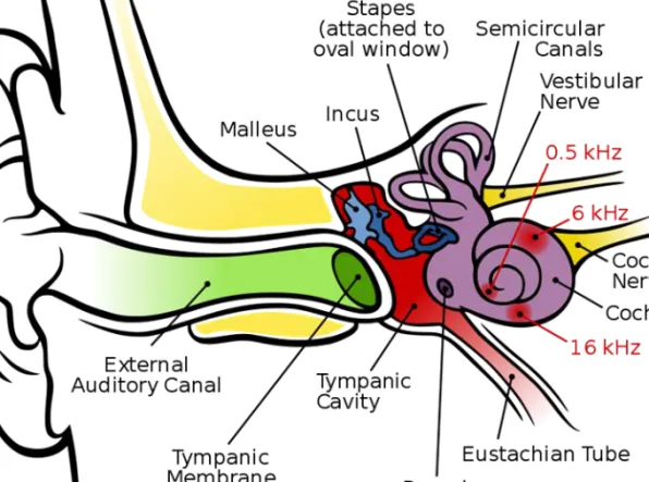
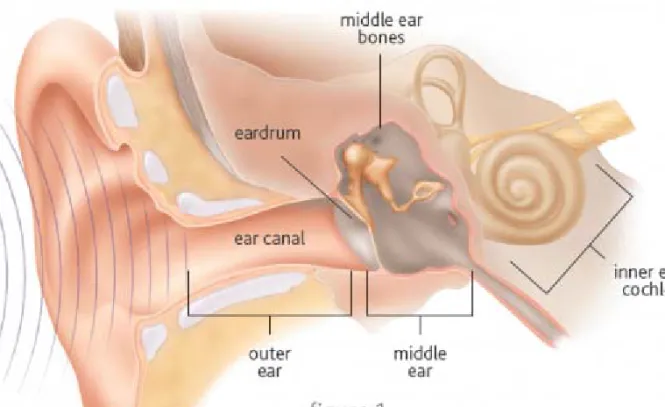
1. Outer ear: Starts from the burr, through the ear canal (external auditory canal) until the eardrum (tympanic membrane). The function of the outer ear is to collect sound waves. (green on Figure 1.)
2. Middle ear: is an air-filled cavity, containing the flexibly jointed ossicles (malleus, incus, stapes). The stapes is attached to the oval window, this is the connection point between the middle and inner ear. Lateral border: eardrum, medial border: osseous labyrinth. The function of the middle ear is the amplification of the sound and impedance fitting. (red and blue on Figure 1.) 3. Inner ear: Contains the balance (semicircular canals) and hearing (cochlea) sensory organs. Consists of a bony part (osseous labyrinth, which can be considered to be a type of capsule) and a membranous part (membranous labyrinth) which contains the receptor cells of the organ of balance and hearing. (violet on Figure 1.) The auditory and vestibular nerve transmits the electric signals from the inner ear to the brain.
THE STRUCTURE OF THE HUMAN EAR
www.itk.ppke.hu
Figure 1. Anatomy of the human ear
(commons.wikimedia.org)
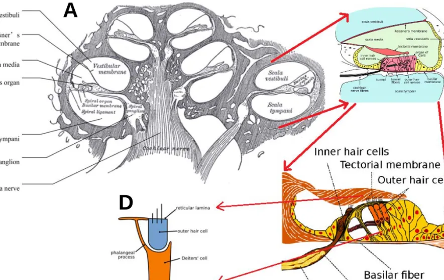
The cochlea is a spiral-shaped organ (Figure 1., 2.A), localized in the anterior part of the osseous labyrinth (the balance sensory system is localized posteriorly). Its function is the transduction of mechanic waves into electric signals.
It has an osseous capsule and a membraneous part that contains the organ of Corti (Figure 2.B, C) which is the key of hearing. The organ of Corti is made up of two types of hair cells (inner and outer, Figure 2.D), supporting cells, basilar membrane and tectorial membrane. The movement of the basilar membrane makes the stereocilia of the hair cells touch the tectorial membrane. This process results in the bending of the stereocilia, that opens certain ion channels, and consequently depolarizes the cells (Figure 3.,4.).
That is the point of, where mechanical movement is being transformed into electrical activity which is transmitted through the auditory nerve to the brain.
www.itk.ppke.hu
THE STRUCTURE OF THE COCHLEA
• The are approximately 15000 hair cells and 30000 nerve fibers in the cochlea.
• The 3500 inner hair cells are innervated by the 95% of the nerve fibers
• The 12000 outer hair cells are placed in three rows and are innervated by the rest (5%) of the nerve fibers.
• The resting membrane potential of the hair cells is around -60 mV.
• The 15% of the ion channels in the hair cells are open.
• Receptor potential: 100 nm deflection of the stereocilium corresponds to the 90% of the response range.
• Normal stimulus: ~±1º deflection (less than the diameter of the stereocilium)
• Deflection due to thermal noise: 3 nm
www.itk.ppke.hu
NUMBERS RELATED TO THE COCHLEA
www.itk.ppke.hu
(commons.wikimedia.org)
Figure 2. The cross section and the building blocks of the human cochlea
A B
D C
www.itk.ppke.hu
THE MECHANISM OF HEARING
1. Sound waves collected by the outer ear reach the eardrum, and cause its vibration. (mechanic wave, air conduction)
2. Mechanic waves resulting from the vibration of the eardrum are transmitted to the ossicle-system (malleus, incus, stapes; bone conduction).The footplate of stapes is joined directly to the round oval window, which is covered by a flexible membrane.
3. Via the footplate of the stapes the movement of the ossicle-chain leads to the vibration of the oval window, thus the mechanical vibration is transmitted to the liquid of the inner ear.
4. The waves in the liquid cause vibration spreading longitudinally on the basal membrane of the Corti organ (fluid conduction), that makes the stereocilia of hair cells to touch the membrana tectoria. This results in depolarisation of the cells, that induces electric potential in hearing nerve fibres (mechano-electric transduction) .
www.itk.ppke.hu
THE ACTIVITY OF HAIR CELLS
The most important part of the hair cells are the stereocilia (Figure 3.). When sound waves enter the cochlea, the stereocilia touch the tectorial membrane because of the vibrating liquid, which results in bending of these. Stereocilia are connected together via tip links (Figure 4.). Displacement of the stereocilia towards the tallest produces a cell depolarization as ionic current flows (potassium ions) in through the mechanoelectric-transducer channels. These ion channels close shortly after the increase of calcium ion concentration.
Auditory nerves show a spontaneous activity, meaning that there is a continuous discharging with a given frequency. If the hair cells are depolarized (bent towards the tallest stereocilium), this spontaneous spike activity is increased (Figure 4.). In case of hyperpolarization (stereocilia are bent towards the smallest one) the effect is the inverse: the frequency of the spontaneous activity is decreased (Figure 4.).
www.itk.ppke.hu
Figure 3. The effect of the movement of the stereocilia on the spontaneous activity
www.itk.ppke.hu
Figure 4. The working mechanism of the stereocilia: if the stereocilia are bent towards the tallest one, the ion channels open, potassium ions enter and the hair cell depolarizes
www.itk.ppke.hu
Tonotopy in the auditory system begins in the cochlea. Different regions of the chord-like basilar membrane vibrate at different sinusoidal frequencies, due to the membrane’s different width and tightness at it’s each point.
Because of the mechanical properties of the basilar membrane within the snail-shaped cochlea, high frequencies will produce a vibration peak near the oval window, whereas low frequencies will stimulate receptors near the apex of the cochlea (Figure 5.). According to the frequency of the sinusoid, there will be a point on the basilar membrane, which is mostly sensitive to the signal frequency (so-called characteristic frequency). The maximum amplitude of the signal will be exactly at the part of the membrane pointed out by the characteristic frequency. This is the point where the wave becomes extincted, and the response is maximal. The auditory nerves that transmit information from different regions of the basilar membrane therefore encode frequency tonotopically. This tonotopy is maintained in
TONOTOPY IN THE AUDITORY SYSTEM
www.itk.ppke.hu
(commons.wikimedia.org) Figure 5. Tonotopy in the uncoiled cochlea. High-pitched sounds are processed at the base while low-pitched sounds at the apex of the cochlea.
www.itk.ppke.hu
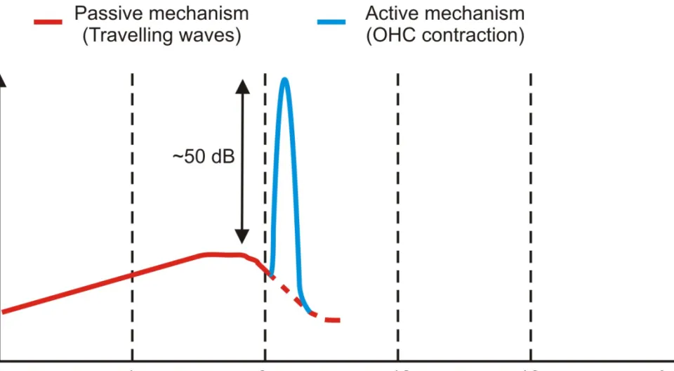
The tonotopy described before is a passive tonotopy, but there is also an active mechanism of the outer hair cells (OHC) which ampilifies the signal originating from passive tonotopy. On Figure 6. the effect of the active mechanism of the outer hair cells is shown in case of a travelling wave on the basilar membrane of the guinea pig caused by a pure tone with 10 kHz frequency. The outer hair cells shift the maxima of the wave with approximately ½ octave and OHCs also amplify the amplitude of the travelling wave with around 50 dB. This active mechanism is because the OHC responds to the depolarization by changing length. Ultrastructural analysis of cell plasma membrane indicates that this electromotility may be based upon conformational changes of a dense array of integral membrane proteins.
TONOTOPY IN THE AUDITORY SYSTEM
www.itk.ppke.hu
Figure 6. The effect of the active mechanism of outer hair cell contraction on the amplitude of the sound waves.
www.itk.ppke.hu
FUNCTION OF THE ORGAN OF CORTI
1. Sounds waves move the basilar membrane (BM) up and down.
2. Mechano-transduction at OHCs: Stereocilia of the OHCs, implanted in the tectorial membrane (TM), are bent, potassium ions enter the cell which causes depolarization.
3. Electromotility and reverse transduction: Depolarisation of the cell membrane results in OHC contraction which magnifies the motion. This is an active mechanism.
4. Mechano-transduction at inner hair cells (IHC): Due to the tight coupling of OHCs with the BM and reticular lamina the active mechanism in (3.) feeds energy back into the organ of Corti. Inner hair cells (IHCs) are excited possibly via TM activation of IHC stereocilia.
5. IHC-auditory nerve synapse activation: The depolarized IHCs activate the IHC-auditory nerve synapse and an auditory message is sent to the brain via the type I afferent nerve.
www.itk.ppke.hu
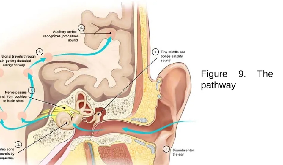
THE AUDITORY SYSTEM AND PATHWAY
The sound coming from the outer world enters the ear, than travels through the ear canal to the eardrum (air conduction in the outer ear). The pressure of the sound makes the eardrum (or tympanic membrane) to vibrate, which sets the small bones in the middle ear (malleus, incus, stapes) into motion. The sound is amplified and travels through the bones to the oval window (bone conduction). The oval window is the connection point between the middle ear and the inner ear or cochlea. In the cochlea, the vibrations travel via fluid conduction, causing the hair cells to move, which convert this movement into electrical impulses. (Figure 7.) These electrical signals are transmitted by the vestibulocochlear nerve to the cochlear nuclei and superior olivary complex of the brainstem and to the inferior colliculus of the midbrain. The next stage of information processing is the medial geniculate body of the thalamus, where the signals are relayed to the primary auditory cortex (Brodmann area 41, 42), which is located in the temporal lobe of the human brain. (Figure 8.)
www.itk.ppke.hu
Figure 7. The first part of the auditory pathway from the ear to the cochlea
http://www.cochlear.com/
www.itk.ppke.hu
http://www.cochlear.com/
Figure 8. The second part of the auditory pathway from the cochlea to the auditory cortex.
www.itk.ppke.hu
Figure 9. The auditory pathway
(commons.wikimedia.org)
www.itk.ppke.hu
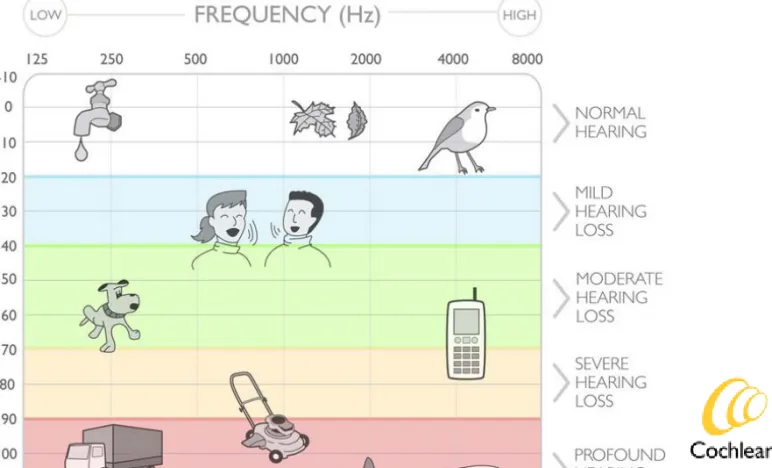
QUANTIFICATION OF HEARING LOSS
The severity of a hearing impairment is ranked according to the loudness a sound must be before being detected by an individual. It is measured in decibels (dB).
• Mild:
• for adults: between 27 and 40 dB
• for children: between 20 and 40 dB
• Moderate: between 41 and 55 dB
• Moderately severe: between 56 and 70 dB
• Severe: between 71 and 90 dB
• Profound: 90 dB or greater
These intervals are displayed on the example of an audiogram. (Figure 11.)
The decibel scale is logarithmic, which means that a 10 dB difference between two sounds means a tenfold difference in the loudness, 20 dB means 100x, 30 dB means 1000x difference in the loudness between the two sounds.
www.itk.ppke.hu
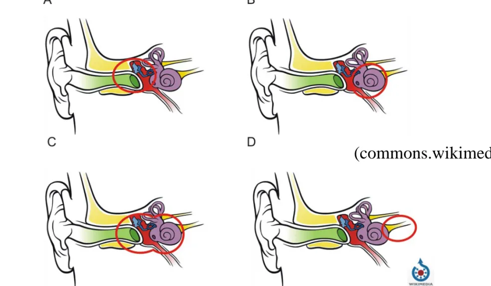
HEARING DISORDERS
Conductive hearing loss - Any problem in the outer or middle ear that prevents sound from being conducted properly is known as a conductive hearing loss. It can be caused by excessive earwax, ear infections, tympanic membrane perforation, otosclerosis or tumours. The degree of conductive hearing loss is mild or moderate and may be helped with medication, surgery, hearing aids or middle ear implants. (Figure 10. A)
Sensorineural hearing loss - Sensorineural hearing loss results from missing or damaged sensory cells (hair cells) in the cochlea and can be mild, moderate, severe or profound. Mild to severe sensorineural hearing loss can often be helped with traditional hearing aids or middle ear implants.
Cochlear implants are often a solution for severe or profound hearing loss.
Partial deafness means that the sensorineural hearing loss is only in the high frequencies: here a combined electric and acoustic stimulation, the EAS can help. Sensorineural hearing loss may be congenital or acquired. (Figure 10. B)
www.itk.ppke.hu
HEARING DISORDERS
Mixed hearing loss - A mixed hearing loss is a combination of both conductive and sensorineural hearing loss. The inner, outer and middle ear is damaged at the same time for the same reason or for different reasons.
Medication, surgery, hearing aids, middle ear implants and cochlear implants can be treatment options. (Figure 10. C)
Neural hearing loss – Results from the damage of the auditory nerve or other brain structures related to the auditory pathway. Neural hearing loss is usually permanent and profound. Experimental prostheses like the auditory brainstem implant, placed into the cochlear nuclei or the auditory midbrain implant, placed into the inferior colliculus may be a possibility for treatment.
(Figure 10. D)
Unilateral Profound Sensorineural Hearing Loss (Single Sided Deafness, SSD) – Means sensorineural deafness in one ear, while the other ear has good hearing. It is usually severe and permanent.
www.itk.ppke.hu
Figure 10. Damaged structures in different types of hearing disorders. A – Conductive hearing loss, B – Sensorineural hearing loss, C – Mixed hearing
(commons.wikimedia.org)
www.itk.ppke.hu
AUDIOGRAM
Hearing sensitivity varies according to the frequency of sounds. To take this into account, hearing sensitivity can be measured for a range of frequencies and plotted on an audiogram. On the x-axis of an audiogram are the frequencies ranging from about 125 Hz to 8 kHz (or 16 kHz), usually on a logarithmic scale. On the vertical axis the loudness of the sounds is represented, measured in decibels ranging from -10 dB to 120 dB. (Figure 11.)
An audiogram can be obtained using a behavioral hearing test called audiometry. This means presenting different tones at a specific frequency and loudness (intensity). When the person hears the sound they raise their hand or press a button so that the tester knows that they have heard it. The lowest intensity sound they can hear is recorded. With normal hearing all frequencies are heard at low intensities (Figure 12. A), in case of deafness no sign of sound hearing is present, or just at high intensities above 80 dB (Figure 12. B).
Partial deafness affects only the high frequencies, people who suffer from partial deafness need higher intensities to hear sounds above 4000 Hz.
www.itk.ppke.hu
Figure 11. The audiogram. The classification of the hearing loss is shown on the right side of the figure. The loudness of some sound sources with
http://www.cochlear.com/
www.itk.ppke.hu
Figure 12. A - The audiogram of a normal hearing B – The audiogram of deafness (commons.wikimedia.org)
www.itk.ppke.hu
To generate a psychoacoustical tuning curve, an arbitrary frequency is chosen, and the amplitude is lowered such that the subject can barely detect it (~2dB above threshold). This probe tone will be intermittent. Then, the masker tone is presented at a different frequency, and the amplitude is increased as high as possible such that the subject can still detect the probe tone. This process is repeated for multiple frequencies, and the line graph of the tuning curve is derived. In Figure 13. we see two psychoacoustical tuning curves - one for a normal cochlea and one for a cochlea with damaged outer hair cells. Notice that the damaged cochlea is both less sensitive (the line is lower) and less selective (the curve is not sharp). Thus, to restore hearing in someone with outer hair cell damage, simple amplification is not enough, since that raises inaudible, indistinguishable sounds to the level of audible, indistinguishable sounds. The selectivity problem needs to be solved to have useful hearing.
TUNING CURVE
www.itk.ppke.hu
(commons.wikimedia.org) Figure 13. The tuning curve of a normal cochlea (blue) and of a cochlea
with damaged outer hair cells (red).
www.itk.ppke.hu
HEARING LOSS AND AGE
The age at which hearing loss occurs is crucial for the acquisition of a spoken language:
•Pre-lingual deafness: a hearing impairment that is sustained prior to the acquisition of language, which can occur as a result of a congenital condition or through hearing loss in early infancy.
•Post-lingual deafness: a hearing impairment that is sustained after the acquisition of language, which can occur as a result of disease, trauma, or as a side-effect of a medicine. Post-lingual deafness is far more common than pre- lingual deafness.
www.itk.ppke.hu
OBJECTIVE TESTS
Audiometry is a subjective test, it needs a response from the the examined patient. However there are also objective tests, which do not need any response from the subjects and it can be performed in sleep or in awake state.
• Auditory Brainstem Response Testing (ABR): It is used, when subjective tests can not be performed, for example in infants. Click stimuli are given through earphones and the electrical responses are recorded from the auditory nerve and brainstem. This method can provide reliable threshold information for frequencies up to 2kHz.
• Transient Otoacoustic Emission (TOAE): It is a quick method, to test if there is any cochlear damage (e.g. loss of outer hair cells). It is often used to test the hearing condition of neonatals. About Otoacoustic emission you can read more a few slides later.
• Immittance Audiometry (tympanometry): It is a diagnostic method, to test if there is any middle ear related hearing disorder present. The compliance and pressure of the middle ear are assessed.
www.itk.ppke.hu
COCHLEAR MICROPHONIC
Cochlear microphonic (CM) is an electric potential generated by the cochlear hair cells (mainly outer hair cells) and can be recorded with intra- tympanic and extra-tympanic electrocochleography (ECochG) in humans and in experimental animals (e.g. guinea pig). In case of extra-tympanic ECochG a thin needle electrode is placed through the tympanic membrane onto the promontory, near the round window and the cochlear potentials are recorded.
CM can also obtained from surface recordings. The cochlear microphonic is originating primarily from the basal portions of the cochlea and it closely resembles the sound stimulus, it is a reflection of the alternating component.
CM has a limited clinical use, it can be used in the identification of auditory neuropathy, where the auditory nerve fibers are damaged because of demyelination, axonal loss or lesion. However the outer hair cell integrity is preserved, which can be examined by CM.
www.itk.ppke.hu
OTOACOUSTIC EMISSION (OAE)
The otoacoustic emission (OAE) is a low-intensity sound which is generated from within the inner ear, mostly by the cochlear outer hair cells, as they expand and contract. OAEs disappear after the inner ear has been damaged, so they can be used to determine the cochlear status and the hair cell function. Otoacoustic emission has a great clinical importance, because it is the basis of a simple, non-invasive test for hearing disorders in neonates, infants and in children who are too young to cooperate in conventional hearing tests.
Because hearing tests with OAEs do not require any behavioral response, it can be performed also on sleeping or on comatose patients. Spontaneous OAEs are present in only 40-50% of the people, who have normal hearing, and can be considered as a sign of cochlear health, but the absence of spontaneous OAEs is not necessarily a sign of abnormality.
There are also researches to utilize OAEs for biometric identification, but still several difficulties have to be solved until it can became useable in the everyday life.
www.itk.ppke.hu
OTOACOUSTIC EMISSION (OAE)
There are 4 types of otoacoustic emissions, one spontaneous and three evoked:
• Spontaneous otoacoustic emissions (SOAEs) - Sounds emitted without an external stimulus (i.e. spontaneously).
• Transient otoacoustic emissions (TOAEs) or transient evoked otoacoustic emissions (TEOAEs) - Sounds emitted in response to an acoustic stimuli of very short duration (e.g. clicks or tone-bursts).
• Distortion product otoacoustic emissions (DPOAEs) - Sounds emitted in response to 2 simultaneous tones of different frequencies.
• Sustained-frequency otoacoustic emissions (SFOAEs) - Sounds emitted in response to a continuous tone.
www.itk.ppke.hu
TRADITIONAL HEARING AIDS
Traditional hearing aids are designed to amplify the sound in frequency ranges, where the hearing loss is the greatest. Usually it builds up from a microphone, an amplifier, a processor, a speaker and a battery (Figure 14.).
The sound is received through a microphone, which transmits it to the amplifier in electric from. The amplifier increases the power of the signals and then sends them to the processor for modulation. Finally the signal gets to the speaker, where the amplified and modulated sound enters the ear canal.
Hearing aids can be analog or digital. Programmable hearing aids also make it possible to change the settings of the hearing aid according to different listening enviroments. Digital circuitry makes programming more flexible, for example a hearing aid can be programmed to amplify some frequencies more than others.
Hearing aids are the most effective in case of conductive hearing loss with mild and moderate level, or in case of sensorineural hearing loss, where a proportion of the outer hair cells is damaged.
www.itk.ppke.hu
Figure 14. Schematic of the traditional hearing aid
• Behind-the-ear (BTE) hearing aid: A hard plastic case, where the electronics take place, is worn behind the pinna. This is connected with a tube to a plastic earmold, that fits into the ear canal. The sound is routed from the case to the earmold via the tube. BTE hearing aid is used by people of all ages for mild to profound hearing loss. A subtype of the BTE hearing aid is the open-fit hearing aid or „mini” BTE: it fits behind the ear completely and a narrow tube is inserted into the ear canal. The canal remains open, which allows sound to enter the ear without being amplified.
This is advantageous for people with high frequency hearing loss.
• In-the-ear (ITE) hearing aid: It fits completely inside the outer ear bowl and is used for mild to severe hearing loss.
• Canal hearing aids: It fits into the ear canal. The in-the-canal (ITC) hearing aid is made to fit the size and shape of a person’s ear canal. A completely-in-canal (CIC) hearing aid is nearly hidden in the ear canal.
Both types are used for mild to moderately severe hearing loss.
www.itk.ppke.hu
TYPES OF TRADITIONAL HEARING AIDS
(Figure 15.)www.itk.ppke.hu
(commons.wikimedia.org)
www.itk.ppke.hu
TRADITIONAL HEARING AIDS
Other types:
• Invisible-in-canal hearing aids (IIC) – Placed deep in the ear canal
• Receiver-in-the-Canal/Ear (RIC/RITE) – Speaker placed inside the ear canal
• Extended wear hearing aids – Continuously worn, invisible Technological developments:
• Wireless hearing aids and FM listening systems
• Directional microphones to improve the speech recognition in noise
• Telecoils used with telephones, FM systems and induction loop systems (hearing loops) - A telecoil is a small magnetic coil that allows users to receive sound through the circuitry of the hearing aid. It helps people hear in public facilities that have installed induction loop systems.
• Direct Audio Input (DAI) – direct connection to external audio sources
www.itk.ppke.hu
IMPLANTABLE HEARING DEVICES (IHD)
• Bone anchored hearing apparatus (BAHA)
• Middle ear implantable hearing devices (MEIHD)
• Cochlear implants (CI)
• Electric Acoustic Stimulation (EAS)
• Auditory brainstem implants (ABI)
• Auditory midbrain implants (AMI)
www.itk.ppke.hu
INTRODUCTION TO IHDs
Auditory prostheses are one of the most successful neural prostheses that restore human sensory function. They have been used to partially restore hearing in more than 60000 hearing-impared people worldwide. There are several solutions of IHDs according to the place of the damage in the auditory system or type of hearing loss (Figure 16.). The most successful one is the cochlear implant which will be detailed in the next lecture. When conductive hearing loss is present, a bone conducting hearing device like the BAHA seeks to restore hearing by vibratory conduction through the skull to the inner ear.
Sensory hearing impairment is also treated by MEIHDs which amplificate the sound signals through implanted transducers wich can be either piezoelectric crystals or electromagnetic transducers. The Vibrant Soundbridge System produced by Med-El will be demonstrated in details in this section. Devices in experimental phase like the auditory brainstem implant (ABI) and auditory midbrain implant (AMI) will be discussed also in the next lecture.
www.itk.ppke.hu
Figure 16. Targets (red) of different types of hearing aids (blue). On the horizontal arrows the method of signal generation of the respective hearing aid is presented. Next to the vertical arrows, the ways of transmitting the sound- signals originating from the outside world are shown.
EAS – Electric Acoustic Stimulation
BAHA - Bone anchored hearing apparatus
MEIHD - Middle ear implantable hearing devices ABI - Auditory brainstem implants
www.itk.ppke.hu
BONE ANCHORED HEARING APPARATUS(BAHA)
The BAHA is a hearing aid based on bone conduction: it transmits sound by direct conduction through bone to the inner ear, bypassing the outer ear and middle ear. It is used by people with conductive, mixed or unilateral hearing loss (single sided deafness). A sound processor is fixed through an abutment to a surgically implanted titanium implant, which is embedded into the skull of the patient. The abutment is exposed outside the skin. The titanium fixture has to bond with the bone. This process is called osseointegration and lasts for a few months. After osseointegration the BAHA can be used. During working, the system vibrates the skull and the inner ear, which stimulates the cochlear nerve. Two companies have solutions using bone conduction: Cochlear (Baha System, Figure 17-19.) and Oticon Medical (Ponto, Ponto Pro). Cochlear registered the acronym Baha as a trademark. BAHA has some drawbacks: the open wound means a risk of infection, so a daily care is required; batteries last only for 6 to 14 days. Several accessories are available for BAHA: telecoil, audio adapter (to connect to external audio sources) and FM receiver.
www.itk.ppke.hu
Figure 17. Working mechanism of a BAHA 1. A sound processor picks up sound vibrations.
2. An abutment is attached to the sound processor and the implant. The abutment transfers the sound vibrations from the processor to the implant.
3. A small titanium implant is placed in the bone behind the ear where it fuses with the living bone (osseointegration). The implant transfers the sound vibrations
http://www.cochlear.com/
www.itk.ppke.hu
http://www.cochlear.com/
Figure 18. Parts of the Cochlear Baha system
www.itk.ppke.hu
Figure 19.
Left – Direct bone conduction:
1. Sound waves are received by Baha sound processor
2. Sound waves bypass middle ear function and are delivered directly to the working cochlea in both ears
Right – Single Sided Deafness
1. Sound waves are received by the Baha sound processor
2. Sound waves travel by bone conduction to the functioning cochlea on the opposite side
http://www.cochlear.com/
www.itk.ppke.hu
MIDDLE EAR IMPLANTABLE HEARING DEVICES
MEIHDs were developed to treat conductive or sensorineural hearing loss with a degree from mild to severe. MEIHDs can be partially-implanted or totally-implanted. Totally-implanted solutions, where the device is completely concealed in the body, are fully functioning also during swimming or bathing, and there is no occlusion effect present (Occlusion effect: when an object fills the ear canal, and the person perceives "hollow" or "booming" echo-like sounds of their own voice. It is mostly experienced in case of traditional hearing aids). MEIHDs directly stimulate the ossicles in the middle ear, and because the sensor and the driver is uncoupled, the feedback (which is another annoying effects of conventional aids) is almost completely eliminated. These devices have to fulfil some important requirements: the transducer must be attached to the tympanic membrane, the long process of the incus, or the head of the stapes, because otherwise, the transmitted force is reduced. The battery life must be sufficient to avoid frequent replacements requiring reoperation.
Some middle ear implants are also compatible with MRI.
www.itk.ppke.hu
MIDDLE EAR IMPLANTABLE HEARING DEVICES
Middle ear devices can be classified into two groups: there are piezoelectric and electromagnetic solutions.
Piezoelectric devices operate by passing an electric current into a piezoceramic crystal, which changes its volume (by deformation or bending).
This deformation provides the mechanical energy to stimulate the ossicular chain or inner ear, it produces a vibratory signal. The power output is directly related to the size of the crystal.
Electromagnetic hearing devices function by passing an electric current into a coil, which creates a fluctuating magnetic field (corresponding to the acoustic input). This causes a small magnet (samarium cobalt or neodymium iron boron), which is attached to the vibratory structures of the middle ear, to vibrate. This vibration, in turn, causes the movement of these structures resulting in the sensation of hearing. The coil may be separate from the magnet or integrated with it. The coil and magnet must be close, because the power is
SOLUTIONS FOR MEIHDs
www.itk.ppke.hu
Name Manufacturer Transducer Implantable
TICA (Totally Integrated Cochlear Amplifier)
Cochlear (device developed by
Implex AG) Piezoelectric Fully
Envoy Esteem System Envoy Medical (originally St. Croix
Medical) Piezoelectric Fully
MET (Middle Ear
Transducer) Otologics LLC Electromagnetic Semi
Carina MET Otologics LLC Electromagnetic Fully
Vibrant Soundbridge Med-El (device developed by
Symphonix Devices) Electromagnetic Semi
Direct System Soundtec Electromagnetic Semi
MAXUM System Ototronix Electromagnetic Semi
www.itk.ppke.hu
THE VIBRANT SOUNDBRIDGE SYSTEM
This middle ear hearing device was developed by Symphonix Devices. The technology was later purchased by Med-El. It was the first FDA-approved implantable MEIHD (2000) and got also the European CE-mark (1998). Now it is implanted worldwide. Vibrant Soundbridge is a semi-implantable device with an electromagnetic transducer and consists of an external sound processor and amplifier (Amadé), and an internal vibrating ossicular prosthesis (VORP), which is surgically implanted. Sound passes into a microphone on the postauricular audio processor and is transmitted through the skin to the receiver on the VORP. The VORP, which is implanted postauricularly, conducts the sound to a magnet surrounded by a coil, called the floating mass transducer (FMT). The transducer is crimped around the long process of the incus by a titanium clip. Vibration of the device is transmitted to the ossicles, thereby delivering stimuli to the middle and, subsequently, to the inner ear.
(Figure 20.)
www.itk.ppke.hu
THE VIBRANT SOUNDBRIDGE SYSTEM – FEATURES AND BENEFITS
• improved hearing quality
• improved speech understanding (particularly in noisy environment)
• ear canal remains completely open (no occlusion effect)
• no feedback or irritating whistling
• wind-noise reduction
• speech and noise management with the Amadé audio processor
• high wearing comfort
• reliable and flexible
• suitable for children
www.itk.ppke.hu
Audio
Processor Receiver (VORP)
Conductor lead
Floating Mass Transducer
(FMT)
http://www.medel.com/
www.itk.ppke.hu
VIBRANT SOUNDBRIDGE - TERMINOLOGY
• AP - Audio Processor (Amadé)
The external signal processor containing microphone, battery and electronics.
It is attached to the patient’s head with a magnet and can be worn discreetly underneath the hair. Three different programs are available, which can be adapted to fit personal preferences. (Figure 21.)
• VORP - Vibrating Ossicular Prosthesis
The complete implant device, including the receiver coil, attachment magnet, receiver electronics and FMT. It is placed underneath the skin and the attachment magnet holds the audio processor over the implant. (Figure 22.)
• FMT - Floating Mass Transducer
The electromagnetic transducer that generates the vibratory motion. It conducts a wide frequency range (up to 8,000 Hz), which is particularly relevant for listening to or playing music. This tiny magnet consists of just five components. It can be attached to the incus, to the round or to the oval window. (Figure 23. and Figure 24.)
www.itk.ppke.hu
Figure 21. The Amadé Audio Processor
http://www.medel.com/
www.itk.ppke.hu
Figure 22. The Vibrating Ossicular Prosthesis (VORP) Demodulator
Receiver coil Magnet
Silicone Floating Mass Transducer
http://www.medel.com/
www.itk.ppke.hu
Figure 23. The Floating Mass Transducer (FMT) - The FMT is attached to the long Titanium clip
2 mm
http://www.medel.com/
www.itk.ppke.hu
Figure 24. The FMT reproduces and amplifies the natural movement of the ossicular chain. The FMT should be in close contact and parallel to the stapes.
http://www.medel.com/
www.itk.ppke.hu
THE VIBRANT SOUNDBRIDGE SYSTEM – WORKING MECHANISM
1. Sounds are picked up by the microphone of the audio processor.
2. The audio processor converts environmental sounds into electrical signals.
3. The electrical signals are transmitted across the skin to the implanted part.
4. The implant relays the signal down to the FMT.
5. The FMT converts the signal into mechanical vibrations that
directly stimulate a middle ear structure (e.g., the ossicular chain) causing it to vibrate.
6. These vibrations then conduct sound to the inner ear where they are passed on to the brain and are perceived as sound.
www.itk.ppke.hu
THE FUTURE PROSPECTS OF HEARING AIDS
•
Totally Implant system•
Perfectly hide•
External control using wireless communication•
More convenient•
Miniaturized•
High-performance•
Will be popularized in near futurewww.itk.ppke.hu
WEBPAGES OF COMPANIES RELATED TO IMPLANTABLE HEARING AIDS
• www.oticonmedical.com
• www.cochlear.com
• www.medel.com
• www.advancedbionics.com
• www.envoymedical.com
• www.otologics.com
• www.ototronix.com
www.itk.ppke.hu
LINKS RELATED TO HEARING AIDS
Traditional hearing aids:
http://www.nidcd.nih.gov/health/hearing/hearingaid.asp http://www.hearingaids1000.com/index.shtml
BAHA:
http://www.entkent.com/baha.php MEIHD:
http://emedicine.medscape.com/article/860444-overview http://www.middleearimplants.com/
www.itk.ppke.hu
RECOMMENDED LITERATURE
• Cochlear implants: Auditory Prostheses and Electric Hearing; Fang-Gang Zeng, Arthur N.
Popper, Richard R. Fay; 2004; Springer
• Handbook of Neuroprosthetic Methods; Warren E. Finn, Peter G. LoPresti; 2003, CRC Press
• Neuroprosthetics: Theory and Practice; Kenneth W. Horch, Gurpreet S. Dhillon; 2004;
World Scientific Publishing
• Cochlear And Brainstem Implants (Advances in Oto-Rhino-Laryngology); A.R. Møller;
2006; Karger
• Neural Prostheses for Restoration of Sensory and Motor Function; John K. Chapin, Karen A. Moxon; 2000; CRC Press
• More than 500 books related to implantable hearing aids implants on www.amazon.com
www.itk.ppke.hu
REVIEW QUESTIONS
• What are the three main parts of the human ear?
• What is the structure and working mechanism of the cochlea?
• How do you make a tuning curve?
• What are the main processing stations in the auditory pathway?
• What type of hearing disorders do you know?
• What is the audiogram good for?
• How is the phenomenon of otoacoustic emission generated?
• How are traditional hearing aids helping people with hearing impairment?
• What are the main types of implantable hearing devices?
• What is the working mechanism of a BAHA? What are its main parts?
• How can be the MEIHDs grouped? What parts build up the Vibrant Soundbridge system? Can you mention another MEIHD solutions?