László Bitó
First Department of Medicine, Albert Szent-Györgyi Health Center University of Szeged, Korányi fasor 8–10
HU–6720 Szeged (Hungary) bito6725@gmail.com
Single Case
PAX2 Mutation-Related
Oligomeganephronia in a Young Adult Patient
László Bitóa Tibor Kalmárb Zoltán Marótib Sándor Turkevi-Nagyc Csaba Bereczkib Béla Iványic
aFirst Department of Medicine, Albert Szent-Györgyi Health Center, University of Szeged, Szeged, Hungary; bDepartment of Pediatrics and Pediatric Health Center, Albert Szent-Györgyi Health Center, University of Szeged, Szeged, Hungary;
cDepartment of Pathology, Albert Szent-Györgyi Health Center, University of Szeged, Szeged, Hungary
Keywords
Congenital anomalies of the kidney and urinary tract · Focal segmental glomerulosclerosis · Oligomeganephronia · Paired box protein 2 mutation · Renal coloboma syndrome
Abstract
Oligomeganephronic hypoplasia, commonly referred to as oligomeganephronia (OMN), is a rare pediatric disorder characterized by small kidneys. Histologically a paucity of nephrons is observed which show compensatory enlargement. Hyperfiltration injury leads to end-stage kidney disease. Here we report a 23-year-old Caucasian female patient who presented with a 7-year history of nonnephrotic proteinuria, slow worsening of renal function, normal-sized kid- neys, normal blood pressure, healthy weight, and normoglycemia. Evaluation of a kidney bi- opsy specimen revealed sparsely distributed and markedly enlarged glomeruli (glomerular density 0.63/mm2, glomerular diameter 268 µm), focal segmental glomerulosclerosis (FSGS), and 70% effacement of the foot processes. The glomerular basement membrane was normal
(mean thickness 285 nm). The genetic analysis of 19 genes known to cause FSGS identified a heterozygous de novo nonsense mutation of PAX2 in exon 4 (NM_003990.3:c.430C>T and NP_003981.2:p.Gln144Ter). Clinical investigations ruled out optic nerve coloboma, hearing loss, and vesicoureteral reflux. Magnetic resonance imaging of the urogenital tract found the uterus to be bicornuate. Based on these data, OMN in nonhypoplastic kidneys and adaptive FSGS related to PAX2 mutation was diagnosed. Her kidney function worsened during the 30-month follow-up (last visit: eGFR-EPI 32 mL/min/1.73 m2) despite angiotensin-converting enzyme in- hibitor treatment. To our best knowledge, our patient is the seventh in the English-language literature with a biopsy diagnosis of OMN in an adult, the first observed with normal-sized kidneys, and the first in whom a specific etiologic genetic diagnosis was established. Nonsense PAX2 mutations between the paired domain and the octapeptide domain appear to manifest
in renal-limited phenotype. © 2020 The Author(s)
Published by S. Karger AG, Basel
Introduction
Oligomeganephronic hypoplasia, commonly referred to as oligomeganephronia (OMN), is a pediatric anomaly characterized by small kidneys with reduced number of renal lobes. His- tologically, the nephrons are structurally intact, but are present in markedly low number and exhibit compensatory hypertrophy. End-stage renal failure occurs months after birth or until early to mid adolescence. Mutations in PAX2 transcription factor gene can play a role in the etiology of OMN because heterozygous PAX2 mutations have been found in one-third of chil- dren with OMN [1, 2].
The PAX2 gene is essential in embryonic development of the kidney, the eye, the inner ear, the midbrain/hindbrain, the spinal cord, and the urogenital system. The phenotypic spectrum of heterozygous PAX2 mutations is broad and can manifest in congenital anomalies of the kid- ney and urinary tract (CAKUT), in renal disorders such as OMN or focal segmental glomerulo- sclerosis (FSGS) [3, 4], or in papillorenal syndrome (OMIM #120330) [5, 6]. The latter consists of a wide spectrum of renal anomalies, optic nerve dysplasia, and occasionally hearing loss.
The renal anomalies include hypoplasia, renal cysts, multicystic dysplastic kidneys, OMN, and childhood- or adulthood-onset sporadic or familial FSGS. Vesicoureteral reflux, ureteropelvic junction obstruction, pyeloureteral duplication, anomalous renal pelvis, horseshoe kidney, or renal malrotation may be associated findings. The majority of patients with PAX2 mutations have normal intellect [3–7].
Recently, we evaluated an adult patient in whom investigations led to the diagnosis of PAX2 mutation-related OMN. Only 6 adult patients with OMN have been published in the En- glish-language literature so far [8–11], and genotype-phenotype correlations were not inves- tigated in these publications. In the present communication, the clinicopathologic features of adult-onset OMN were aggregated by supplementing the findings of our patient to those pub- lished earlier.
Case Report
In November 2017, a 23-year-old Caucasian female patient was referred to our depart- ment for renal biopsy evaluation. The patient had been born at full term and her growth and development had been normal. She had healthy, nonconsanguineous parents and her family history was negative for renal diseases. A 100 mg/dL proteinuria had been detected at the age of 16. At age 21, impaired renal function had been observed (proteinuria >300 mg/dL, serum creatinine 110 μmol/L, eGFR-EPI 62 mL/min). On admission, she had no complaints and physical findings and blood pressure values were normal. Her blood chemistry val- ues were as follows: glucose, normal; urea nitrogen, 7.9 mmol/L; creatinine, 145 µmol/L;
eGFR-EPI, 43 mL/min/1.73 m2; albumin, 34 g/L; cholesterol, 6.07 (H) mmol/L; triglyceride, 1.36 mmol/L; IgG, 10.01 g/L; IgA, 2.31 g/L; and IgE, 10 IU/mL. The IgM value could not be interpreted for technical reasons. The autoimmune panel (among others anti-double- stranded DNA, anti-SSA, anti-SSB, and anti-neutrophil cytoplasmic antibodies) did not reveal any anomaly. The complement 3 and 4 levels were in the normal range. Serum viral hepatitis markers were negative. Serum intact parathormone value was in the normal range. Urinalysis revealed proteinuria of 2.5 g/day; microhematuria or casts were not detected. Abdominal ul- trasound (US) disclosed a right kidney of 108 × 38 × 15 mm, a left kidney of 124 × 55 × 15 mm, and a hyperechogenic parenchyma. The calices, the pyelon, and the ureter on both sides were normal. A US-guided percutaneous biopsy of the left kidney was performed.
Renal Biopsy Findings
Thirty-one tissue levels were prepared from the 10% formalin-fixed and paraffin-embed- ded specimen. Light microscopic evaluation revealed altogether seven patent and three glob- ally sclerotic glomeruli; one to four sparsely distributed and markedly enlarged patent glo- meruli per level (Fig. 1a, b) were observed, two of which showed FSGS (Fig. 1c, d). The tubular profiles were enlarged. Streaks of interstitial fibrosis and tubular atrophy in the range of 20–
25% and focal mononuclear interstitial infiltrates with occasionally small groups of foam cells accompanied the glomerular lesions. The interlobular arteries were normal; two arteriolar profiles exhibited eccentric subendothelial hyalinosis. The total renal chronicity score of four indicated mild chronic changes. The frozen sections prepared for immunofluorescence evalu- ation (IgG, IgA, IgM, C3, C1q, kappa, lambda, and fibrinogen) contained four patent and three completely sclerotic glomeruli. One patent glomerulus displayed segmental sclerosis, with C3 and IgM positivity in the area of sclerosis. The resin-embedded specimen contained two pat- ent glomeruli. Ultrastructurally, the glomerular basement membrane was normal (mean thickness 285 nm, range 200–467 nm). The degree of effacement of podocyte foot processes was 70%.
The pathology report was signed out as FSGS suspicious for mutation-induced FSGS. Upon receiving the result of genetic analysis, quantitative measurements were carried out. The total cortical area ranged between 6.19 and 7.26 mm2 in the various tissue levels. The mean glo- merular density, calculated by dividing the number of patent and globally sclerotic glomeruli by the total cortical area read in tissue levels 3, 19, and 31, was 0.63/mm2 (range 0.42–
0.82/mm2). The mean glomerular diameter, obtained by measuring the maximum glomerular diameter in profiles exhibiting a hilus in the section plane, was 268 µm (range 252–297 µm).
Genetic Analysis
After obtaining written informed consent, genomic DNA from the patient and her parents was extracted from peripheral blood according to standard procedures. The exonic (and the proximal intronic regions) sequence of 19 genes known to cause FSGS was investigated to identify possible mutations by using a clinical exome sequencing kit (TruSightTM One). The following genes were tested: ACTN4, APOL1, ARHGAP24, CDA2P, CDKN1C, COL4A3, CYP11B2, INF2, MED12, MYO1C, MYO1E, NPHS1, NPHS2, PAX2, PLCE1, PTPRO, SYNPO, TRPC6, and WT1.
Mutation was observed only in PAX2 (Fig. 2). The C/T heterozygous mutation in exon 4 is rare and has not been detected in 360 Hungarian control samples, nor is it included in gnomAD (http://gnomad.broadinstitute.org), Exom Variant Server (http://evs.gs.washington.edu), ClinVar (www.ncbi.nlm.nih.gov/clinvar/), or HGMD (www.hgmd.cf.ac.uk). To clarify the ori- gin of the heterozygous mutation, targeted bidirectional sequencing of only exon 4 (and its flanking intronic regions) of PAX2 was tested in the DNA samples of the parents. Since the mutation was not detected in the parent samples, the de novo nonsense mutation in the pro- band was regarded as pathogenic. The case was concluded as OMN and adaptive FSGS (MalaCard fsgs7) related to de novo PAX2 mutation (see Discussion).
Investigations after the Biopsy Procedure and Follow-Up
The PAX2 protein expression pattern (clone EP3251, 1:250, pH 9; Abgent, USA) was as- sessed in an archived paraffin-embedded slide. Six renal biopsy cases of native kidney dis- eases with no known genetic mutations served as controls. Definite positivity was observed in the nuclei of distal nephron segments; borderline positivity or negativity was seen in the nuclei of proximal tubular epithelial cells. The nuclei of podocytes and mesangial cells were negative, and the nuclei of parietal epithelial cells were either negative or barely positive. In the controls a similar finding was noted, with the exception that the nuclei of proximal tubular cells were more frequently positive and the nuclei of glomerular parietal cells were unequiv- ocally positive (Fig. 1e, f).
Vesicoureteral reflux detection with 99mTc-EC renal scintigraphy and separated clear- ance determination revealed normal renogram bilaterally, normal-sized kidneys, and nonsig- nificant ptosis of the right kidney. Vesicoureteral reflux was not detected. EC clearance (mL/min/1.73 m2 body surface area): right kidney 80, left kidney 71; the global EC clearance was lowered (cutoff 347). Native and contrast-enhanced abdominal magnetic resonance im- aging: normal-sized kidneys and normal ureters and urinary bladder. The uterus was bicor- nuate and smaller; the ovaries were normal. The patient had a history of mild myopia (right eye –3.25 D, left eye –2.75 D) corrected completely with contact lenses. Funduscopic exami- nation by slit lamp microscopy did not demonstrate any optic nerve head anomalies, and au- diologic examination did not confirm hearing loss.
After the kidney biopsy evaluation, therapy of angiotensin-converting enzyme inhibitor (ramipril 1.25 mg/day) and atorvastatin was started. Nevertheless, the patient’s renal func- tion slowly decreased. Laboratory values at 30 months after biopsy were as follows: serum creatinine 185 µmol/L, eGFR-EPI 32 mL/min/1.73 m2, proteinuria 3.46 g/day, and urinary protein/creatinine ratio 214.5 mg/mmol. Abdominal US examination still revealed normal- sized kidneys. Her blood pressure values were normal during visits.
Discussion
The biopsy evaluation of our patient with a 7-year history of nonnephrotic proteinuria, slowly worsening renal function, and normal-sized, hyperechogenic kidneys revealed paucity of glomeruli, glomerular enlargement, FSGS, and nonextensive effacement of podocyte foot processes. Decreased glomerular density and increased glomerular diameter were demon- strated quantitatively. Conditions leading to glomerulomegaly, such as obesity, diabetes, hy- pertension, or prematurity, did not exist in our patient. Since she had normal-sized kidneys and the calyces and pelvis did not exhibit alteration, she definitely did not have pediatric type of OMN [1]. The distinctive biopsy features and the lack of hypertension did not favor the di- agnosis of oligonephronia [1]. The case was concluded as OMN in nonhypoplastic kidney with adaptive FSGS related to PAX2 mutation. We assume that the number of nephrons available for her postnatal life was not strikingly low, and the hemodynamic injury to the nephrons manifested only from age 16. Her renal function slowly worsened during the follow-up of 9 years, not influenced by the angiotensin-converting enzyme inhibitor therapy administered for 30 months.
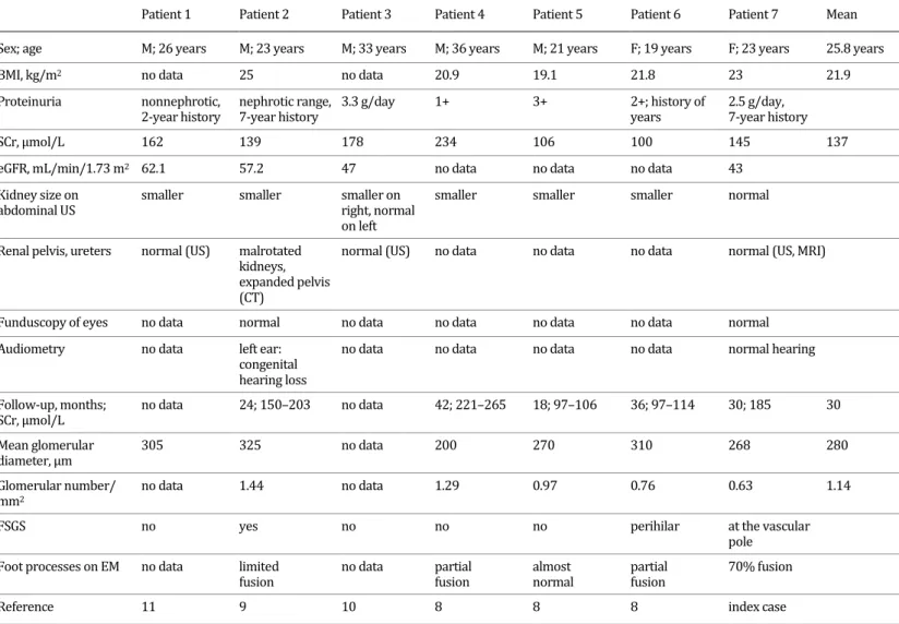
The pathologic and clinical features of adult-onset OMN are summarized in Table 1. We retrospectively calculated the values of mean glomerular density and mean glomerular diam- eter (1.14/mm2 and 280 µm, respectively), and these were out of the range of normal glomer- ular density (mean 3.5/mm2, range 1.2–8.1/mm2) [9] and normal glomerular diameter (mean + SD: 201 ± 28 µm) [8]. Thus, the quantitative data supported the original histopathologic di- agnosis of OMN. The ultrastructural examination did not reveal diffuse effacement of foot pro- cesses, therefore the FSGS lesion was the result of an adaptive process in response to a signif- icantly reduced nephron mass. OMN occurred more frequently in males (male:female ratio = 2.5:1) and the mean age at diagnosis was 25.8 years. Significant proteinuria and elevated se- rum creatinine value were present in all patients. However, full-blown nephrotic syndrome was not recorded in any of the patients. Hematuria was not observed. Patients 4 and 5 had mildly elevated blood pressure values at the time of biopsy. Imaging investigations disclosed bilaterally small kidneys in 5 patients, which could well be due to mild hypoplasia. However, the number of renal lobes, the criterion of hypoplasia, was not addressed in the publications.
A slow decrease in renal function was observed in all patients. Nevertheless, end-stage renal disease was not reached in the cohort during the follow-up of a mean of 30 months. These data suggest that the reduction in nephron mass in adult-onset OMN is much less than that in pediatric OMN [2].
Our genetic analysis revealed a new mutation in PAX2 that we regard responsible for the interruption of nephrogenesis and the markedly reduced number of nephrons. A single nucle- otide variant in exon 4 was identified, which led to a nonsense mutation between the highly conserved paired domain (encoded by exons 2–4, amino acids 16–142, which has known DNA-binding properties) and the octapeptide domain (in exon 5 encoded by amino acids 185–
192, having a role in repression of transactivation of target genes) [5]. Nevertheless, it is not clear how the mutation influences the function of the gene. So far 59 missense/nonsense path- ogenic PAX2 mutations have been reported according to The Human Gene Mutation Database (www.hgmd.cf.ac.uk/). It appears that the nonsense mutations between the paired domain and the octapeptide domain associate with the FSGS phenotype, similar to the p.Gln144Ter mutation in our patient with OMN, secondary FSGS, and normal-sized kidneys. The nonsense
mutations at other locations were described with either papillorenal syndrome or renal hy- podysplasia phenotypes [4, 12, 13].
In the publication on PAX2 mutations and OMN in children, 9 patients with renal hypo- plasia and the clinical diagnosis of OMN were evaluated genetically [2]. The diagnosis of OMN was confirmed by renal histology evaluation of nephrectomy specimens in 5 patients who were transplanted. Heterozygous PAX2 mutations involving the paired domain were identi- fied in 3 patients [2]. Since limited optic nerve coloboma or very mild optic disc dysplasia were secondarily detected in patients with PAX2 mutations, the cases appear to be examples of pap- illorenal syndrome in retrospect. Our case was assigned as PAX2-related isolated OMN be- cause the clinical investigations excluded coloboma, hearing loss, vesicoureteral reflux, and renal malformations. Nonetheless, abdominal magnetic resonance imaging revealed a bicor- nuate uterus, and the condition can be the subtle extrarenal manifestation of PAX2 mutation.
Regarding the cases of adult OMN, clinical investigations towards CAKUT, eye and ear anom- alies, etc. were not performed systematically. However, malrotated kidneys, expanded pelvis, unilateral complete hearing loss (congenital), and no anomaly on fundus examination were documented in patient 2 [9], and the authors considered the possibility of genetic factors in the etiology of OMN in their patient.
The immunohistochemical expression of PAX2 in normal adult kidneys is confined to the nuclei of parietal cells of Bowman’s capsule and nuclei in distal tubules and collecting ducts [14]. In the present communication, this expression pattern was seen in biopsy samples of native kidney diseases with no known genetic mutations. In the case of OMN, PAX2 expression was negative in glomerular parietal cells. The significance of the finding remains unknown.
In summary, the renal biopsy evaluation of a young adult patient with nonnephrotic pro- teinuria, impaired renal function, and normal-sized kidneys revealed a markedly reduced number of glomeruli. The glomeruli were enlarged and exhibited FSGS lesion. The findings were interpreted as adult-onset OMN and adaptive FSGS. The genetic analysis identified a novel heterozygous nonsense PAX2 mutation in exon 4. Since the existence of papillorenal syndrome was ruled out clinically, isolated renal disease characterized the PAX2 disorder.
Nonsense PAX2 mutations between the paired domain and the octapeptide domain seem to manifest in renal-limited phenotype.
Acknowledgments
The authors are grateful to Mr. Mihály Dezső, Ms. Erika Németh, and Mr. Krisztián Daru for their technical assistance. Ms. Fiona Molnár was the language editor. We thank the review- ers for their comments, especially reviewer 3.
Statement of Ethics
All investigations and procedures were in accordance with the ethical standards of the Institutional Committee and the World Medical Association Declaration of Helsinki. Written informed consent for publication including images was obtained from the participants in- volved in the study.
Conflict of Interest Statement
The authors declare that they have no conflict of interest.
Funding Sources
This study was supported by the GINOP-2.3.2-15-2 grant (T. Kalmár, Z. Maróti) provided by the National Research, Development and Innovation Office (Hungary). The state funders played no role in the study design, data collection and analysis, decision to publish, or in the preparation of the manuscript.
Author Contributions
L. Bitó and T. Kalmár, the two first authors, equally contributed to the paper. L. Bitó man- aged the clinical evaluation, the treatment and the follow-up of the patient, and wrote the manuscript. T. Kalmár performed the genetic test, analyzed the mutation, and wrote the man- uscript. Z. Maróti performed the genetic test. S. Turkevi-Nagy (junior pathologist) performed the quantitative measurements. C. Bereczki consulted the clinical evaluation. B. Iványi (senior pathologist) diagnosed the case, documented it for publication, and critically reviewed the manuscript. All authors read and approved the final manuscript.
References
1 Bonsib SM. Renal hypoplasia, from grossly insufficient to not quite enough: consideration for expanded concepts based upon the author’s perspective with historical review. Adv Anat Pathol. 2020 Sep;27(5):311–
30.
2 Salomon R, Tellier AL, Attie-Bitach T, Amiel J, Vekemans M, Lyonnet S, et al. PAX2 mutations in oligomeganephronia. Kidney Int. 2001 Feb;59(2):457–62.
3 Vivante A, Chacham OS, Shril S, Schreiber R, Mane SM, Pode-Shakked B, et al. Dominant PAX2 mutations may cause steroid-resistant nephrotic syndrome and FSGS in children. Pediatr Nephrol. 2019 Sep;34(9):1607–13.
4 Barua M, Stellacci E, Stella L, Weins A, Genovese G, Muto V, et al. Mutations in PAX2 associate with adult- onset FSGS. J Am Soc Nephrol. 2014 Sep;25(9):1942–53.
5 Bower M, Salomon R, Allanson J, Antignac C, Benedicenti F, Benetti E, et al. Update of PAX2 mutations in renal coloboma syndrome and establishment of a locus-specific database. Hum Mutat. 2012 Mar;33(3):457–
66.
6 Schimmenti LA. Renal coloboma syndrome. Eur J Hum Genet. 2011 Dec;19(12):1207–12.
7 Okumura T, Furuichi K, Higashide T, Sakurai M, Hashimoto S, Shinozaki Y, et al. Association of PAX2 and other gene mutations with the clinical manifestations of renal coloboma syndrome. PLoS One. 2015 Nov;
10(11):e0142843.
8 Kawanishi K, Takei T, Kojima C, Moriyama T, Sugiura H, Itabashi M, et al. Three cases of late-onset oligomeganephronia. NDT Plus. 2011 Feb;4(1):14–6.
9 Fuke Y, Hemmi S, Kajiwara M, Yabuki M, Fujita T, Soma M. Oligomeganephronia in an adult without end stage renal failure. Clin Exp Nephrol. 2012 Apr;16(2):325–8.
10 Alves RJ, Oppermann K, Schein LE, Pêgas KL. A case of late-onset oligomeganephronia. J Bras Nefrol. 2012 Oct–Dec;34(4):392–4.
11 Yang XD, Shi W, Li D, Peng T. Oligomeganephronia: case report and literature review. Srp Arh Celok Lek.
2014 Nov–Dec;142(11–12):732–5.
12 Iwafuchi Y, Morioka T, Morita T, Yanagihara T, Oyama Y, Morisada N, et al. Diverse renal phenotypes observed in a single family with a genetic mutation in paired box protein 2. Case Rep Nephrol Dial. 2016 Apr;6(1):61–9.
13 Zhang L, Zhai SB, Zhao LY, Zhang Y, Sun BC, Ma QS. New PAX2 heterozygous mutation in a child with chronic kidney disease: a case report and review of the literature. BMC Nephrol. 2018 Sep;19(1):245–50.
14 Ohtaka A, Ootaka T, Sato H, Soma J, Sato T, Saito T, et al. Significance of early phenotypic change of glomerular podocytes detected by Pax2 in primary focal segmental glomerulosclerosis. Am J Kidney Dis.
2002 Mar;39(3):475–85.
L. Bitó and T. Kalmár are both first authors and contributed equally to this paper.
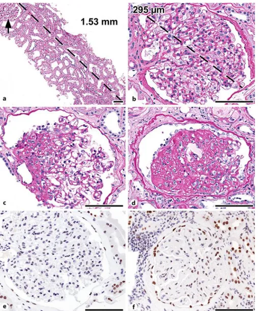
Fig. 1. Histopathology of OMN. a–d Tissue sections stained with periodic acid-Schiff. b–f The micrographs represent a 40× visual field; the empirical histologic feature of glomerular enlargement is that the glomer- ulus photographed almost fills the field. a Only one glomerular profile (arrow) is located in the 1.53-mm- long cortical sample, indicating a reduced number of glomeruli. b Glomerular hypertrophy, confirmed by the enlarged value of glomerular diameter. c Segmental glomerulosclerosis at the vascular pole. d Ad- vanced segmental glomerulosclerosis, with capping of epithelial cells overlying the sclerotic tufts. e PAX2 protein expression in OMN: the nuclei of parietal cells of Bowman’s capsule were negative; the nuclei of distal nephron segments were positive. f Native kidney disease control: PAX2 protein is expressed in the nuclei of parietal cells of Bowman’s capsule and in the nuclei of distal nephron segments. Scale bars, 100 µm. OMN, oligomeganephronia.
Fig. 2. A heterozygous PAX2 mutation (exon 4; NM_003990.3:c.430C>T and NP_003981.2:p.Gln144Ter) in the proband. A single nucleotide change c.430C>T was identified in our patient whereas both parents were homozygous for normal c.430C alleles. The Sanger sequence traces show the nucleotide change position (indicated by a blue highlight).
Table 1. Literature review: clinical and pathologic findings in adult-onset OMN
Patient 1 Patient 2 Patient 3 Patient 4 Patient 5 Patient 6 Patient 7 Mean
Sex; age M; 26 years M; 23 years M; 33 years M; 36 years M; 21 years F; 19 years F; 23 years 25.8 years
BMI, kg/m2 no data 25 no data 20.9 19.1 21.8 23 21.9
Proteinuria nonnephrotic,
2-year history nephrotic range,
7-year history 3.3 g/day 1+ 3+ 2+; history of
years 2.5 g/day, 7-year history
SCr, µmol/L 162 139 178 234 106 100 145 137
eGFR, mL/min/1.73 m2 62.1 57.2 47 no data no data no data 43
Kidney size on
abdominal US smaller smaller smaller on
right, normal on left
smaller smaller smaller normal
Renal pelvis, ureters normal (US) malrotated kidneys, expanded pelvis (CT)
normal (US) no data no data no data normal (US, MRI)
Funduscopy of eyes no data normal no data no data no data no data normal
Audiometry no data left ear:
congenital hearing loss
no data no data no data no data normal hearing
Follow-up, months;
SCr, µmol/L no data 24; 150–203 no data 42; 221–265 18; 97–106 36; 97–114 30; 185 30
Mean glomerular
diameter, µm 305 325 no data 200 270 310 268 280
Glomerular number/
mm2 no data 1.44 no data 1.29 0.97 0.76 0.63 1.14
FSGS no yes no no no perihilar at the vascular
pole Foot processes on EM no data limited
fusion no data partial
fusion almost
normal partial
fusion 70% fusion
Reference 11 9 10 8 8 8 index case
BMI, body mass index; CT, computed tomography; eGFR, estimated glomerular filtration rate; EM, electron microscopy; FSGS, focal segmental glomerulosclerosis; MRI, magnetic resonance imaging; OMN, oligomeganephronia; SCr, serum creatinine; US, ultrasound.