The review focuses on transcriptomic changes following treatment with serotonin reuptake inhibitor (SSRI) antidepressants. We aimed to overview results of the most established meth- ods for the investigation of the gene expression alterations including northern blotting, in situ hybridization, quantitative reverse transcriptase polymerase chain reaction (qRT-PCR), microarray and RNAseq in various brain regions and after chronic treatment protocols.
In spite of some measurable changes in serotonin system mRNA expression, serotonin transporter levels remained mostly unaltered following various treatment protocols. In contrast, tryptophan hydroxylase 2 appeared to be downregulated in serotonergic nuclei, and upregulated in the midbrain regions. Alterations in serotonin receptors lack clear con- clusions and changes probably reflect animal strain/substance related- and brain region dependent effects. Brain derived neurotrophic factor was upregulated following many, but not all chronic treatment regimens. GABA and glutamate genes also showed heterogeneous changes, with a surprising NMDA receptor downregulation in areas including the striatum and amygdala, known to be involved in depressive states and stress reactions. The review of the above studies suggests alterations in multiple processes, reflecting the heterogene- ity of the action depending on brain area and type of SSRI, and raises the possibility of a novel grouping of antidepressant medications based on their chronic molecular profile rather than on their initial actions.
(Neuropsychopharmacol Hung 2019; 21(1): 26–35)
Keywords: SSRI, BDNF, rat, transcriptomics, 5-HTT
S
ahelK
umar1, Z
SofiaG
al1, X
eniaG
onda2,3,4, r
obinJ h
uSe1, G
abriellaJ
uhaSZ1,2,5,6, G
yorGyb
aGdy1,2,4andP
eterP
etSchner1,2,41 Department of Pharmacodynamics, Faculty of Pharmacy, Semmelweis University, Budapest, Hungary
2 MTA-SE Neuropsychopharmacology and Neurochemistry Research Group, Hungarian Academy of Sciences, Semmelweis University, Budapest, Hungary
3 Department of Psychiatry and Psychotherapy, Semmelweis University, Budapest, Hungary
4 NAP-2-SE New Antidepressant Target Research Group, Semmelweis University, Budapest, Hungary
5 SE-NAP2 Genetic Brain Imaging Migraine Research Group, Hungarian Brain Research Program, Semmelweis University, Budapest, Hungary
6 Neuroscience and Psychiatry Unit, The University of Manchester and Manchester Academic Health Sciences Centre, Manchester, United Kingdom
INTRODUCTION
Antidepressants (ADs) are on the market since the 1950s and in wide use since the 1960s for the treat- ment of depression. Following tricyclic antidepres- sants (TCAs) and monoamine oxidase inhibitors as the first antidepressants, selective serotonin reuptake inhibitors (SSRIs) have been introduced towards the
end of the 1980s in an attempt to employ antidepres- sants with more selective action mechanisms to limit burdensome side effects, but retaining therapeutic efficacy. Subsequently several other groups of antide- pressants were introduced based on the monoamine theory of depression aimed at selectively targeting one or more of such systems, including for example serotonin–norepinephrine reuptake inhibitors (SN-
RIs) and norepinephrine reuptake inhibitors (NRIs).
Nevertheless, SSRIs remain the most prescribed medications for the treatment of major depressive disorders (Bobo et al. 2019).
Major depressive disorder (MDD) is a complex and heterogeneous disorder both in its manifesta- tion and in its neurobiology, and accordingly, the pathogenesis can’t be easily determined. Genetic, environmental and biological factors together play a role in the development of depression (see for review (Gonda et al. 2019).
The acute mechanism of action of the SSRIs con- stitutes the blockade of the serotonin transporter di- rectly elevating serotonin concentration in the syn- aptic cleft which, together with a similar blockade of noradrenaline reuptake, was thought to be the primary mechanism responsible for antidepressant effects ever since the observation that tricyclic anti- depressants block these transporters (Coppen 1967;
Schildkraut 1965). While this theory is not up to date, the monoaminergic systems remained in the focus of antidepressant pharmacotherapy, and a more complex mechanism of how monoaminergic antide- pressants influence neurotransmitter function have been described.
Functional effects after the administration of SSRIs are diverse and biphasic, also reflected in the temporal development of both side effects and ther- apeutic effects in the course of treatment. The mo- lecular changes underlying these functional effects can be measured by gene expression analysis (Volle et al. 2018). Four common methodical approaches are used to identify alterations of gene expression including northern blot, in situ hybdridization (ISH), quantitative reverse transcription polymerase chain reaction (qRT-PCR), RNA microarrays and RNA se- quencing (RNA-Seq).
In this review, after a short overview on current methods in gene expression analysis we summarize animal studies assessing transcriptomic changes fol- lowing chronic SSRI administration.
METHODS FOR GENE EXPRESSION ANALYSIS Northern blot
Northern blotting is one of the oldest methods in analyzing gene expression. Electrophoresis is used to separate the RNAs of the sample by size with a very low throughput, usually after separation of those with a polyA tail (mRNAs) from the other RNA content (Kevil et al. 1997). Then, the mRNA containing gel
has to be transferred onto a nylon membrane with the help of the electrophoretic blotter. Under UV light, the RNAs of different sizes will be covalently bound to this membrane. Afterwards, oligomers, usually of complementary DNA sequences (transcribed from RNA with reverse transcriptase) will be used for hy- bridization to the RNA of interest. After washing out, the hybridization, which is from the labelled oligonu- cleotides sequences or probes being complementary to the target transcript, will be detected by X-Ray.
In this way, northern blot is useful to determine the RNA transcript size and also its sequence through the probes (VanGuilderet al. 2008). In northern blotting, a larger amount of RNA compared to qRT-PCR is required and is much less sensitive with respect to mRNA expression compared to qRT-PCR (Fehr et al. 2000).
In situ hybridization
In situ hybridization is a method using labelled DNA or RNA probes which can attach selectively to the nucleic acid of interest even in a cellular environment, thus, for example in a brain sample. Advantage of the method is the information about the specific cellular localization of the transcript and the fine (or even manual) evaluation of the signals that can provide additional details about the expression patterns (see e.g. Kirilly et al. 2008). The method is, nevertheless, unfeasible for transcriptional analysis on a larger scale.
qRT-PCR
Real-Time Quantitative Reverse Transcription PCR (polymerase chain reaction) is a method based on PCR. First RNA will be converted to cDNA with the use of reverse transcriptase. After this step the expo- nential amplification of cDNA takes place with PCR.
A specific primer, or probe, will be attached on the template cDNA strand in order to start the replication trough polymerase enzyme. A probe is a modified primer with for example a fluorescent molecule (Heid et al. 1996), allowing relative quantification of specific genes, through sensors at a certain wavelength, during each PCR cycle (Cooper 2000).
Thus with this method, it is possible to quantify multiple genes with a high throughput single reac- tion. Trancriptome-wide gene expression analysis is however difficult (in case of some animal species), or even absolutely unfeasible (e.g. in humans) to realize (Teo et al. 2016).
Microarray
In differential expression analysis RNA is purified, extracted and is transcribed to cDNA. After tran- scription, cDNA samples are labeled with different fluorescent colors and applied on the microarray chip. The microarray chip is already printed with short, complementary probes of genes of interests and, thus, the cDNAs hybridize with these comple- mentary sequences on the chip. Gene expression can be identified and compared through the different fluorescent intensity on the chip (Trevinoet al. 2007).
Microarrays has been the technology of choice for large-scale studies of gene expression, since it is pos- sible to measure the gene expression of thousands of known or putative transcripts or even genome-wide transcription simultaneously (Teo et al. 2016).
RNA-seq
RNA sequencing is the newest method for gene expression analysis and uses the next generation sequencing (NGS) platform. The main advantage of this method is the massively parallel sequencing and thus the possibility of detailed and transcrip- tome-wide analysis. There are several methods to perform RNA-seq that are different in their sequenc- ing and amplification steps. From RNA, cDNA will be transcribed and will be cut randomly into more or less equally sized fragments. Fragments are ligated with sequence adaptors and amplified. For example, in case of Illumina platforms, amplification is made through bridge amplification on so-called flow cells, which create clusters of the same DNA fragments on the flow cell (www.illumina.com). After amplification the fragments are sequenced to produce reads. Again, in Illumina platforms this is based on the sequential addition of labelled bases that upon integration in the synthesis of the strand emit light, which is detected by a camera after each step. Thus, millions of cDNA copies are sequenced by synthesis in parallel. The number of cycles determines the read length. After- wards in silico analysis is performed by various tools, the first step in the analysis pipeline is to map reads to a known reference genome. Which software exactly has to be used to map and align the reads depends on the sequencing protocol (sequencing data type). In RNA-seq for example the tool Tophat is used for the alignment (D’Antonio et al. 2015; Al Seesi et al. 2016).
The next step depends on the scientific question. With different tools it is possible for example to identify chimeric transcripts, splicing junctions or alternate
splicing in addition to differential gene expression analysis. The advantage of RNA-seq is that the whole transcriptome can be analyzed and discovery of novel transcripts is also possible (Teo et al. 2016).
TRANSCRIPTOMIC CHANGES FOLLOWING SSRI ADMINISTRATION
There have been several studies focusing on transcrip- tomic changes following SSRI administration. Since antidepressants are taken long-term and not acute for their therapeutic effects to manifest, an investiga- tion with a single dose may not reflect the long-term gene expression changes. Therefore, in our review we confined our research on transcriptomic changes after chronic in vivo treatments in animals (rats, if not stated otherwise). Genetically modified animals were excluded (except one study for a particular sero- tonin receptor) as they could not represent the whole spectrum of targets.
Effects of chronic SSRI administration on expression of genes involved serotonin synthesis and serotonin transport
The prime acute target of SSRIs is the serotonin trans- porter (5-HTT). Nevertheless, 5-HTT mRNA levels were unaffected in the hippocampus after four weeks of citalopram (CIT) (10mg/kg) and fluoxetine (FLX) (10mg/kg) treatments (Cardamone et al. 2014). The same result was demonstrated after 12 d FLX (10mg/
kg) treatment in the dorsal raphe (DRN) (Volle et al. 2018), after 21d treatment with FLX (3mg/kg) (Neumaier et al. 1996) and 4 week-long CIT (30mg/
kg) treatment in the same region (Abumaria et al.
2007). Another study reported different results in the midbrain and brainstem after 2, 4 and 8 week-long treatments with FLX (7,5mg/kg), showing decreased 5-HTT expression (Shishkina, Kalinina, and Dygalo 2007).
Other studies focused on tryptophan hydroxylase 2 (TPH2) expression, which is the rate limiting en- zyme in serotonin synthesis. A recent human study reported significantly decreased TPH2 mRNA ex- pression in MDD patients who also attempted suicide (Zhang et al. 2015). The authors linked decreased TPH2 expression with DNA methylation of the TPH2 promoter region in MDD patients. In rat studies, FLX (7,5mg/kg) administration normalized the mRNA ex- pression of TPH2 in the midbrain of stressed rodents (Shishkina et al. 2007). In the rat studies, following four-week CIT (30mg/kg) treatment downregulation
of TPH2 in the DRN was reported (Abumaria et al.
2007) and decreased expression in the brainstem was also observed in case of a two week long FLX (25mg/
kg) treatment (Dygalo et al. 2006). Based on the above, changes in TPH2 expression following chronic SSRI treatment appear to be heterogeneous and no clear conclusions can be drawn. For detailed results see Table 1.
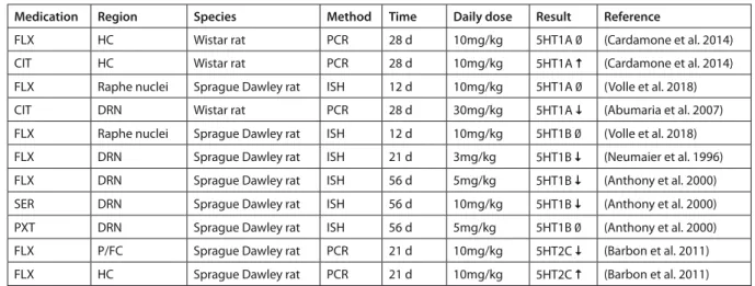
Effect of chronic SSRI administration on expression of serotonin receptor genes
While mainly the 5HT1A receptor has been empha- sized in the action of SSRIs, the serotonin receptor family includes 14 receptors (Segi-Nishida 2017), some of which showed expression changes following chronic SSRI administration. During SSRI treatment the negative feedback mechanism limiting the sero- tonin release is inactivated due to the desensitization of the raphe 5-HT1A (and partially the 5-HT1B) au- toreceptors that changes the discharge of serotoner- gic neurons (Hamon and Blier 2013). This process is assumed to take several weeks. Cardamone et al.
(2014) reported no change in 5-HT1A expression after a four week long FLX treatment in the HC of rats while 5HT1A receptor upregulation was observed in the same time period and region following CIT (10mg/kg) treatment. We have to note that in this study rats underwent kindling to investigate kindling epileptogenesis and the effects of ADs in this process, which might have influenced mRNA expression (Car- damone et al. 2014).
Surprisingly, no expression changes were detect- ed in the raphe nuclei after FLX (10mg/kg) for 12 days (Volle et al. 2018), while 5-HT1A mRNA was downregulated after 28 day-long treatment with CIT (30mg/kg) (Abumaria et al. 2007). Thus, studies show contradictory results on 5HT1A expression both in the HC and DRN. It is possible that different medica- tions, treatment protocols, strains, and methodologies used to identify gene expression are responsible for these contradictory findings. Interestingly, desensi- tized 5HT1A receptors in the DRN, without a change in the receptor levels after FLX (Le Poul et al. 1995) and sertraline (SER) (Rossi et al. 2008) treatment, were observed in healthy rodents (Le Poul et al. 1995;
Rossi et al. 2008).
The serotonin 1B receptor (5-HT1B) is located mainly presynaptically locally regulating 5-HT re- lease [for review see (Sari 2004)]. It is possibly im- plicated in the pathophysiology of MDD. For ex- ample, a combination of a 5HT1B antagonist and a SSRI showed a rapid antidepressant effect (Sari 2004). 5-HT1B mRNA expression of rats in DRN was downregulated following 21 days of FLX (3mg/kg) treatment while no changes were observed in the ra- phe nuclei after 12 days of FLX (10mg/kg) treatment (Neumaier et al. 1996; Volle et al. 2018). The exper- iment from (Neumaier et al. 1996) also reported no significant 5-HT1B expression changes in the frontal cortex, striatum or HC. Another research team inves- tigating this receptor in rats found decreased levels of 5-HT1B mRNA in the DRN with FLX (5mg/kg) and SER (10mg/kg) after eight week-long administrations,
Table 1 Expression changes in serotonin transporter and synthesis
Medication Region Species Method Time Daily dose Result Reference
FLX HC Wistar rat PCR 28 d 10mg/kg 5-HTT 0 (Cardamone et al. 2014)
CIT HC Wistar rat PCR 28 d 10mg/kg 5-HTT 0 (Cardamone et al. 2014)
FLX DR Sprague Dawley rat ISH 12 d 10mg/kg 5-HTT 0 (Volle et al. 2018)
CIT DRN Wistar rat PCR 28 d 30mg/kg 5-HTT 0 (Abumaria et al. 2007)
FLX DRN Sprague Dawley rat ISH 21 d 3mg/kg 5-HTT 0 (Neumaier et al. 1996)
FLX Midbrain Wistar rat PCR 14, 28 and 56 d 7,5mg/kg 5-HTT (Shishkina et al. 2007) FLX Brainstem Wistar rat PCR 14, 28 and 56 d 7,5mg/kg 5-HTT (Shishkina et al. 2007)
CIT DRN Wistar rat PCR 28 d 30mg/kg TPH2 (Abumaria et al. 2007)
FLX Midbrain Wistar rat PCR 4 and 8 weeks 7,5mg/kg TPH2 (Shishkina et al. 2007)
CIT DRN Wistar rat PCR 28 d 30mg/kg TPH2 (Abumaria et al. 2007)
FLX Brainstem Wistar rat PCR 14 d 25mg/kg TPH2 (Dygalo et al. 2006)
HC: Hippocampus; DR: Dorsal raphe; DRN: Dorsal raphe nucleus; 0: no changes observed; : upregulation observed; : downregulation observed
while paroxetine (PXT) (5mg/kg) treatment for 56 days didn’t contribute to any significant decrease in 5-HT1B expression in this region. All three SSRIs failed to alter 5-HT1B expression in the HC (Anthony et al. 2000).
The serotonin 2C receptor (5-HT2C) has been im- plicated as a potential target of therapeutic efforts for various psychiatric disorders (Higgins and Fletcher 2003). Elevated protein levels of 5-HT2C in suicide victims was demonstrated in the prefrontal and fron- tal cortices when compared to controls (Pandey et al. 2006). MRNA levels were postmortem increased in patients with schizophrenia and bipolar disorder (Castensson et al. 2003; Iwamoto and Kato 2003). In a rat experiment using FLX (10 mg/kg) administration, 5-HT2C showed a downregulation in the prefrontal cortex (P/FC) and elevated mRNA levels were detect- ed in the HC (Barbon et al. 2011).
A (partial) agonism at the serotonin 4 receptors (5HT4) showed rapid effects on depression-related behaviors and on hippocampal neurogenesis sug- gesting a possible role in MDD (Mendez-David et al.
2014; Vidal et al. 2014). Since transcriptomic changes are lacking in the literature, we discuss here, as an exception a study with transgenic mice. This exper- iment revealed that chronic treatment in transgenic mice with FLX elevated 5HT4 receptor expression levels in cortical neurons (Schmidt et al. 2012). P11 is a protein product of the S100a10 gene and has been involved in the mediation of antidepressant responses and depression-like states (Svenningsson 2006). Cor- tical neurons express p11 and 5HT4 receptors and
may have behavioral effects in concert with SSRIs [we recommend reading (Schmidt et al. 2012) for further information about the connection between p11 and 5-HT4].
Besides the ones described above, there are other serotonin receptors with possible roles in depression and antidepressant pharmacotherapy that lack tran- scriptomic data following SSRI treatments, which is not to say that they couldn’t be of etiological or therapeutic importance. Some papers, for example, mention the 5HT6 and 5HT7 receptors as useful tar- gets in the treatment of affective disorders (Carr et al.
2011; Nikiforuk 2015). For an overview of the studies discussed above see Table 2.
Effect of chronic SSRI treatment on expression of neuroplasticity factors
Brain-derived neurotrophic factor (BDNF) is widely implicated both in the etiopathology of depression and in the mechanism of its treatment. BDNF stimu- lates neuroplasticity during development and also in adulthood (Huang and Reichardt 2001; McAllister et al. 1999). Deficits in neuroplasticity factors can lead to depression (Fossati et al. 2004). Decreased BDNF levels may be a factor for depression, and in a me- ta-analysis decreased serum BDNF was suggested to be a relevant biomarker in MDD patients (Polyakova et al. 2015). Several studies showed an upregulation while others showed no effect on BDNF levels with different SSRIs [for a review see (Duman and Mon- teggia 2006)].
Table 2 Expression changes in the serotonin transporter and its receptors
Medication Region Species Method Time Daily dose Result Reference
FLX HC Wistar rat PCR 28 d 10mg/kg 5HT1A 0 (Cardamone et al. 2014)
CIT HC Wistar rat PCR 28 d 10mg/kg 5HT1A (Cardamone et al. 2014)
FLX Raphe nuclei Sprague Dawley rat ISH 12 d 10mg/kg 5HT1A 0 (Volle et al. 2018)
CIT DRN Wistar rat PCR 28 d 30mg/kg 5HT1A (Abumaria et al. 2007)
FLX Raphe nuclei Sprague Dawley rat ISH 12 d 10mg/kg 5HT1B 0 (Volle et al. 2018)
FLX DRN Sprague Dawley rat ISH 21 d 3mg/kg 5HT1B (Neumaier et al. 1996)
FLX DRN Sprague Dawley rat ISH 56 d 5mg/kg 5HT1B (Anthony et al. 2000)
SER DRN Sprague Dawley rat ISH 56 d 10mg/kg 5HT1B (Anthony et al. 2000)
PXT DRN Sprague Dawley rat ISH 56 d 5mg/kg 5HT1B 0 (Anthony et al. 2000)
FLX P/FC Sprague Dawley rat PCR 21 d 10mg/kg 5HT2C (Barbon et al. 2011)
FLX HC Sprague Dawley rat PCR 21 d 10mg/kg 5HT2C (Barbon et al. 2011)
DR: Dorsal raphe; HC: Hippocampus; DRN: Dorsal raphe nucleus; P/FC: Prefrontal cortex; ISH: In situ hybridization; 0: no changes observed;
: upregulation observed; : downregulation observed
Considering findings from studies focusing on the effect of chronic SSRI treatment on BDNF ex- pression, FLX failed to alter BDNF levels in a 3-week long chronic treatment in rats in doses of 10 and 11 mg/kg (Hanson et al. 2011; Larsen et al. 2008), but another study showed elevations with FLX (10mg/kg) treatment following a similar treatment protocol in the HC and P/FC and after a one-week washout period the expression level was still significantly higher than basal levels (Musazzi et al. 2009). At the same time, the lower dose of 5 mg/kg in rats administered for 2 weeks in- duced elevated BDNF expression in the frontal cortex (FC), but not in the HC (Rogóż et al. 2017). The same study examined the effects in the same time period of escitalopram (ESCIT) (10mg/kg) and demonstrated an upregulation in the HC but no changes in the FC (Rogóż et al, 2017). Another study showed an upreg- ulation following 3-week FLX (10mg/kg) treatment (Musazzi et al. 2009). SER, after treatment for 3 weeks with a 10 mg/kg dose in rats, increased mRNA levels of both BDNF and its receptor trkB, in the HC and FC (Nibuya, Morinobu, and Duman 1995). See Table 3 for a summary of the reviewed studies.
Effects of chronic SSRI administration on the expression of genes related to GABA neurotransmission
Dysfunctions of the GABAergic system are proposed to be associated with mood disorders. The GAB- Aergic hypothesis by Emrich (Emrich et al. 1980)
proposes a lack of GABA in the brain as a potential contributing factor in the background of mood dis- orders suggesting that elevated levels in the brain might be of therapeutic relevance in affective symp- tomatology. We only found one study reporting on mRNA expression of glutamic acid decarboxylase (GAD), the enzyme involved in GABA synthesis, which fulfilled our search criteria. SER in rats with a dose of 10 mg/kg downregulated the expression of GAD in the P/FC, nucleus accumbens (NAcc), olfac- tory tubercle (OT) and the thalamic reticular nucleus (TRN) (Giardino et al. 1996), suggesting decreased GABA synthesis in these regions following treat- ment. This result, however, is contradictory because in depressed humans GABA deficits normalized af- ter SSRI intake (Sanacora et al. 2002) and it also opposes the original hypothesis. We didn’t find any further transcriptomic studies that could confirm or debate these findings, suggesting that studies would be needed to deepen our understanding concerning the involvement of GABA in mood disorders and their treatment. See Table 4 for an overview of the discussed result.
Effect of chronic SSRI administration on the expression of genes related to the glutamate system
Glutamate is rising as a prime target of interest in the treatment of several psychiatric disorders including major depression, especially considering the approval
Table 3 Expression changes of neuroplasticity factors
Medication Region Species Method Time Daily dose Result Reference FLX HC Sprague Dawley rat ISH 21 d 10mg/kg BDNF 0 (Larsen et al. 2008)
FLX HC Wistar rats PCR 14 d 5mg/kg BDNF 0 (Rogóż et al. 2017)
FLX FC Wistar rats PCR 14 d 5mg/kg BDNF (Rogóż et al. 2017)
ESCIT HC Wistar rats PCR 14 d 10mg/kg BDNF (Rogóż et al. 2017)
ESCIT FC Wistar rats PCR 14 d 10mg/kg BDNF 0 (Rogóż et al. 2017)
FLX HC Sprague Dawley rat PCR 21 d 10 mg/kg BDNF (Musazzi et al. 2009) FLX P/FC Sprague Dawley rat PCR 21 d 10 mg/kg BDNF (Musazzi et al. 2009) SER FC Sprague Dawley rat Northern blot 21 d 10 mg/kg BDNF (Nibuya et al. 1995) SER HC Sprague Dawley rat Northern blot 21 d 10 mg/kg BDNF (Nibuya et al. 1995)
FLX HC Sprague Dawley rat ISH 21 d 11 mg/kg BDNF 0 (Hanson, Nemeroff, and Owens 2011) SER FC Sprague Dawley rat Northern blot 21 d 10 mg/kg trkB (Nibuya et al. 1995)
SER HC Sprague Dawley rat Northern blot 21 d 10 mg/kg trkB (Nibuya et al. 1995)
HC: Hippocampus; P/FC: Prefrontal cortex; FC: Frontal cortex; ISH: In situ hybridization; 0: no changes observed; : upregulation observed;
: downregulation observed
of esketamine as a treatment in treatment resistant depression (www.jnj.com, accessed on the 28th of February, 2019). In contrast to the selective serotonin and noradrenalin reuptake blocker venlafaxine, which upregulated NR2A and NR2B subunits of NMDA glutamate receptors after chronic treatment in the FC (Tamási et al. 2014), CIT (20mg/kg) for 16 days downregulated the mRNA expression of almost all NMDA-R subunits in many regions with the excep- tion of the HC in mice, where its levels remained unchanged (Boyer et al., 1998). In detail, in the amyg- dala NR1 and NR2A-B subunits were downregulated, whereas NR2C showed no change. NR1 and NR2A subunits showed downregulation in thalamus. Inves- tigations in the striatum reported downregulation of NR1 and NR2A subunits but no changes in NR2B.
Furthermore, in the cerebellum NR1 was downregu- lated and NR2A-C showed no significant alterations.
In summary, surprisingly, no upregulations of NMDA receptor subunits could have been observed in any of the relevant regions following chronic SSRI treat- ment (Boyer, Skolnick, and Fossom 1998). All this suggests that upregulation of glutamatergic genes is not the main mechanism through which SSRIs exert their antidepressant properties. See Table 4 for detailed results.
CONCLUSIONS
Transcriptomic changes after chronic treatment could be orientating in uncovering the hidden mechanism of action of SSRI antidepressants. The current stud- ies, however, rather demonstrate just how broad the spectrum of molecular mechanism of chronic SSRI treatments may be, in contrast to their similar initial effects. In the serotonergic system, 5-HTT expression showed mostly no change, while TPH2 mRNA was downregulated in some brain regions including the DRN. Serotonin receptor gene expression was vari- able. Chronic CIT treatment downregulated 5-HT1A receptors, which corresponds to the desensitization of the autoregulatory feedback mechanism, but FLX and SER downregulated 5-HT1B, with the former lacking effects at 5-HT1A in the DRN. FLX and CIT seemed to complement each other’s effect in the HC and FC, with FLX more reliably inducing BDNF ex- pression elevations in the FC and CIT in the HC at least in the discussed transcriptomic studies. Only SER was able to induce upregulations of both BDNF and its receptor in the two regions. With GAD and NMDA receptor expressions unchanged or rather downregulated, GABAergic and glutamatergic mech- anisms also seem to be contradictory to the many
Table 4 Expression changes related to the neurotransmitter GABA and glutamate
Medication Region Species Method Time Daily dose Result Reference
SER P/FC Sprague Dawley rat ISH 28 d 10 mg/kg GAD (Giardino et al. 1996)
SER NAcc Sprague Dawley rat ISH 28 d 10 mg/kg GAD (Giardino et al. 1996)
SER OT Sprague Dawley rat ISH 28 d 10 mg/kg GAD (Giardino et al. 1996)
SER TRN Sprague Dawley rat ISH 28 d 10 mg/kg GAD (Giardino et al. 1996)
CIT HC NIH-Swiss mice ISH 16 d 20 mg/kg NR1 0 (Boyer et al. 1998)
CIT HC NIH-Swiss mice ISH 16 d 20 mg/kg NR2A-C 0 (Boyer et al. 1998)
CIT Amygdala NIH-Swiss mice ISH 16 d 20 mg/kg NR1 (Boyer et al. 1998)
CIT Amygdala NIH-Swiss mice ISH 16 d 20 mg/kg NR2A-B (Boyer et al. 1998)
CIT Amygdala NIH-Swiss mice ISH 16 d 20 mg/kg NR2C 0 (Boyer et al. 1998)
CIT Thalamus NIH-Swiss mice ISH 16 d 20 mg/kg NR1 (Boyer et al. 1998)
CIT Thalamus NIH-Swiss mice ISH 16 d 20 mg/kg NR2A (Boyer et al. 1998)
CIT Striatum NIH-Swiss mice ISH 16 d 20 mg/kg NR1 (Boyer et al. 1998)
CIT Striatum NIH-Swiss mice ISH 16 d 20 mg/kg NR2A (Boyer et al. 1998)
CIT Striatum NIH-Swiss mice ISH 16 d 20 mg/kg NR2B 0 (Boyer et al. 1998)
CIT Cerebellum NIH-Swiss mice ISH 16 d 20 mg/kg NR1 (Boyer et al. 1998)
CIT Cerebellum NIH-Swiss mice ISH 16 d 20 mg/kg NR2A-C 0 (Boyer et al. 1998)
P/FC: Prefrontal cortex; NAcc: Nucleus accumbens; HC: Hippocampus; OT: Olfactory tubercle; TRN: Thalamic reticular nucleus; ISH: In situ hybridization; 0: no changes observed; : upregulation observed; : downregulation observed
theories involving these neurotransmitter systems in depression. While we have to mention among the many limitations that the reviewed studies employed different rat strains, and that mRNA levels not neces- sarily represent protein levels, these results point to the enormous heterogeneity of action mechanisms behind the seemingly “homogeneous” class of SSRI ADs. In addition, the results of this review also under- line the need for the development of novel ADs with more reliable clarity in their actions on a molecular level and raises the possibility of a novel grouping of antidepressants based rather on their chronic molec- ular changes, than on their initial effects.
Acknowledgements: This work was supported by NEW- MOOD (LSHM-CT-2004-503474); TAMOP-4.2.1.B 09/1/
KMR-2010-0001; KTIA_13_NAP-A-II/14, KTIA_NAP_13- 1-2013-0001; KTIA_NAP_13-2-2015-0001 (MTA-SE-NAP B Genetic Brain Imaging Migraine Research Group: Hungarian Academy of Sciences, Semmelweis University and the Hungar- ian Brain Research Program); 2017-1.2.1-NKP-2017-00002;
MTA-SE Neuropsychopharmacology and Neurochemistry Research Group; OTKA 119866; the ÚNKP-17-4-I-SE-8 by the New National Excellence Program of the Ministry of Human Capacities. Xenia Gonda is recipient of the Janos Bolyai Re- search Fellowship of the Hungarian Academy of Sciences and is supported by ÚNKP-18-4-SE-33 New National Excellence Program of the Ministry of Human Capacities.
Corresponding author: Petschner Péter Semmelweis Egyetem Gyógyszerhatástani Intézet 1089 Budapest, Nagyvárad tér 4.
E-mail: petschner.peter@pharma.semmelweis-univ.hu
REFERENCES
1. Abumaria, Nashat, Rafal Rygula, Christoph Hiemke, Eberhard Fuchs, Ursula Havemann-Reinecke, Eckart Rüther, and Gabriele Flügge. 2007. Effect of Chronic Citalopram on Serotonin-Relat- ed and Stress-Regulated Genes in the Dorsal Raphe Nucleus of the Rat. European Neuropsychopharmacology 17(6–7):417–29.
2. Anthony, J. P., T. J. Sexton, and J. F. Neumaier. 2000. Antide- pressant-Induced Regulation of 5-HT1b MRNA in Rat Dorsal Raphe Nucleus Reverses Rapidly after Drug Discontinuation.
Journal of Neuroscience Research 61(1):82–87.
3. Baldwin, David S., Stuart A. Montgomery, Rico Nil, and Mal- colm Lader. 2007. Discontinuation Symptoms in Depression and Anxiety Disorders. The International Journal of Neuropsy- chopharmacology 10(01):73.
4. Barbon, Alessandro, Cesare Orlandi, Luca La Via, Luca Carac- ciolo, Daniela Tardito, Laura Musazzi, Alessandra Mallei, Mas- simo Gennarelli, Giorgio Racagni, Maurizio Popoli, and Sergio Barlati. 2011. Antidepressant Treatments Change 5-HT2C Re- ceptor MRNA Expression in Rat Prefrontal/Frontal Cortex and Hippocampus. Neuropsychobiology 63(3):160–68.
5. Bobo, William V, Brandon R. Grossardt, Maria I. Lapid, Jona- than G. Leung, Cynthia Stoppel, Paul Y. Takahashi, Robert W.
Hoel, Zheng Chang, Christian Lachner, Mohit Chauhan, Lee Flowers, Scott M. Brue, Mark A. Frye, Jennifer St Sauver, Wal- ter A. Rocca, and Bruce Sutor. 2019. Frequency and Predictors of the Potential Overprescribing of Antidepressants in Elderly Residents of a Geographically Defined U.S. Population. Phar- macology Research & Perspectives 7(1):e00461.
6. Boyer, Pierre-Alain, Phil Skolnick, and Linda H. Fossom. 1998.
Chronic Administration of Imipramine and Citalopram Alters the Expression of NMDA Receptor Subunit MRNAs in Mouse Brain. Journal of Molecular Neuroscience 10(3):219–33.
7. Brambilla, P., J. Perez, F. Barale, G. Schettini, and J. C. Soares.
2003. GABAergic Dysfunction in Mood Disorders. Molecular Psychiatry 8(8):721–37.
8. Cardamone, Lisa, Michael R. Salzberg, Amelia S. Koe, Ezgi Ozturk, Terence J. O’Brien, and Nigel C. Jones. 2014. Chronic Antidepressant Treatment Accelerates Kindling Epileptogen- esis in Rats. Neurobiology of Disease 63:194–200.
9. Carr, Gregory V., Lee E. Schechter, and Irwin Lucki. 2011. An- tidepressant and Anxiolytic Effects of Selective 5-HT6 Recep- tor Agonists in Rats. Psychopharmacology 213(2–3):499–507.
10. Castensson, Anja, Lina Emilsson, Rolf Sundberg, and Elena Jazin. 2003. Decrease of Serotonin Receptor 2C in Schizophre- nia Brains Identified by High-Resolution MRNA Expression Analysis. Biological Psychiatry 54(11):1212–21.
11. Cooper, Geoffrey M. 2000. The Cell: A Molecular Approach.
Structure of the Plasma Membrane. Sunderland (MA): Sinauer Associates. Detection of Nucleic Acids and Proteins. Available from: https://www.ncbi.nlm.nih.gov/books/NBK9916/.
12. Coppen, A. 1967. The Biochemistry of Affective Disorders.
The British Journal of Psychiatry : The Journal of Mental Sci- ence 113(504):1237–64.
13. Craske, Michelle G. and Murray B. Stein. 2016. Anxiety. The Lancet 388(10063):3048–59.
14. D’Antonio, Mattia, Paolo D’Onorio De Meo, Matteo Pallocca, Ernesto Picardi, Anna Maria D’Erchia, Raffaele A. Calogero, Tiziana Castrignanò, and Graziano Pesole. 2015. RAP: RNA- Seq Analysis Pipeline, a New Cloud-Based NGS Web Applica- tion. BMC Genomics 16(Suppl 6):S3.
15. Duman, Ronald S. and Lisa M. Monteggia. 2006. A Neuro- trophic Model for Stress-Related Mood Disorders. Biological Psychiatry 59(12):1116–27.
16. Dygalo, N. N., G. T. Shishkina, T. S. Kalinina, A. M. Yudina, and E. S. Ovchinnikova. 2006. Effect of Repeated Treatment with Fluoxetine on Tryptophan Hydroxylase-2 Gene Expres- sion in the Rat Brainstem. Pharmacology Biochemistry and Behavior 85(1):220–27.
17. Emrich, H. M., D. Zerssen, W. Kissling, H. J. Müller, and A.
Windorfer. 1980. Effect of Sodium Valproate on Mania. Archiv Für Psychiatrie Und Nervenkrankheiten 229(1):1–16.
18. Fehr, J. E., G. W. Trotter, J. T. Oxford, and D. A. Hart. 2000.
Comparison of Northern Blot Hybridization and a Reverse Transcriptase-Polymerase Chain Reaction Technique for Measurement of MRNA Expression of Metalloproteinases and Matrix Components in Articular Cartilage and Synovial Mem- brane from Horses with Osteoarthritis. American Journal of Veterinary Research 61(8):900–905.
19. Fossati, Philippe, Andrei Radtchenko, and Patrice Boyer. 2004.
Neuroplasticity: From MRI to Depressive Symptoms. Euro- pean Neuropsychopharmacology 14:S503–10.
20. Giardino, Luciana, Massimo Zanni, Carla Bettelli, Maria A.
Savina, and Laura Calzà. 1996. Regulation of Glutamic Acid Decarboxylase MRNA Expression in Rat Brain after Sertraline Treatment. European Journal of Pharmacology 312(2):183–87.
21. Gonda, Xenia, Peter Petschner, Nora Eszlari, Daniel Baksa, Andrea Edes, Peter Antal, Gabriella Juhasz, and Gyorgy Bagdy.
2019. Genetic Variants in Major Depressive Disorder: From Pathophysiology to Therapy. Pharmacology & Therapeutics 194:
22–43.
22. Hamon, Michel and Pierre Blier. 2013. Monoamine Neurocir- cuitry in Depression and Strategies for New Treatments. Pro- gress in Neuro-Psychopharmacology and Biological Psychiatry 45:54–63.
23. Hanson, Nicola D., Charles B. Nemeroff, and Michael J. Owens.
2011. Lithium, but Not Fluoxetine or the Corticotropin-Releas- ing Factor Receptor 1 Receptor Antagonist R121919, Increases Cell Proliferation in the Adult Dentate Gyrus. The Journal of Pharmacology and Experimental Therapeutics 337(1):180–86.
24. Heid, C. A., J. Stevens, K. J. Livak, and P. M. Williams. 1996.
Real Time Quantitative PCR. Genome Research 6(10):986–94.
25. Herring, Bruce E. and Roger A. Nicoll. 2016. Long-Term Po- tentiation: From CaMKII to AMPA Receptor Trafficking. An- nual Review of Physiology 78(1):351–65.
26. Higgins, Guy A. and Paul J. Fletcher. 2003. Serotonin and Drug Reward: Focus on 5-HT2C Receptors. European Journal of Pharmacology 480(1–3):151–62.
27. Hillhouse, Todd M. and Joseph H. Porter. 2015. A Brief His- tory of the Development of Antidepressant Drugs: From Mon- oamines to Glutamate. Experimental and Clinical Psychophar- macology 23(1):1–21.
28. Huang, Eric J. and Louis F. Reichardt. 2001. Neurotrophins:
Roles in Neuronal Development and Function. Annual Review of Neuroscience 24r(1):677–736.
29. Iwamoto, Kazuya and Tadafumi Kato. 2003. RNA Editing of Serotonin 2C Receptor in Human Postmortem Brains of Major Mental Disorders. Neuroscience Letters 346(3):169–72.
30. Kevil, Christopher G., Loren Walsh, F. Stephen Laroux, Theo- dore Kalogeris, Matthew B. Grisham, and J. S. Alexander. 1997.
An Improved, Rapid Northern Protocol. Biochemical and Bio- physical Research Communications 238(2):277–79.
31. Kew, James N. C. and John A. Kemp. 2005. Ionotropic and Me- tabotropic Glutamate Receptor Structure and Pharmacology.
Psychopharmacology 179(1):4–29.
32. Larsen, Marianne H., Anders Hay-Schmidt, Lars C. B. Rønn, and Jens D. Mikkelsen. 2008. Temporal Expression of Brain- Derived Neurotrophic Factor (BDNF) MRNA in the Rat Hip- pocampus after Treatment with Selective and Mixed Monoam- inergic Antidepressants. European Journal of Pharmacology 578(2–3):114–22.
33. Marsden, W. N. 2011. Stressor-Induced NMDAR Dysfunc- tion as a Unifying Hypothesis for the Aetiology, Pathogenesis and Comorbidity of Clinical Depression. Medical Hypotheses 77(4):508–28.
34. McAllister, A. Kimberley, Lawrence C. Katz, and Donald C. Lo.
1999. “Neurotrophins and Synaptic Plasticity.” Annual Review of Neuroscience 22(1):295–318.
35. Mendez-David, Indira, Denis J. David, Flavie Darcet, Melody V Wu, Saadia Kerdine-Römer, Alain M. Gardier, and René Hen.
2014. “Rapid Anxiolytic Effects of a 5-HT₄ Receptor Agonist Are Mediated by a Neurogenesis-Independent Mechanism.”
Neuropsychopharmacology : Official Publication of the Amer- ican College of Neuropsychopharmacology 39(6):1366–78.
36. Musazzi, Laura, Annamaria Cattaneo, Daniela Tardito, Ales- sandro Barbon, Massimo Gennarelli, Sergio Barlati, Giorgio Racagni, and Maurizio Popoli. 2009. “Early Raise of BDNF in Hippocampus Suggests Induction of Posttranscriptional Mechanisms by Antidepressants.” BMC Neuroscience 10:48.
37. Neumaier, J., Daniel C. Root, and Mark W. Hamblin. 1996.
“Chronic Fluoxetine Reduces Serotonin Transporter MRNA
and 5-HT1B MRNA in a Sequential Manner in the Rat Dorsal Raphe Nucleus.” Neuropsychopharmacology 15(5):515–22.
38. Nibuya, Masashi, Shigeru Morinobu, and Ronald S. Duman.
1995. Regulation of BDNF and TrkB MRNA in Rat Brain by Chronic Electroconvulsive Seizure and Antidepressant Drug Treatments. Vol. 75.
39. Nikiforuk, Agnieszka. 2015. “Targeting the Serotonin 5-HT7 Receptor in the Search for Treatments for CNS Disorders: Ra- tionale and Progress to Date.” CNS Drugs 29(4):265–75.
40. Pandey, Ghanshyam N., Yogesh Dwivedi, Xinguo Ren, Hoori- yah S. Rizavi, Gabor Faludi, Andrea Sarosi, and Miklos Palko- vits. 2006. “Regional Distribution and Relative Abundance of Serotonin2c Receptors in Human Brain: Effect of Suicide.”
Neurochemical Research 31(2):167–76.
41. Petschner, Peter, Viola Tamasi, Csaba Adori, Eszter Kirilly, Ro- meo D. Ando, Laszlo Tothfalusi, and Gyorgy Bagdy. 2018. “Gene Expression Analysis Indicates Reduced Memory and Cognitive Functions in the Hippocampus and Increase in Synaptic Reor- ganization in the Frontal Cortex 3 Weeks after MDMA Admin- istration in Dark Agouti Rats.” BMC Genomics 19(1):580.
42. Polyakova, Maryna, Katharina Stuke, Katharina Schuemberg, Karsten Mueller, Peter Schoenknecht, and Matthias L. Schro- eter. 2015. “BDNF as a Biomarker for Successful Treatment of Mood Disorders: A Systematic & Quantitative Meta- Analysis.” Journal of Affective Disorders 174:432–40.
43. Le Poul, Emmanuel, Nora Laaris, Edith Doucet, Anne Marie Laporte, Michel Hamon, and Laurence Lanfumey. 1995. “Early Desensitization of Somato-Dendritic 5-HT1A Autorecep- tors in Rats Treated with Fluoxetine or Paroxetine.” Naunyn- Schmie deberg’s Archives of Pharmacology 352(2):141–48.
44. Preskorn, Sheldon H., John Preston Feighner, Christina Y.
Stanga, and Ruth. Ross. 2004. Antidepressants: Past, Present and Future. Springer Berlin Heidelberg.
45. Rogóż, Zofia, Katarzyna Kamińska, Patrycja Pańczyszyn- Trzewik, and Magdalena Sowa-Kućma. 2017. “Repeated Co- Treatment with Antidepressants and Risperidone Increases BDNF MRNA and Protein Levels in Rats.” Pharmacological Reports 69(5):885–93.
46. Rossi, Dania V., Teresa F. Burke, Melissa McCasland, and Julie G. Hensler. 2008. “Serotonin-1A Receptor Function in the Dorsal Raphe Nucleus Following Chronic Administration of the Selective Serotonin Reuptake Inhibitor Sertraline.” Journal of Neurochemistry 105(4):1091–99.
47. Sanacora, Gerard, Graeme F. Mason, Douglas L. Rothman, and John H. Krystal. 2002. “Increased Occipital Cortex GABA Concentrations in Depressed Patients After Therapy With Se- lective Serotonin Reuptake Inhibitors.” American Journal of Psychiatry 159(4):663–65.
48. Sangkuhl, Katrin, Teri E. Klein, and Russ B. Altman. 2009. “Se- lective Serotonin Reuptake Inhibitors Pathway.” Pharmacoge- netics and Genomics 19(11):907–9.
49. Sari, Youssef. 2004. “Serotonin 1B Receptors: From Protein to Physiological Function and Behavior.” Neuroscience and Biobehavioral Reviews 28(6):565–82.
50. Schildkraut, J. J. 1965. “The Catecholamine Hypothesis of Affective Disorders: A Review of Supporting Evidence.” The American Journal of Psychiatry 122(5):509–22.
51. Schmidt, Eric F., Jennifer L. Warner-Schmidt, Benjamin G.
Oto palik, Sarah B. Pickett, Paul Greengard, and Nathaniel Heintz. 2012. “Identification of the Cortical Neurons That Me- diate Antidepressant Responses.” Cell 149(5):1152–63.
52. Al Seesi, Sahar, Fei Duan, Ion I. Mandoiu, Pramod K. Srivas- tava, and Angela Kueck. 2016. “Genomics-Guided Immuno- therapy of Human Epithelial Ovarian Cancer.” Pp. 237–50 in Translational Cardiometabolic Genomic Medicine. Elsevier.
53. Segi-Nishida, Eri. 2017. “The Effect of Serotonin-Targeting Antidepressants on Neurogenesis and Neuronal Maturation of the Hippocampus Mediated via 5-HT1A and 5-HT4 Recep- tors.” Frontiers in Cellular Neuroscience 11:142.
54. Shishkina, G. T., T. S. Kalinina, and N. N. Dygalo. 2007. “Up- Regulation of Tryptophan Hydroxylase-2 MRNA in the Rat Brain by Chronic Fluoxetine Treatment Correlates with Its An- tidepressant Effect.” Neuroscience 150(2):404–12.
55. Svenningsson, P. 2006. “Alterations in 5-HT1B Receptor Func- tion by P11 in Depression-Like States.” Science 311(5757):77–80.
56. Tamási, Viola, Peter Petschner, Csaba Adori, Eszter Kirilly, Ro- meo D. Ando, Laszlo Tothfalusi, Gabriella Juhasz, and Gyorgy Bagdy. 2014. “Transcriptional Evidence for the Role of Chronic Venlafaxine Treatment in Neurotrophic Signaling and Neuro- plasticity Including Also Glutatmatergic- and Insulin-Medi- ated Neuronal Processes” edited by L. Lu. PLoS ONE 9(11):
e113662.
57. Teo, Zhi Ling, Peter Savas, and Sherene Loi. 2016. “Gene Expres- sion Analysis: Current Methods.” Pp. 107–36 in Molecular Pa- thology in Cancer Research. New York, NY: Springer New York.
58. Trevino, Victor, Francesco Falciani, and Hugo A. Barrera-Sal- daña. 2007. “DNA Microarrays: A Powerful Genomic Tool for
Biomedical and Clinical Research.” Molecular Medicine (Cam- bridge, Mass.) 13(9–10):527–41.
59. VanGuilder, Heather D., Kent E. Vrana, and Willard M. Free- man. 2008. “Twenty-Five Years of Quantitative PCR for Gene Expression Analysis.” BioTechniques 44(5):619–26.
60. Vidal, Rebeca, Elena Castro, Fuencisla Pilar-Cuéllar, Jesús Pas- cual-Brazo, Alvaro Díaz, María Luisa Rojo, Raquel Linge, Alicia Martín, Elsa M. Valdizán, and Angel Pazos. 2014. “Serotonin 5- HT4 Receptors: A New Strategy for Developing Fast Acting An- tidepressants?” Current Pharmaceutical Design 20(23):3751–62.
61. Volle, Julien, Tatiana Bregman, Brian Scott, Mustansir Diwan, Roger Raymond, Paul J. Fletcher, José N. Nobrega, and Clem- ent Hamani. 2018. “Deep Brain Stimulation and Fluoxetine Ex- ert Different Long-Term Changes in the Serotonergic System.”
Neuropharmacology 135:63–72.
62. Zhang, Yuqi, Zaohuo Chang, Jionghua Chen, Yang Ling, Xi- aowei Liu, Zhang Feng, Caixia Chen, Minghua Xia, Xingfu Zhao, Wang Ying, Xu Qing, Guilin Li, and Changsong Zhang.
2015. “Methylation of the Tryptophan Hydroxylase-2 Gene Is Associated with MRNA Expression in Patients with Major De- pression with Suicide Attempts.” Molecular Medicine Reports 12(2):3184–90.
Krónikus szelektív szerotoninvisszavétel-gátló kezelés hatására bekövetkező transzkripciós változások: az állatkísérletek
áttekintése
A közlemény a szelektív szerotoninvisszavétel-gátló (SSRI) antidepresszívumok hatására bekövetkező transzkriptomikai változásokat tekinti át. Célunk az volt, hogy összefoglaljuk a különböző agyterületeken krónikus kezelések hatására bekövetkező, a génexpressziós válto- zások vizsgálatának leggyakrabban használt módszereivel – így Northern blottal, in situ hibri- dizációval, kvantitatív reverz transzkriptáz-polimeráz láncreakcióval (qRT-PCR), microarray-jel és a RNS szekvenálással – végzett kísérletek eredményeit. Annak ellenére, hogy a szerotonerg rendszer génjeiben krónikus SSRI kezelés hatására mRNS szinten mérhető változások voltak megfigyelhetők, a szerotonintranszporter szintje legtöbbször változatlan maradt. Ugyanak- kor a triptofán-hidroxiláz-2 downregulálódott a szerotonerg magokban és upregulálódott a középagyi területeken. A szerotoninreceptorokat érintő változások nem mutattak egységes képet, és a leírt változások valószínűleg törzstől, hatóanyagtól, illetve a különböző vizsgált agyterületektől függő hatásokat is tükröznek. A legtöbb krónikus kezelés növelte az agy-ere- detű neurotróf faktor expresszióját. A GABA- valamint a glutamátreceptorok génjei szintén heterogén változásokat mutatnak; meglepő módon az NMDA receptorok downregulációja volt megfigyelhető a depresszió és a stresszreakciók kialakításában részt vevő olyan agyterüle- teken, mint például a striatum és az amygdala területén. A fenti kutatások áttekintése számos eltérésre hívja fel a figyelmet, és hangsúlyozza a vizsgált agyterülettől és SSRI gyógyszertől is függő hatások heterogenitását. Mindeközben azonban az eredmények felvetik annak lehetőségét is, hogy az antidepresszívumokat – akut hatásmechanizmusuk helyett – talán célszerűbb volna krónikus molekuláris hatásaik alapján csoportosítani.
Kulcsszavak: SSRI, BDNF, patkány, transzkriptomika, 5-HTT