Studies on the Isolated Membrane and Cytoplasm of Amoeba proteus in Relation to Ameboid Movement
L . WOLPERT, C. M . THOMPSON, AND C. H . O ' N E I L L Zoology Department, King's College, University of London, London, England
Introduction
It is a standard approach in experimental biology to try and isolate functional and structural systems from the intact organism so as to make them more amenable to study. In cell biology the isolation of subcellular constituents has been particularly fruitful; this is evident from the ad- vances following from the isolation of mitochondria, mitotic apparatus, and muscle proteins. It is thus surprising that, despite notable excep- tions, so little attention should have been paid to the isolation of the subcellular components in the study of ameboid movement. One of the difficulties peculiar to the planning of an isolation of the systems in- volved in ameboid movement is that one does not have well-defined structural or chemical criteria on which to base the isolation. There is nothing in the ameba to which one can point with confidence and say:
"If we isolate that structure or that biochemical system we shall have the components responsible for movement." There is no compelling reason to think that most of the protein is involved in movement as in muscle; nor are there clearly recognizable structures as in cilia, or the mitotic apparatus. Worse still, we do not even know whether the site of the motive force resides within the surface membrane or cyto- plasm. This point is particularly important since movement involves change in form of both cytoplasm and surface membrane. For such reasons one must rely largely on isolating a system that will show some of the phenomena seen in the living cell; for example, isolated surface membranes must change their form or a cytoplasmic fraction must show streaming.
There is another approach which has received considerable support, and that is to base the isolation procedure on the assumption that cell movement involves a system very similar to that responsible for muscu- lar contraction. A persuasive analogy may be drawn, and this has been strengthened by the pioneer studies of Loewy (1952) and later workers
143
144 L. WOLPERT, C. M. THOMPSON, AND C. H. O'NEILL
who isolated an actomyosin-like protein from slime molds, and also of Hoffmann-Berling (1960) who prepared glycerol-extracted models of fibro- blasts which resembled in many ways those of glycerol-extracted muscle, and an actomyosin-like protein fraction from tissue cells. However, such studies and their extension have not really established that the ideas on muscular contraction can really be applied to problems of cell move- ment. Suggestive though it may be, the isolation of a system showing viscosity changes with adenosine triphosphate (ATP) in no way guaran- tees that such a system is responsible for cell movement.
We have, therefore, adopted a somewhat different approach. At the time our studies began, about 4 years ago, the most satisfactory theory of ameboid movement was the extension and modification of Mast's (1926) ideas by Goldacre (1952). This had two main postulates: (a) a contractile gel capable of sol-gel transformations, contraction being ac- companied by solation; and (b) the surface membrane initiates contrac- tion in the gel, is resorbed at the tail of the ameba, and is re-formed at the advancing end, the whole membrane being formed anew each time the cell passed through its own length. We, therefore, set out to isolate the contractile gel and the surface membrane. For the gel, the criterion would be its ability to contract or at least to undergo sol-gel transforma- tions. With respect to the membrane, we felt that if this could be isolated, one might be better able to study its dynamics, particularly the proposed rapid turnover and the mechanism of its resorption and re-formation.
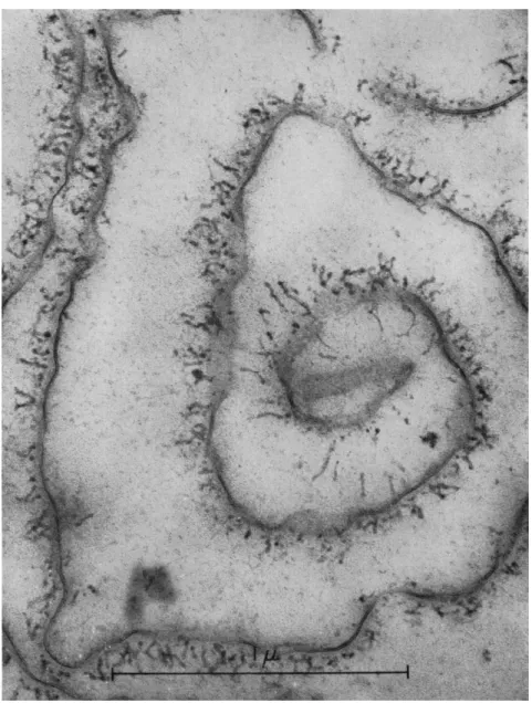
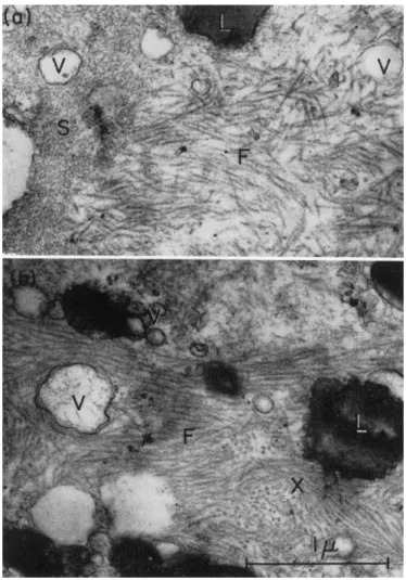
Dynamics of the Surface Membrane during Cell Movement The idea that the cell membrane plays an active part in ameboid movement goes back a long time and is, in many ways, attractive. Early theories suggesting that changes in surface tension would provide the basis for ameboid movement were discounted when it was realized that at the cell surface there was a structural membrane that showed elastic properties. Electron microscope studies of the membrane of Amoeba proteus suggest that it is made up of a typical plasma membrane with a thick outer coat (Pappas, 1959; Mercer, 1959). T h e plasma membrane appears as a double layer about 80 A thick which is consistent with the bimolecular lipid leaflet with protein on either side, postulated by Davson and Danielli (1942), and termed the "unit membrane" by Robert- son (1959). The surface coat appears as a fibrillar layer about 0.2 μ thick and is probably composed of mucoprotein (O'Neill, 1963). These struc- tures can be clearly seen in membranes of A. proteus isolated in bulk (Fig. 1) (O'Neill and Wolpert, 1961).
The membrane may play both physiological and mechanical roles in
Isolated Membrane and Cytoplasm of A. proteus 145
FIG. 1. Electron micrograph of isolated cell surface membrane of ameba. The fibrillar surface coat can be seen overlying the double-layered unit membrane ( 8 0 A thick) characteristic of the surfaces of cells. No other components of the cell can be detected. (Photograph by Dr. Ε. H. Mercer.)
ameboid movement. A most significant example of the former is the possibility that changes in membrane potential may control the initiation of pseudopods and the rate and direction of streaming (Bingley and
146 L. WOLPERT, C. M. THOMPSON, AND C. H. O'NEILL
Thompson, 1962). Here we are mainly concerned with the mechanical role of the membrane.
It has been suggested that the apparent mechanical properties of the membrane are not clue to the plasma membrane but to a relatively thick (1-2 μ) gel-like cortex beneath the plasma membrane (Marsland, 1956;
Mitchison, 1952, 1956), although this has been questioned (Wolpert, 1960b; Mercer and Wolpert, 1962). No evidence can be found for a cortex of this type in A. proteus either in intact cells or in isolated membranes.
Λ 2 3 4\ /2 3 4
iN /£
4T~?\
rAΓ ? £\
Β
(à % \ t)
C» ΤI
SJ (,ê
7 \ %) Ce 7 6 s )a b e d a b e d e a b e d e b e d e
C
/ I 2 Γ — T \ fi i 2 Γ \ / Τ Ι i T \ / T - ^ β—TN
7 β Vj 6 S V Ve S 4 3Y VS 4 3 2/
a b e d b e d e c d e f d e f g FIG. 2. Diagram to show possible ways the membrane can change its shape during
cell movement. Sites on the membrane are distinguished by numbers, and sites on the substratum by letters. Three possibilities arise from geometrical considerations. ( A ) Membrane is supplied to the surface at the front, and withdrawn from the surface at the rear; the surface elsewhere is stationary. (B) T h e membrane is alternately extended and contracted. Expansion occurs while the cell is in contact with the substratum at the rear, and contraction occurs while the cell is in contact with the substratum at the front. ( C ) T h e membrane flows to accommodate changes in shape. T h e membrane flows forward over the upper surface of the cell, while it remains stationary at the points of contact with the substratum.
There is a well-defined problem as to how the membrane changes its form as the cell moves forward. Three main possibilities exist, which may be derived from consideration of the geometry of the moving cell (Fig. 2).
(a) The membrane is stationary over most of the cell and change in shape involes formation and resorption of membrane (Fig. 2A). This is the requirement of the theories of Goldacre (1952, 1961) and apparently Bell (1961). The cell contents move through a stationary tube of mem- brane, new surface being formed at the front, while there is withdrawal
Isolated Membrane and Cytoplasm of A. proteus 147 of membrane from the surface at the rear. Goldacre suggests that the force for movement is provided by contraction of the ectoplasmic gel at the rear, whereas Bell suggests that the formation of new surface itself provides the force. It is a consequence of the theory of Goldacre at least, that the rate of turnover of the membrane between the surface and the interior must be high during movement and the cell may be expected to renew its membrane completely each time it passes through its own length, namely about every 5 min.
(b) The membrane is reversibly extensible (possibly elastic) and localized changes in shape may take place by localized increase or decrease in surface area without the formation or removal of surface material (Fig. 2B). This is the basis of the theory for the movement of amphibian embryonic cells put forward by Holtfreter (1949) and for fibroblast move- ment by Ambrose (1961). No such theory has been put forward for ame- boid movement.
(c) The membrane of the cell flows freely, and this will often be seen as forward motion over the upper surface when the cell is attached to the substratum over the whole of its length (Fig. 2C) (Mast, 1926; Griffin and Allen, 1960). In this case membrane turnover will be low.
These examples illustrate the main ways that the membrane may change in form during locomotion, and numerous variations are, of course, possible. For example, it is important to note that cell movement by pseudopod extension and shortening (cf. Gustafson and Wolpert,
1963) may involve any of the three possibilities.
From these considerations three experimentally testable questions concerning the behavior of the surface during locomotion can be posed.
(1) Is there localized rapid membrane turnover? (2) Does the surface extend reversibly? (3) Does the surface flow forward? There is remarkably little unequivocal evidence that allows us to answer these questions.
With respect to ameboid movement, only possibilities (1) and (3) have been put forward. On the one hand, Goldacre (1952, 1961), from observa- tions on the behavior of large obstacles such as oil drops attached to the membrane and from the accumulation of neutral red at the tail, has claimed that turnover is rapid and that membrane is resorbed at the tail and formed anew at the front. But these observations are open to other interpretations. For example, the fact that an oil drop attached to the surface does not move forward may only show that membrane flow in the region of attachment has been stopped (Wolpert and O'Neill, 1962).
On the other hand, from studies of particles attached to the surface, Mast (1926) and Griffin and Allen (1960) have suggested that the mem- brane is fluid and flows forward. However, Goldacre (1961) has suggested that this is open to other interpretations.
148 L. WOLPERT, C. M. THOMPSON, AND C. H. O'NEILL
In order to try and resolve this controversial issue we considered it highly desirable to mark the cell surface by a method not subject to the uncertainties inherent in the observation of the movement of adhering particles or the accumulation of dyes, in order to follow the behavior during movement. Such a marker is provided by antibody specific to the cell surface in combination with fluorescent dye.
Fluorescent-labeled antibody, specific to the cell surface of A. proteus, has been prepared from cell membranes isolated in bulk. The gross chemical analysis of the membranes indicates the presence of 32% lipid, 25% protein, and 15% polysaccharide (O'Neill, 1963). Antisera to this fraction contain a single major antibody, together with two minor ones.
This major antibody gives exceptionally intense lines in double diffusion tests, and is almost certainly directed against the mucoprotein coat (O'Neill, 1963; Wolpert and O'Neill, 1962).
EXPERIMENTS WITH ANTIBODY-LABELED CELLS
The labeled antibody may be applied to living amebae in a concen- tration of 0.05% globulin, without damage, for at least 5 min, and after repeated washing the cell surface alone can be seen in the fluorescence microscope to be brilliantly stained. Such cells soon begin to move normally and are capable of pinocytosis and phagocytosis. The fluores- cence at the surface remained very strong during the first hour after staining, but the intensity at the surface slowly decreased during the sub- sequent hours, eventually no longer being detectable after 12 hr had elapsed. The half-life of the label at the surface was judged to be 5 hr.
This loss of fluorescence from the surface was paralleled by the appear- ance of fluorescence within the cell, where it wTas initially confined to the surface of small vesicles. This suggested that it was entering on the sur- face of pinocytic vesicles' and on occasion pinocytic channels, brilliantly fluorescent, were seen at the tail. With time, all the fluorescence accumu- lated in the large vacuoles which were defecated (Holter, 1961). The cells tolerate this treatment well, and have been relabeled at daily intervals for 6 days.
Careful examination with N.A. 0.95 objectives of the whole periphery of the motile cell showed substantially uniform staining at all stages, and, in particular, the tips of advancing pseudopods could always be clearly seen to bear a flourescent surface film.
Local areas of the surface of cells could also be stained by drawing most of the cell into a micropipette and placing the exposed end in 0.5%
concentrated fluorescent antibody. After less than 1 min, the cell was expelled into a large volume of fresh culture medium. Cells treated in such a way in all cases showed brilliantly fluorescent tails, while the major
Isolated Membrane and Cytoplasm of A. proteus 149 part of the cell surface remained unlabeled. T h e stained tail surfaces re-
mained evident for an hour or more. Intermittently during this period, islets of labeled surface could be observed to break off from the main body of the surface at the tail, and these rapidly dispersed while moving forward over the upper surface of the cell.
The four main conclusions that can be drawn from such studies (Wolpert and O'Neill, 1962) are:
(a) The surface of the ameba behaves like a fluid layer, flowing forward over the hyaline layer to accommodate the changing shape of the cell as described in Fig. 2C, and this in in agreement with observa- tions on particles attached to the surface.
(b) There is no evidence for rapid turnover of the surface or for formation of new surface at the tip of advancing pseudopods as suggested by the theories of Goldacre (1961) and Bell (1961). These two conclusions are strictly applicable to the surface coat only, since the antibody proba- bly attaches to it and not to the plasma membrane. We cannot exclude the possibility that the surface coat is flowing over a stationary plasma membrane which is being resorbed at the rear and formed at the front.
Such a possibility seems highly improbable since it requires shear in the surface coat which has a well-defined structure, orientated perpendicular to the surface. T h e viscous drag in this case may be many times greater than if the whole membrane flows forward and the zone of shear is in the fluid hyaline layer between the membrane and the ectoplasmic gel.
(For Newtonian flow, the total force required will be inversely propor- tional to the thickness of the layer, and proportional to the viscosity.
The coat is at least 10 times thinner than the hyaline layer. T h e hyaline layer is very fluid, as shown by Brownian movement, and while no figure is available for the coat, its structure and composition suggest a higher viscosity.) Furthermore, it would be necessary to assume that the plasma membrane separates from the fibrillar coat at the rear and is withdrawn into the cell interior by a mechanism different from that of pinocytosis as observed in the ameba, and that new plasma membrane is formed by intussusception at the front. There is no evidence or plausible mechanism for this at present. Only if these improbable postulates are accepted do mechanisms involving rapid membrane turnover (Fig. 2A) remain tenable in their essence. Such additional complexity is difficult to justify and we thus prefer, at this stage, the simpler hypothesis that both coat and plasma membrane move in unison and flow forward.
(c) There is a slow turnover of the membrane, and its half-life is about 5 hr. This would correspond to a time of 8 hr for replacing an area of surface equal to the total area of the cell, which is equivalent to a turn- over of 0.2%/min of the total surface area. This is consistent with ob-
150 L. WOLPERT, C. M. THOMPSON, AND C H. O'NEILL
servations on the time required to recover from pinocytosis (Chapman- Andresen, 1963).
(d) The tail behaves differently from the rest of the cell, and the surface there does not flow forward. Pinocytosis probably occurs there and this accounts for the slow turnover of the surface. The state of the membrane at the tail is similar to that of the whole membrane of a pinocytosing cell. This view of the tail is in marked contrast to that of Goldacre (1952) who suggested that it is a site of membrane contraction and resorption. It should be noted that the pinocytosis at the tail might have been induced by the application of the antibody, and when cells are left in the antibody solution for more than 20 min pinocytosis commences, although the total concentration of protein is some 10 times lower than that normally required to elicit pinocytosis. Thus, in unlabeled cells the turnover may be even slower. The differentiation of the membrane at the tail may be similar to that at points of contact with the substratum.
The membrane must not flow in such regions if a mechanical force is to be exerted on the substratum so as to bring about movement. It is, of course, possible that the flow is prevented in such regions by adhesion to the substratum or the underlying gel.
These conclusions, emphasizing the fluidity and slow turnover of the membrane, relate to our experiments on A. proteus, and it now becomes necessary to consider briefly the problem in more general terms and in relation to other cells.
CHANGES IN AREA OF THE C E L L MEMBRANE
One of the problems outlined previously is whether the cell mem- brane, during movement, undergoes reversible changes in area as illus- trated in Fig. 2B, and, particularly, whether this could provide the force for cell movement. One of the best studied systems is the change in form at cell cleavage, and although there is evidence that a contraction occurs in the region of the cell surface, particularly in the furrow, without neces- sarily a change in area, it has been argued that the origin of this force is probably not in the membrane but in the cytoplasm just beneath it (Wolpert, 1963a). There is quite good evidence that the membrane of the cell is elastic, can resist tension, and can form quite a tough mechanical barrier and yet have some fluid properties (reviewed Wolpert, 1960a), although little is known about the mechanical properties of the ameba membrane. That the membrane is elastic means only that the deforma- tion produced by an applied force is proportional to the force and not that a deformation involves a change in surface area. There is, in fact, very little evidence to show that the cell membrane can alter its area without the removal or introduction of surface material. For the red
Isolated Membrane and Cytoplasm of A. proteus 151 blood cell the change in form between disc and sphere occurs without
change in area, any swelling of the spherical form leading to hemolysis (Ponder, 1948). Apparent increases in surface area accompanying swelling of a rounded cell, e.g., the sea urchin egg, do not necessarily involve a real increase in area of the surface, since the membrane of the sea urchin egg and other cells may be highly convoluted. Again, the appearance of moving cells certainly suggests that changes in area are taking place, but there is surprisingly little evidence that this is true.
Consideration of the molecular structure of the plasma membrane forces one to a similar conclusion. Haydon and Taylor (1963), discussing the stability of bimolecular leaflets, have emphasized that the lipid mole- cules must be tightly packed. From their considerations it also seems that the plasma membrane could not change its area significantly without disruption or introduction or removal of further lipid.
FORMATION OF N E W SURFACE MEMBRANE
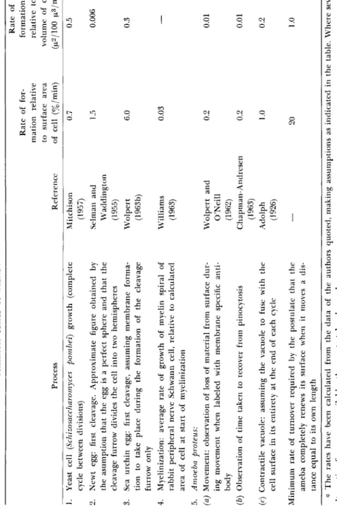
Although our studies suggest that during ameboid movement surface membrane turnover is slow and that changes in surface area would also require turnover, it is relevant to ask if there is evidence from other cells showing that membrane turnover is ever rapid. Turnover of cell mem- brane, i.e., the formation or resorption of membrane, occurs in a variety of cellular processes such as growth, cell cleavage, pinocytosis, phagocy- tosis, and vacuolar secretion. Although quantitative measurements are, in the main, lacking, calculations from some of the available data are summarized in Table I. From this table it can be seen that the highest rate of formation is 6%/min for cleavage of the sea urchin egg, which is a factor of 3 less than the 20%/min required on the theory that the ameba renews its surface each time it passes through its own length, and 30 times more than the observed rate. However, even a 1%/min rate of increase does show that, in principle, membrane formation could be rapid enough to provide new membrane for cell movement requiring slower rates of locomotion or pseudopod formation.
There are two special cases where it does seem that rapid formation of new surface occurs. The first is the so-called surface precipitation reac- tion (Chambers and Chambers, 1961) that results in new surface forma- tion when the surface is torn apart. However, for the ameba at least, the evidence is by no means conclusive that new surface is, in fact, being formed rather than that the existing surfaces come together and fuse. T h e second is the observation that amebae and tissue cells become rounded, with apparently unconvoluted surfaces, when subjected to high hydrostatic pressures, and that on release of the pressure the cells rapidly resume their ameboid forms. As Landau (1961; Landau and Thibodeau, 1962)
TABLE I MAXIMUM RATES OF MEMBRANE FORMATION IN VARIOUS SYSTEMS« Rate of Rate of for-formation mation relative relative to to surface area volume of cell Process Reference of cell (%/min) (μ2/100 μ3/ππη) 1. Yeast cell (Schizosaccharomyces pombei) growth (complete Mitchison 0.7 0.5 cycle between divisions) (1957) 2. Newt egg: first cleavage. Approximate figure obtained by Selman and 1.5 0.006 the asumption that the egg is a perfect sphere and that the Waddington cleavage furrow divides the cell into two hemispheres (1955) 3. Sea urchin egg: first cleavage, assuming membrane forma-Wolpert 6.0 0.3 tion to take place during the formation of the cleavage (1963b) furrow only 4. Myelinization: average rate of growth of myelin spiral of Williams 0.03 — rabbit peripheral nerve Schwann cell, relative to calculated (1963) area of cell at start of myelinization 5. Amoeba proteus: (a) Movement: observation of loss of material from surface dur-Wolpert and 0.2 0.01 ing movement when labeled with membrane specific anti-O'Neill body (1962) (b) Observation of time taken to recover from pinocytosis Chapman-Andresen 0.2 0.01 (1963) (c) Contractile vacuole: assuming the vacuole to fuse with the Adolph 1.0 0.2 cell surface in its entirety at the end of each cycle (1926) Minimum rate of turnover required by the postulate that the — 20 1.0 ameba completely renews its surface when it moves a dis- tance equal to its own length α The rates have been calculated from the data of the authors quoted, making assumptions as indicated in the table. Where several alternative figures are available, the greatest has been chosen.
WITHDRAWAL OF MEMBRANE FROM THE SURFACE Entry of membrane by phagocytosis and pinocytosis
OUTER MUCOPROTEIN COAT PLASMA MEMBRANE Precursor pool The amino acids, sugars, etc., from which the molecular components of the membrane are synthesized I
Vesicular pool Interior membranes of cell Phagocytic and pinocytic vacuoles, lysosomes, endoplasmic reticulum, Golgi apparatus SUPPLY OF NEW MEMBRANE Membrane1 synthesis ' in situ '
Molecular pool Elaborated molecular components of the membrane, In the form of micelles or in solution, not yet organized as membranes Direct | formation ι Supply of new membrane in vesicular form Secretion by vacuoles: defecation, and function of contractile vacuole
CELL INTERIOR PLASMA MEMBRANE llllllllllllllllllllllllllllllllllllllllllllllllllllllllllllllllllllllllllllllllllllllllll^ OUTER MUCOPROTEIN COAT FIG. 3. Diagram of interchange between cell surface and cell interior. Continuous lines represent pathways whose exist- ence is believed to be established; dotted lines represent possible pathways. The diagram shows the entry of membrane in vesicular form, and the possible supply of new plasma membrane to the surface in the form of vesicles. Three pools are postulated in the cytoplasm: (a) precursors providing the material for the elaboration of (b) the molecular components of the membrane, which themselves become organized (c) in vesicles. For simplicity arrows have been drawn in one direction only.
154 L. WOLPERT, C. M. THOMPSON, AND C. H. O'NEILL
points out, this certainly suggestive of rapid resorption and re-formation of the membrane.
Our knowledge of the mechanisms by which new membrane is formed or resorbed is certainly poor, but various possibilities are illustrated in Fig. 3 which shows that membrane can leave or enter the surface in the form of vacuoles (as in pinocytosis and vacuolar secretion). Entry of mem- brane during pinocytosis is well established (Nachmias and Marshall, 1961) and so is formation of new surface during vacuolar secretion (Pal- ade, 1959). It is not clear whether this mode of formation of new mem- brane occurs in the ameba, but a pathway by which the contents of pinocytosis vacuoles ultimately appear in defecation vacuoles has been described by Brandt and Pappas (1962). Our experiments with the fluo- rescent antibody indicate a similar pathway. The possibility of entry (or exit) in molecular or micellar units is also illustrated, although there is as yet little evidence for such a mechanism. It is also possible that the membrane may be synthesized in situ by enzymatic sites forming part of the membrane itself, particularly as the internal membranes of the cell have many synthetic functions. The problem of membrane formation is one of general importance, which could have significant implications for cell movement. Now that we can obtain isolated membranes we are hope- ful that we may be able to investigate this problem using radioisotopes, and particularly to determine the turnover rate of the plasma membrane.
The available evidence leads us to the conclusion that the surface membrane of the ameba flows freely during movement. It probably is also elastic, but cannot undergo large changes in surface area without addition or removal of membrane material, both of which are relatively slow processes. It seems that, for the moment, the membrane must be assigned a relatively passive mechanical role in movement, al- though it probably plays a very important role in the control of move- ment. We must thus look to the interior of the cell for the motive force.
Studies of the Isolated Cytoplasm
One of the earliest observed characteristics of protoplasm was its apparent contractility. Since Mast's classic studies on amebae, "contrac- tile" processes in the cytoplasm have been emphasized as the most prob- able basis for cell movement, and particular attention has been focussed on sol-gel transformations. It has more recently been established that cytoplasm is capable of autonomous streaming, for which the crucial evi- dence is the demonstration by Allen et al. (1960) that naked cytoplasm from ruptured amebae continues to stream.
As already pointed out, the analogy between muscular contraction
Isolated Membrane and Cytoplasm of A. proteus 155 and cytoplasmic motility has dominated attempts to isolate the active
components. Table I I summarizes some of the features of the systems extracted from nonmuscular cells for comparison with muscle. It should be noted that all the extracts are made in high salt concentrations. Al- though some of these results support the muscle protein analogy, namely, the changes of viscosity of myxomyosin and the superprecipitation of sarcoma cell protein (both A T P dependent), there is no compelling reason to believe that the motile system has yet been isolated in a form which can be related to what is seen in the intact cell. T h e results of Sakai (1962) indicate that the contraction of reconstituted threads may not require an ATP-dependent system.
A further difficulty, and one which also weakens the analogy, is that studies on the fine structure of ameboid cells have not, so far, brought to light any structures that can with confidence be assigned a role in motil- ity and compared with myofilaments. T h e only possible exception to this is the recent demonstration of fibers in electron micrographs of the slime mold by Wohlfarth-Bottermann (1962). Amebae contain a great quantity of vesicular material resembling smooth endoplasmic reticulum (Mercer, 1959; Pappas, 1959; Daniels and Roth, 1961), but its function is not known. T h e electron microscope morphology of the cytoplasm is per- plexing in that there is no detectable difference even between different regions of the cytoplasm. For example, there is still no clear indication of the nature of the boundary between the hyaline layer and the ecto- plasm, or between endoplasm and ectoplasm.
ISOLATION OF A MOTILE CYTOPLASMIC FRACTION
In our own attempts to isolate the motile system of A. proteus we tried, in the first instance, to obtain an actomyosin-like fraction using variations on the extraction methods with strong salt solutions found successful with muscle, sarcoma, and slime mold. In no case, however, did we obtain a preparation which fulfilled the necessary criterion, namely, unequivocal reversible change of viscosity with added ATP.
These early experiments led us to conclude that if present, such a sys- tem was highly labile. We also reasoned that even if we brought an ex- traction procedure to the point of duplicating the results achieved with, say, the slime mold (Ts'o et al., 1956a,b, 1957a,b), we should be little nearer to the molecular basis of motility in the intact cytoplasm. For these two reasons, we began to look for a way of isolating the system not by a chemical extraction procedure but by a primarily physical one which would yield cytoplasm with as many as possible of its intracellular char- acteristics intact, that is, the power to stream, to contract, and to undergo
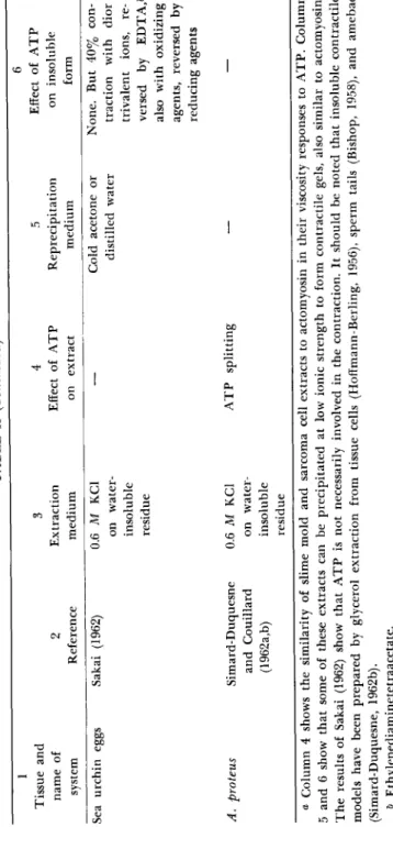
TABLE II SUMMARY OF RESULTS OF SALT EXTRACTIONS OF MOTILE CELLS FOR COMPARISON WITH MUSCLE» 1 Tissue and name of system Reference Extraction medium Effect of ATP on extract Reprecipitation
Effect of ATP on insoluble form Muscle, actomvosin Sarcoma cells Slime mold, myxomyosin Slime mold, myosin Β Sperm tails
Weber and Portzehl (1952) Hoffmann- Berling (1956) Ts'o et al. (1956a,b) Nakajima (1960) Pautard (1962)
0.6 M KCl, pH 8-9 0.6 M KCl + ATP 1.4 M KCl 0.6 M KCl, pH 8-9 0.5 M KCl, pH 8-9
Fall of viscosity; reversed as ATP is split Fall of viscosity; reversed as ATP is split Fall of viscosity; reversed as ATP is split Fall of viscosity; reversed as ATP is split
As gel or load bearing fibers below 0.2 M KCl Flocculent precipitate 0.05-0.1 M KCl 25-40% saturated ammonium sulfate Below 0.1 M KCl 0.05 M KCl, pH 5.2
Rapid contraction of fibers. Superprecip- itation of gel Slow shrinkage of pieces of gel Slow superprecipita- tion Formation of stream- ing network of gel; contraction, rhyth- mic in places
TABLE II (Continued) 1 Tissue and name of system Sea urchin eggs
Reference Sakai (1962) Extraction medium 0.6 M KCl on water- insoluble residue Effect of ATP on extract Reprecipitation medium
Effect of ATP on insoluble form Cold acetone or distilled water None. But 40% con- traction with dior trivalent ions, re- versed by EDTA,ö also with oxidizing agents, reversed by reducing agents A. proteus Simard-Duquesne 0.6 M KCl ATP splitting — — and Couillard on water- (1962a,b) insoluble residue α Column 4 shows the similarity of slime mold and sarcoma cell extracts to actomyosin in their viscosity responses to ATP. Column 5 and 6 show that some of these extracts can be precipitated at low ionic strength to form contractile gels, also similar to actomyosin. The results of Sakai (1962) show that ATP is not necessarily involved in the contraction. It should be noted that insoluble contractile models have been prepared by glycerol extraction from tissue cells (Hoffmann-Berling, 1956), sperm tails (Bishop, 1958), and amebae (Simard-Duquesne, 1962b). & Ethylenediaminetetraacetate.
158 L. WOLPERT, C. M. THOMPSON, AND C. H. O'NEILL
sol-gel changes. These we regarded as better criteria for recognizing the active component than viscosity changes or ATPase activities.
In general, the cytoplasm of an ameba, burst in a watery medium, either coagulates or disperses rapidly, destroying the possibility of deli- cate sol-gel changes. Our first objective was, therefore, to find a medium in which the cytoplasm remained undamaged as regards motility. We set out to test the effect of various media on the cytoplasm of amebae before, during, and after rupture. This was done by compressing a dense suspension of cells with a cover slip and watching the changes in the behavior and appearance of the cytoplasm. Although crude, this method gave relatively consistent and quite valuable results. Of particular in- terest was the observation that sometimes the cytoplasm from several cells would run together on the slide and remain without dispersing for several minutes. During this time, granules could be observed to move about in different directions by sudden displacements in such a way as to suggest a twitching network of oppositely directed streams. Sometimes whole areas would contract and tear in jerks from other areas. This behavior was taken as a sign of the survival, in disorganized form, of the motile mechanism outside the cell. As a rule addition of salts, es- pecially calcium, prevented the twitching, whereas A T P and SH com- pounds tended to favor it. Cysteine reversed, to some extent, the dis- persing effect of high potassium chloride concentration. Distilled water was as effective in prolonging twitching as ATP, but the contractions appeared less vigorous. T h e conclusion was that the most useful con- ditions for preparing ameba homogenates would be in the presence of A T P and SH compounds, and the exclusion of salts; but addition of either or both, before homogenization, did not yield bulk homogenates that showed detectable motility. Homogenization in total absence of medium and subsequent addition to A T P was also ineffective under the conditions tried. We, therefore, decided to dispense with homogenization altogether, a possibility hinted at by Marshall et al. (1959) and the ex- periments of Allen et al. (1960). After many trials we found that an active preparation showing dramatic streaming and syneresis can be obtained if the cells are first cooled at 4°C for 24 hr and then centrifuged whole at 35,000 g for 10 min. All the larger inclusions such as nuclei, food vacuoles and crystals are pinched off at the heavy pole; they pass to the bottom of the tube and are discarded. T h e cells remain tightly packed as smooth bags with a stratified contents of fat droplets, mitochondria, and various small vesicles. The membranous bags are emptied of their contents by gentle homogenization and spun out in the cold at 1200 g.
The resulting extract, whose preparation is described in greater detail elsewhere (Thompson and Wolpert, 1963) is called extract I.
Isolated Membrane and Cytoplasm of A. proteus 159
MOTILITY OF EXTRACTS
Extract I is heterogeneous and contains fat droplets, granules, and vesicles visible under the phase-contrast microscope. It is difficult to avoid slight contamination by surface membranes (10-100 membranes/ml of extract). Motility is judged by observing the behavior of granules in a drop of extract in a sealable chamber 50 to 100 μ deep. At 4°C or at room tem- perature only Brownian motion of granules can be seen. There are no signs of displacements of granules which could be distinguished from those of a colloidal suspension of inert particles of comparable size.
However, the addition of neutralized A T P under appropriate conditions produces striking effects which have been observed and filmed many times. The sequence of events may be described as follows. T h e first signs of movement, appearing after about 30 sec, are sudden "saltatory"
displacements of individual granules in any direction. T h e distance covered may be only a few microns, but the displacements are quite dis- tinct from Brownian motion. Displacements continue in any direction but gradually become more widespread, whole areas moving as single blocks. T h e entire field appears to twitch. T h e moving blocks of cyto- plasm appear to become connected to each other, so that the motion of one area is transmitted to others through a constantly changing network of oppositely directed streams of granules. The movement appears to be- come more definitely orientated into one of two alternative patterns:
(a) Streams of granules become arranged in parallel lines moving in opposite directions through the bulk of the cytoplasm. T h e streams per-
sist for up to 10 min and die out slowly, leaving a uniform distribution of granular material over the whole area of the chamber. There is no net transfer of material in any direction, although streams of particles may be moving in opposite directions to each other at 80 μ/sec for distances of 650 μ. In general these movements are very similar to those observed by Allen et al. (1960) in single ruptured cells.
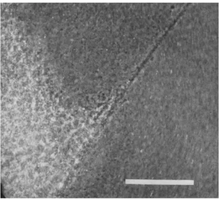
(b) Granules cease to move in oppositely directed streams. Instead, large areas of the field appear to gel into sheets which contract quickly to a small fraction of their original area (Fig. 4). This type of movement does not persist as long as the streaming. As gelled areas form and con- tract, many larger inclusions are trapped and drawn together, including any membrane fragments. In one particular case, at the edge of a con- tracting gel area, particles were seen to be moving toward the center at a speed of 150 μ/sec relative to the slide. At the same time, other granules were moving in the opposite direction at 50-100 μ/sec. It seemed they were being squeezed out from the interstices of the contracting gel net- work. In this pattern of movement there is always a net transfer of material as dense localized masses are formed. When two sheets of gel
160 L. WOLPERT, C. M. THOMPSON, AND C. H. O'NEILL
FIG. 4. T h e edge of a contracted gel mass formed from extract I in the presence of 2.5 mM A T P . T h e preparation was photographed in a chamber and shows part of a gel strand connected to another gelled region. Scale mark is 100 μ.
The second type of movement is seen particularly well when a mix- ture of extract and A T P is introduced into a capillary, of diameter about 1000 μ. T h e central mass then takes the shape of a thread which can conveniently be blown out for examination or fixed for electron micros- copy (discussed later).
CONDITIONS FOR MOTILITY AND DEPENDENCE ON A T P
T h e types of motility described have been induced so far only by the addition of A T P or adenosine diphosphate (ADP), ADP showing less contract in different directions and tear apart from each other, the con- nections between them become drawn out and sometimes clearly visible strands (see Fig. 4) are formed which can extend for 500 μ. These thin strands have been observed to shorten and pull in smaller clumps of contracted material toward the main mass over a distance of several hundred microns. If a thin strand breaks, the two ends snap apart like rubber.
Isolated Membrane and Cytoplasm of A. proteus 1 6 1 activity. In the absence of A T P or ADP no motility has been observed.
Formation of contracted masses in capillary tubes occurs when the final concentration of A T P lies in the range 1 - 5 mM; 1 0 mM is inhibitory.
Adenosine monophosphate (AMP) and all other compounds so far tested are without effect (pyrophosphate, orthophosphate, SH compounds, and salts). Salts, particularly potassium chloride and traces of calcium, abolish the effect of ATP, and 1 - 5 mM calcium chloride causes irreversible clumping of the vesicles. T h e induction of motility is dependent upon temperature. The capacity of extracts stored at 0 ° C to respond to added ATP gradually diminishes and disappears after 1-2 hr, and A T P does not have any effect on extracts kept at 4 ° or 2 2 ° C . Motility is observed only if extracts previously mixed with A T P in the cold are allowed to come to room temperature.
PARTIAL PURIFICATION OF ACTIVE COMPONENT
Spinning extract I (Thompson and Wolpert, 1963) for 1 0 min at 9 0 0 0 g in the cold removes a large proportion of the granules. T h e re- sulting extract II shows almost identical behavior with A T P to that of extract I. T h e preparation is, however, more stable (up to 6 hr). Further spinning at 3 5 , 0 0 0 g for 1 0 min results in the disappearance of ATP- induced motility from the supernatant. Surprisingly, the pellet is also in- active provided that care has been taken to wash traces of the supernatant away from the sides of the tube with cold distilled water. T h e super- natant is largely free of granules and vesicles, but still contains drops of coalesced lipid which make it possible to detect motility. The pellet contains many vesicles and granules, and when resuspended in distilled water looks like extract II. It is found that when equivalent quantities of resuspended pellet and supernatant are mixed with cold A T P the usual response is restored, although neither are active alone. Spinning the 3 5 , 0 0 0 g supernatant a further 8 0 min at 1 5 0 , 0 0 0 g removes very little extra material and leaves a supernatant that will also give A T P responses when combined with the 9 - 3 5 , 0 0 0 g pellet. The response to ATP is lost if either pellet or supernatant are warmed to 1 0 0 ° C . T h e pellet and supernatant can be stored separately in the deep freeze and retain some activity after a week, but the pellet does not survive a second freezing.
ELECTRON MICROSCOPY OF THE CONTRACTED G E L
Pieces of the thread formed by contraction of extract I in capillaries were prepared for electron microscopy, in collaboration with Dr. Ε. H.
Mercer, by fixing in osmium (Palade's fixative) and embedding in Aral- dite (Mercer and Birbeck, 1961). The contracted mass is very heteroge-
162 L. WOLPERT, C. M. THOMPSON, AND C. H. O'NEILL
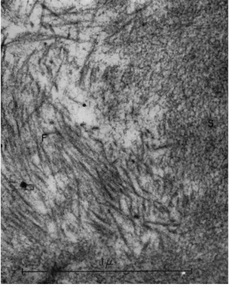
neous and contains fat droplets, vesicles both small and large, mitochon- dria, and occasional fragments of cell membranes. T h e most striking features are arrays of well-defined and partially orientated fibers and a more or less homogeneous region with a spongy appearance (Fig. 5).
Such spongy regions are continuous with the fibers (Fig. 6). T h e fibers are about 120 A thick and about 0.5 μ long. There is no evidence of
FIG. 5. Electron micrographs of the contracted gel obtained from extract I after the addition of A T P . T h e gel is heterogeneous and contains vesicles (V), lipoid droplets (L), and fibers (F). T h e fibers are about 120 A wide and 0.5 μ long. In (a) a spongy region (S) can be seen to be continuous with the fibers. Note in (6) the parallel align- ment of the fibers. Fibers appear in cross section at (X) in (b). Magnification: χ 30,000.
(Photograph by Dr. Ε. H. Mercer.)
Isolated Membrane and Cytoplasm of A. proteus 163
FIG. 6. Electron micrograph of spongy region (S) and fibers (F) of gel prepared as in Fig. 5. T h e fibers seem to be continuous with the spongy region which contains small fibrils and particles. Magnification: χ 80,000. (Photograph by Dr. Ε. H. Mercer.)
banding. Under high power the spongy region appears to contain some small particulates and small fibrils. The fibers seem to fuse with the spongy material, which suggests that it might be the precursor of the fibers.
Electron micrographs of gel prepared from extract II show it to be less heterogeneous than that from extract I, many of the vesicles having
164 L. WOLPERT, C. M. THOMPSON, AND C. H. O'NEILL
been spun out. The fibrils now appear somewhat different (Fig. 7). They are about 80-90 A thick and, although they do not show banding, they do appear denser in some regions than in others. They also, on occasion, give the impression of being coiled.
FIG. 7. Electron micrograph of fibers in gel obtained from extract I I by the addi- tion of ATP. T h e fibers are about 90 A wide. (Photograph by Dr. Ε. H. Mercer.)
Discussion
The results obtained so far should be regarded as preliminary, and detailed interpretation postponed until more data have been accumu- lated. There are, nevertheless, some general points that merit attention.
The crude cytoplasmic extracts exhibit four main properties when brought to room temperature in the presence of A T P : (1) "gelation";
(2) streaming of particles often in localized regions and sometimes for long distances; (3) contraction and the ability to transmit tension; (4) syneresis. All these properties have been ascribed to the cytoplasm of whole amebae or are demanded by current theories of movement. T h e