34(2007) pp. 103–109
http://www.ektf.hu/tanszek/matematika/ami
Complex Fiber Visualization
Henrietta Tomán
a, Róbert Tornai
b, Marianna Zichar
ca Department of Computer Graphics University of Debrecen e-mail: toman@inf.unideb.hu
bDepartment of Computer Graphics University of Debrecen e-mail: rtornai@inf.unideb.hu
cDepartment of Computer Graphics University of Debrecen e-mail: zicharm@inf.unideb.hu
Submitted 6 October 2007; Accepted 18 December 2007
Abstract
In this paper we give a short overview about how the known, different methods (DTI, WMT) can be integrated as new components into a multi functional system. Our exact aim was to develop a well applicable implemen- tation for the DTI method and Fiber Tracking. The final goal is to integrate the fibers or ellipsoids into the surface model clipped by examination planes.
Furthermore, the stream of the molecules (the fibers) can be modelled by a particle system. These techniques are available by the new graphic cards and the standards they support.
Keywords: Diffusion Tensor Imaging, fiber tracking, diffusion ellipsoid, shader languages, parallelized algorithms
MSC:68U05, 92C55
1. Introduction
The white matter of human brain has a complex structure and plays an essential role in brain function.
In spite of the fact, that a fair amount of information is available today about white matter, not all the aspects of its structure are completely known and un- derstood. We know even less about how the white matter structure is affected by neurological diseases, tumors or traumas.
103
Diffusion Tensor Imaging (DTI) is an emerging Magnetic Resonance Imaging (MRI) technique based on water diffusion. Fiber tracking, also called White Matter Tractography (WMT), uses the directional information of diffusion tensor maps to estimate connection pathways in white matter.
The method is presented in the PhD dissertation of Mariana Lazar (see [2]).
Our aim is to reconstruct the fiber tracts of the human brain from measure- ments of fiber orientation and visualize them on the image of the brain. Generally the programs show the surface model clipped by orthogonal sections (coronal, ax- ial and sagital). The current version of our software is capable to visualize the surface model clipped by (even more than the usual three) planes having arbitrary directions. The software component is able to promote the recognition of the brain diseases and after the diagnosis it helps to select the appropriate way of cure. When surgical intervention is needed, the point and the direction of the permeation can be determined more exactly. The software component can be parallelized by using the shading languages (see [6]).
The visualization procedure belongs to a complex software system developed by a team led by Miklós Emri. This project is part of a new partnership between the Department of Computer Graphics and the PET Center of the Medical School and Health Science Center.
2. Theoretical background
During an MRI scanning process radio waves are sent through the brain which are 10 000 to 30 000 times stronger than Earth’s magnetic field. This forces the nuclei into a different position and when they move back into their place they send out radio waves of their own. The scanner picks up these signals and records their strength as numeric values into a file.
Diffusion MRI measures the diffusion of water molecules in biological tissues.
In an isotropic medium (e.g.: inside a glass of water) water molecules naturally move randomly according to Brownian motion. In biological tissues however, the diffusion may be anisotropic that is to say the diffusion properties vary with orien- tation. The recent development of Diffusion Tensor Imaging (DTI) (see [7]) enables diffusion to be measured in multiple directions and the fractional anisotropy in each direction to be calculated for each voxel. The most important base concepts are reviewed, while a complex overview of anisotropic water diffusion is presented by Beaulieu (see [1]).
2.1. Diffusion tensor
It is well known that the diffusion in white matter is the largest along fiber directions. When diffusion is anisotropic, a scalar diffusion measure is insufficient for describing diffusion properties. It has been shown that the diffusion in this case can be described by a second-order diagonally symmetric tensor, called the
diffusion tensor. This tensor model of diffusion can be used well to describe the directional diffusion information.
D=
Dxx Dxy Dxz Dyx Dyy Dyz Dzx Dzy Dzz
The six independent elements of the diffusion tensor can be estimated from a series of diffusion-weighted images. When diffusion weighted measurements are performed along N directions, the following matrix equation can be constructed (see [5]):
B ~dT =A~T where
A~ =
lnSS10 lnSS20 . . . lnSSN0 is the vector of the corresponding logarithmic signal ratios and
B=
b~1 b~2 ... b~N
includes the influences of all the encoding gradients.
3. The process of calculation
In practice 25 files are provided usually containing the results of diffusion weight- ed measurements, and we need also a sepatare file describing the baseline data. All of them serve as input data of the algorithm and they are referred as volume of voxels further on. The input files have an extention ’mnc’ because they have a special inner structure. A plug-in is responsible for writing and reading of this file type.
3.1. Diffusion tensor
During the calculation special volume iterators are used, that makes more con- venient to reach all the data belonging to the same voxel at same time in a very effective way. This makes also possible that the algorithm remains independent from the size of volumes. The names of input files are used as command line pa- rameters, that is why the program are able to handle dinamically the number of input volumes. A function was constructed to determine the six tensor elements of a voxel, which works in the following way.
1. If the input data make possible the gradients themselves are yielded or default values are used instead.
2. The components of vector A are calculated based upon the signal values. (S0 denotes the value derived from the baseline data.)
3. The equation itself is solved by using the appropriate function of GSL. The GNU Scientific Library (GSL) is a numerical library for C and C++ program- mers, which is a free software under the GNU General Public License and provides a wide range of mathematical routines. The selected GSL function is able to find the least squares solution to our overdetermined system.
4. Finally the resulted 6 values, that is to say the 6 independent elements of diffusion tensor are to be stored in appropriate position of new volumes.
3.2. Parameters of diffusion ellipsoid
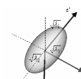
A principal frame of directions (x’, y’ and z’) can be defined by the eigenvectors of the diffusion tensor for each voxel. The diffusion displacement profile may be represented as an ellipsoid with the length of principal axes described by the tensor eigenvalues λ1,λ2 andλ3 (principal diffusivities) and the directions given by the tensor eigenvectors (e~1,e~2ande~3). Figure 1 shows the geometric meanings of the computed values.
Figure 1: The diffusion ellipsoid.
The diffusion eigenvectors are generally not aligned with the laboratory frame.
In the principal component frame, the displacements along x’, y’, and z’ appear uncorrelated and the diagonal elements of the tensor are equal to tensor eigenval- ues. The major axes are given by the diffusion tensor eigenvectors. The length of the ellipsoid along the axes is proportional with the square root of the tensor eigenvalues.
Depending on the relation between the eigenvalues volume, three types of dif- fusion and corresponding ellipsoidal shapes can be differentiated:
a) Isotropic diffusion: λ1≈λ2≈λ3
Diffusion in gray matter and fluids generally appears isotropic. The corre- sponding diffusion ellipsoid has a spherical shape.
b) Planar diffusion: λ1≈λ2≫λ3
Planar diffusion is generally associated with diffusion in sheets or it may describe regions of crossing fibers. The corresponding diffusion ellipsoid has a special disk shape.
c) Prolate diffusion: λ1≫λ2> λ3
Prolate or uniaxial diffusion is observed in highly organized white matter regions. The corresponding diffusion ellipsoid has a prolate shape.
White matter structures (corpus callosum and corticospinal tract) are generally characterized by uniaxial diffusion. Planar diffusion is dominant in regions of cross- ing or fanning fibers. For the visualization of the stream directions at each voxel ellipsoids are used. These ellipsoids can be incorporated with the brain model.
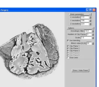
Figure 2 shows how the brain can be clipped by three arbitrary planes that is not common in usual softwares.
Figure 2: Brain clipped by three arbitrary positioned planes.
From the computed diffusion ellipsods fibers can be built up (see [4]). Current algorithms do not handle the fiber crossings. In this case they follow the stronger
track only. For the dynamic modelling of the fibers, we apply a particle system (see [3]). The molecule groups inspected by the measurements can be modelled by points moving along the actual fiber. These fibers make up a complex brain structure. For example the tumors distort the fibers, so a change in the fiber structure prognosticate the illness.
Moreover, our software is capable to handle more pictures (e.g.: one individual and one picture that is common for a population) and blend them together in any ratio. Besides, we can enhance a strip of the brain around any given intensity value.
The above work uses the OpenGL system, particularly its multitexturing, 3D texturing and alpha blending subsystems.
4. Conclusion and further research
The standard algorithms are improved and new methods are developed by our team considering the branching and merging problems also. We plan to massively parallelize the algorithms by using heavily the shader languages (vertex, geometry and pixel shaders).
Beyond the completed new methods, we want to combine them as the final step. We will integrate the ellipsoids and the particle system with the brain model having arbitrary clipping planes.
References
[1] Beaulieu, C.,The basis of anisotropic water diffusion in the nervous system,NMR Biomed., Vol. 15 (2002), 435–455.
[2] Lazar, M., White matter tractography: an error analysis and human brain fiber tract reconstruction study (PhD dissertation, University of Utah) (2003).
[3] McReynolds, T., Blythe, D.,Advanced Graphics Programming Using OpenGL Morgan Kaufmann(2005)
[4] Mori, S., van Zijl, P.C.M.,Fiber tracking: principles and strategies - a technical review,NMR Biomed., Vol. 15 (2002), 468–480.
[5] Papadakis, N.G., Xing, D., Huang, C.L., Hall, L.D., Carpenter, T.A., A comparative study of acquisition schemes for diffusion tensor imaging using MRI,J.
Magn. Reson., Vol. 137 (1999), 67–82.
[6] Rost, R.J.,OpenGLR Shading Language, Second EditionAddison Wesley Profes- sional (2006).
[7] Taylor, A.J.,Diffusion tensor imaging: evaluation of tractography algorithm per- formance using ground truth phantoms (Thesis, Virginia Polytechnic Institute and State University) (2004).
Henrietta Tomán
Department of Computer Graphics University of Debrecen
Egyetem tér 1.
H-4010 Debrecen, Hungary Róbert Tornai
Department of Computer Graphics University of Debrecen
Egyetem tér 1.
H-4010 Debrecen, Hungary Marianna Zichar
Department of Computer Graphics University of Debrecen
Egyetem tér 1.
H-4010 Debrecen, Hungary