https://doi.org/10.1007/s00044-019-02472-9
RESEARCH
O R I G I N A L R E S E A R C H
Structural modi fi cation of ellipticine derivatives with alkyl groups of varying length is in fl uential on their effects on human DNA
topoisomerase II: a combined experimental and computational study
M. Kuskucu1●V. Akyildiz2●Á. Kulmány3●Y. Ergün2●S. Zencir4●I. Zupko3●S. Durdagi5●M. Zaka5●K. Sahin5● H. Orhan6●Z. Topcu1
Received: 3 June 2019 / Accepted: 6 November 2019 / Published online: 20 November 2019
© Springer Science+Business Media, LLC, part of Springer Nature 2019
Abstract
The compounds reducing tumor cell viability and disrupting DNA topoisomerase reactions have been widely used in anticancer drug development. Ellipticine (5,11-dimethyl-6H-pyrido[4,3-b]carbazole) is a potent intercalating agent that interferes with nucleic acid processing through interaction with DNA topoisomerase II. Although ellipticine is a well- characterized compound, it is not a widely-accepted drug due to the adverse effects detected upon administration. We have previously reported two novel ellipticine derivatives, N-methyl-5-demethyl ellipticine (ET-1) and 2-methyl-N-methyl-5- demethyl ellipticinium iodide (ET-2) as potent compounds targeting DNA topoisomerase II. This study covers an extended synthesis, characterization, and activity data forfive new salts of N-methyl 5-demetyl ellipticine (Z-1, Z-2, Z-4, Z-5 and Z-6) having several organic halides and their effects on human topoisomerase II enzymes. Moreover, combined in silico studies were conducted for better understanding of modes of action of studied molecules at the binding pocket of target. Our results showed that three of the derivatives (Z-1, Z-2, and Z-6) have considerable effect on the catalytic activity of human topoisomerase II implying the influence of alkyl groups added to the parental structure of ellipticine.
Keywords DNA topoisomerase II●Ellipticine derivatives ●Anticancer drugs
Introduction
DNA topoisomerases play fundamental roles in overcoming topological problems of DNA by introducing transient breaks in the duplex during replication, transcription, recombination, and chromosomal segregation (Wang1971).
Given that the inherent functions of topoisomerases are crucial for genomic integrity, the interfere with these enzymes or generating enzyme-mediated DNA damage is an effective strategy in development of chemotherapeutic agents (Coban et al.2008; Gul et al.2009; Senarisoy et al.
2013; Ashley and Osheroff 2014; Ketron and Osheroff 2014; Zupkó et al.2014; Vann et al.2016a,2016b; Delgado et al.2018).
Topoisomerase targeting agents act primarily in two ways: Topoisomerase poisons stabilize the transient phos- photyrosine covalent bond between the enzyme–DNA complexes and kill the cells by increasing the levels of truncated-DNA covalent intermediates (Champoux 2001;
Topcu2001). Alternatively, topoisomerase inhibitors act by disrupting the essential catalytic functions of the enzymes. It
* Z. Topcu
zeki.topcu@ege.edu.tr
1 Department of Pharmaceutical Biotechnology, Faculty of Pharmacy, Ege University, Izmir 35100, Turkey
2 Department of Chemistry, Faculty of Science, Dokuz Eylul University, Izmir 35160, Turkey
3 Institute of Pharmacodynamics and Biopharmacy, Faculty of Pharmacy, University of Szeged, Szeged 6720, Hungary
4 Department of Medical Biology, Faculty of Medicine, Pamukkale University, Denizli 20070, Turkey
5 Computational Biology and Molecular Simulations Laboratory, Department of Biophysics, School of Medicine, Bahcesehir University, Istanbul 34734, Turkey
6 Department of Pharmaceutical Toxicology, Faculty of Pharmacy, Ege University, Izmir 35100, Turkey
Supplementary informationThe online version of this article (https://
doi.org/10.1007/s00044-019-02472-9) contains supplementary material, which is available to authorized users.
1234567890();,: 1234567890();,:
has also been shown that catalytic inhibitors affect various stages of the catalytic cycle of topoisomerase II, including the cleavage of DNA while some compounds manifest both DNA breakage patterns as well as enzyme inhibition (Topcu2001).
The natural product ellipticine (5,11-dimethyl-6H-pyr- ido-[4,3-b]carbazole) is one of the carbazole alkaloids with a potent anticancer activity. Several key mechanisms of action, including DNA intercalation and topoisomerase II inhibition, bio-oxidation, adduct formation, mitochondrial membrane modulation, inhibition of p53 and interaction with kinases such as c-Kit and AKT contribute to the anticancer activity of ellipticine (Vann et al.2016b; Garbett and Graves2004; Kuo et al. 2005; Thompson et al. 2008;
Chikamori et al.2010). Like several other topoisomerase- targeting drugs, a number of adverse side effects such as xerostomia, weight loss, hemolysis, and renal toxicity are also associated with ellipticine administration mostly due to its cytochrome P450 (CYP)- and/or peroxidase-mediated activation yielding covalent DNA adducts (Rouëssé et al.
1993; Lichota and Gwozdzinski 2018). Therefore, over the last years, obtaining new natural or synthetic derivatives to replace the known topoisomerase-targeting compounds gained a significant attention.
Several derivatives of ellipticine have been investigated (Moody et al.2007; Miller et al. 2012; Deane et al.2013;
Iacopetta et al. 2017). We previously reported two novel ellipticine derivatives, N-methyl-5-demethyl ellipticine and 2-methyl-N-methyl-5-demethyl ellipticinium iodide that both were potent topoisomerase II-targeting compounds (Vann et al.2016b). We now synthesized and characterized five new derivatives of ellipticine having alkyl groups of different lengths with increased carbon number (Z-1 and Z- 2) and substituted alkyl groups (cyanoalkyl) (Z-4 and Z-5) as well as a derivative with an inserted amide alkyl group (Z-6). We monitored their effect on tumor cell growth and human topoisomerase II activities and also conducted in silico studies.
Materials and methods Chemistry
We investigated side chain substituent effect at the N-2 position in order to increase the solubility and activity of the compounds. The salts of N-methyl 5-demetyl ellipti- cine were synthesized using N-methyl 5-demetyl ellipti- cine and several alkyl halides, cyano alkyl halides, and amide alkyl halide (Fig.1). Melting points were measured in sealed tubes using an electro thermal digital melting point apparatus (Gallenkamp, London, UK). Fourier
transform infrared spectroscopy (FTIR) analyses using PerkinElmer Spectrum BX-II Model FTIR spectro- photometer were carried out for characterization of syn- thesized molecules. The samples within KBr pellets were measured in the range of 4000–400 cm−1. 1H-NMR and
13C-NMR spectra were obtained on a Varian AS-400 NMR spectrometer with tetramethylsilane as an internal standard (Oxon, UK).
Synthesis of N
2-methyl N-methyl 5-demethyl ellipticine salts
N-Methyl 5-demethyl-ellipticine (2 mmol) was suspended in dimethylformamide (5 mL), and organic halide (2.2 mmol) was added (Vann et al. 2016b). The reaction mixture was stirred at room temperature for 5 h, quenched with cold ether (5 mL), and the resulting precipitate was collected with vacuum filtration. The precipitate was washed with cold ethanol (2.5 mL) to yield N-methyl 5- demethyl ellipticine salts (Z-1, Z-2, Z-4, Z-5, Z-6) (Fig.1).
N
2-Ethyl N-methyl 5-demethyl ellipticinium bromide (Z-1)
Yield: 45%; mp: 296 °C; IR (KBr)νmax: 3020 (CH), 2974 (CH), 1619 (C=N) cm−1; 1H-NMR (DMSO-d6, 400 MHz):δ1.60 (t, 3H,+NCH2CH3), 2.97 (s, 3H, CH3), 3.74 (s, 3H, NCH3), 4.67 (q, 2H, +NCH2CH3), 7.25 (t, 1H, J=7.6 Hz, ArH), 7.43 (d, 1H, J=7.6 Hz, ArH), 7.52 (d, 1H,J=7.2 Hz, ArH), 7.73 (s, 1H, ArH), 8.09 (d, 1H,J=7.6 Hz, ArH), 8.13 (d, 1H,J=6.4 Hz, ArH), 8.36 (d, 1H, J=6.4 Hz, ArH), 9.82 (s, 1H, ArH); 13C-NMR (DMSO-d6, 100 MHz): 15.22, 16.89, 29.58, 55.74, 101.41, 109.82, 119.83, 121.34, 121.35, 124.17, 124.24, 125.55, 128.97, 131.17, 134.75, 135.05, 143.21, 145.38, 145.77.
N
2-Hexyl N-methyl 5-demethyl ellipticinium bromide (Z-2)
Yield: 52%; mp: 132 °C; IR (KBr) νmax: 3047 (CH), 2929 (CH), 1622 (C=N) cm−1;1H-NMR (DMSO-d6, 400 MHz):
δ0.85 (t, 3H,J=7.2 Hz, CH3), 1.24–1.45 (m, 6H, 3xCH2), 1.96–2.08 (m, 2H, CH2), 3.17 (s, 3H, CH3), 3.85 (s, 3H, NCH3), 4.67 (t, 2H,J=7.6 Hz,+NCH2), 7.35 (t, 1H, J= 7.6 Hz, ArH), 7.61–7.69 (m, 2H, ArH), 7.96 (s, 1H, ArH), 8.27 (d, 1H,J=6.8 Hz, ArH), 8.31 (d, 1H,J=8 Hz, ArH), 8.47 (d, 1H, J=6.8 Hz, ArH), 10.02 (s, 1H, ArH); 13C- NMR (DMSO-d6, 100 MHz): 14.17, 15.08, 22.27, 25.61, 29.45, 30.96, 31.18, 60.24, 100.82, 109.66, 119.66, 121.13, 121.30, 124.02, 124.09, 125.43, 128.90, 131.31, 134.62, 134.87, 143.02, 145.22, 145.54.
N
2-Cyanomethyl N-methyl 5-demethyl ellipticinium iodide (Z-4)
Yield: 28%; mp: 189 °C; IR (KBr)νmax: 3042 (CH), 2924 (CH), 2218 (CN), 1619 (C=N) cm−1;1H-NMR (DMSO-d6, 400 MHz):δ3.20 (s, 3H, CH3), 3.89 (s, 3H, NCH3), 5.97 (s, 2H, +NCH2CN), 7.38–7.42 (m, 1H, ArH), 7.62–7.69 (m, 2H, ArH), 8.02 (s, 1H, ArH), 8.36 (d, 2H,J=6.8 Hz, ArH), 8.51 (d, 1H, J=6.8 Hz, ArH), 10.10 (s, 1H, ArH); 13C- NMR (DMSO-d6, 100 MHz): 14.09, 29.87, 62.41, 100.81, 109.52, 119.67, 120.08, 121.14, 121.32, 123.45, 124.09, 125.41, 128.87, 131.37, 134.76, 135.11, 142.97, 145.28, 146.27.
N
2-(3-Cyanopropyl) N-methyl 5-demethyl ellipticinium chloride (Z-5)
Yield: 35%; mp: 116 °C; IR (KBr)νmax: 3047 (CH), 2924 (CH), 2246 (CN), 1619 (C=N) cm−1;1H-NMR (DMSO-d6, 400 MHz): δ 2.32–2.40 (m, 2H, CH2), 2.75 (t, 2H, J=
6.8 Hz, CH2), 2.99 (s, 3H, CH3), 3.68 (s, 3H, NCH3), 4.76 (t, 2H,J=6.8 Hz,+NCH2), 7.25 (t, 1H, J=7.2 Hz, ArH), 7.45 (d, 1H, J=8 Hz, ArH), 7.53 (t, 1H,J=8 Hz, ArH), 7.74 (s, 1H, ArH), 8.10 (d, 1H,J=8 Hz, ArH), 8.16 (d, 1H, J=6.8 Hz, ArH), 8.41 (d, 1H, J=6.4 Hz, ArH), 9.99 (s, 1H, ArH); 13C-NMR (DMSO-d6, 100 MHz): 14.08, 15.17, 26.79, 29.46, 58.68, 100.85, 109.69, 119.69, 120.16, 121.12, 121.29, 123.56, 124.07, 125.37, 128.86, 131.35, 134.73, 135.09, 142.99, 145.26, 146.25.
N
2-(Carboxyaminoethyl) N-methyl 5-demethyl ellipticinium bromide (Z-6)
Yield: 21%; mp: 119 °C; IR (KBr) νmax: 3058 (CH), 2935 (CH), 1673 (C=O), 1619 (C=N) cm−1;1H-NMR (DMSO- d6, 400 MHz):δ2.76 (t, 2H,J=6.8 Hz, CH2), 3.20 (s, 3H, CH3), 3.88 (s, 3H, NCH3), 4.89 (t, 2H,J=6.8 Hz,+NCH2), 7.05 (m, 1H, ArH), 7.40 (m, 1H, ArH), 7.56 (m, 1H, ArH), 7.68 (s, 1H, NH2), 7.99 (m, 1H, ArH), 8.28 (d, 1H, J= 7.6 Hz, ArH), 8.36 (d, 1H,J=7.6 Hz, ArH), 8.46 (d, 1H,J Fig. 1 Ellipticine derivatives
covered in this study
=6.4 Hz, ArH), 10.04 (s, 1H, ArH);13C-NMR (DMSO-d6, 100 MHz): 15.51, 29.92, 36.31, 56.39, 101.58, 110.32, 120.02, 121.61, 121.82, 123.89, 124.71, 126.09, 129.21, 132.16, 135.18, 135.67, 142.76, 146.02, 147.50, 171.40.
Biology
MaterialspBR322 plazmid DNA, kinetoplast DNA (kDNA), recom- binant human topoisomerase II were purchased from Inspiralis (Inspiralis, Norfolk, UK). Human cervix adeno- carcinoma (HeLa and SiHa) and breast adenocarcinoma (MCF-7 and MDA-MB-231) cancer cell lines were pur- chased from European Collection of Cell Cultures (Salisbury, UK).
Cell culturing and viability assay
Cells were maintained in minimal essential medium supplemented with 10% fetal bovine serum, 1% non- essential amino acids and an antibiotic–antimycotic mixture (all from Lonza Group Ltd., Basel, Switzerland).
The antiproliferative properties of the prepared analogs were determined by means of MTT (3-(4,5-dimethyl- thiazol-2-yl)-2,5-diphenyltetrazolium bromide, Sigma Aldrich Ltd. (Budapest, Hungary) assay (Mosmann 1983). Briefly, near-confluent cancer cells were seeded onto a 96-well microplate (5000/well) and, after an overnight standing, new medium, containing the tested compounds was added. After incubation for 72 h in humidified air with 5% CO2at 37 °C, the viability of the cells were determined by the addition of 44 µL of 5 mg/
mL MTT solution. The living cells metabolized the MTT and the produced formazan was precipitated as purple crystals during a 4-h contact period. The medium was next removed and the formazan was dissolved in 100 µL of dimethylsulfoxide (DMSO) during a 60-min period of shaking at 37 °C. Finally, the reduced MTT was assayed at 545 nm (Stat Fax-2100, Awareness Technologies Inc., Palm City, FL, USA), sigmoidal concentration–response curves were fitted to the determined data and the IC50 values were calculated by means of GraphPad Prism 4.0 (GraphPad Software, San Diego, CA, USA). All in vitro experiments were carried out on two microplates with five parallel wells. Stock solutions of the tested com- pounds (10 mM) were prepared in DMSO. The highest DMSO content of the medium (0.3%) did not have any substantial effect on the cell proliferation. Cisplatin (Ebewe Pharma GmbH, Unterach, Austria) was used as a reference agent. The IC50 value is defined as the con- centration of a compound that gives half-maximal response.
Topoisomerase activity assays
Decatenation assays were carried out as described (Vann et al. 2016a). Reaction mixtures contained 0.2 µg of kine- toplast DNA (kDNA) fromCrithidia fasciculataand 75 nM and human topoisomerase II alfa in afinal volume of 20 µL of 50 mM Tris-Cl, pH 8.0, 120 mM KCl, 10 mM MgCl2, 0.5 mM ATP, and 0.5 mM dithiothreitol. Reactions were carried out in the absence or presence of compounds for 15 min at 37 °C and terminated with 5% sarkosyl, 0.0025%
bromophenol blue, and 25% glycerol. Samples were mixed with 2 µL of agarose loading dye (60% sucrose in 10 mM Tris-HCl pH 7.9, 0.5% bromophenol blue, and 0.5% xylene cyanol FF), heated for 2 min at 45 °C and subjected to electrophoresis using 1% agarose gels in 40 mM Tris- borate, pH 8.3 and 2 mM EDTA containing 0.5μg/mL ethidium bromide (Et-Br). DNA bands were visualized under UV light and average band intensities were calculated from three independent reactions using GeneSys program (SynGene, Frederick MD, USA). DNA decatenation was monitored by the conversion of large catenated networks that remained at the origin to decatenated minicircles. One unit of human topoisomerase II was corresponding to dec- atenation the 200 ng of kDNA substrate for 15 min at 37 °C in the indicated reaction buffer. Supercoil relaxing activity of the enzyme using supercoiled plasmid DNA substrate, pBR322 was also covered for correlation purpose (Topcu et al.2008).
Cleavage of Plasmid DNA
DNA cleavage assays contained 10 nM negatively super- coiled pBR322 DNA and 150 nM human topoisomerase II of either alfa or beta isoforms in afinal volume of 20 µL of 10 mM Tris-HCl, pH 7.9, 5 mM MgCl2, 100 mM KCl, 0.1 mM EDTA, and 2.5% (v/v) glycerol (Senarisoy et al.
2013; Vann et al. 2016a). Mixtures were incubated for 6 min at 37 °C. Enzyme-DNA cleavage complexes were trapped by the addition of 2μL of 5% SDS followed by 2μL of 250 mM EDTA, pH 8.0. Proteinase K (2μL of a 0.8 mg/mL solution) was added, and samples were incu- bated for 30 min at 45 °C to digest the enzyme. Samples were mixed with 2 µL of agarose loading dye, heated for 2 min at 45 °C and subjected to electrophoresis as above.
DNA cleavage was monitored by the conversion of super- coiled plasmid to linear molecules.
DNA intercalation
Human DNA topoisomerase I (0.5 U) and 300 ng of relaxed pBR322 were incubated in 10 mM Tris-HCl, pH 7.5, 10 mM KCl, 2 mM MgCl2, 0.02 mM EDTA, 0.1 mM dithiothreitol, and 6 µg/mL bovine serum albumin in afinal
volume of 20 µL (Vann et al.2016b). Reactions contained either ellipticine derivatives or Et-Br (10 µM), a well- characterized intercalator, used as positive control. Mixtures were incubated for 15 min at 37 °C. Following the addition of 3μL of stop solution (0.77% SDS 77.5 mM EDTA, pH 8.0), samples were extracted using 20 µL of phenol:
chloroform:isoamyl alcohol (25:24:1), and the aqueous layer was mixed with 2 µL of agarose loading dye and heated for 5 min at 45 °C. Intercalation products were subjected to electrophoresis in a 1% agarose gel in 100 mM Tris-borate and 2 mM EDTA. DNA bands were visualized as described above. DNA intercalation was monitored by the conversion of relaxed DNA to supercoiled plasmid populations of different superhelical densities.
In silico simulations
Ligand preparationThe 2D structures of studied ligands were sketched by 2D Sketcher program under Maestro molecular modeling package and prepared by Ligand Preparation module (Lig- Prep). OPLS3 force field was used to obtain low energy conformers of ligands. Protonation states of ligands were investigated at physiological pH by Epic module. To obtain the lowest energy conformers of ligands, a conformational search method was used in which all possible conforma- tions were generated by a systematic Monte Carlo search algorithm. The effect of slightly higher pH values (i.e., pH 8) to the protonation did not reveal any change.
Protein preparation
The crystal structure of DNA-bound human topoisomerase II alpha was retrieved from Protein Data Bank (PDB ID:
4FM9, 25 (Wendorff et al.2012)). The protein preparation module of Maestro molecular modeling package was used.
The missing side chains of the protein were fixed, proto- nation states of the amino acids were investigated at the physiological pH 7.4 by PROPKA code. Water molecules within the catalytic domain were kept using protein pre- paration wizard of Maestro molecular modeling suite. Both backbone and side chain atoms were minimized with OPLS2005 forcefield to refine the protein.
Molecular docking
Molecular docking studies were performed by Glide and GOLD docking programs (Friesner et al. 2004; Verdonk et al.2003). The binding interactions were calculated using a genetic algorithm in GOLD. The following steps were applied: (i) a population of potent binding poses at a defined binding pocket is set up at random; (ii) each member of the
population is encoded as a “chromosome”, which contains information about the mapping of protein−ligand interac- tions; (iii) each chromosome is assigned a fitness score based on its predicted binding affinity, and the chromo- somes within the population are ranked according tofitness;
and (iv) the population of chromosomes is iteratively opti- mized. The following genetic algorithm parameters were used (populations size, 50; selection pressure,1.1; number of islands, 5; migrate, 10; mutate, 95; crossover, 95; niche size, 2; number of operations, 125,000 and search efficiency 200%). The Glide/standard precision (SP), extra precision (XP), induced-fit docking (IFD), and quantum-polarized ligand docking approaches implemented into the Maestro molecular modeling package were also used. The IFD method provides more flexibility to the binding pocket residues. All the ligands were docked into the binding pocket of target using Glide/SP, and then complexes with high docking scores were forwarded to next steps. Amino acids of complexes within the 5 Å of the docked ligands were optimized by Prime module of Maestro. Finally, all ligands were redocked using Glide/XP. In QPLD, initially, Glide/XP docking was carried out to generate ten poses per docked compound. These poses were submitted to QM charge calculations which uses the 6-31G*/LACVP* basis set, B3LYP density functional, and “Ultrafine” SCF accuracy level.
Molecular dynamics (MD) simulations
MD simulations were performed using the Desmond pro- gram with the OPLS2005 forcefield and RESPA integrator (Desmond Molecular Dynamics System 2017). The com- plex generated from top-docking pose of Glide/SP, was immersed in orthorhombic water box and SPC solvent models were used in the preparation of the system and 0.15 M NaCl salt were added to the box in order to neu- tralize the used systems. In all MD simulations, NPT ensemble at 310 K with Nose–Hoover temperature coupling (Hoover 1985) and at constant pressure of 1.01 bar via Martyna–Tobias–Klein pressure coupling (Martyna 1994) were used. 2.0 fs time step was used in simulations and 10- ns MD simulations were performed both for selected molecule Z-1 and positive control.
MetaCore/MetaDrug binary QSAR models
MetaDrug uses the property of Tanimoto Prioritization to find the similarity between analyzed compounds and com- pound sets in the quantitative structure–activity relation- ships (QSAR) models based on elements found in the structure (Metacore/Metadrug platform, Clarivate Analytics 2019). These models were prepared with a diverse set of compounds based on experimental evidence of their
activity/function on a certain protein of interest, and then tested with validation sets. The accuracy of each model depends on the number of compounds used to create it and can be estimated by the correlation coefficient (R2) and root mean squared error (RMSE), where a higher R2 and low RMSE indicate higher model accuracy. The QSAR model with the highest specificity, sensitivity, accuracy, and the Matthews Correlation Coefficient was selected in MetaDrug for each particular activity or toxicity tested. The prediction of a therapeutic activity or toxic effect is calculated based on the ChemTree ability to correlate structural descriptors to that property using the recursive partitioning algorithm. The ChemTree parameters that gave the best results were as follows: path length 5, max segments 3,p value threshold Bonnferoni 0.99,pvalue multiway split 0.99 and number of random trees 50. The training set used in MetaCore/Meta- Drug includes molecules that possess the property (posi- tives) and chemicals that do not have such property (negatives) in approximately equal numbers. The marketed drugs were used if their number was greater than 100 in the disease QSAR models; drug candidates in clinical trials and preclinical compounds with in vivo activity have been added to the training set. The drugs that have been anno- tated to cause a particular toxic effect were used for the prediction of toxic effects.
Results and discussion
The antiproliferative capacity of ellipticine and its deriva- tives were characterized using in vitro MTT assay against four human tumor cell lines of gynecological origin (Table1).
Calculated IC50 values showed that ellipticine exhibited more pronounced action compared to the reference agent, cisplatin. The alkyl substituted analogs (Z-1 and Z-2) eli- cited substantially higher efficacy than parental Ellipticin against all of the utilized cell lines, followed by Z-6 while the other congeners proved to be roughly equipotent (Z-4 and Z-5) or less active when compared to the natural alkaloid. Ellipticine and its derivatives are aromatic
compounds with intercalating properties and used as anticancer agents. The cytotoxicity of these compounds is due to their effect on the topoisomerase II enzyme as well as the hydroxyl group present in the 9 position of the pyr- idocarbazole ring in their structure (Fig.1). The OH group in the ellipticine derivatives allows binding of the molecules to the major groove on DNA and stabilize the triple com- plex by hydrogen bonding with the enzyme (Fosse et al.
1992).
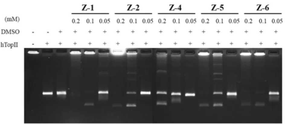
Given that vast majority of compounds, covering a number of ellipticine derivatives effective on tumor cells were reported to exert their effects through interfering with topoisomerase II reactions we screened the activities of the derivatives against DNA topoisomerase II reactions (Senarisoy et al. 2013; Ashley and Osheroff 2014; Miller et al. 2012; McClendon and Osheroff2007). We therefore employed decatenatiation, intercalation, and cleavage assays. Decatenation assay is specific for topoisomerase II activity. It is based on the detection of kinetoplast DNA, the mitochondrial DNA of Crithidia fasciculata found as a catenated network of DNA rings (Senarisoy et al.2013). All of the derivatives, Z-1, Z-2, Z-5, and Z-6, inhibited dec- atenation activity of topoisomerase II at 0.2 mM con- centration. Among the compounds, Z-1 and Z-6 (Fig. 2, lanes 5 and 17, respectively), and Z-2 (Fig.2, lane 8) par- tially retained inhibition at 0.1 mM concentration. Z-1 was found to be the most effective one in repeated densitometric analyses by revealing an avarage IC50 of 16μM. On the other hand Z-2 and Z-6 gave rise to average IC50 values of 30 and 20μM, respectively while Z-4 and Z-5 were rela- tively less effective as the interference caused by these compounds were not detectable upon dilution. Further dilutions revealed disappearance of the interference on decatenations (Fig.2, lanes 6, 9, 12, 15 and 18 for Z1, Z3, Z4, Z5, and Z6, respectively). These results correlated with supercoil relaxing activity of DNA topoisomerase II using supercoiled plasmid DNA substrate, pBR322 (data not shown).
Although decatenation assay is a reliable method to investigate the effect of a given compound on human topoisomerase II, it is not solely informative about their mode of action. We next determined if the detected activ- ities of the compounds were because of the formation of DNA strand breaks. We incubated the pBR322 and the alfa and beta isoforms of the enzyme in the presence of test compounds and terminated the reactions using SDS and then monitored DNA breaks following Proteinase-K treat- ment. Figure3 shows a representative result obtained with the compounds and the control agents, Etoposide and mAMSA with both topoisomerase IIαand βisoforms (Fig.
3 upper and lower panels, respectively). Supercoiled pBR322 substrate (Fig. 3, upper and lower panels lane 1) was partially relaxed with topoisomerase IIα(Fig. 3, upper Table 1 The IC50values determined from cytotoxicity assays
Compounds Calculated IC50values (µM)
HeLa SiHa MCF-7 MDA-MB-231
E 3.13 2.06 2.46 2.30
Z-1 0.06 0.63 0.91 0.77
Z-2 0.09 0.46 0.42 0.34
Z-4 54.39 19.21 4.33 27.09
Z-5 3.68 8.53 3.63 3.57
Z-6 2.42 4.92 1.72 2.99
Cisplatin 12.43 7.84 5.78 19.13
panel, lane 2) andβ(Fig.3, lower panel, lane 2) without an effect caused by DMSO (Fig. 3, upper and lower panels lane 3). None of the compounds (Fig. 3, upper and lower panels lanes 5, 6, 7, 8 and 9 for MK-1, -2, -4, -5 and -6, respectively) and parental ellipticine (Fig. 3, upper and lower panels lane 10) gave rise to a double strand breakage pattern with the method we employed while both Etoposide and mAMSA gave rise to linear DNA bands (Fig.3, upper and lower panels lanes 11 and 12) with a migration patter
similar linearized pBR322 (Fig.3, upper and lower panels lane 4).
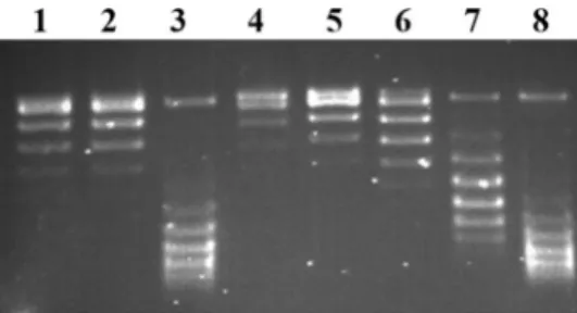
We, then, investigated the intercalative potential of the compounds through the shift of DNA bands from relaxed to negatively supercoiled DNA populations. As seen in Fig.4, the test compounds Z1 and Z6 showed a remarkable inter- calation (Fig. 4. Lanes 3 and 7, respectively) somewhat similar to the intercalation of Etd-Br (Fig. 4, lane 8). The band shifting patterns for Z2, Z4, and Z5 were neglectable Fig. 2Representative Topoisomerase II alfa decatenation assay in the
presence of test compounds. Agarose gel photograph indicates the effect of the compounds Z-1, -2, -4, -5 and -6 on decatenation activity of human topoisomerase II. Lane 1, kDNA; lane 2, kDNA with 1 u topo II; lane 3, the same as lane 2 in the presence of DMSO, lanes 4–6,
decatenation assays in the presence of 0.2, 0.1 and 0.05 mM Z1, respectively. The other test compounds, Z2 (lanes 7–9), Z4 (lanes 10–12), Z5 (lanes 13–15) and Z6 (lanes 16–18) were assessed at the same concentrations
Fig. 3 A representative DNA strand break assay in the absence or the presence of test
compunds. The assays were carried out either with human topoisomerase IIα(upper panel) orβ(lower panel) isoform. The numbers correspond to Z-1, Z-2, Z-4, Z-5, and Z-6, respectively.
See text for detailed description of applications
as they were comparable with relaxed pBR322 (Fig.4, lane 1) with human topoisomerase II (Fig.4, lane 2).
We next used in silico simulations for five molecules using Glide/IFD, Glide/QPLD, Glide/SP, and Glide/XP docking protocols. Four standard inhibitors were also stu- died for comparison. Table2shows top docking scores of the test compounds at the binding pocket of human DNA topoisomerase II alpha. Different docking algorithms were used in docking simulations. Although GOLD Fitness and ChemScore functions were not able to distinguish the docking scores significantly between the studied molecules as well as ellipticine, molecules had similar binding inter- actions with ellipticine and they have betterfitness scores compared with positive control. Glide/SP and XP scoring functions did not result in dramatic scoring difference within the compounds set and reference molecules. How- ever, all docking algorithms showed that the molecules have similar or better docking scores than reference molecules.
Higher docking scores of compound Z-1 compared with reference molecules were observed in IFD and QPLD docking. It’s known that both IFD and QPLD have better
performances compared with Glide/SP and XP modules. An induced fit protocol is used to assist with increasing the flexibility of atoms of the target protein at the active site and thus accurately locates the ligand with a reasonable orien- tation. Successful docking approaches require the accurate calculations and modeling of the partial atomic charges that ligand is bearing. Quantum mechanics (QM) modeling may give the highest level of docking precision when specifi- cally, the induced charge polarization of the binding pocket residues was considered. For the underlying consideration regarding accuracy of docking results, QPLD, based on ab initio charge calculations was used. The 2D and 3D ligand interaction diagrams of Z-1 at the binding site. Crucial amino acids were found as Glu461, Gly462, Asp463, Leu486, Arg487, Gly488, Tyr757, His759, Gly760, Glu761, Met762, Ser763, Leu764, and Met766. When Z1, Z4, and Z5 docking poses were compared, it can be seen that bioactive conformation of Z1 is totally different than Z4 and Z5 (Fig. S1, Supporting Materials).
MD simulations were performed for Z1 and ellipticine to see the structural and dynamical profiles throughout the simulations. (Figs. S2, S3, Supporting Materials). RMSD and RMSF plots were compared for Z1 and ellipticine in Fig. S2. Binding interactions analyses of Z1 showed that especially two residues Met762 and Met766 are crucial for ligand-target interactions.
Pharmacokinetic profiles of studied molecules Z-1, Z-2, and Z-6 were also performed by binary QSAR models of MetaCore/MetaDrug platform. Results showed that within 26 toxicity QSAR models, Z-1 and Z-2 showed toxicity in only three models (AMES, Anemia, and cardiotoxicity).
Compound Z-6 showed toxicity profile only in four models (AMES, Anemia and liver cholestasis and neurotoxicity).
However, it must be noted that three out of four results of toxic profiles in compound Z6 were in near threshold (0.5).
Toxicity predictions were also compared with known Fig. 4 DNA intercalation assay. Intercalation by ellipticine derivatives
was based on the detection of the band shift from relaxed to negatively supercoiled DNA. Assays that contained only the relaxed DNA sub- strate or relaxed DNA and topoisomerase I with no compound are shown in lanes 1 and 2, respectively. The derivatives Z-1, Z-2, Z-4, Z- 5, and Z-6 are applied on the lanes 3–7, respectively. Strong inter- calator agent, Et-Br is shown at the lane 8
Table 2 Docking scores of the test compounds (kcal/mol)
Compounds IFD
Docking score
QPLD Docking score
Glide/XP Docking score
Glide/SP Docking score
GOLD Docking score Fitness score Chem
score (kcal/
mol)
Z-1 −12.378 −7.428 −4.766 −7.344 41.21 −5.22
Z-2 −12.337 −6.917 −5.888 −6.911 45.38 −6.14
Z-4 −11.616 −7.208 −4.491 −7.025 41.85 −5.18
Z-5 −12.521 −7.288 −5.860 −7.043 46.12 −5.21
Z-6 −12.306 −7.061 −6.293 −7.345 43.60 −4.71
Ellipticine −8.901 −6.952 −6.428 −8.420 34.54 −5.38
Ciprofloxacin −6.246 −6.431 −6.467 −6.680 NA NA
Ofloxacin −6.358 −6.523 −4.909 −7.140 NA NA
Mitonafide −5.107 −5.893 −5.010 −6.450 NA NA
inhibitor ellipticine. Ellipticine showed toxicity in five models (AMES, carcinogenicity in male mouse, carcino- genicity in rat female, cardiotoxicity, and genotoxicity) out of 26 different toxicity QSAR models (Table S1, Support- ing Materials).
Taken together, our results showed that the com- pounds Z1, Z2, and Z6 have significant potential as compounds targeting human DNA topoisomerase II.
Given that the alfa and beta isoforms of human DNA topoisomerase II is expressed in a different pattern in normal and cancer cells, differentiating between these two isoforms is an important aspect of topoisomerase- targeting drug development. However, the absence of DNA breaks in either isoform when compared to the reference compounds revealed that ellipticine derivatives could affect inhibition of catalytic activity rather than stabilizing the covalent complex.
Data accessibility
The datasets supporting this study have been uploaded as part of the electronic supplementary material.
Acknowledgements This study is supported by the grant of TUBI- TAK 115Z349 (PI; ZT).
Author contributions MK, enzyme assays; VA, synthesis and char- acterization of test compounds; AK and IZ, mammalian cell viability assays; MZ and KS, in silico analyses; SD, in silico analyses and editing manuscript; HO, consultation and data analyses; YE, designing experiments, synthesis and characterization of test compounds, inter- pretation of data; SZ, designing experiments, enzyme assays, editing the manuscript; ZT, designed the project, enzyme assays, interpretation of data and writing manuscript.
Compliance with ethical standards
Conflict of interest The authors declare that they have no conflict of interest.
Publisher’s note Springer Nature remains neutral with regard to jurisdictional claims in published maps and institutional affiliations.
References
Ashley RE, Osheroff N (2014) Natural products as topoisomerase II poisons: effects of thymoquinone on DNA cleavage mediated by human topoisomerase IIα. Chem Res Toxicol 27:787–793 Champoux JJ (2001) DNA topoisomerases: structure, function, and
mechanism. Annu Rev Biochem 70:369–413
Chikamori K, Grozav AG, Kozuki T, Grabowski D, Ganapathi R, Ganapathi MK (2010) DNA topoisomerase II enzymes as mole- cular targets for cancer chemotherapy. Curr Cancer Drug Tar 10:758–777
Coban G, Zencir S, Zupkó I, Réthy B, Gunes HS, Topcu Z (2008) Synthesis and biological activity evaluation of 1H- benzimidazoles via mammalian DNA topoisomerase I and cytostaticity assays. Eur J Med Chem 44:2280–2285
Deane FM, O’Sullivan EC, Maguire AR, Gilbert J, Sakoff JA, McCluskey A, McCarthy FO (2013) Synthesis and evaluation of novel ellipticines as potential anti-cancer agents. Org Biomol Chem 1:1334–1344
Delgado JL, Hsieh CM, Chan NL, Hiasa H (2018) Topoisomerases as anticancer targets. Biochem J 475:373–398
Desmond molecular dynamics system, D.E.S.R., New York, NY, 2017.
Fosse P, Rene B, Charra M, Paoletti C, Saucier J (1992) Stimulation of topoisomerase II-mediated DNA cleavage by ellipticine deriva- tives structure–activity relationship. Mol Pharm 42:590–595 Friesner R, Banks JL, Murphy RB, Halgren TA, Klicic JJ, Mainz DT,
Repasky MP, Knoll EH, Shelley M, Perry JK, Shaw DE, Francis P (2004) Glide: a new approach for rapid, accurate docking and scoring. 1. Method and assessment of docking accuracy. J Med Chem 47:1739–1749
Garbett NC, Graves DE (2004) Extending nature’s leads: the antic- ancer agent ellipticine. Curr Med Chem Anticancer Agents 4:149–172
Gul HI, Cizmecioglu M, Zencir S, Gul M, Canturk P, Atalay M, Topcu Z (2009) Cytotoxic activity of 4’-hydroxychalcone derivatives against Jurkat cells and their effects on mammalian DNA topoi- somerase I. J Enzym Inhib Med Chem 24:804–807
Hoover WG (1985) Canonical dynamics: equilibrium phase-space distributions. Phys Rev A 31:1695–1697
Iacopetta D, Rosano C, Puoci F, Parisi OI, Saturnino C, Caruso A et al.
(2017) Multifaceted properties of 1,4dimethylcarbazoles: focus on trimethoxybenzamide and trimethoxyphenylurea derivatives as novel human topoisomerase II inhibitors. Eur J Pharm Sci 96:263–272
Ketron AC, Osheroff N (2014) Phytochemicals as anticancer and chemopreventive topoisomerase II poisons. Phytochem Rev 13:19–35
Kuo PL, Hsu YL, Chang CH, Lin CC (2005) The mechanism of ellipticine-induced apoptosis and cell cycle arrest in human breast MCF-7 cancer cells. Cancer Lett 223:293–301
Lichota A, Gwozdzinski K (2018) Anticancer activity of natural compounds from plant and marine environment. Int J Mol Sci 19:1–38
Martyna GJ (1994) Remarks on “Constant-temperature molecular dynamics with momentum conservation”. Phys Rev E 50:3234–3236
McClendon AK, Osheroff N (2007) DNA topoisomerase II, geno- toxicity, and cancer. Mutat Res 623:83–97
Metacore/Metadrug platform, Clarivate Analytics (2019)https://portal.
genego.com/
Miller CM, O’Sullivan EC, Devine KJ, McCarthy. CM (2012) Synthesis and biological evaluation of novel isoellipticine deri- vatives and salt. Org Biomol Chem 10:7912–7921
Moody DL, Dyba M, Kosakowska-Cholody T, Tarasova NI, Michejda CJ (2007) Synthesis and biological activity of 5-aza-ellipticine derivatives. Bioorg Med Chem Lett 17:2380–2384
Mosmann T (1983) Rapid colorimetric assay for cellular growth and survival: application to proliferation and cytotoxicity assays. J Immunol Methods 65:55–63
Rouëssé J, Spielmann M, Turpin F, Le Chevalier T, Azab M, Mondésir JM (1993) Phase II study of elliptinium acetate salvage treatment of advanced breast cancer. Eur J Cancer 29:856–859
Senarisoy M, Canturk P, Zencir S, Baran Y, Topcu Z (2013) Gossypol interferes with both type I and type II topoisomerase activities without generating strand breaks. Cell Biochem Biophys 66:199–204
Thompson D, Miller C, McCarthy FO (2008) Computer simulations reveal a novel nucleotide-type binding orientation for ellipticine- based anticancer c-kit kinase inhibitors. Biochemistry 47:10333–10344
Topcu Z (2001) DNA topoisomerases as targets for anticancer drugs. J Clin Pharm Ther 26:405–416
Topcu Z, Ozturk B, Kucukoglu O, Kilic E (2008) Inhibition of mammalian type I DNA topoisomerase by Helichrysum pam- phylicum is correlated with the total antioxidant capacities of its individualflavonoids. Z Naturforsch C 63:69–74
Vann KR, Ekiz G, Zencir S, Bedir E, Topcu Z, Osheroff N (2016a) Effects of secondary metabolites from the fungus Septofusidium berolinense on DNA cleavage mediated by human topoisomerase IIα. Chem Res Toxicol 29:415–420
Vann KR, Ergun Y, Zencir S, Oncuoglu S, Osheroff N, Topcu Z (2016b) Inhibition of human DNA topoisomerase IIαby two novel ellipticine derivatives. Bioorg Med Chem Lett 26:1809–1812
Verdonk ML, Cole JC, Hartshorn MJ, Murray CW, Taylor RD (2003) Improved protein-ligand docking using GOLD. Proteins 52:609–623
Wang JC (1971) Interaction between DNA and anEscherichia coli protein omega. J Mol Biol 55:523–533
Wendorff TJ, Schmidt BH, Heslop P, Austin CA, Berger JM (2012) The structure of DNA-bound human topoisomerase II alpha:
conformational mechanisms for coordinating inter-subunit inter- actions with DNA cleavage. J Mol Biol 424:109–124
Zupkó I, Molnár J, Réthy B, Minorics R, Frank E, Wölfling J et al.
(2014) Anticancer and multidrug resistance-reversal effects of solanidine analogs synthetized from pregnadienolone acetate.
Molecules 17:2061–2076