Tandem mass spectrometric study of annelation isomers of the novel thieno[3 ′ ,2 ′ :4,5]pyrido[2,3- d ]pyridazine ring system
Gábor Krajsovszky,a Eszter Tóth,b Krisztina Ludányic*
aDepartment of Organic Chemistry, Semmelweis University, Hőgyes E. u. 7, H-1092 Budapest, Hungary
bMS Proteomics Laboratory, Institute of Organic Chemistry, Research Centre for Natural Sciences, Hungarian Academy of Sciences
H-1117 Budapest, Magyar tudósok körútja 2, Hungary
cDepartment of Pharmaceutics, Semmelweis University, Hőgyes E. u. 7, H-1092 Budapest, Hungary
E-mail: ludanyi.krisztina@pharma.semmelweis-univ.hu DOI: http://dx.doi.org/10.3998/ark.5550190.0015.500
Abstract
In the present communication, we describe tandem combinations of Suzuki – condensation ring closure reactions to obtain a novel thieno[3′,2′:4,5]pyrido[2,3-d]pyridazine ring system. These compounds were studied by tandem mass spectrometry in detail and characteristic fragmentations were observed, which can be useful to confirm the presence of N-substituted pyridazine ring in synthesized compounds.
Keywords: Halopyridazin-3(2H)-one, Suzuki – condensation tandem reactions, mass spectrometry, organic fragmentations, thieno[3′,2′:4,5]pyrido[2,3-d]pyridazines
Introduction
In our previous studies1-4 we reported the synthesis of fused nitrogen-containing ring systems, several tandem reactions included.5-7 In this paper the application of Suzuki – condensation ring closure tandem reaction is described for the synthesis of a novel thieno[3′,2′:4,5]pyrido[2,3- d]pyridazine ring system. Among starting material of our previous works also 5-azido-4- bromopyridazin-3(2H)-ones derivatives have been applied for synthesis route azide→iminophosphorane→Suzuki-aza-Wittig tandem reaction. The 4-halo-5-iminophosphoranes were prepared from the reaction of the related azides with triphenylphosphine and followed by the reaction with 2-formylarylboronic acid to obtain a tricyclic ring system. The 5-azido-4-halo precursors could be selectively obtained from dihalo compounds. On the contrary, the 4-azido-5- bromopyridazin-3(2H)-ones regioisomers could not be prepared (under apolar conditions by sodium
azide, anhydrous toluene, 15-crown-5, heating) from the appropriate 4,5-dibromopyridazine derivatives, so the other regioisomer of this previously tricyclic system could not be obtained from haloazide precursor. That is why haloamines of pyridazinones were applied in our present work as starting materials, which could be synthesized from the appropriate dibromo-pyridazinones. After reaction with aqueous ammonia the regioisomer haloamines could be separated by recrystallization, respectively by flash chromatography purification of the mother liquor. From these two isolated isomers were carried out separately Suzuki-condensation tandem reactions for synthesis of two regioisomers of novel thieno[3′,2′:4,5]pyrido[2,3-d]pyridazine ring system.
Moreover, MS investigations of the obtained target compounds were performed, similar to published MS study.8 Main aim of these examinations is the observation of the similarity and differences in LC-MS behaviour of these two tricyclic regioisomers. Following some relevant articles have been shown the different cleavages of pyridazine derivatives. The ring contraction reactions of pyridazines were supposed by density functional theory computations.9 Other papers are concerning on thermal cleavages and photolysis of pyridazines10 as well as mass spectrometric investigation of pyridazine derivatives, whereby fragments were identified, which have been produced by UV photons.11
Results and Discussion
(i) Synthesis of target compounds
In this case haloamines 2 and 3 - obtained in first step from the N-benzyl-4,5-dibromopyridazinone 1 through halogen displacements by ammonia - were transformed to thieno[3′,2′:4,5]pyrido[2,3- d]pyridazines 4 and 5 via Suzuki – condensation tandem reaction using 2-formyl-3-thiophene- boronic acid12 (Scheme 1).
N N
O Br
Br
N N
O Br
NH2
N N
O NH2
Br
N N
O
N S
N N
O N
S S
O B O H O H S
O B O H O H
+
1 2 3
4 5
NH4OH
Pd[PPh3]4 Pd[PPh3]4
DME, Na2CO3 DME, Na2CO3
1 2 3
4 4a
5 6
7 6a 9 8 9a 9b
1 2 3
4 4a
5 6 6a
7
9 8 9a 9b 1'
2' 3' 4'
5' 6'
1' 2' 3' 4'
5' 6'
Scheme 1. Halogen displacements on halopyridazinones by ammonia and subsequent Suzuki- condensation tandem reactions of pyridazinones.
(ii) HPLC-MS and MS/MS analysis of the target compounds
HPLC-MS analysis was performed to investigate the purity of compounds 4 and 5 (we found that the purity > 99%). Mass spectra of the two compounds were recorded with the aim of distinguishing the isomers from each other. On the chromatogram of isomer 4 the retention time of this compound is 8.07 min. On the mass spectrum m/z=294 belongs to this peak.
On the chromatogram of compound 5, the retention time of this compound is 6.33 min, m/z=294, m/z=316, m/z=587 and m/z=609 belong to this peak. The main difference between the mass spectra of the two isomers is the presence of some special ions in the spectrum of 5 besides the main signal. The main components are the sodium adduct of 5 (m/z=316), the protonated dimer of 5 (m/z=587), and the sodium adduct of this dimer (m/z=609). These adducts have significant signals besides the signal of compound 5 and they can be found only as minor components in the spectrum of the other isomer (4).
HPLC-MS/MS spectrum of compound 4 displays m/z=189 as a main fragment and some minor fragments e.g. m/z=106, m/z=135, m/z=147, m/z=175, which are not observed in the spectrum of compound 5. The structure of this fragment and its exact mass are shown in Scheme 2.
The components have been identified with 3 ppm mass accuracy.
Scheme 2. The mass spectrometric fragmentation process of isomer (4).
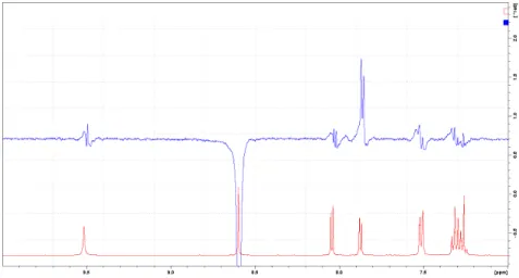
These MS data are according with the results of NMR structure determination. The signal assignment was based on 1H, 13C, HSQC and HMBC NMR experiments. The differentiation of the two regioisomers was based on NOE difference experiment. In case of compound 5, H-1 was irradiated. In the difference spectrum NOE intensity increasement was observed on H-9. (Figure 1.) An additional proof is the chemical shift difference of H-9s in the two regioisomers. In case of compound 4, where H-9 is in the vicinity of the carbonyl group a huge downfield chemical shift (0.92 ppm) can be observed. (Figure 2.)
Figure 1. NOE difference spectrum of compound 5 (H-1 was irradiated).
Figure 2. 1H NMR spectra of compound 4 and 5 (aromatic section).
(iii) Tandem mass spectrometric study of compounds 4, 5 and of further isomers
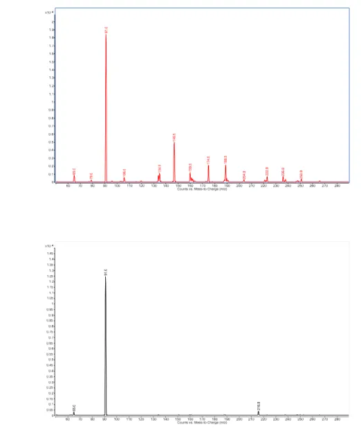
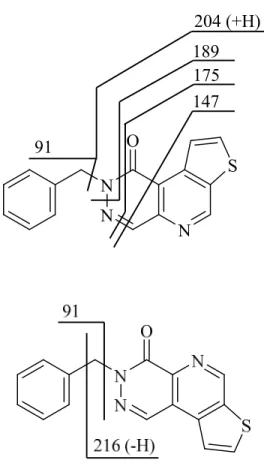
The fragmentation behaviour of the isomers (compounds 4 and 5) have been studied by tandem mass spectrometry (Figure 3.). For both isomers poor fragmentation was observed, the benzyl cation formation (m/z=91) is the largest peak in the tandem mass spectra. There are, however, significant difference in the fragmentation pattern of the isomers close to the N-2 atoms. Schematic diagram of the tandem mass spectrometric fragmentation of isomers is shown on Figure 4.
Compound 4 relatively easily forms cross ring fragments (m/z=189, m/z=147), which cannot be observed in the case of compound 5. On the other hand, fragment m/z=216 can only be noticed in the case of compound 5, which is probably produced by cleavage of the aromatic ring. These fragments make it possible to discriminate the 2 isomers on the basis of their tandem mass spectrometric behaviour.
LC-MS study of compounds 4 and 5 shows, that the isomers can be discriminated by their retention time on reversed phase HPLC and these results confirms that by tandem mass spectrometry as well.
3a
3b
Figure 3. Tandem mass spectra of isomers (Figure 3a: compound 4, Figure 3b: compound 5).
4a
N N
O
N S 91
204 (+H) 189 175 147
4b
Figure 4. Fragmentation schemes of isomers (Figure 4a: compound 4, Figure 4b: compound 5).
It is instructive to compare the fragmentation behaviour of compound 4 and 5 to other similar compounds (Figure 5.). The benzyl group containing compounds (compound 61,5 and 71,5) show similar fragmentation. The formation of benzyl cation (m/z=91) is the highest peak in the tandem mass spectra and its counter ion can also be observed in the case of compound 6 (m/z=198).
The location of the CO group significantly determines the fragmentation behaviour of the compounds. If the O is connected to the position 1 (eg. compound 6 and 4) intensive cross ring fragments are formed. If the O is at the position 4 (eg. compound 7 and 5) no cross ring fragments can be observed.
On the other hand if the benzyl group is missing (compounds 81,5 and 91,5) the fragmentation pattern of the 2 molecules are similar (m/z=155, m/z=153, m/z=141, m/z=128), but the intensities of fragment ions are completely different and the two isomers can also be distinguished. The peak at m/z=128 is the most intense peak in the case of compound 8, while m/z=153 is the base peak in the tandem mass spectrum in the case of compound 9.
These special characteristics of the tandem mass spectra can be utilized for the distinction of various regioisomers.
5a. 2-Benzylpyridazino[4,5-c]isoquinolin-1(2H)-one.
CH2
N O
N
N 198 (+H)
91
141 (-H) 129
5b. 3-Benzylpyridazino[4,5-c]isoquinolin-4(3H)-one.
5c. 2-Methylpyridazino[4,5-c]isoquinolin-1(2H)-one.
5d. 3-Methylpiridazino[4,5-c]isoquinolin-4(3H)-one.
CH3
N
O
N N
153 (-2H) 155 141 128
Figure 5. Fragmentation schemes of isomers (Figure 5a: compound 6, Figure 5b: compound 7, Figure 5c: compound 8, Figure 5d: compound 9).
Conclusions
We applied Suzuki cross-coupling methodology for the facile synthesis of a novel thieno[3′,2′:4,5]pyrido[2,3-d]pyridazine ring system, and elaborated an LC-MS investigation procedure for the regioisomer compounds. The main aim of the present manuscript was to investigate the fragmentation behaviour of fused pyridazine type compounds. Major fragment of isomer 4 (m/z=189), which characterizes the fragmentation of pyridazine part, was not observed in the case of isomer 5. The number of fragments and mass of pyridazine ring fragments largely depend on the attachment of thienopyridine moiety. The isomer 4 showed a high degree of fragmentation as compared to the isomer 5. In the case of the other isomer pair (compounds 8, 9) similar fragmentation could be observed during the cleavage of pyridazin part (m/z=155, m/z=141, m/z=128), which represents methyl substituent containing pyridazine ring. In contrast, benzyl substituent containing isomers (compounds 6 and 7) showed far less fragmentation compared to the N-methyl substituted analogue compounds. This method can be used for study of substituent- dependent, N-substituted pyridazine rings and for confirmation of the presence of pyridazine ring in synthesized compounds.
Experimental Section
General. 1H and 13C-NMR spectra were recorded on a Varian MERCURY plus (1H NMR 400 MHz; 13C NMR 100 MHz) spectrometer in the solvent indicated at room temperature, using the 2H signal of the solvent as the lock. The 1H and 13C chemical shifts were referred to TMS (δTMS=0 ppm). Chemical shifts (δ) are given in ppm and coupling constant (J) values in Hz. The structures of
compounds were elucidated using COSY, HSQC, HMBC (NMR) methods. Melting points were determined in a Büchi B-540 Melting Point apparatus and the values are uncorrected. IR spectra were recorded in potassium bromide pellets with a Perkin-Elmer 1600 FT-IR spectrophotometer.
Solvent mixtures used for chromatography are always given in a vol/vol ratio. HPLC purity measurements were run on a JASCO LC-Net II/ADC system with a JASCO MD-2010 Plus diode array detector, on a Gemini NX C18 RP 25 cm × 4.6 mm 5 µm column, if not indicated otherwise, with the conditions given below (flow rate: 0.5 mL/min). Commercially available solvents were purified by standard procedures prior to use, whereas reagents (Reanal, Budapest or Sigma-Aldrich Kft., Budapest) were used as received. Standard flash chromatography on silica (Kieselgel 60, Aldrich, 0.040-0.063 mm) and/or recrystallization as indicated, was applied for purification of crude products. Thin layer chromatography was done on commercially available silica plates (Silica gel 60 F254, Merck).
Compounds 1,13 2,14 and 35 were synthesized according to the literature.
General procedure for the synthesis of 4, 5 via Suzuki-reaction from the appropriate haloamines. A round-bottom flask was purged with argon and charged with the amino derivative 2 or 3 (1.05 g, 3.70 mmol), tetrakis(triphenylphosphine)-palladium(0) (0.27 g, 0.23 mmol), and dry 1,2-dimethoxyethane (40 mL). While stirring, the mixture was flushed with argon for approximately 20 min. Subsequently 2-formyl-3-thiophene-boronic acid (0.88 g, 5.64 mmol) and sodium carbonate solution (2 M, 3.5 mL) were added, and the reaction mixture was heated at 110 °C for 12 hours. The reaction mixture was poured onto ice-water (25 mL), was extracted with dichloromethane (3x25 mL) and the combined organic layers were dried over anhydrous sodium sulphate. The solvent was evaporated in vacuum and the residue after grinding by diethyl-ether was purified by flash column chromatography and crude solid product was recrystallized from dry ethanol.
2-Benzylthieno[3′,2′:4,5]pyrido[2,3-d]pyridazin-1(2H)-one (4). Starting from 5-amino-2-benzyl- 4-bromopyridazin-3(2H)-one (2)14, eluent used for flash column chromatography: chloroform. Pale brown crystals, yield 40%, 0.434 g, mp 142.2-142.8°C; Rf (chloroform): 0.22. IR (νmax, cm-1): 3426, 3114, 3062, 2924, 2856, 1646, 1488, 1452, 1314, 1264, 1216, 750, 696. δH (CDCl3 400 MHz): 9.47 (s, 1H, H-6), 8.80 (d, J8,9 6 Hz, 1H, H-9), 8.57 (s, 1H, H-4), 8.05 (d, 1H, H-8), 7.50 (d, J2′,3′ 12 Hz, 2H, H-2′,6′), 7.34 (m, 2H, H-3′,5′), 7.28 (m, 1H, H-4′), 5.51 (s, 2H, CH2); δC (CDCl3 100 MHz):
159.9 (C-1), 150.7 (C-6), 143.4 (C-4a), 142.3 (C-9a)*, 140.2 (C-4), 139.4 (C-6a)*, 137.3 (C-1′), 136.0 (C-8), 129.3 (C-3′,5′), 129.2 (C-2′,6′), 128.5 (C-4′), 126.3 (C-9), 119.8 (C-9b), 55.7 (CH2). H′, C′ are phenyl protons and carbons. MS: 189, 294. HRMS (ESI): C16H11N3OS [M+H]+: calculated 294.0701; measured 294.0702.
*signals are interchangeable
3-Benzylthieno[3′,2′:4,5]pyrido[2,3-d]pyridazin-4(3H)-one (5). Starting from 4-amino-2-benzyl- 5-bromopyridazin-3(2H)-one (3)5, eluent used for flash column chromatography: chloroform:ethyl acetate=4:1. Pale gray crystals, yield 30%, 0.326 g, mp 258-258.5°C; Rf (chloroform:ethyl acetate=4:1): 0.22. IR (νmax, cm-1): 3074, 3034, 2950, 1654, 1584, 1546, 1492, 1458, 1430, 1356, 1196, 760, 694. δH (CDCl3 400 MHz): 9.50 (s, 1H, H-6), 7.86 (d, J8,9 6 Hz, 1H, H-9), 8.60 (s, 1H,
H-1), 8.03 (d, 1H, H-8), 7.52 (d, J2′,3′ 12 Hz, 2H, H-2′,6′), 7.32 (m, 2H, H-3′,5′), 7.27 (m, 1H, H-4′), 5.54 (s, 2H, CH2); δC (CDCl3 100 MHz): 159.8 (C-4), 149.1 (C-6), 140.4 (C-9a)*, 140.2 (C-6a)*, 139.8 (C-4a), 137.3 (C-1′), 136.0 (C-8), 134.2 (C-1), 129.5 (C-2′,6′), 129.2 (C-3′,5′), 128.5 (C-4′), 121.8 (C-9b), 121.4 (C-9), 55.6 (CH2). H′, C′ are phenyl protons and carbons. MS: 189, 294, 316, 587, 609. HRMS (ESI): C16H11N3OS [M+H]+: calculated 294.0701; measured 294.0703.
*signals are interchangeable
HPLC-MS measurement
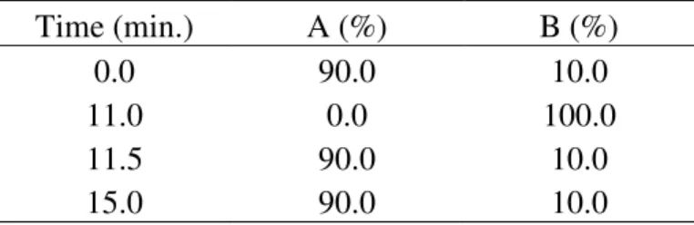
The two isomers were separated before the MS measurement by AcquityTM UPLC System (Waters, Milford, MA, USA), which consisted of a column manager [column: Acquity BEH 1.7 µm, C18 1.0 x 150 mm (Waters, Milford, MA, USA)], sample manager, binary solvent manager, photodiode array detector (wavelength here: 210–450 nm DAD) and MassLynx Software 4.1. The concentration of the sample was ~ 0.2 mg/mL, the solvent was in both cases acetonitrile:water = 50:50. Sample injection volume was 5 µL, the flow rate was 0.10 mL/min and the run time was 15 min. Gradient elution was applied, eluent A contained 85% water, 5% acetonitrile and 10% 0.1 M ammonium acetate solution, eluent B contained 90% acetonitrile and 10% 0.1 M ammonium acetate solution. The gradient table can be seen in Table 1.
Table 1. Gradient table of the title 4, 5 (from each other separated) compounds
Time (min.) A (%) B (%)
0.0 90.0 10.0
11.0 0.0 100.0
11.5 90.0 10.0
15.0 90.0 10.0
Mass spectra were taken by Waters Q-TOF Premier mass spectrometer (Waters, Milford, MA, USA). Ionization mode was ES+, V-mode, capillary voltage: 2800 V, sampling cone: 30 V, extraction cone: 3.5 V, desolvation temperature: 500 °C, source temperature 120 °C. The scan time was by MS1 1.0 s, the inter scan delay 0.1 s, by MS2 0.3 s, and 0.05 s. Collision energy by MS1 was 2.5 V, by MS2 15 – 30 V.
MS/MS measurement
Tandem mass spectrometric measurements were made with an Agilent 6460 QQQ instrument. Jet Stream ESI ion source was used in positive mode. Fragmentor voltage: 135 V, gas temperature: 300
°C, gas flow rate: 13 l/min, nebulizer gas flow: 45 psi, sheathgas flow: 11 l/min, sheathgas temperature (heater) 400 °C, capillary voltage was 3500 V. The collision energy was 30 eV in both cases.
Acknowledgements
The authors gratefully thank to Prof. Péter Mátyus for his continual support and guidance throughout this work, to Tamás Gáti for recording the NMR spectra, as well as for helpful discussions, to László Drahos for HRMS measurements, and to Péter Tétényi for recording the IR spectra.
Financial support by the Hungarian Scientific Research Fund (K73389), the Medical Research Council (ETT 099-03/2009) and the National Development Agency (TÁMOP-4.2.1/B-09/1/KMR- 2010-0001) is gratefully acknowledged.
References
1. Riedl, Zs.; Maes, B. U. W.; Monsieurs, K.; Lemiére, G. L. F.; Mátyus, P.; Hajós Gy.
Tetrahedron 2002, 58, 5645.
http://dx.doi.org/10.1016/S0040-4020%2802%2900531-8"
2. Mátyus, P.; Maes, B. U. W.; Riedl, Zs.; Hajós, Gy.; Lemiére, G. L. F.; Tapolcsányi, P.;
Monsieurs, K.; Éliás, O.; Dommisse, R. A.; Krajsovszky, G. Synlett 2004, 7, 1123. See references therein.
http://dx.doi.org/10.1055/s-2004-822894
3. Riedl, Zs.; Monsieurs, K.; Krajsovszky, G.; Dunkel P.; Maes, B. U. W.; Tapolcsányi, P.; Egyed, O.; Boros, S.; Mátyus, P.; Pieters, L.; Lemière, G. L. F.; Hajós, Gy. Tetrahedron 2006, 62, 121.
http://dx.doi.org/10.1016/j.tet.2005.09.136
4. Dajka-Halász, B.; Földi, Á. A.; Ludányi, K.; Mátyus, P. Arkivoc 2008, (iii), 102.
http://www.arkat-usa.org/get-file/22943/
5. Krajsovszky, G.; Károlyházy, L.; Dunkel, P.; Boros, S.; Grillo A.; Mátyus, P. Arkivoc 2011, (x), 229.
http://dx.doi.org/10.3998/ark.5550190.0012.a19 6. Fresneda, P. M.; Molina, P. Synlett 2004, 1.
http://dx.doi.org/10.1055/s-2003-43338
7. Molina, P.; Vilaplana, M. J. Synthesis 1994, 1197.
http://dx.doi.org/10.1055/s-1994-25672
8. Jerkovich, Gy.; Mátyus, P. Organic Mass Spectrometry 1989, 24, 195.
http://dx.doi.org/10.1002/oms.1210240312 9. Silva, P. J. J. Org Chem. 2012, 77, 4653.
http://dx.doi.org/10.1021/jo300448d
10.Kvaskoff, D.; Bednarek, P.; Wentrup, C. J. Org Chem. 2010, 75, 1600.
http://dx.doi.org/10.1021/jo902570d
11.Ilosera, G. V.; Coreno, M.; Erman, P.; Huels, M. A.; Jakubowska, K.; Kivimäki, A.; Rachlew, E.; Stankiewicz, M. Int. J. of Mass Spectrometry 2008, 275, 55.
http://dx.doi.org/10.1016/j.ijms.2008.05.019
12.Pellicciari, R.; Moroni, F. WO 02/36599 A1 2002. US Patent 2004/82789 A1 2004.
13.Yamasaki, T.; Kawaminami, E.; Yamada, T.; Okawara, T.; Furukawa, M. J. Chem. Soc., Perkin Trans. 1: Org. Bioorg. Chem. (1972-1999) 1991, 991.
http://pubs.rsc.org/en/content/articlelanding/1991/p1/p19910000991#!divAbstract
14.Fischer, A.; Kropp, R.; Reicheneder, F. Ger. Offen. DE 1912770 1970, Chem. Abstr. 1971, 74, 53824f.