XII./2.: Malignant tumours of the nose and paranasal sinuses
Balázs Szabó Md.
XII./2.1.: Characteristic features
Tumours of the nose and paranasal sinuses constitute 10% of all head-neck tumours, in Asia and Africa this type of tumour is more frequent. Men are 1.5 times more affected than women, 80% of the patients are between 45-85 years of age. 60-70% of the tumours affect the maxillary sinus, 20-30% the nasal cavity, 10-15% the ethmoidal cells, the least affected are the frontal sinus and the sphenoidal sinus. The most frequent histological type (80%) is squamous cell carcinoma and its variations , 10-15% of the tumours arise from the small salivary glands and histologically they are adenocarcinoma, mucoepidermoid carcinoma or adenoid cystic carcinoma (Table 1, sinus tumours.) Regional metastasis is rare (20%), generally affecting the I-III.neck region or the retropharyngeal lymph node region. Distant metastasis is also rare, characteristically the lungs, the liver and the bones are affected.
From among the histologically benign tumours inverted papilloma needs to be highlighted. It has a clinically aggressive course and during years (on average 63 months) 10-15%-of the cases turn into planocellular carcinoma.
The 5 years survival rate is about 40% ( at the ethmoidal cell tumours are worse), is mainly due to the frequency of local recurrence.
Nasal cavity:
Incidence in Hungary in 2009: man: 39, woman: 20 (National Cancer Registry)
Mortality in Hungary in 2009: man: 4, woman: 6 (Hungarian Central Statistical Office)
Sinuses:
Incidence in Hungary in 2009: man: 66, woman: 40 (National Cancer Registry)
Mortality in Hungary in 2009: man: 23, woman: 15 (Hungarian Central Statistical Office)
Etiology and risk factors
Smoking and alcohol as risk factors have only a small role. Chronic industrial injury (2-4 decades of exposure) has a major role. Nickel, sawdust, leather,
formaldehyde, mineral oils, chrome, varnish colours, gases and vapours formed when soldering and welding can be associated with epithelial carcinoma, while hard-wood dust can cause ethmoidal cell adenocarcinoma.
The tumour can develop on the basis of chronic rhinitis, recurrent nasal polyosis, inverted papilloma, and the role of EBV, HPV infection needs to be considered too.
XII./2.2.: Clinical symptoms
Obvious symptoms are late signals of primary paranasal tumours. At an early stage tumours are mostly discovered by random checking, when due to an inflammation or trauma led to an examination. Symptoms: nasal bleeding on one side, blocked nose, sinusitis, foetid nasal discharge, headache, swollen palate, disengaging teeth. Locally advanced tumours may often cause upper lip anaesthesy, double vision, proptosis, lockjaw, face and eye swelling or palpable tumour on the neck.
Figure 1.
XII./2.3.: Diagnosis
Diagnostic algorithm during the examination of the nose and paranasal tumours:
History
-Physical examination (general, otorhinolaryngological, dental, protetical)
-Direct endoscopic (fibroscopic) examination of the nasal cavity, the pharynx and the larynx
-Histological examination from the primary tumour, aspiration cytology of the neck metastasis
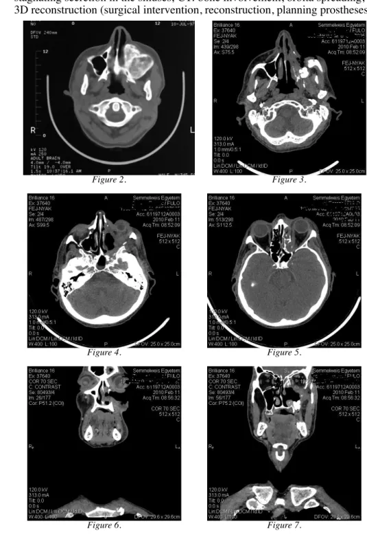
-Imaging examinations: face and skull, neck CT and/or MR, neck US, chest X-ray. (CT), abdominal US, (if needed CT, MR), bone scintigraphy. The use of PET/CT with nose and sinus tumours is still questionable. (In advanced stage tumours PET/CT is part of staging examinations for detecting distant metastases. PET/CT is
recommended in the case of Gr 2-3 histology grade and N 2-3 neck lymph node state. PET/CT is useful for interpreting therapy
response, relapse examination, and restaging, too.)
Most recommendations in the diagnosis of nose and paranasal tumours suggest joint skull CT and MR (MR: perineural, cranial, intracranial, masticator space, parapharyngeal spreading, separating tumour and
stagnating secretion in the sinuses; CT: bone involvement, orbita spreading, 3D reconstruction (surgical intervention, reconstruction, planning prostheses)
Figure 2. Figure 3.
Figure 4. Figure 5.
Figure 6. Figure 7.
Laboratory tests: routine blood count, blood type, detailed coagulogram and clinical chemistry, in the case of non-epithelial carcinoma tumour marker tests are also necessary.
XII./2.4.: Staging (AJCC – TNM staging (7th edition, 2010))
TNM staging
TNM staging for carcinomas. Staging systems are made for sinus maxillaris and ethmoidal region carcinomas only.
T Primary tumour
Tx Primary tumour cannot be assessed T0 No evidence of primary tumour.
Tis Carcinoma in situ
Maxillary sinus
T1 The tumour limited to maxillary sinus mucosa with no erosion or destruction of bone
T2 The tumour causing bone erosion or destruction including
extension into the hard palate and/or the middle nasal meatus, except extension to the posterior wall of the maxillary sinus and pterygoid plates
T3 The tumour invades any of the following: bone of the posterior wall of the maxillary sinus, subcutaneous tissues, the medial wall or floor of the orbit, pterygoide fossa, ethmoidal sinuses.
T4a Moderately advanced local disease. The tumour invades anterior orbital contents, skin of cheek, pterygoid plates, infratemporal fossa, cribriform plate, sphenoid or frontal sinus
T4b Very advanced local disease. The tumour invades any of the following: orbital apex, dura, brain, middle cranial fossa, cranial nerves other than maxillary division of trigeminal nerve(V2) nasopharynx, clivus
Nasal cavitiy and ethmoid sinus
T1 The tumour restricted to any one subsites, with or without bony invasion
T2 The tumour invading two subsites in a single region or extending to involve an adjacent region within the nasoethmoideal complex, with or without bony invasion.
T3 The tumour extends to invade the medial wall or floor of the orbit, maxillary sinus, palate, or cribriform plate
T4a Moderately advanced local disease. Tumour invades any of the following: anterior orbital contents, skin of nose or cheek, minimal extension to anterior cranial fossa, pterygoid plates, sphenoid or frontal sinuses
T4b Very advanced local disease. Tumour invades any of the following: orbital apex, dura, brain, middle cranial fossa, cranial nerves other than (V2), nasopharynx, or clivus
Classification of regional lymph nodes (N) and distant metastases (M), the staging system is the same as written for meso and hypopharynx tumours.
Classification of nasal and sinus tumours according to different other localisations:
On the basis of Öhngren’s line: suprastructural, infrastructural tumours.
Figure 8.
According to Sébileau: lower, middle and high - étage tumours
Figure 9.
XII./2.5.: Therapy
Nasal cavity and sinus tumours when in an advanced state require complex oncotherapy. Planning the therapy the following need to be equally taken into consideration: the advanced state of the illness, the general state of the patient (performance status), personality, and the side effects of the planned therapy, and defect states. Specialists of the following fields need to cooperate in planning the therapy: otorhinolaryngology, head and neck surgery, oncology, oncoradiology, plastic surgery, neurosurgery, ophtalmalogy, dentistry, maxillo-facial prosthetics, pathology. For the treatment of patients surgical intervention, chemotherapy (classical citotoxic and biological therapy), radiotherapy and the concerted application of all the above come into question.
XII./2.5.1.: The treatment algorithm of nasal and sinus tumours. (NCCN 2011)
XII./2.5.1.1. The treatment algorithm of maxilla tumours
Lymphomas: treatment according to the Non-Hodgkin lymphoma T1, T2 N0
All histologies except adenod cystic cc.:
Complete surgical resection:
tumour negative margin: follow up
perineural invasion: RT or chemo/RT (II.B)
tumour positive margin: surgical re-resection (if possible), in the case of negative margin RT, in the case of positive margin chemo/RT (II.B)
In the case of adenoid cystic cc.:
Complete surgical resection: with suprastructural tumours postop. RT, with infrastructural tumours observation or postop. RT
T3, T4a N0
Complete surgical resection:
no risk factors: postop. RT (II.B) (primary tumour + neck region ) with risk factors (perineural invasion, tumour positive margin, extracapsular spreading) postoperative chemo/RT (II.B) T4b, any N
Clinical trial (treatment in clinical studies) or definitve RT or chemo/RT.
T1-T4a, N+
Surgical resection + neck blockdissection:
no risks factors: postop. RT(II.B) (primary tumour + neck region).
with risk factors: postop. chemo/RT (II.B).
XII./2.5.1.2.: Therapeutic algorithm of ethmoidal cell tumours Lymphomas: According to Non-Hodgkin lymphomas.
Newly diagnosed T1, T2
1. either surgical resection (recommended), followed by - postop RT in case of T1 observation (II.B)
in case of risk factors chemo/RT (II.B) 2. or definitive radiation therapy
Newly diagnosed T3, T4a
1. either surgical resection (recommended), followed by - postop RT in case of T1 observation (II.B)
in case of risk factors chemo/RT (II.B) 2. or definitive chemo/RT
Newly diagnosed T4b, or the patient declines surgery Clinical trial(recommended) or definitive RT or
chemo/RT
Diagnosis after incomplete tumour removal ( polypectomia, FESS) with gross residual tumour:
1. either surgical (re-)resection (recommended), and then postop RT
in case of risk factors chemo/RT (II.B) 2. or radiation therapy
3. or chemo/RT
Diagnosis after incomplete tumour removal ( polypectomia, FESS) without demonstrable residual tumour ( physical examination, endoscopy, imaging)
1. either radiation therapy 2. or (re-) resection, and then
postop RT
In case of T1 observation (II.B) In case of risk factors chemo/RT (II.B)
XII./2.5.2.: Surgical treatment
Operations can be divided into three groups:
1) Partial maxilla-, or frontal bone resection 2) Total maxillectomy
Figure 10.
3) Radical maxillectomy + exenteratio orbitae
Neck dissection is only done in the case of manifest lymph node metastasis.
XII./2.5.3.: Systemic therapy (squamous cell cc.)
Induction chemotherapy:
taxans/cisplatin/5-FU
Concurrent chemo-radiotherapy:
Cisplatin by itself(I.)
Cetuximab(I.) 5-FU/hydroxyurea Cisplatin/paclitaxel Cisplatin/5-FU Carboplatin/5-FU
Postoperative indication chemo/RT:
Chemotherapy: Cisplatin by itself , weekly 100 mg/m2/3
Intra-arterial chemotherapy by itself or paired with radiotherapy at the same time, in the case when no cranial destruction can be detected, it can be done with thiosulfate and cisplatin.
XII./2.5.4.: Radiotherapy
Definitive treatment (irradiation and chemoirradiation):
Primary tumour and positive neck:
66-74 Gy (conventional fractioning, 2 Gy/fraction during 7 weeks ) Negative neck:
44-64 Gy (1,6 - 2 Gy/ fraction) Postoperative treatment:
The time lag between the operation and radiotherapy is ≤6 weeks.
Primary tumour:
60-66 Gy (2 Gy/fraction) Neck:
Positive neck (lymph nodes): 60-66 Gy (2 Gy/fraction) Negative neck: 44-64 Gy (1,6-2 Gy/ fraction)
Postoperative chemoirradiation:
Cisplatin 100 mg/m2, on days 1., 22., 43. (in the case of reduced renal functioning-Carboplatin.)
IMRT radiotherapy is recommended.
The treatment of irresecable and recurrent tumours, the follow up recommendations matches the ones described under pharynx tumours, see there.