ORIGINAL ARTICLE
Acral lentiginous melanoma: a single-centre retrospective review of four decades in East-Central Europe
I. Csanyi,1 N. Houshmand,1M. Sz}ucs,2H.Ocsai, 1,3L. Kemeny,1,4J. Olah,1,5, E. Baltas1,*
1Department of Dermatology and Allergology, University of Szeged, Szeged, Hungary
2Department of Medical Physics and Informatics, University of Szeged, Szeged, Hungary
3Outpatient Department of Dermato-Oncology, Bekes County Central Hospital, Kalman Pandy Subdivision, Gyula, Hungary
4Dermatological Research Group, Hungarian Academy of Sciences, University of Szeged, Szeged, Hungary
5Department of Oncotherapy, University of Szeged, Szeged, Hungary
*Correspondence: E. Baltas. E-mail: ebaltas@gmail.com
Abstract
Background Acral lentiginous melanoma (ALM) occurs on the palms, soles and subungual surface and has poor prognosis. It is uncommon in the Caucasian population and has remained unreported in East-Central Europe.
Objectives Our aim was to collect data from East-Central Europe by analysing the demographic and clinicopathologic features of patients with ALM and comparing data with the reports in literature.
Methods We conducted a single-centre, retrospective review between 1976 and 2016 at one of the largest melanoma referral centres in Hungary.
Results We identified 176 patients with ALM (3.83%) from 4593 patients with melanoma (mean age: 66.2 years). The tumours were mainly located on the lower extremities (88.63%). The mean Breslow tumour thickness was 3.861 mm, 37.50% of the tumours were thicker than 4.00 mm, and 71.6% exhibited microscopic ulceration. Nearly one-third of the patients underwent sentinel lymph node (SLN) biopsy, and 60.3% of the biopsies were positive for metastasis. The positive SLN status was associated with significantly thick tumours and reduced survival. Patients with ALM had 5- and 10-year overall survival rates of 60.5% and 41.6%, respectively. The mean delay in diagnosis was 18 months after the discovery of skin tumours. In multivariate analyses, age, tumour thickness and distant metastasis were independent risk factors for poor survival (P<0.001).
Conclusions Our study, which is thefirst single-centre report in East-Central Europe focusing on ALM, confirms that patient and tumour characteristics and prognostic factors are similar with previous literature data involving Caucasians;
however, tumour thickness and survival suggest even worse prognosis.
Received: 12 August 2019; Accepted: 17 December 2019
Conflicts of interest
None declared.
Funding source
None.
Introduction
Acral lentiginous melanoma (ALM) is a unique subtype of mela- noma, which arises from the glabrous skin of the palms, soles and nail apparatus.1–3ALM is the least common subtype of cuta- neous malignant melanoma (CMM) in Caucasians, accounting for less than 10% of cases, but it is the most common melanoma subtype in dark-skinned and Asian population.4–6
ALM has poor prognosis compared with other histologic sub- types or anatomical locations of melanoma.4,7–9Age, ulceration, tumour thickness and tumour spread at diagnosis are substantial prognostic factors in ALM.4,10–13Given that these factors are
consistent with other CMM subtypes, the relatively poor prog- nosis in ALM can be attributed to the delay in diagnosis.2,12Mis- diagnosis due to various clinical appearances, different dermoscopic features, high rate of amelanosis and low awareness can contribute to the delay in diagnosis.2,5,10,14,15
Clinical subtypes of melanoma have differences in pathogene- sis and genetic background; however, ALM as a biologically aggressive subtype remains debatable.16–19Ultraviolet radiation does not play a role in ALM pathogenesis. Compared with the mutations in other CMM subtypes, activating BRAF and NRAS mutations are less frequent in ALM (13–20% and 12–25%,
respectively), whereas KIT mutations are more common (5–
36%).3,20–23
Published single institution case series and population-based studies from different countries involving different skin types for ALM are few, and none are available from the East-Central Eur- ope region.1,2,4,6,10 Therefore, we chose to study the demo- graphic and clinicopathological features and survival rates of patients diagnosed with ALM at one of the dermato-oncological referral centres in Hungary. We aimed to compare our results with the data from the international literature for Caucasian patients and to understand further this rare and aggressive sub- type of cutaneous melanoma.
Patients and methods
This study is a retrospective review of patients diagnosed with ALM at a single institution over a 40-year period (1976–2016).
It was conducted at the Department of Dermatology and Aller- gology in Szeged and was approved by the ethics committee and institutional review board of the University of Szeged in Hun- gary. We investigated nearly 25 000 histopathology reports and included only those patients for whom the diagnosis of ALM was confirmed by histopathological examination and for whom the melanoma was located on the glabrous skin (non-hair-bear- ing skin) of the palms, soles or subungual area. Patients present- ing melanoma at an acral location but not of an acral lentiginous histological subtype were excluded from this study.
Clinical data of patients with ALM were collected from the indi- vidual handwritten patient records (1976–1996) and from the electronic database of our institute (1997–2016).
We aimed to collect and analyse data about the demographics of the patients, clinical and histological characteristics of the pri- mary tumour (location, macroscopic appearance, ulceration, Breslow tumour thickness and Clark level), status of the sentinel lymph node (SLN), stage of the disease (AJCC 8th ed.) and details of the treatment.24We were interested whether the mean tumour thickness or the mean age of the patients with ALM has changed in the diagnostic delay and in the prognostic factors of ALM in our region over the investigated time period.
The survival probabilities were estimated by Kaplan–Meier analysis. The differences in the survival of the groups were exam- ined by a log-rank test with Bonferroni correction. The mean Breslow tumour thickness between the SLN-positive and -nega- tive groups was investigated by Student’st-test. Meanwhile, the trends during the examined time period in terms of age of the patients and Breslow tumour thickness were analysed by Poisson regression andANOVAmodels. In addition,P <0.05 was consid- ered statistically significant. The influence of different risk fac- tors on the survival was analysed using univariate and multivariate Cox proportional hazard models. The risk factors for survival were considered to be gender (male vs. female), Clark invasion depth (II/III vs. IV/V), ulceration (absent vs. pre- sent), dermal mitotic rate≥1/mm2(absent vs. present), tumour
site (hand vs. foot), status of SLN (negative vs. positive), nodal status (negative vs. positive) and presence of distant metastases (absent vs. present). Age and Breslow thickness were calculated as continuous variables. Factors with P-values<0.05 on uni- variate analysis were included in multivariate analysis, whereas P-value<0.001 was considered to indicate statistical signifi- cance. For the statistical analyses, R software (version 3.2.1) was used.
Results
Between January 1976 and December 2016, 4593 patients were diagnosed with CMM at our centre. Among them, 176 patients had histologically confirmed ALM located on the palms, soles or subungual area.
Patient’s demographics and primary tumour characteristics
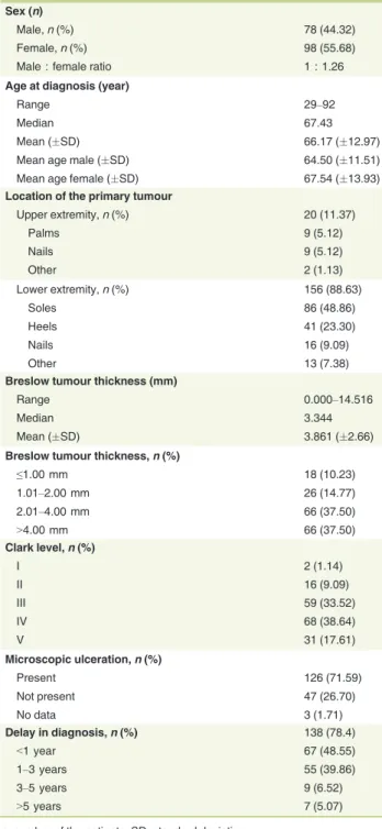
All patients were Caucasians. The mean age at diagnosis was 66.17 years (29–92,12.97 years). Most of the cases (73.29%) were diagnosed after 60 years old. The average age for males and females was 64.50 (11.51) and 67.54 (13.93) years, respec- tively. The male : female ratio was 1 : 1.26, and 55.68% of the tumours occurred in female patients above 60 years old (Table 1).
Of the 88.63% of tumours found on the lower extremities, 72.16% occurred on the soles and heels. Subungual melanoma was diagnosed in 25 cases (14.21%).
The mean Breslow tumour thickness was 3.861 mm (0.000–
14.516, 2.66 mm); specifically, 75.00% of the tumours were thicker than 2 mm, and 37.50% were thicker than 4 mm. Clark invasion depth levels IV–V were described in 56.25% of the tumours. Microscopic ulceration occurred in 126 cases (71.59%), as observed by histological examination. Macroscopi- cally, 35.79% of the tumours were ulcerated, and 14.20% were bleeding (Fig. 1).
Sentinel lymph node biopsy (SLNB) has been performed routinely at our centre since 1999. In our study group, 32.95%
of the patients underwent SLN biopsy, and 60.30% of these biopsies were positive. SLN-negative patients were predomi- nantly females (1 : 4), and these patients had significantly lower mean Breslow tumour thickness and survival rates than SLN- positive patients (2.020.36 mm vs. 4.660.45 mm;
P <0.001). Meanwhile, no differences were observed between the SLN-positive and -negative groups with respect to the mean age of the patients, location of the tumours and amount of time after noticing the tumour before consulting a physician (diag- nostic delay).
Survival analysis
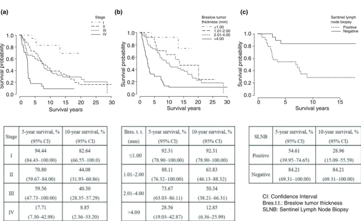
The 5- and 10-year overall survival rates of patients with ALM were 60.51% (95% CI, 53.27–68.75) and 41.59% (95% CI, 34.10–50.72), respectively.
Patients with ALM at tumour stage I had a significantly (P<0.001) better disease-specific survival rate than those at tumour stages II, III and IV (Fig. 2). No significant differences were noted in the disease-specific survival rates between patients with stage II and III tumours, but both of these patient groups
had significantly (P<0.001) better disease-specific survival rates than those with stage IV tumour.
Analysis according to Breslow tumour thickness revealed that the 5-year disease-specific survival rate of patients with T1 tumours was 92.31% (95% CI, 78.90–100.00), whereas that of patients with T4 tumours was 28.56% (95% CI, 19.03–42.87). In addition, patients with T4 tumours had significantly (P<0.001) worse disease-specific survival rates than those with T1–T3 tumours (Fig. 2).
The 5-year disease-specific survival rate in the SLN-negative group (84.21%; 95% CI, 69.31–100.00) was significantly better (P<0.001) than that in the SLN-positive group (54.61%; 95%
CI, 39.95–74.65; Fig. 2).
The results of the Cox univariate and multivariate analyses for disease-specific survival are shown in Table 2. In univariate anal- ysis, age (P <0.001), gender (P=0.03), Breslow tumour thick- ness (P <0.001), Clark level (P <0.001), ulceration (P <0.001), positive SLN (P=0.004), positive nodal status (P=0.008) and presence of distant metastasis (P<0.001) were statistically sig- nificant (P<0.05) and were, therefore, associated with worse survival. On the other hand, dermal mitoses and anatomical sites were not associated with worse survival. In subsequent multivari- ate analysis, patients’ age [hazard ratio (HR) 1.058, 95% confi- dence interval (CI) 1.035–1.083], Breslow tumour thickness (HR 1.187, 95% CI 1.099–1.282) and presence of distant metastasis (HR 3.002, 95% CI 1.850–4.871) remained statistically significant (P<0.001) and became independent risk factors for survival.
Delay in the diagnosis of ALM
Of the 176 patients with ALM, 138 had data describing the delay in the diagnosis. The delay between the time the patient noticed the tumour, and the time of diagnosis was extremely variable and ranged from 1 month to 10 years (mean time: 18 months).
Approximately half (51.45%) of the patients waited more than 1 year after noticing the skin lesion before seeking medical help.
Changes over four decades in the number of diagnosed cases, Breslow thickness of the tumours and age of the patients
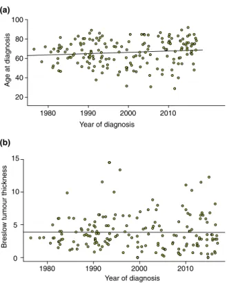
During the 40 years of analysis, no significant change was noted either in the mean Breslow tumour thickness (P=0.964) of the Table 1 Demographic and tumour characteristics of patients with
acral lentiginous melanoma (ALM) in the Hungarian study Sex (n)
Male,n(%) 78 (44.32)
Female,n(%) 98 (55.68)
Male : female ratio 1 : 1.26
Age at diagnosis (year)
Range 29–92
Median 67.43
Mean (SD) 66.17 (12.97)
Mean age male (SD) 64.50 (11.51)
Mean age female (SD) 67.54 (13.93)
Location of the primary tumour
Upper extremity,n(%) 20 (11.37)
Palms 9 (5.12)
Nails 9 (5.12)
Other 2 (1.13)
Lower extremity,n(%) 156 (88.63)
Soles 86 (48.86)
Heels 41 (23.30)
Nails 16 (9.09)
Other 13 (7.38)
Breslow tumour thickness (mm)
Range 0.000–14.516
Median 3.344
Mean (SD) 3.861 (2.66)
Breslow tumour thickness,n(%)
≤1.00 mm 18 (10.23)
1.01–2.00 mm 26 (14.77)
2.01–4.00 mm 66 (37.50)
>4.00 mm 66 (37.50)
Clark level,n(%)
I 2 (1.14)
II 16 (9.09)
III 59 (33.52)
IV 68 (38.64)
V 31 (17.61)
Microscopic ulceration,n(%)
Present 126 (71.59)
Not present 47 (26.70)
No data 3 (1.71)
Delay in diagnosis,n(%) 138 (78.4)
<1 year 67 (48.55)
1–3 years 55 (39.86)
3–5 years 9 (6.52)
>5 years 7 (5.07)
n, number of the patients; SD, standard deviation.
Figure 1 Acral lentiginous melanoma (ALM) on the thumb (71-year-old male, pT1a), great toe (44-year-old male, pT1a), palm (85-year-old female, pT3b) and sole (81-year-old female, pT3b).
tumours or in the mean age of the patients at the time of diag- nosis (P =0.157; Fig. 3). We diagnosed nearly the same number of ALM cases in each decade. When analysing all patients with CMM, we noticed a significant (P<0.001) decrease in the pro- portion of patients with ALM over the past 40 years, possibly because of the dramatic increase of the superficial spreading melanoma (SSM) subtype (Fig. 4).
Treatment of ALM patients
All 176 ALM patients in our study underwent wide local excision of the primary tumour with different safety margins during the study period of 40 years (Table S1). Regional lymph node dis- section (RLND) was performed with an elective intent until 1998 in 24 patients (28.5%). From 1999, after the introduction of SLN biopsy at our department, 58 patients with ALM
0 5 10 15 20 25 30
0.0 0.2 0.4 0.6 0.8 1.0
Survival years
Survival probability
Stage I II III IV
Breslow tumor thickness (mm)
≤1.00 1.01-2.00 2.01-4.00
>4.00
Sentinel lymph node biopsy
Positive Negative
0 5 10 15 20 25 30
0.0 0.2 0.4 0.6 0.8 1.0
Survival years Survival years
Survival probability
(a) (b) (c)
0 5 10 15
0.0 0.2 0.4 0.6 0.8 1.0
Survival probability
CI: Confidence Interval Bres.t.t.: Breslow tumor thickness SLNB: Sentinel Lymph Node Biopsy
Figure 2 Kaplan–Meier disease-specific survival curves and rates stratified by TNM stage (a), Breslow primary tumour thickness (b) and SLN status (c) in our patients with acral lentiginous melanoma (ALM).
Table 2 Results of Cox proportional hazard models of covariates
Univariate Multivariate
HR (95% CI) P-value HR (95% CI) P-value
Age 1.046 (1.027–1.065) <0.001 1.058 (1.035–1.083) <0.001
Female gender 0.652 (0.443–0.960) 0.030 0.506 (0.326–0.784) 0.002
Breslow thickness (mm) 1.253 (1.173–1.339) <0.001 1.187 (1.099–1.282) <0.001
Clark level IV/V (vs. II/III) 2.842 (1.871–4.316) <0.001 1.768 (1.071–2.918) 0.026
Presence of ulceration 3.625 (1.934–6.795) <0.001 2.591 (1.302–5.154) 0.006
Presence of dermal mitoses≥1/mm2 1.149 (0.601–2.195) 0.674 Anatomical site: foot (vs. hand) 1.471 (0.714–3.032) 0.296
SLNB†positivity 6.087 (1.799–20.600) 0.004
Positive nodal status 1.714 (1.154–2.156) 0.008 2.585 (1.088–6.139) 0.031
Presence of distant metastasis 2.834 (1.840–4.364) <0.001 3.002 (1.850–4.871) <0.001
†Analysis of 58 patients.
Results of Cox proportional hazard models of covariates (significantP-value marked with bold).
underwent SLNB, followed by complete RLND in 26 patients (28.2%). In patients with clinically detectable lymph node metastases, RLND was performed in 20 patients (23.8%) until 1998 and in seven patients (7.6%) from 1999. Regarding adju- vant systemic treatment, dacarbazine was mainly used until 1998 (19.0%), whereas interferon-alpha (22.8%) was used from 1999.
For irresectable and/or metastatic disease, the systemic treatment
of choice was chemotherapy until 2015 (until 1998: 11.9%; from 1999: 20.6%). Since 2015, novel treatment options, such as tar- get and immunooncological therapies, became available and were used in a total of 14 patients (imatinib: 2; dabrafenib and trametinib: 1; ipilimumab: 4; nivolumab: 4; pembrolizumab: 3).
Radiotherapy was used in 16.5% of our patients, either in an adjuvant or a metastatic (cutaneous: 7; lymph node: 2; brain:
11) setting. From 2007, electrochemotherapy was performed in 2.2% of our patients for loco-regional cutaneous metastases of melanoma. Based on the changes in the treatment strategies and available drugs, we identified three important time periods (1976–1998, 1999–2014 and 2015–2016) during the investigated 40 years. The sample size allowed us to compare the first two periods. No statistically significant differences were observed between the two groups with respect to demographic data (age, male : female ratio and Breslow tumour thickness); however, clinically detectable lymph node metastases (20 vs. 5 patients) were numerically higher in the first group (1976–1998) than in the second group (1999–2014). Using the Kaplan–Meier analy- sis, 5- and 10-year overall survival rates were 74.33 (95% CI, 66.10–83.59) and 63.72 (54.36–74.70), respectively, with a signif- icantly better (P<0.001) survival in the patients treated between 1999 and 2014 (Fig. S1).
Discussion
Acral lentiginous melanoma in Caucasians is a rare subtype of CMM, accounting for less than 10% of all CMM cases.2,7ALM is often detected in advanced age and clinical stage and is associ- ated with poor survival. During the past 40 years, 3.83% of the 4593 patients with CMM presenting at our department were diagnosed with ALM. Our results confirmed that ALM mainly affects elderly people, considering that most of the tumours occurred at 70 years of age.2,4,7,8Similar to the study of Ter- amotoet al.,12the mean age of the patients with ALM in the present study was 66 years. Data in the literature concerning sex predominance for ALM are conflicting.10 The study of Phan et al.10reported a female predominance in patients with ALM, and female sex was an independent prognostic factor for sur- vival. Other studies did not find significant sex differences in the occurrence of ALM. In our study, the male : female ratio was 1 : 1.26.2,7,8
The mean Breslow tumour thickness of ALM tumours in our study was nearly 4 mm, and 75% of the primary tumours were thicker than 2 mm and were microscopically ulcerated. In more than half of the tumours, the invasion depth was at Clark level IV or V. In previous studies, the number of ALM lesions arising on the foot was 5–16 times higher than that on the hand.10In our study, melanoma on the lower extremities was exhibited in nearly 90% of the patients, occurring mainly on the soles (48.86%) and heels (23.30%). Subungual melanoma is seemingly more common on the fingernails than on the toenails.10In our study, 14.20% of the melanomas were subungual, appearing
1980 1990 2000 2010
20 40 60 80 100
Year of diagnosis
Age at diagnosis
(a)
(b)
1980 1990 2000 2010
0 5 10 15
Year of diagnosis
Breslow tumour thickness
Figure 3 Distribution of the mean age at diagnosis (a) and Bres- low primary tumour thickness (b) in our acral lentiginous melanoma (ALM) cases during the past 40 years.
0 20 40 60 80 100 120 140 160 180 200
1976 1981 1986 1991 1996 2001 2006 2011 2016
Superficial spreading Nodular Acral lenginous Lengo maligna Amelanoc
Figure 4 Distribution of cutaneous melanoma by subtypes (4593 patients) during the past 40 years.
mostly on the lower extremities. The mean age of patients with ALM at the time of diagnosis and the location of the tumours in our study were similar to the results published by other authors.6,10,14However, the mean Breslow tumour thickness was significantly (P<0.001) higher in our study than most in other literature data for Caucasian patients.2,10,12,25
Patients with ALM have poor survival and unfavourable prog- nosis. In the study of Bradfordet al.,4the 5- and 10-year overall survival rates of patients with CMM are 91.3% and 87.5%, respectively, whereas those of patients with ALM have signifi- cantly worse survival rates. Comparing our results with other studies involving Caucasians, we concluded that both the 5- and 10-year overall survival rates (70–80% and 56–67%, respectively) of our patients with ALM are lower.4,8,10
After investigating the relationship between survival and the TNM stage of the disease, we found that progression from one stage to the next stage corresponded with significantly low chances of survival, and this difference is the highest between stages I and III and between stages I and IV. Therefore, survival rate is highly influenced by the lymph node metastasis (stage III) and presence of distant metastases (stage IV). With regard to tumour thickness, the most significant difference in the survival rate is found between the tumours thinner than 2 mm (T1–T2) and the tumours thicker than 4 mm (T4). Tumours thicker than 4 mm correspond to a significantly lower survival rate in every case than thinner tumours.
SLN biopsy has been performed routinely at our centre since 1999, and 32.95% of the patients in our study underwent SLN biopsy. SLN-positive cases occurred at a significantly (P <0.001) higher proportion than the results from Pavri et al.26
Delay in ALM diagnosis is well documented and is reportedly greater than 7 months on average.10,15Factors contributing to the delay might include the age and/or the cognitive state of the patients, hidden location, unusual clinical presentation, low public awareness, and misdiagnosis and mistreatment by health- care professionals, especially when pigmentation is absent. Rex et al.and Teramotoet al.attributed the poor survival in patients with ALM to the diagnostic delay rather than to a difference in
biological behaviour of this melanoma subtype.2,12In our study, more than half of the patients waited more than 1 year, and nearly 12% waited more than 3 years before seeking medical help. In a study by Phanet al.,10the delay between the time the patient first noticed the lesion and the time they were diagnosed ranged from 2 months to 30 years; in our study, this delay ran- ged from 1 month to 10 years.
Besides the parameters of malignant disease and patients’
characteristics, treatment is definitely the most important factor that may influence the survival of melanoma sufferers. In patients with malignant melanoma, treatment strategies changed quite extensively during the investigated 40 years of our study.
Based on the Kaplan–Meier analysis, survival improved with the introduction of new treatment strategies; however, due to the various and constantly changing treatment options, major con- clusions cannot be achieved from this analysis.
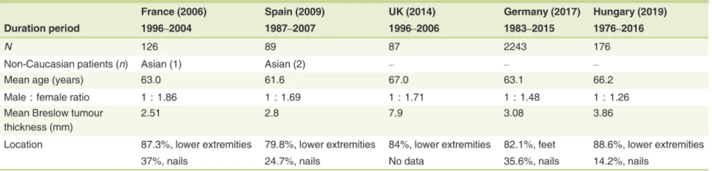
We compared our data with the literature for Caucasian patients with ALM from Europe (Table 3). In all the chosen studies, ALM occurred after 60 years old, with a female and lower-extremity predominance. The mean Breslow tumour thickness was greater than 2 mm in every study, but the thick- ness (nearly 4 mm) observed in our country was second only to that in the United Kingdom.2,12,15
Our study confirmed that patient and tumour characteristics and prognostic factors are similar with those found in previous literature data involving Caucasians; however, tumour thickness and survival suggest even worse prognosis.7,8,10,25
More than 2000 new patients with melanoma are diagnosed in Hungary annually, and one dies every day due to this malig- nant disease [National Cancer Registry, http://www.ksh.hu].
Based on our observations at our dermato-oncology centre, the number of patients diagnosed with melanoma increased dramat- ically in the past few decades. This increase, especially with respect to SSM and LMM, can be explained at least partially with the increasing popularity of tanning and the lack of appropriate photoprotection.
When analysing the tendencies of the past four decades, with respect to the incidence of the disease, we diagnosed the same number of ALM cases each decade, and we did not find major
Table 3 Comparison of our data with other studies involving Caucasians2,10,12,15
France (2006) Spain (2009) UK (2014) Germany (2017) Hungary (2019)
Duration period 1996–2004 1987–2007 1996–2006 1983–2015 1976–2016
N 126 89 87 2243 176
Non-Caucasian patients (n) Asian (1) Asian (2) – – –
Mean age (years) 63.0 61.6 67.0 63.1 66.2
Male : female ratio 1 : 1.86 1 : 1.69 1 : 1.71 1 : 1.48 1 : 1.26
Mean Breslow tumour thickness (mm)
2.51 2.8 7.9 3.08 3.86
Location 87.3%, lower extremities 79.8%, lower extremities 84%, lower extremities 82.1%, feet 88.6%, lower extremities
37%, nails 24.7%, nails No data 35.6%, nails 14.2%, nails
changes in the Breslow tumour thickness or in the mean age of the patients at diagnosis. As UV-induced pathogenesis has increased during this same period, this constant rate of diagnosis is unlikely to be associated with UV exposure.
Our study, which is the first single-centre report in East-Cen- tral Europe focusing on ALM, found that despite the improve- ment in early diagnosis of melanoma and improved public awareness in general, ALM in our region remains a subtype of melanoma that is diagnosed with high Breslow tumour thickness and advanced stage.27Therefore, we would like to emphasize the importance of early diagnosis of ALM by increasing disease awareness through education and prevention programmes.
Acknowledgement
This work was supported by the Janos Bolyai Research Scholar- ship of the Hungarian Academy of Sciences.
References
1 Kuchelmeister C, Schaumburg-Lever G, Garbe C. Acral cutaneous mela- noma in caucasians: clinical features, histopathology and prognosis in 112 patients.Br J Dermatol2000;143: 275–280.
2 Rex J, Paradelo C, Mangas C, Hilari JM, Fernandez-Figueras MT, Ferran- diz C. Management of primary cutaneous melanoma of the hands and feet: a clinicoprognostic study.Dermatol Surg2009;35: 1505–1513.
3 Furney SJ, Turajlic S, Fenwick Ket al.Genomic characterisation of acral melanoma cell lines.Pigment Cell Melanoma Res2012;25: 488–942.
4 Bradford PT, Goldstein AM, McMaster ML, Tucker MA. Acral lentigi- nous melanoma: incidence and survival patterns in the United States, 1986–2005.Arch Dermatol2009;145: 427–434.
5 Mahendraraj K, Sidhu K, Lau CSM, McRoy GJ, Chamberlain RS, Smith FO. Malignant melanoma in African-Americans: a population-based clin- ical outcomes study involving 1106 African-American patients from the surveillance, epidemiology, and end result (SEER) database (1988–2011).
Medicine (Baltimore)2017;96: e6258.
6 Wada M, Ito T, Tsuji Get al.Acral lentiginous melanoma versus other melanoma: a single-center analysis in Japan.J Dermatol2017;44: 932–
938.
7 Egger ME, McMasters KM, Callender GGet al.Unique prognostic factors in acral lentiginous melanoma.Am J Surg2012;204: 874–879; discussion 879–880.
8 Bello DM, Chou JF, Panageas KSet al.Prognosis of acral melanoma: a series of 281 patients.Ann Surg Oncol2013;20: 3618–3625.
9 Durbec F, Martin L, Derancourt C, Grange F. Melanoma of the hand and foot: epidemiological, prognostic and genetic features. A systematic review.Br J Dermatol2012;166: 727–739.
10 Phan A, Touzet S, Dalle S, Ronger-Savle S, Balme B, Thomas L. Acral lentiginous melanoma: a clinicoprognostic study of 126 cases.Br J Derma- tol2006;155: 561–569.
11 Phan A, Dalle S, Touzet S, Ronger-Savle S, Balme B, Thomas L. Dermo- scopic features of acral lentiginous melanoma in a large series of 110 cases in a white population.Br J Dermatol2010;162: 765–771.
12 Teramoto Y, Keim U, Gesierich Aet al.Acral lentiginous melanoma: a skin cancer with unfavourable prognostic features. A study of the German central malignant melanoma registry (CMMR) in 2050 patients.Br J Der- matol2018;178: 443–451.
13 Carrera C, Gual A, Dıaz Aet al.Prognostic role of the histological sub- type of melanoma on the hands and feet in Caucasians.Melanoma Res 2017;27: 315–320.
14 Duarte CA, Florez JP, Lopez HG, Meneses MX, de Vries E. Survival of acral lentiginous melanoma in the National Cancer Institute of Colombia.
J Eur Acad Dermatol Venereol2017;31: 438–442.
15 Boriani F, O’Leary F, Tohill M, Orlando A. Acral Lentiginous Melanoma–
Misdiagnosis, referral delay and 5 years specific survival according to site.
Eur Rev Med Pharmacol Sci2014;18: 1990–1996.
16 Whiteman DC, Pavan WJ, Bastian BC. The melanomas: a synthesis of epi- demiological, clinical, histopathological, genetic, and biological aspects, supporting distinct subtypes, causal pathways, and cells of origin.Pigment Cell Melanoma Res2011;24: 879–897.
17 Curtin JA, Fridlyand J, Kageshita Tet al.Distinct sets of genetic alter- ations in melanoma.N Engl J Med2005;353: 2135–2147.
18 Cust AE. Prognostic features for acral lentiginous melanoma.Br J Derma- tol2018;178: 311–312.
19 Asgari MM, Shen L, Sokil MM, Yeh I, Jorgenson E. Prognostic factors and survival in acral lentiginous melanoma.Br J Dermatol2017;177:
428–435.
20 Maldonado JL, Fridlyand J, Patel Het al.Determinants of BRAF muta- tions in primary melanomas.J Natl Cancer Inst2003;95: 1878–1880.
21 Curtin JA, Busam K, Pinkel D, Bastian BC. Somatic activation of KIT in distinct subtypes of melanoma.J Clin Oncol2006;24: 4340–4346.
22 Yaman B, Akalin T, Kandiloglu G. Clinicopathological characteristics and mutation profiling in primary cutaneous melanoma.Am J Dermatopathol 2015;37: 389–397.
23 Lee JH, Choi JW, Kim YS. Frequencies of BRAF and NRAS mutations are different in histological types and sites of origin of cutaneous melanoma:
a meta-analysis.Br J Dermatol2011;164: 776–784.
24 Gerschenwald JE, Scolyer RA, Hess KRet al.Melanoma staging: evi- dence-based changes in the American Joint Committee on Cancer eighth edition cancer staging manual.CA Cancer J Clin2017;67: 472–492.
25 Phan A, Touzet S, Dalle S, Ronger-Savle S, Balme B, Thomas L. Acral lentiginous melanoma: histological prognostic features of 121 cases.Br J Dermatol2007;157: 311–318.
26 Pavri SN, Han G, Khan S, Han D. Does sentinel lymph node status have prognostic significance in patients with acral lentiginous melanoma?J Surg Oncol2019;119: 1060–1069.
27 Ito T, Wada M, Nagae Ket al.Acral lentiginous melanoma: who benefits from sentinel lymph node biopsy?J Am Acad Dermatol2015;72: 71–77.
Supporting information
Additional Supporting Information may be found in the online version of this article:
Table S1. Treatment of ALM patients according to the three time periods.
Figure S1.Survival probability of patients treated between 1976 and 1998 vs. those between 1999 and 2014.