Chapter 8 Folates in Human Nutrition
Louis W. SULLIVAN1
Department of Medicine (Division of Hematology) New Jersey College of Medicine and Dentistry Jersey City, New Jersey
I. Introduction 366 II. General and Chemical 366
A. Historical 366 B. Isolation and Structure 367
C. Analogs and Derivatives 367 D. Folate Assay Methods 368 E. Dietary Sources of Folates 371 III. Biochemical Functions of Folic Acid 372
A. Biosynthesis of Purines and Thymidine 372
B. Biosynthesis of Methionine 373 C. Metabolism of Other Amino Acids 374 IV. Folate Absorption, Tissue Distribution, and Excretion 376
A. Absorption 376 B. Tissue Uptake and Distribution of Folate in Man 377
C. Excretion 378 V. Minimal Daily Requirement for Folic Acid 379
A. Requirements in Adults 379 B. Requirements in Pregnancy and during Lactation 380
C. Effects of Folate Deficiency on the Placenta and on the Fetus. 383 D. Effects of Folate Deficiency on Growth and Development . . . . 383
E. Requirements in Infancy 383 F. Requirements in Chronic Hemolytic Anemia 383
G. Folic Acid Requirements in Miscellaneous Diseases 384 H. Effect of Alcohol on Folic Acid Requirements 384
I. Role of Anticonvulsant Drugs 387 VI. Clinical and Laboratory Findings in Folate Deficiency 389
A. Signs and Symptoms of Folate Deficiency 389
B. Laboratory Findings 389 C. Principles of Therapeutic Trial 391
D. Etiological Classification of Folate Deficiency 392
E. Therapy 393 VII. Interrelations of Folate with Vitamin BJ2 and Ascorbate 394
References 397 P r e s e n t address: Department of Medicine (Section of Hematology), University
Hospital, Boston University School of Medicine, Boston, Massachusetts.
365
366
I. INTRODUCTION
In recent years, there has been increased interest in the role of folate compounds in human nutrition. Significant catalysts of this interest have been the development of microbiological assays capable of measuring the small amounts of folate in human plasma (1-3) and the introduction of clinically applicable tests for folate deficiency and deranged folate metabolism (4-6). As in other areas of human biology, our knowledge and understanding of the role of these compounds in human nutrition is progressing ever more rapidly. Recent general reviews include those of Herbert (7-9), Johns and Bertino (10), Luhby and Cooperman (11), Silber (12), Metz (13), O'Brien (14), Mollin (15), and Girdwood (16).
The biochemistry of folate compounds has been reviewed recently by Stokstad and Koch (16a), Jaenicke (17), Friedkin (18), Rabinowitz (19), Huennekens and Osborn (20), and Hutner et al. (21). In 1965, in the second volume of this series, Stokstad and Oace (22) presented a survey of the biochemistry of folic acid and some of the effects of folate deficiency in animals.
This chapter is concerned mainly with recent developments in our knowledge of the biochemistry of folate compounds, with emphasis on folate deficiency in man.
The term folate is used here to connote the various forms and deriva- tives of pteroylglutamic acid found in nature which serve as factors for growth and hematopoiesis in man and animals, and growth of cultures of microorganisms. The term folic acid refers only to pteroylglutamic acid.
I I . GENERAL AND CHEMICAL
A. Historical
After the demonstration by Minot and Murphy (23) that feeding whole liver was effective in inducing remissions in patients with pernicious anemia, Castle, in a series of elegant studies (24-26) showed that a similar hematopoietic response could be obtained in these patients by the administration of beef muscle (extrinsic factor) with normal gastric juice (intrinsic factor). Almost twenty years later, Castle's extrinsic factor was crystallized and given the name vitamin Bi 2 (27, 28). A comprehensive review of intrinsic factor appeared in Volume I of this series (29).
The possible existence of other substances effective in therapy of macrocytic (megaloblastic) anemias was first suggested by the studies of Wills (30) in India. Wills and her associates observed hematopoietic responses to marmite, an antolyzed yeast preparation, and to "crude'1 liver extracts, but not to refined extracts (31-34). Subsequent workers
8. FOLATES IN HUMAN NUTRITION 367 (35) have indicated that the amount of folic acid in the "Wills factor"
preparations given to these patients was probably on the order of 0.5-1.0 mg. Meanwhile, studies by others indicated that a syndrome of anemia, leukopenia, and stomatitis in monkeys was corrected by the adminis
tration of a factor (vitamin M) present in liver and yeast (36, 37).
Substances in yeast and liver found to be essential for normal growth and hematopoiesis in chicks (38-43) and a factor essential for growth of Lactobacillus casei (44) were variously named vitamins Bc, Bc conjugate, Bio, Βn, R, S, U norite eluate factor, and Lactobacillus casei factor.
B. Isolation and Structure
In 1941, Mitchell, Snell, and Williams (45) obtained a substance from spinach leaves (hence named folic acid) which stimulated the growth of Streptococcus faecalis and L. casei. It was subsequently found that folic acid and the various growth factors noted above had a similar nucleus, 4-(2-amino-4-hydroxy-6-pteridyl)methylaminobenzoic acid, with one or more glutamic acid residues (Fig. 1) attached in a γ-peptide linkage (16, 19, 46, 47). It was also noted that the growth-enhancing effects of these folic acid derivatives were frequently similar in various animals and in some microbiological assay systems (16).
The formula and the chemical synthesis of folic acid were announced in 1946 by Angier and co-workers (46, 47). Details of its structure and physicochemical properties and those of its known analogs were described by Stokstad and Oace (22).
C. Analogs and Derivatives
Mainly from studies of the growth requirements of various micro
organisms, most notably strains of S. faecalis (Streptococcus lactis R.), L. casei and Pediococcus cerevisiae (formerly erroneously designated as Leuconostoc citrovorum), and more recently from various chemical pro
cedures, it is now recognized that there are many derivatives of folic acid in nature. These include pteroyltriglutamic acid in many bacteria (48-50), pteroylheptaglutamic acid in yeast (51), V5-formyltetrahydrofolic acid (folinic acid) in liver (52, 53) and human leukocytes (54), V5-methyl- tetrahydrofolic acid in mammalian liver (55-57) in avian liver (58), and in human plasma (59), and an unidentified form of tetrahydrofolate in human erythrocytes which may be a polyglutamyl derivative of iV5- methyltetrahydrofolic acid (60, 61). Other folic acid derivatives have been identified as intermediates in various biochemical reactions in which folic acid participates, and reference will be made to some of these below.
Very little is known about the structure of naturally occurring folates in plants, though the available evidence suggests that they are largely
368 L O U I S W . SULLIVAN
polyglutamyl derivatives of tetrahydrofolic acid, with three to ten glutamic acid residues in γ-glutamyl peptide linkages (19, 62).
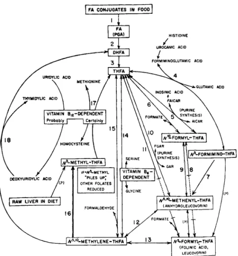
Figure 2 outlines in schematic form the presently known folate- dependent enzymes and their interrelations in one-carbon metabolism
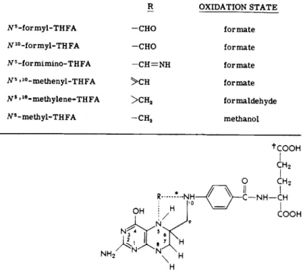
R OXIDATION STATE
i V5- f o r m y l - T H F A - C H O f o r m a t e tf10-formyl-THFA - C H O f o r m a t e Ν5 -f or m i m i n o - TH FA — C H = N H f o r m a t e JV5 >1 0- m e t h e n y l - T H F A > C H f o r m a t e Ns »1 0- m e t h y l e n e - T H F A > C H2 formaldehyde
tf5-methyl-THFA - C H3 methanol
+COO H
I
C H2
Ή
•Broken lines indicate the N5 and/or Ν10 site of attachment of various 1-carbon
units for which THFA acts as a carrier. ' 5,6,7,8-Tetrahydrofolic acid (THFA) (FHJiR = —H)
FIG. 1. Structure of tetrahydrofolic acid and its one-carbon adducts.
t The natural conjugated forms have one or more additional glutamyl residues attached by linkage at this site. Adapted from Herbert (262).
(63). These are listed in Table I according to the reaction number in Fig. 2. Those reactions that have not yet been shown definitively to occur under physiological conditions are noted by question marks. Excellent reviews of folate coenzymes are to be found in recent articles by Jaenicke (17), Friedkin (18), and Stokstad and Oace (22).
D. Folate Assay Methods
Chemical methods of measuring folates in biological materials have not been widely applied because of their relative insensitivity to the
369
FIG. 2. Interrelations of folate and vitamin B i2. Adapted from Herbert (262).
small amounts of folate present. Reduction of folic acid in acid with zinc yields p-aminobenzoylglutamic acid, which is measured chemically (64).
Allfrey et al. (65) introduced a fiuorometric assay based upon the observa
tion that folic acid is oxidized to 2-amino-4-hydroxypteridine-6-carboxylic acid in alkaline solution, which fluoresces strongly at 470 ηΐμ when irradiated by light of wavelength 365 ηΐμ. This latter technique is the more sensitive and will detect folic acid in concentrations of 0.01 to 10 Mg/ml, but is not sensitive enough to determine accurately the folate content of most biological materials.
Animal assays have the advantage that folic acid and its biologically active derivatives can be measured, especially the conjugated forms (which have limited activity in microbiological assays). Folic acid defi
ciency has been experimentally produced by dietary deficiency (combined
Enzyme Reaction No. in Fig. 2
Conjugase 1
Folate reductase 2
Dihydrofolate reductase 3
Formiminotransf erase 4
Aminoimidazolecarboxamide ribotide transformylase 5 Formate-activating enzyme (tetrahydrofolate formylase) 6
Cyclodeaminase 7
iV1 0-Formyltetrahydrofolate isomerase 8
Cyclohydrolase 9
Glycinamide ribotide formylase 10
Formyltr ansf erase 11
iV5,iV1 0-Methylenetetrahydrofolate dehydrogenase 12 AT5,JV10-Methenyltetrahydrofolate reductase 13
Serine hydroxymethyltransferase 14
Formaldehyde-activating enzyme 15
AT5,iV1 0-Methylenetetrahydrofolate reductase 16
Methionine synthetase 17
Thymidylate synthetase 18
in some instances with the administration of sulfonamides or folate antagonists) in fowl (66-72), rats (73-81), mice (82, 83), guinea pigs (84, 85), swine (86, 87), monkeys (88, 89), and man (90). Growth and hematopoiesis in the chick are markedly affected by folate deficiency, and the chick assay has been the most widely used. However, animal assays are tedious and expensive and are thus not practical for clinical use.
Microbiological assays have enjoyed the widest application in assess- ment of folate nutriture in man (8). Toennies et al. (91) reported that almost all the measurable folic acid activity in whole blood was found to reside in the erythrocytes. Until recently, it was generally accepted that the amounts of folate-active compounds in serum were too small for reliable assay by microbiological means (92, 93). In 1959, however, Herbert, Baker, and associates reported that, using Lactobacillus casei (ATCC No. 7469) as the test organism and with ascorbic acid in the assay medium, measurable amounts of folate were found in serum of normal individuals and patients with pernicious anemia (1, 2, 94). Patients with nutritional megaloblastic anemia which subsequently responded to ad- ministration of small amounts of folic acid had significantly lower serum folate levels. This assay has been widely adopted with many minor modi- fications (3, 95-99). The "aseptic addition" method (3, 99a) appears to have achieved the greatest utilization because of its simplicity. The assay with L, casei takes 18 hours and growth is read turbidimetrically. The
T A B L E I
FOLATE-DEPENDENT ENZYMES
371 ready availability of stable commercial "dry mixes" of the basal medium (Baltimore Biological Laboratories, Baltimore, Maryland; Difco Labora
tories, Detroit, Michigan) has eliminated much of the tedium (and some of the potential sources of error) in the folate assay. Lactobacillus casei grows well on all known forms of folate in human serum, whereas S. faecalis does not grow on iV5 methyl forms and P. cerevisiae does not grow in iV5-methyl tetrahydrofolate or nonreduced forms (22); this underscores the necessity of using L. casei to accurately assess the folate nutriture of humans.
This also explains in part the prior failures by various workers to obtain meaningful and reproducible results with S. faecalis assays of serum.
Herbert (3) and Waters and Mollin (95) have indicated that amounts of ascorbic acid in the range of 0.15 to 1.0 gm/100 ml afford optimal protec
tion of labile forms of serum folate against oxidative destruction during autoclaving in the assay; they therefore recommend using ascorbate concentrations in this range. Whether the addition of ascorbate to serum prior to storage at — 20°C affords significant additional protection of serum folate is disputed (95, 100). Studies of the nature of serum folate have shown that it is mainly iV5-methyltetrahydrofolic acid (59) and is thus closely related to the major folate fraction of human erythrocytes (60, 61) and of mammalian and avian liver (55-57).
E. Dietary Sources of Folates
Folates are widely distributed in various foodstuffs. Most measure
ments of the folate concentrations of native foods have been made with microbiological assays utilizing S. faecalis (101, 102), without ascorbate protection. These values are therefore low in relation to the total folate content of the foods assayed (103). Herbert found much higher values for selected foods assayed withL. casei and ascorbate-containing buffer (103).
Using S. faecalis assays, Chung et al. (104) noted that the daily folic acid content of high-cost and low-cost American diets was 193 μg and 157 Mg , respectively. Butterworth and co-workers (105) reported than an
"ordinary" American diet contained 52 Mg of "free" folic acid and 184 Mg of "total" folic acid (after conjugase digestion) per day, as determined by assays with S. faecalis. In addition to the limitations of faecalis assays without ascorbate protection, as discussed above, the ability of the human small intestine to absorb food folates (many of which exist as polyglutamyl derivatives) is unknown. Although the use of tritium- labeled pteroylglutamic acid has proved to be of interest in studies of small intestine function in sprue and other small bowel disorders (106, 107), the relevance of these data to the absorptive capacity of the small bowel for folates in food is unknown. Streiff and Rosenberg (107a) re
ported that following the oral administration of a purified conjugated form of folate (from yeast) to normal volunteers, serum folate levels rose
372 LOUIS W . SULLIVAN
to the same degree as occurred following the oral administration of pteroylglutamic acid. The absorbed folate appeared to be a monogluta- mate. Patients with small bowel disease had significant elevations of serum folate levels after the administration of oral pteroylglutamic acid, but not after oral administration of a polyglutamate form of folate. These workers suggest that conjugated forms of folate in nature are first broken down to triglutamate or monoglutamate forms prior to absorption, and that the defect causing folate deficiency in some patients with small bowel disease is a loss in ability of the small intestine to deconjugate natural forms of folate.
More data are needed concerning the folates in food and their absorp- tion and utilization by the human. Since metabolic balance studies are impractical because of the synthesis of folic acid by intestinal bacteria, many of these problems must await the successful incorporation of suitable radioisotopic labels into food folates. A table of the UL. casei" folate content of a limited number of foods may be found in the paper by Herbert (103), and tables of folate content of foods as determined by S. faecalis assay were published by Toepfer et al. (101) and by Stokstad (108). Foods having the highest concentration of folates include liver, kidney, nuts, fresh green vegetables, legumes, citrus fruits, berries, and cheese (101, 103, 108). Cooking, canning, or other processing may destroy 50 to 95% of the folate content in food.
I I I . BIOCHEMICAL FUNCTIONS OF FOLIC ACID
The recent reviews of Stokstad (22), Huennekens (109), and Friedkin (18) afford detailed accounts of developments in our knowledge of the biochemistry of folic acid. Thus, a brief discussion of this area will be given, with emphasis on the more significant recent findings.
Folic acid has a central role in the metabolism of one-carbon units, at three levels of oxidation—methyl, formyl and formate, respectively (18). Folate coenzymes are thus involved in the synthesis of purines and the synthesis of methionine and serine. A folate-containing coenzyme is also involved in the catabolism of histidine in man (110).
A. Biosynthesis of Purines and Thymidine
Reactions involving the transfer of one-carbon units in purine syn- thesis include the formylation of glycinamide by iV5'1 0-methenyl-FH4 (110) and the formylation* of aminoimidazolecarboxamide ribotide (AICAR) by iV1 0-formyl-FH4 (111), thus introducing carbons 8 and 2, respectively, of the purine ring. In folate deficiency, there is increased excretion of AICAR in the urine (112), presumably the result of a block in purine synthesis, or related to an enlarged purine precursor pool. Similar findings
8. FOLATES IN HUMAN NUTRITION 373 were noted in vitamin Bi2-deficient patients (112, 113) and in patients with hemolytic anemia and liver disease (112).
Folate serves as the one-carbon donor in the synthesis of the methyl group of thymidylic acid (18) as indicated by reaction 18 in Fig. 2. The dihydrofolate resulting from this reaction must be reduced to tetra- hydrofolate by dihydrofolate reductase before it can be reutilized in one-carbon transfer reactions (114). The folate antagonists aminopterin and amethopterin block this reduction by irreversibly binding the enzyme dihydrofolate reductase (115). The therapeutic effect of these folate antagonists appears to reside in their ability to prevent the reduction of folate and dihydrofolate to tetrahydrofolate (116). Recent reviews of folate antagonists include those of Bertino (116), Delmonte and Jukes
(117), and Hitchings and Burchall (118).
B. Biosynthesis of Methionine
One of the more significant recent developments was the isolation and characterization of iV5-methyltetrahydrofolate as a naturally occurring folate in mammalian liver (119-121), serum (59), and erythro- cytes (60, 61). It has been shown that iV5-methyltetrahydrofolate trans- fers its methyl group to homocysteine, to form methionine (122). The generation of iV5-methyltetrahydrofolic acid from iV5'1 0-methylenetetra- hydrofolic acid has not been shown to be reversible in vivo (123). Because the subsequent reaction involving transfer of the methyl group to homo- cysteine is the only reaction in mammalian systems in which both vitamin B12 and folic acid have been shown to participate directly, Herbert and Zalusky (63) and Buchanan et al. (122) have postulated that the reaction in Eq. 1 is of crucial importance in folate metabolism. That is, the "pile
up" of folate as 5-methyl-FH4 secondary to vitamin Bi 2 deficiency leads to the relative depletion of other forms of folate, as outlined in Fig. 2. In bacteria, two different enzyme systems involved in the synthesis of methionine have been found. One uses a triglutamate form of folate only and does not require vitamin Bi 2:
AT5-Methyltetrahydropteroyltriglutamate tetrahydropteroyltriglutamate iST5-Methyl-FH4 F H4
(1)
+ (2)
h o m o c y s t e i n e methionine
This enzyme has been found in a wild-type Escherichia coli (124), E.
coli PA-15 (125), and Aerobacter aerogenes (126). The second enzyme
LOUIS W . SULLIVAN
system, requiring vitamin Bi 2 and closely resembling the mammalian enzyme system, has been found in the vitamin Bi2-requiring mutant E. coli 113-3 (127, 128):
J V5- m e t h y l - F H4
iV5-methyltetrahydropteroyltriglutamate
h o m o c y s t e i n e
FADH2, A T P , M g2+
B1 2- e n z y m e S-adenosylmethionine
+
methionine (3)
This enzyme can utilize the monoglutamate or triglutamate forms of JV~6-methyl-FH4.
C. Metabolism of Other Amino Acids
1. Histidine Catabolism
Histidine is catabolized through a series of steps to formiminoglu- tamic acid (FIGLU):
H — C = C — C H 2 — C H — C O O H
Η Ν Ν I H C = C — C H = C H - C O O H
• ΗΝ I I Η H i s t i d i n e
HOOC CH - CH2 — C H2— COOH HN NH
^ C "
F o r m i m i n o g l u t a m i c a c i d FH*
A ^5- f o r m i m i n o - F H 4 H O O C — C H — C H2— C I V - C O O H
NHo
U r o c a n i c a c i d
(4)
0 = C CH — CH2— CKj—COOH
Η
I m i d a z o l o n e p r o p i o n i c a c i d
G l u t a m i c a c i d
The enzyme formiminotransferase, involved in the catabolism of FIGLU, is folate dependent. When there is a deficiency of folate, the conversion of
FIGLU to glutamic acid is impaired and FIGLU is excreted in the urine (129, 130). The excretion of FIGLU is further increased by the adminis- tration of a loading dose of histidine (131).
The excretion of FIGLU after histidine loading has also been observed in folate-deficient and vitamin Bi2-deficient chicks (132) and rats (133) and after the administration of folate antagonists (134). Such similarities in the effects of vitamin B i2 deficiency and folate deficiency on the metabolism of FIGLU are consistent with the hypothesis that vitamin B i2 deficiency results in a relative deficiency of F H4 caused by "trapping"
of folate as iV5-methyl-FH4 (63, 135).
A FIGLU excretion test following a histidine load was developed by Luhby et al. (11, 134) and has been used extensively as a test for folate deficiency in man. However, the value of this test in the differential diagnosis of megaloblastic anemias has been impaired by the finding that from 19 to 6 1 % of patients with vitamin Bi 2 deficiency (136-139), and some patients with leukemia, myelofibrosis, hemolytic anemia, carcino- matosis, liver disease, and sarcoidosis have increased excretion of FIGLU in the urine following a histidine load (138).
In a study of megaloblastic anemia in pregnancy, Chanarin et al. (140) found that of 41 patients, 12 (29%) had normal FIGLU excretion tests with a histidine load. This failure to observe increased FIGLU excretion in these patients was attributed to altered histidine metabolism in pregnancy. In spite of the relative lack of specificity of this test, its limitations in megaloblastic anemia of pregnancy, and the greater sensitivity of the L. casei folate assay (20), the FIGLU excretion test is still of definite, though limited, value in the diagnosis of megaloblastic anemia. It is useful as a parameter of tissue folate stores and of degrees of metabolic folate deficiency resulting from impaired folate utilization due to vitamin Bi 2 deficiency and the use of folate antagonists.
Of interest is the observation that, similar to vitamin-deficient chicks (132) or rats (133), the abnormal excretion of FIGLU in folate or Bi2-deficient man can be reversed by the oral administration of methionine (141). Whether this effect is due to a "sparing action" of methionine on the pathway involving iV5-methyl-FH4 or to other mechanisms remains to be ascertained.
2. Serine-Glycine Inter conversions
In animals, the major source of glycine is from serine (142). The serine-glycine conversion is a reversible reaction in which the coenzyme F H4 accepts the formyl group from serine, thus generating iV"5'10-methyl- ene-FH4 (143-145).
376 LOUIS W . SULLIVAN
Serine Glycine
CH2OH
s e r i n e hydroxymethylase
CH—COOH - — C H2— C O O H
NH2
J V ^ ^ - m e t h y l e n e - F r ^ NH2
Herbert and Sullivan (141) noted that the administration of glycine to patients with folate or vitamin Β12 deficiency resulted in a drop in FIGLU excretion similar to the effect of methionine administration.
The effect of glycine administration was postulated to be due to the increased generation of F H4 which was then available for the catabolism
I V . FOLATE ABSORPTION, TISSUE DISTRIBUTION, AND EXCRETION
A considerable amount has been learned about the gastrointestinal absorption of pteroylglutamic acid (PGA). However, since food folates are various reduced monoglutamate and polyglutamate derivatives of PGA, the absorption data may be limited in their applicability to absorp
tion of food folate (see Section II,E). Nevertheless some food folates appear to be converted to PGA during the process of cooking (105).
Pteroylglutamic acid can be rapidly absorbed from the entire length of the small intestine, but it is absorbed primarily from its proximal portions (146-149). Absorption is by an energy-dependent process for physiological quantities of the vitamin, but larger doses appear to be absorbed by passive diffusion (147, 148). Studies with tritium-labeled folic acid indicate that normally upwards of 80% of the dose is absorbed when given in physiological (200 Mg ) amounts (150), and peak serum levels occur within 1 to 2 hours of administration (150). Because of active synthesis of folate by bacteria in the large intestine, absorption studies prior to the introduction of tritium-labeled folic acid (151) con
sisted of either measuring by microbiological assay (S. faecalis) the rise in serum folic acid activity (92) or measuring the urinary excretion of folic acid (152) following an oral dose of the vitamin. Such studies were done after "saturation" of body folate stores by oral or parental administration of folic acid over a 3-day period. Detailed review of the methods may be found in the reports of Girdwood (152) and Chanarin et al. (92). Such studies showed abnormal absorption tests in patients with a variety of conditions affecting the small intestine, including tropical and nontropical sprue, Whipple's disease, lymphoma, macroglobulinemia, leukemia, carcinomatosis, and postoperative extensive small bowel resection (92, of FIGLU.
A. Absorption
8. FOLATES IN HUMAN NUTRITION 377 152-154). The differentiation of subjects with malabsorption from those with dietary folate deficiency require that the test be repeated and that the results following oral folic acid be compared with those following parenteral folic acid. While such studies are of value and are generally reliable, degrees of overlap do occur. Other disadvantages include the necessity of administering supraphysiological doses of folic acid (as noted above, the apparent physiological mechanism for folic acid absorption appears to be of significance only in the absorption of small "physiological'' doses), which may elicit a hematological response in anemic vitamin Bi2-deficient patients, and the necessity of complete urine collections and/or multiple venipunctures.
B. Tissue Uptake and Distribution of Folate in Man
In normal subjects, after the intravenous injection of pteroylglutamic acid (15 μg per kilogram of body weight), Chanarin et al. (154) found that the mean serum folic acid level with the S. faecalis assay was 127, 40, and 20 ng/ml at 3, 15, and 30 minutes, respectively, after injection.
In megaloblastic anemia due to deficiency of folic acid or vitamin Bi 2, idiopathic steatorrhea, pregnancy, some cases of leukemia, lymphoma, megaloblastic anemia in patients receiving anticonvulsant drugs and in patients with advanced malignant disease, rapid clearance rates have been reported (16, 63, 92, 93, 152). These rates were interpreted as indicative of tissue depletion of folate in such subjects. The report of Herbert and Zalusky (63) suggested, however, that in patients with vitamin BJ 2 deficiency intravenously injected folic acid was rapidly changed to a form (A^-methyl-FH^ that could be utilized by L. casei}
but not by S. faecalis. This resulted in rapid folic acid "clearances"
when S. faecalis was used, but normal or slower than normal clearances with the L. casei assay.
Intravenously injected folic acid is rapidly taken up by the cells of the body (155), but once within the cell, it is slowly converted to reduced forms of folate (156). The latter observation may be due to the fact that folic acid is not the natural substrate for the enzyme dihydrofolate reductase (156). Johns and Bertino (10) suggest that the slow intracellular uptake of methotrexate as compared with the rapid intracellular uptake of folic acid indicates that cellular uptake of folate is a relatively specific active process rather than passive diffusion.
The major storage organ of folate is the liver (22, 121, 157, 158) with concentrations of 5 to 9 μg/gm. Significant folate concentrations are also found in the kidneys (3 μg/gm) (157), but folate concentrations in erythrocytes (60, 90, 159, 160) and leukocytes (161) are only one- twentieth to one-tenth those in the liver. It has been estimated that
378 LOUIS W . SULLIVAN
total body folate stores in the human are in the range of 5 to 10 mg (9).
The major form of folate in liver (135) and serum (59) has been identified as iV5-methyl-FH4. Studies of erythrocyte folates suggest that a poly- glutamyl derivative of ^V5-methyl-FH4 is the major folate form (60, 61).
The results of differential microbiological assays of kidney folates, as reported by Grossowicz et al. (157) utilizing L. casei, P. cerevisiae, and S. faecalis suggest that iV6-methyl-FH4 may predominate in this organ also. Studies of leukocyte folates reported some years ago indicated that folinic acid was the predominant folate form (161). However, the necessity of precautions to protect natural folates from oxidative destruction during the preparation of samples for assay was not then appreciated, and these values therefore need to be reassessed by more recent methods.
C. Excretion
Under normal circumstances, only traces of folic acid (5 μg in 24 hours) are found in the urine (162-164). Register and Sarett (165) showed that urinary folate excretion was increased during ingestion of a high-purine (and thus, folate-rich) diet, whereas lower than normal values were found during consumption of a milk diet (low in folate content) (165). Patients with folate deficiency due to poor diets (164) or to tropical sprue (163) excrete less than 3 μg of folic acid in 24 hours.
When large (5 mg) doses of folic acid are injected or given by mouth to normal subjects, 2 to 3 mg of this are excreted in the urine (166), whereas patients with folate deficiency usually excrete less than 1.5 mg.
Patients with pernicious anemia, chronic infections and various malig
nancies may also excrete smaller than normal amounts of folate in the urine (167, 168). When 200 μg of tritium-labeled folic acid was given to normal subjects, along with 15 mg of nonradioative folic acid, 4 1 % of the administered radioactivity was excreted in the urine and 20% was excreted in the feces (151).
Goresky and associates (169) found that folic acid in the glomerular filtrate is reabsorbed by the tubules by an active process. This reabsorp- tive mechanism was inhibited by amethopterin. With infusions of folic acid resulting in plasma levels greater than 10 Mg/ml, Johns et al. (155) noted that the renal clearance of folic acid was independent of the plasma level and averaged about 51 ml/minute. This, was attributed to glomerular filtration and urinary excretion of unbound folic acid. Fol
lowing infusions of tritium-labeled folic acid (3H-PGA) to normal sub
jects and to patients with megaloblastic anemia, Chanarin et al. (170) found that the jnajor peak of radioactivity which appeared in the urine was associated with L. casei folate activity (presumably, iV5-methyl-FH4).
A minor amount of the radioactivity was associated with material
8. FOLATES IN HUMAN NUTRITION 379 active for S. faecalis. In contrast, Mollin, Waters, and Harriss (139) found that the tritium excreted in the urine after intravenous injection of 3H-PGA was associated with material which supported the growth of S. faecalis. Baker et al. (171) and Herbert (171a) recently reported that biliary excretion of folic acid may be an important consideration in man.
They found that in normal and folate-deficient subjects, the concentration of folate in the bile was several times that in the serum, and suggested that there may be an enterohepatic circulation of folic acid and that, in subjects with defective folate absorption, the excretion of folate in the bile may contribute to the development of folate deficiency.
V . MINIMAL DAILY REQUIREMENT FOR FOLIC ACID
Only in the past twelve years have significant data been gathered concerning the minimal daily folate requirement in man. Such studies have been done almost exclusively with folic (pteroylglutamic) acid, and the conclusions therefrom must be interpreted in that light. Very little is known about the requirements for natural forms of folate in foods.
A. Requirements in Adults
In 1953, Jandl and Gabuzda (172) reported brisk hematological re- sponses to 125 and 250 Mg of folic acid in two patients with megaloblastic anemia and scurvy. In subsequent studies of alcoholics with megaloblastic anemia, Jandl and Lear (164) obtained hematological responses to 250 to 500 Mg of PGA. This suggested that the requirements for adult man were probably less than 200 to 300 Mg- In 1961, Sheehy et al. (173) reported^that hematological responses occurred with doses of 25 Mg of folic acid given daily, by mouth tojpatients with tropical sprue, although the patients' diets had contained between 1000 and 1500 Mg of total food folate. This finding suggested that very little of the dietary folate was absorbed by such patients. Using a synthetic diet totally devoid of folate activity, Zalusky and Herbert (174) noted a good hematological response to 50 Mg of PGA in a patient with megaloblastic anemia resulting from folate deficiency. In subsequent studies, utilizing a thrice-boiled diet con- taining less than 5 Mg of total folate (103), Herbert was able to produce folate deficiency in a previously healthy adult (90). The sequence of events noted was (a) fall of serum folate level to deficient levels (less than 3 ng/ml) in 3 weeks, (b) hypersegmentation of the nuclei of neutro- p h i l s after 7 weeks, (c) increased urine formiminoglutamate excretion after 13 jweeks, (d) low erythrocyte folate levels after 4 months, followed in a few days by ;(e) the appearance of oval macrocytes in the peripheral blood, (f) megaloblastic jnarrow morphology, and (g) anemia after 4)^
months. Further studies of three normal adult females consuming the
380 LOUIS W . SULLIVAN
folate-deficient diet, suggested that 50 to 100 μg of PGA was sufficient to maintain normal serum folate levels, whereas the ingestion of 25 μg was accompanied by a fall in serum folate levels (175). Sullivan and Herbert (176, 177) found that the administration of 75 Mg of PGA to patients with megaloblastic anemia secondary to folate deficiency resulted in brisk hematological improvement, conversion of marrow from megalo
blastic to normoblastic, rapid fall in serum iron, correction of elevated FIGLU excretion, and, much later, rises in serum folate levels (Figs. 3 and 4). These latter two observations indicated, that, when small doses of folic acid are given to folate-depleted subjects, serum levels rise only after significant tissue repletion has occurred. Other studies have sup
ported the efficacy of 100 to 200 Mg of folic acid in folate-deficient megalo
blastic anemia (159, 178). It thus appears that the adult minimal daily requirement for pteroylglutamic acid is in the range of 25-75 Mg. Whether similar quantities of food folates are required in man remains to be ascertained.
The requirements for folic acid in infancy and childhood, pregnancy, hemolytic anemias, malignancies, and hypermetabolic states (e.g., hyper
thyroidism), infections, and other disease states remain to be established through present clinical evidence suggests that in some, if not all, of these conditions there is an increased folate requirement.
B. Requirements in Pregnancy and during Lactation
A number of investigators have shown that in pregnancy serum folate levels tend to be low (179, 180), injected folic acid is cleared from the plasma rapidly (152, 181, 182), even more rapidly in twin pregnancies (181), and that megaloblastic anemia appears to occur more frequently in pregnant females than in the general population (183). The anemia (183a) frequently remits following termination of pregnancy, without sup
plemental folic acid (7). However, Shapiro et al. (184) reported that in South Africa the highest incidence of folate-deficient megaloblastic anemia in association with parturition occurs after termination of preg
nancy primarily in women who breast-feed their infants. It was postulated that this is due to an increased folate requirement from pregnancy and lactation (185). Lowenstein et al. have presented evidence that supple
mentation with 500 Mg of folic acid during pregnancy significantly reduces the incidence of megaloblastic anemia (185a), and a recent report by Willoughby and Jewell suggests that supplementation with 300 Mg of folic acid daily may be sufficient to meet the folate requirements of pregnancy (185b). Alperin and colleagues (185c) failed to obtain hematologic responses to daily doses of 100 Mg and 200 Mg of folic acid given, respec
tively, to two patients with megaloblastic anemia, whereas prompt
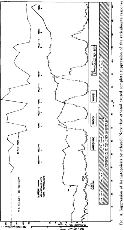
FIG. 3. Suppression of hematopoiesis by ethanol. Note that ethanol caused complete suppression of the reticulocyte response and rise in erythrocyte count during folic acid administration. After Sullivan and Herbert (177).
382 LOUIS W . SULLIVAN
FIG. 4. Partial correction of the effect of ethanol on hematopoiesis by larger doses of folic acid. Note also the fall in leukocyte and platelet counts during ethanol inges- tion, and the rise of these formed elements after the dose of folic acid was increased.
After Sullivan and Herbert (177).
8. FOLATES IN HUMAN NUTRITION 383 hematologic improvement occurred after delivery, suggesting that the folic acid requirement during pregnancy was greater than 200 Mg .
C. Effects of Folate Deficiency on the Placenta and on the Fetus
Streiff and Little have reported that, in pregnant patients with vaginal bleeding due to abruptio placentae, the incidence of folate deficiency is four times greater than in other pregnant patients (185d). In those patients with third trimester bleeding of unknown etiology, the incidence of folate deficiency was twice that of other pregnant patients.
Studies on the effect on the rat embryo of maternal folate deficiency resulting from the administration of folate antagonists have shown that these agents may cause fetal resorption, stillbirths, and congenital malformations (186-188). It is well known that in humans, aminopterin can produce abortions (189). Recent reports suggest that human fetal malformations may also result from the administration of folate antago- nists (190) or from dietary deficiency of folate (191, 192).
D. Effects of Folate Deficiency on Growth and Development
Watson-Williams (193) described four adults (3 female, 1 male), 20 to 30 years old, with folate deficiency and sickle-cell anemia who had short stature, retarded bone age, and immature sexual development.
During therapy with 15 mg of folic acid for 10 months, there was a rapid spurt in growth, increase in body weight, development of gonads and secondary sex characteristics, and increase of radiological bone age.
Martin et al. (194) have reported that urinary estrogen excretion is decreased in pregnant patients with folate deficiency.
E. Requirements in Infancy
In 1946, Zuelzer and Ogden showed that megaloblastic anemia in infancy was often the result of folate deficiency (195). Luhby and Wheeler (196) subsequently obtained hematological responses with 0.8 to 1.2 mg of folic acid given to infants with nutritional megaloblastic anemia.
Recent studies of infants with anemia due to folate deficiency suggest that the minimal daily requirement is in the range of 50 Mg (197). Thus, relative to body weight, the folic acid requirements in infancy is several- fold that of adults.
F. Requirements in Chronic Hemolytic Anemia
There have been a number of reports of folate deficiency with (or without) megaloblastic marrow morphology as a complication of chronic hemolytic anemia. These have included hereditary spherocytosis (198-
201) thalassemia (202-205), hemoglobinopathies (193, 206-211), immune
384
hemolytic anemia (198, 212, 213), paroxysmal nocturnal hemoglobinuria (214, 214a), and erythropoietic porphyria (211). The majority of patients were given pharmacological (5 to 15 mg or more) doses of folic acid, to which they showed a good hematological response. The suggestion that folate deficiency in hemolytic anemia is a result of an increased folate requirement has been directly explored by appropriate clinical investi- gation in only two reports. Jandl and Greenberg (202) found that a patient with thalassemia major and relative bone marrow failure did not respond to the intramuscular administration of 400 Mg of folic acid daily for 4 days, but had good reticulocyte responses (accompanied by rises in erythrocyte count) to pharmacological doses (8 to 32 mg/day, p.o.).
Lindenbaum and Klipstein (210) noted that a patient with megaloblastic anemia complicating sickle-cell anemia did not have a detectable hema- tological response to 50 Mg of folic acid, had a suboptimal response to 200 Mg ; and a good response to 1.0 mg of folic acid. These latter reports suggest that the increased hematopoiesis in hemolytic anemias results in an increased folate requirement.
G. Folic Acid Requirements in Miscellaneous Diseases
The low serum folates found in substantial numbers of patients with malignancies (215-217), myelofibrosis (217, 218), infections (211), after subtotal gastrectomy (217, 219), cirrhosis of the liver (220-222), rheuma- toid arthritis (217), and other chronic diseases (217), have not yet been demonstrated to be the result of an increased folate requirement. These data may be simply a reflection of dietary folate deficiency resulting from anorexia. Baseline serum folate and plasma folate clearance studies by Lindenbaum and Klipstein (223) in patients with hyperthyroidism suggest that there may be an increased folate requirement in this disorder.
H. Effect of Alcohol on Folic Acid Requirements
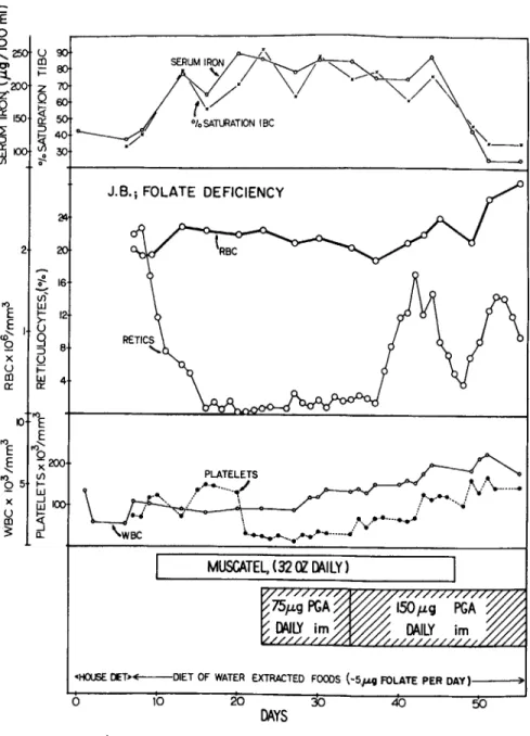
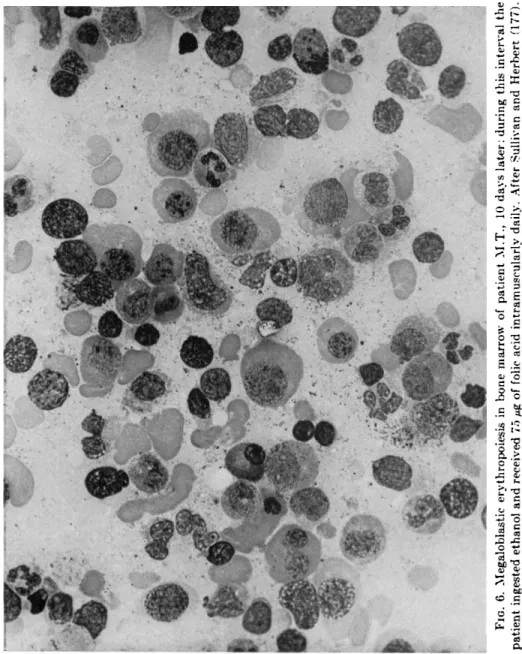
It has been recently demonstrated that alcohol suppresses the hema- topoietic response to minimal doses of folic acid in patients with megalo- blastic anemia secondary to folate deficiency (176, 177). In addition to preventing the expected rise in reticulocytes, leukocytes, and platelets in response to minimal doses (75 Mg ) of folic acid (Figs. 3 and 4), alcohol ingestion prevented the conversion of bone marrow morphology from megaloblastic to normoblastic and, conversely, caused reversion of previously normoblastic marrow morphology to megaloblastic (Figs.
5 and 6), in spite of continued intramuscular injections of folic acid and maintenance of dietary intake (177). The inhibition of hematopoiesis
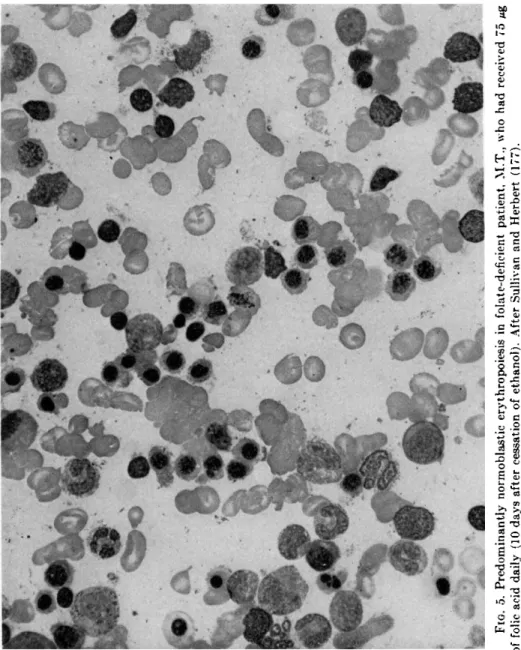
FIG. 5. Predominantly normoblastic erythropoiesis in folate-deficient patient, M.T., who had received 75 Mg of folic acid daily (10 days after cessation of ethanol). After Sullivan and Herbert (177).
FIG. 6. Megaloblastic erythropoiesis in bone marrow of patient M.T., 10 days later; during this interval the patient ingested ethanol and received 75 μg of folic acid intramuscularly daily. After Sullivan and Herbert (177).
8. FOLATES IN HUMAN NUTRITION 387 by alcohol can be partially overcome by larger doses of folic acid; this finding suggests that alcohol increases the folate requirement under these experimental conditions, but the mechanism of this phenomenon is unknown. Alcohol ingestion was subsequently found to have similar effects in patients with pernicious anemia, but in patients with iron deficiency anemia it did not inhibit the hematological response to iron repletion (224). More recently, Sullivan and Liu studied a patient with chronic hemolytic anemia (hereditary elliptocytosis) and normal serum and erythrocyte folate levels (224a). During the ingestion of 312 gm of ethanol, the patient's reticulocyte, leukocyte, and platelet counts fell, utilization of F e69 for erythropoiesis decreased and the bone marrow became megaloblastic, although the patient continued to consume a normal hospital diet. These changes were largely (but not completely) reversed by the administration of 1 mg of folic acid. The urinary excretion of FIGLU and AICAR were not significantly increased during alcohol ingestion.
Bertino et al. (225) have found that alcohol inhibits the incorporation of formate-1 4C, but not of thymidine-3!! into nucleic acids of bone marrow cells in vitro. This effect appears to be due to competitive inhibition of tetrahydrofolate formylase by alcohol in studies with the partially purified enzyme.
I. Role of Anticonvulsant Drugs
The role of anticonvulsants in the etiolgy of folate deficiency and megaloblastic anemia remains unclear. Since the report of Mannheimer et al. (226) of the association of megaloblastic enemia with anticonvulsant therapy, more than 60 cases have been reported. Although a few hemato- logic responses to vitamin Bi 2 have been reported (227, 228), the majority of patients have responded to folic acid with or without interruption of anticonvulsant therapy (228, 229). Some patients have improved simply upon withdrawal of anticonvulsant medication (230), but the possibility exists that improvement in these and other patients was due to ingestion of folates in the diet (229, 230). When six patients with megaloblastic anemia and folate deficiency associated with anticonvulsant drug therapy were followed while on a folate-deficient diet with continued anticonvulsant drug therapy, Soeparman (228) found that doses of 100 to 500 Mgnof folic acid resulted in reticulocyte responses in five of them. The sixth patient did not respond to 100 Mg nor 200 Mg of folic acid daily, but did thereafter show a sustained reticulocyte response and rise in hemoglobin and hema- tocrit when diphenylhydantoin and primidone were discontinued.
388
Klipstein reported that 58% of clinic patients ingesting diphenyl- hydantoin for seizure disorders had subnormal serum folate levels (231), and that the incidence of these low levels was greater in those patients who had taken the drug for more than five years. It is unfortunate that serum folate levels in a control series of other clinic patients was not reported, in view of the recent finding that 45% of all patients admitted to an urban medical center, serving primarily an indigent population, had subnormal serum folate levels (232). The clearance of injected folic acid from the plasma was found to be rapid in some patients (231) but normal in others (233). Girdwood and Lenman (234) and others (63, 231, 235) have pointed out the structural similarities of folic acid and diphenyl- hydantoin, primidone, and phenobarbital, with the suggestion that there might be slight competitive inhibition of folic acid by these compounds.
Herbert and Zalusky (63) suggested that because of the close structural resemblance of diphenylhydantoin to the 5-membered ring of Λ"5·1 0- methenyl-FH4, there may be weak competitive inhibition of the conver
sion of this compound to iV6-methyl-FH4. Klipstein speculated that diphenylhydantoin may displace folic acid from its carrier plasma protein, analogous to the displacement of thyroxine from its carrier protein by diphenylhydantoin (231).
In vitro studies did not produce evidence of inhibition of L. casei growth in cultures to which diphenylhydantoin, primidone, or pheno
barbital was added (231). Studies by Baker et al. (236) indicate that the growth of folate-requiring Crithidia fasciculata (culex strain) and thymine or thymidine-requiring E. coli I are inhibited by primidone. It was noted that this inhibition could be reversed in the former organism by folic acid and in the latter by thymidylic acid. Biswas (237) found that the growth inhibition of E. coli K12 caused by barbituric acid could be reversed by uracil, cytosine, and thymine and their respective nucleotides and nucleosides, whereas orotic and orotidylic acids were without effect. It was suggested that barbituric acid caused a block in the conversion of orotidylic to uridylic acid and thus impaired pyrimidine synthesis. A similar defect in pyrimidine synthesis (congenital oroticaciduria) resulting in megaloblastic anemia has been reported (238) and was shown to be the result of deficiencies of orotidylic pyrophosphorylase and orotidylic decarboxylase, which are necessary for the conversion of orotic to uridylic acid (239).
Soeparman (228) emphasized the fact that most patients with megaloblastic anemia associated with anticonvulsant medications have subsisted on diets inadequate in folate content. It would appear that in man anticonvulsant medications have very weak, if any, antifolate properties and that if such medications do contribute to the development of megaloblastic anemia in man, they do so only in the presence of deficient
8. FOLATES IN HUMAN NUTRITION 389 body stores of folate. This may be similar to the effects of alcohol upon erythropoiesis in folate-deficient humans (177).
V I . CLINICAL AND LABORATORY FINDINGS IN FOLATE DEFICIENCY
A. Signs and Symptoms of Folate Deficiency
The extreme result of folate deficiency in man is a profound disturb- ance in cellular proliferation, the chief manifestation being megaloblastic anemia. The body systems with the most active cellular turnover rates (bone marrow, gut, integument, gonads) are the ones most obviously affected, though the effects of folate deficiency result in a generalized illness which affects almost all body systems directly or indirectly.
Patients with folate deficiency may complain of anorexia, weight loss, weakness, malaise, dyspnea, palpitations, syncope, irritability, and poor memory (7, 90, 217). There may be findings, such as fever, diarrhea, steatorrhea, from associated illnesses (8, 9, 11, 15, 217). A detailed dietary history almost always reveals a poor dietary intake (especially of foods rich in folate content) associated with alcoholism, bizarre food habits, ignorance, or poverty, except when folate deficiency is secondary to a malabsorption syndrome. Examination of the patient may reveal pallor of the skin and mucous membranes, mild icterus of the sclerae and skin, a pale smooth tongue, wasting of subcutaneous tissues, diffuse or focal increased pigmentation of the skin, and enlargement of the liver and spleen (8, 9, 240, 241). Because the patient's history and physical findings are not specific for folate deficiency, definitive diagnosis rests upon laboratory procedures or clinical therapeutic trial.
B. Laboratory Findings
1. Hematological Changes
The sequential changes occurring after the onset of folate deprivation (90) were related in a previous section. As emphasized by Herbert (7), careful evaluation of the peripheral blood smear often provides the first indication of folate deficiency. Hypersegmented polymorphonuclear leukocytes and large oval erythrocytes (macroovalocytes) are found.
Further evaluation may reveal mild to moderate anemia, and when the patient is severely anemic (hematocrit less than 20%), leukopenia and thrombopenia frequently are present. Megaloblasts are found in the bone marrow, along with large granulocyte precursors and megakaryocytes.
Generally, the severity of the bone marrow morphological abnormalities varies directly with the degree of anemia. In patients with severe anemia, the rise in granulocytes in the peripheral blood, following a standard endotoxin challenge, is less than normal suggesting that the bone marrow granulocyte reserve is diminished (241a). Whether this is due to "ineffec-
390
tive leukopoiesis" in megaloblastic anemia or to other factors, remains to be determined. With correction of the folate deficiency, the marrow granulocyte reserve returns to normal (241B).
2. Tests of Gastrointestinal Function
Gastrointestinal function tests are of importance in differentiating megaloblastic anemia due to Addisonian pernicious anemia (lack of gastric intrinsic factor), and malabsorption of vitamin Bi 2, from folate deficiency resulting from inadequate dietary intake or malabsorption of folate. Gastric secretions should be tested for acid and intrinsic factor
(242). Because some apparently normal individuals may not secrete acid (243) or intrinsic factor (244) without stimulation of gastric secretory function, the maximal histamine test should be employed (243). Intrinsic factor secretion is normal in dietary folate deficiency (244). Tests of folic acid absorption may be utilized (16, 150), but they have the disad- vantage that the amount of folic acid administered may induce a hemato- logical response in patients with vitamin B i2 deficiency (159). Using isotopically labeled vitamin Bi2, absorption of this vitamin may be evaluated by measurement of radioactivity excreted in the feces (245), the appearance of radioactivity in the plasma (246-248), uptake into the liver (249) or the whole body (249a), or the excretion in the urine (250, 251). The urinary excretion test is the most widely employed of these procedures; it gives reproducible results (244, 251) but shares a disadvan- tage in common with folate absorption tests—namely, hematological responses to the flushing doses of vitamin Bi2 given may occur in patients with folate deficiency (252, 253).
Although abnormalities in the jejunal mucosa associated with the administration of folate antagonists have been noted in man (254, 255), folate deficiency resulting from restriction of dietary folate intake does not appear to be associated with any morphological alterations in the jejunal mucosa (90, 256, 257); this suggests that in tropical sprue the histological changes in the small bowel are not primarily the result of folate deficiency.
3. Biochemical Findings
The earliest detectable abnormality in the development of folate deficiency is a fall in the serum folate level (90) indicating that plasma folate is the most labile of the body folate compartments.^ Following further folate depletion, erythrocyte folate levels drop to low levels and the excretion of formiminoglutamic acid in the urine is increased (90, 217), an indication of tissue folate deficiency. There may be increased excretion of aminoimidazolecarboxamine (AIC) in the urine (112, 113).
Occasionally in patients with folate deficiency, serum folate levels may be subnormal, but not in the "deficiency" range (9, 217). Whether this is the result of release of folates from tissue stores under certain conditions, as has been shown to occur with vitamin B i2 in liver damage (224), or to other factors, is unknown. A slightly delayed plasma clearance of intravenously injected folic acid was found in one patient during a period of alcohol ingestion, whereas before and after this period folate clearance was normal (224). Hogan et al. (218), in studying patients with myelo
fibrosis, found that plasma clearance of injected folic acid resolved the patients with "borderline" serum folate levels into two groups—those with rapid and those with normal clearance rates. It was suggested that the former group, but not the latter, was deficient in folate. Mollin (217) reported that erythrocyte folate levels and urine formiminoglutamic acid (FIGLU) excretion in this "borderline" group served to differentiate those with low erythrocyte folate concentrations and increased FIGLU excretion and thus folate deficient, from those in whom these parameters were normal.
The excretion of methylmalonate in the urine is normal in folate deficiency, in contrast to the elevated excretion of this metabolite of propionate in vitamin Bi2-deficient subjects (258, 259).
C. Principles of Therapeutic Trial
In many instances, when suitable laboratory facilities are not available or the results of laboratory investigation are inconclusive, therapeutic trial can be a valuable adjunct in the differential diagnosis of megalo
blastic anemia (260, 261). If properly performed, it can provide informa
tion obtainable in no other way.
The requirements for therapeutic trial include:
1. A control period of a few days to establish the baseline state of erythropoiesis as reflected by the reticulocyte count, and erythrocyte count, hemoglobin or hematocrit.
2. A diet devoid of foods that are rich sources of folate or vitamin Β12, since these may result in a hematological response that might erroneously be attributed to the therapeutic agent being tested.
3. The administration of a small dose of the therapeutic agent to avoid the possibility of an "inappropriate" response, e.g., 50 to 200 Mg of folic acid, is given parenterally or by mouth if folate deficiency is sus
pected; 1 to 5 Mg of cyanocobalamin is given parenterally if the patient is thought to be deficient in vitamin Bi2.
4. Daily (or every second day) reticulocyte counts and twice-weekly erythrocyte counts, hemoglobin or hematocrit determinations throughout the control and test periods.
392
A significant rise in reticulocytes, (usually beginning 48 to 96 hours after administration of the deficient agent) indicates that the patient was lacking the hematopoietic agent. If there is no response after 10 days, and the patient is free of infection, renal disease, active inflam- matory diseases (e.g., collagen diseases, active liver disease) and is not taking alcohol, chloramphenicol or other agent known to be capable of suppressing hematopoiesis, then the possibility of deficiency of the agent being tested is excluded.
D. Etiological Classification of Folate Deficiency
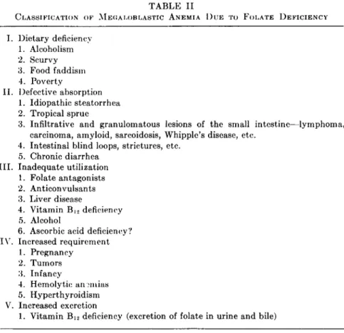
An exhaustive listing of the various conditions known to cause, or to be associated with folate deficiency has been presented by Herbert (8, 9). Table II lists the more common causes of folate deficiency in man.
TABLE II
CLASSIFICATION OF MEGALOBLASTIC ANEMIA D U E TO FOLATE DEFICIENCY
I. Dietary deficiency 1. Alcoholism 2 . Scurvy 3. Food faddism 4. Poverty
II. Defective absorption 1. Idiopathic steatorrhea 2 . Tropical sprue
3. Infiltrative and granulomatous lesions of the small intestine—lymphoma, carcinoma, amyloid, sarcoidosis, Whipple's disease, etc.
4. Intestinal blind loops, strictures, etc.
5. Chronic diarrhea III. Inadequate utilization
1. Folate antagonists 2 . Anticonvulsants 3. Liver disease
4. Vitamin B i2 deficiency 5. Alcohol
6 . Ascorbic acid deficiency?
IV. Increased requirement 1. Pregnancy 2 . Tumors 3. Infancy
4. Hemolytic anemias 5. Hyperthyroidism V. Increased excretion
1. Vitamin B i2 deficiency (excretion of folate in urine and bile)