The cellular basis of rhythmic activity patterns occurring in the CA1 region of the hippocampus
Ph.D. Dissertation Rita Zemankovics Semmelweis University
János Szentágothai School of Ph.D. Studies
Supervisor: Norbert Hájos Ph.D.
Institute of Experimental Medicine Hungarian Academy of Sciences
Laboratory of Network Neurophysiology
Official Reviewers of the Ph.D. Dissertation:
Gábor Czéh Ph.D., D.Sc.
Gábor Gerber Ph.D.
Members of the Final Examination Board:
József Kiss Ph.D., D.Sc.- Chairman József Takács Ph.D.
István Tarnawa Ph.D.
Budapest
2011
2
1. TABLE OF CONTENTS
1. Table of contents………... 2
2. List of abbreviations……….. 5
3. Introduction………... 7
3.1. Review of the literature………... 8
3.1.1. Anatomy of the hippocampus………. 8
3.1.1.1. The cytoarchitecture of the hippocampus……….. 8
3.1.1.1.1. Principal cells……….. 8
3.1.1.1.2. Interneurons……… 11
3.1.1.2 Hippocampal connectivity………... 15
3.1.1.2.1. Cholinergic neuromodulation in the hippocampus.……..….. 18
3.1.2. Behavior related activity patterns of the hippocampus………..……...20
3.1.2.1 Theta oscillations in the hippocampus………. 22
3.1.2.2. Gamma oscillations in the hippocampus………...…………. 25
3.1.3. Possible models of rhythm generation within a network………. 28
3.1.4. In vitro methods to study rhythm-generating mechanisms of the brain………. 29
3.1.4.1. Investigating the resonance properties of single cells………. 29
3.1.4.1.1. Ih (h-current)……… 30
3.1.4.2. Introducing in vitro network oscillation models……...………….. 33
3.1.4.2.1. In vitro gamma oscillation models……….. 34
4. Aims of Thesis………... 36
5. Methods……….. 38
5.1. Ethical approval……… 38
5.2. Tissue preparations……….………... 38
5.3. Tissue storage... 39
5.4. Solutions……….………... 39
5.5. Electrode preparation... 40
5.6. Electrophysiological recordings and data analysis in study I... 40
5.6.1. Data acquisition... 40
3
5.6.2. Characterization of the passive membrane properties of the cells... 41
5.6.3. Characterization of the “sag” potential……… 42
5.6.4. Characterization of neuronal impedance profiles and resonance properties……….. 42
5.6.5. Characterization of Ih in different cell types……… 45
5.6.6. Statistical analyses………... 47
5.7. Computational model used in study I……….. 48
5.8. Electrophysiological recordings and data analysis in study II….……… 49
5.8.1. Data acquisition……….…………... 49
5.8.2. Event detection and analysis……….…… 50
5.8.3. Statistical analyses………..………... 52
5.9. Anatomical identification of the neurons………..………... 53
6. Results……… 54
6.1. Part I.: Single cell resonances in hippocampal CA1 neurons produced by intrinsic membrane characteristics……….. 54
6.1.1. The different cell types investigated in this study….……… 55
6.1.2. Basic electrophysiological characteristics………..………… 56
6.1.3. Distinct features of depolarizing sag in hippocampal neuron types….. 58
6.1.4. The impedance profiles and resonance properties of four types of hippocampal neurons……… 59
6.1.5. The involvement of Ih in impedance profiles and resonance…………. 63
6.1.6. The properties of Ih in the different cell types……….……….. 66
6.1.7. Computational model……….………… 69
6.2. Part II. Network resonances of hippocampal CA1 produced by synaptic synchronization……….74
6.2.1. Methodological requirements for recording high frequency oscillations under submerged conditions………. 75
6.2.2. CCh-induced fast network oscillations………..……. 77
6.2.3. Classification of the investigated cell types……….……….. 77
6.2.4. Firing properties of different cell types during CCh-induced network oscillations in hippocampal slices……… 80 6.2.5. The effects of synaptic inputs on the firing properties of the different cell
4
types during CCh-induced gamma frequency oscillations………... 85
6.2.5.1. The properties of synaptic currents in the different cell classes…. 85 6.2.5.2. Correlations between firing properties and synaptic currents in the different cell types……… 90
6.2.5.3. Phase and time relations between firing and synaptic inputs in the different cell classes………. 94
7. Discussion……….. 97
7.1. Resonance properties of different cell-types of the hippocampal CA1... 97
7.1.1. Physiological relevance………..……… 100
7.2. Gamma-frequency oscillations in the CA1 region of the hippocampus.. 102
7.2.1. The properties of synaptic inputs of the different cell types during CCh-induced gamma oscillation in CA1……….. 105
7.2.2. What determines the firing activity of hippocampal CA1 neurons during CCh-induced in vitro gamma oscillations?... 108
7.2.3. Physiological relevance……..……… 110
7.3. General Discussion……….……….. 112
8. Conclusion………. 114
9. Summary……….………... 115
10. Összefoglalás………116
11. Acknowledgements……….. 117
12. References……… 118
13. List of publications……….. 135
13.1. Publications related to the dissertation……… 135
13.2. Other publications………...………... 135
14. Appendix……….. 136
14.1. Computational models used in study I…………...……….. 136
5
2. LIST OF ABBREVIATIONS
ACh: acetylcholine
ACSF: artificial cerebrospinal fluid AHP: afterhyperpolarization
AMPA: α-amino-3-hydroxy-5-methylisoxazole-4-propionic acid AP: action potential
BIC: Bayesian information criterion CA1-CA3: Cornu Ammonis fields CA1 PC: CA1 pyramidal cell CA1 IN: CA1 interneuron CA3 PC: CA3 pyramidal cell CA3 IN: CA3 interneuron
cAMP: cyclic adenosine monophosphate CB1: cannabinoid receptor type 1 CCh: carbachol
CCK: cholecystokinin
cGMP: cyclic guanosine monophosphate CNBD: cyclic-nucleotide binding domain DG: dentate gyrus
EEG: electroencephalogram EC: entorhinal cortex
eGFP: enhanced green fluorescent protein EPSC: excitatory postsynaptic current EPSP: excitatory postsynaptic potential FFT: Fast Fourier Transform
FS PTI: fast spiking perisomatic region-targeting interneuron GABA: γ-aminobutyric acid
HCN channel: hyperpolarization-activated cyclic nucleotide-gated channel Ih: h-current; a current mediated by HCN channels
IN: interneuron
6 IPSC: inhibitory postsynaptic current
IPSP: inhibitory postsynaptic potential ISI: interneuron-selective interneuron KAR: kainate receptor
KCC2: K+- Cl- cotransporter KW: Kruskal-Wallis test
M1-M5: muscarinic acetylcholine receptor subtypes mAChR: muscarinic acetylcholine receptor
mGluR: metabotropic glutamate receptor MP: membrane potential
MS-DBB: medial septum-diagonal band of Broca nAChR: nicotinic ACh receptor
NMDA: N-methyl-D-aspartate OA IN: oriens-alveus interneuron
OLM: oriens-lacunosum-moleculare cell O-R: oriens-radiatum cell
PB: phosphate buffer PC: pyramidal cell
PIP2: phosphatidylinositol 4,5-bisphosphate PSC: postsynaptic current
PSD: power spectral density PV: parvalbumin
PV+ IN : PV-eGFP positive interneuron RAD IN: radiatum interneuron
REM: rapid eye movement sleep Rp: Rayleigh probability
SCA : Schaffer collateral-associated interneuron SOM: somatostatin
SPW: sharp wave TTX: tetrodotoxin
ZD7288: 4-ethylphenylamino-1,2-dimethyl-6-methylaminopyrimidinium chloride
7
3. INTRODUCTION
The hippocampal formation is known to be involved in many higher order cognitive functions, such as encoding and retrieval of memory (Murray and Mishkin, 1984; Paulsen and Moser, 1998) or spatial navigation (O'Keefe and Recce, 1993). It is also well known that hippocampal circuitry exhibit a wide variety of population patterns, including oscillations at theta (4-7 Hz) or gamma (30-100 Hz) frequencies, or sharp-wave-ripple oscillations (100-200 Hz) under different behavioral states (Vanderwolf, 1969; Vanderwolf et al., 1977; Leung, 1980; Buzsáki et al., 1992;
Chrobak and Buzsáki, 1994). Several lines of studies show that the occurrence of these special behavior-dependent activity patterns strongly correlates with different cognitive functions. There are many theories related to these observations that suggest that the complex dynamics presumably reflects specific information processing states of the networks. However, the exact role of these complex activity patterns in different operations of the brain remains elusive.
One way to get closer to understand the involvement of rhythmic oscillations occurring at different frequencies in certain brain functions is to reveal the underlying cellular mechanisms. Unfolding the contribution each component of the network makes to network oscillations will produce mechanistic as well as functional insights into their generation and role. In this thesis I would like to address the question what mechanisms tune the network to operate at certain frequencies in the hippocampus. To answer this we have to investigate the intrinsic properties of the individual elements of the network as well as the complex properties of the circuitry that makes the system being capable of generating or following rhythmic activity.
One theme in this thesis will be the intrinsic membrane properties of the different types of hippocampal CA1 neurons that can endow them with the capability of operating at certain preferred frequencies. I will review a study in which we showed that different cell classes in the CA1 region of the hippocampus have distinct types and degrees of frequency preference, and that these differences are partly due to variations in the quantity and biophysical properties of the voltage-gated ion channels, which mediate the so-called h-current.
8
The second theme of the thesis regards to the network properties of the hippocampal CA1 that can be important in defining the occurrence of rhythmic population patterns in this region. Here, I present a study that focuses on network oscillations occurring in the gamma frequency range in the hippocampus, and shows that under particular circumstances gamma oscillation in the CA1 region is driven by the activity of the CA3 region via a feed-forward inhibitory pathway.
3.1. Review of the literature
3.1.1. Anatomy of the hippocampus
In order to investigate network activity, a detailed knowledge not only of the components of the network, but also of how individual components are connected is required. The hippocampus in amongst the most intensively studied regions of the mammalian brain and thanks to the works of many outstanding researchers, its anatomy is known in great detail.
Although there are substantial differences in the architecture and in patterns of connectivity of the hippocampal formation between different species, the basic layout of cells and fiber pathway is phylogenetically conserved and much the same even in humans and rodents. Since the experimental work described in this thesis was carried out in rodents, the following description of the hippocampus is limited to the structure of the rodent hippocampus.
3.1.1.1. The cytoarchitecture of the hippocampus 3.1.1.1.1. Principal cells
The hippocampal formation includes the dentate gyrus (DG), the Cornu Ammonis fields (CA1-CA3), the subiculum, the parasubiculum and the entorhinal cortex (EC). In the narrow sense the hippocampus is defined as comprising the DG and the areas CA3-CA1.
9
Hippocampal neurons can be divided into two major categories based on their main neurotransmitters. Eighty five % of the cells are glutamatergic. These cells are called principal cells, since they form the major output pathways from hippocampal subregions. The remaining 15 % of the neurons are mainly GABAergic cells (i.e.
releasing γ-aminobutyric acid as a main neurotransmitter). These cells are usually defined as interneurons, since they thought to be mainly local-circuit neurons, though certain classes of them establish commissural axon collaterals or even extrahippocampal projections.
The laminar structure of the hippocampus is basically defined in each subregion by the morphology of the principal cells and the main termination zones of their glutamatergic inputs arriving via distinct pathways.
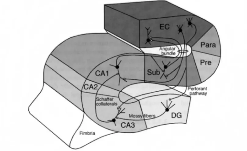
Figure 1. The information flow through the hippocampus. The hippocampus has bidirectional connections with the EC. Cells in the layer II of the EC project to the DG and the CA3 region of the hippocampus via the perforant path. Neurons in layer III of the EC project to the CA1 region of the hippocampus via the temporoammonic pathway. The axons of the granule cells in the DG form the mossy fibers and innervate the CA3 cells. Besides their dense local recurrent collaterals, CA3 pyramidal cells innervate the CA1 neurons via the Schaffer collaterals. CA3 neurons also innervate the contralateral hippocampus via the commissural afferents. Neurons in the CA1 field of the hippocampus and the subiculum project back to the layer V-VI of the EC.
(Adapted from Andersen et al., 2007.)
The principal cells of the DG are the granule cells that are localized in one layer called the granule cell layer. Besides the granule cell layer, the DG has two other layers: the molecular layer and the hilus. The molecular layer is a largely acelullar layer
10
located above the granule cell layer, and consists the dendrites of the granule cells. The hilus is located below the granule cell layer and comprises different polymorphic cells.
The axons of the granule cells innervate some of the neurons of the hilus, such as mossy cells, then exit the hilus and enter the stratum lucidum of CA3 as a bunch of fibers.
These fibers are also called the mossy fibers, because of their special synaptic terminals formed on the CA3 neurons (Hamlyn, 1962) (Fig. 1).
The other excitatory (glutamatergic) cell type of the DG is the mossy cell (Amaral, 1978). Mossy cells project only to the molecular layer of the DG both ipsilaterally and contralaterally. These neurons tend not to project locally, but rather distant septotemporal levels of the DG.
The principal cells of the Cornu Ammonis field are the pyramidal cells. They are located also in one layer called stratum pyramidale. Unlike granule cells, pyramidal cells have dendritic trees extending perpendicularly to the cell layer in both directions.
Based on the size of the pyramidal cells and their synaptic innervations, the Cornu Ammonis field is usually divided into three subfields; CA1, CA2 and CA3 (Lorente de Nó, 1934) (Fig. 1).
The CA3 region can be found near the DG and can be further subdivided into CA3c, b and a (from the hilus towards the CA1, respectively). CA3 contains pyramidal cells with relatively large somata. The apical dendrites of CA3 pyramidal cells traverse three strata: the stratum lucidum, the stratum radiatum and the stratum lacunosum moleculare. The basal dendrites extend into stratum oriens towards the alveus.
The stratum lucidum is unique to CA3 region. It contains the so-called thorny excrescences of the CA3 pyramidal cells, which are complex branched spines engulfed by a single large bouton of mossy fibers(Hamlyn, 1962). The other two regions of the Cornu Ammonis field receive no mossy fiber input, therefore they lack the stratum lucidum; otherwise they have the same laminar structure.
Besides giving rise to the Schaffer collaterals that project to the CA1 region, the axons of CA3 pyramidal cells also form a massive recurrent collateral system within the CA3 and give rise to commissural projections to the contralateral hippocampus.
The CA2 subfield is a very narrow zone of cells inserted between CA3 and CA1.
CA2 pyramidal cells have large cell bodies like CA3 pyramidal cells, but receive no inputs from the DG, like CA1 cells. Like CA3 pyramidal cells, CA2 pyramidal cells
11
also have a recurrent collateral system as well as projections toward the CA1 and to the contralateral hippocampus.
The cell bodies of pyramidal cells in CA1 are smaller than in the other two Cornu Ammonis regions. CA1 pyramidal cells give rise to projections both to the subiculum and to the deep layers of the EC, without any prominent local collateral system (Fig. 1).
3.1.1.1.2. Interneurons
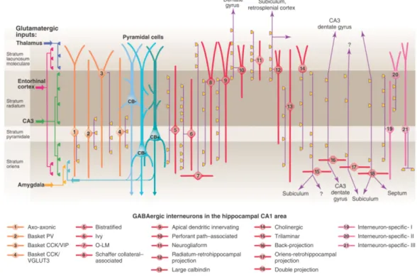
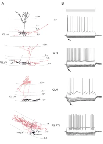
Compared to the relatively homogenous population of pyramidal neurons, interneurons of the hippocampus have been found to be much more diverse. For instance, just in the CA1 region at least 21 different types of GABAergic neuron have been described so far (Klausberger and Somogyi, 2008; Fig. 2). The morphological categorization of the interneurons is based on their dendritic and axonal arborization.
The location of the dendritic tree can reveal the potential sources of inputs to these cells, while the location of the axonal arborization can disclose the possible postsynaptic targets of the interneurons. Therefore, this type of classification can be very useful, if we want to understand the function of the interneurons within the network.
The propensity for interneurons to express so-called neurochemical markers, such as different neuropeptides or Ca2+-binding proteins allow us to refine their classification and can also help us in identifying them more precisely.
Depending on their axonal arborization, three main types of inhibitory cells can be distinguished: perisomatic region targeting interneurons; dendritic region targeting interneurons, and the so-called interneuron-selective inhibitory neurons.
Perisomatic region targeting interneurons innervate the perisomatic region of the pyramidal cells. These interneurons are in a good position to control the action potential generation of pyramidal cells. They are mainly located within or near the stratum pyramidale. Their dendrites project into stratum radiatum and stratum oriens and thus may receive excitatory inputs from Schaffer collaterals, commissural associational fibers and feedback synapses from local pyramidal neurons. There are also mutual inhibitory connections and dense gap junctional coupling among these interneurons (Freund and Buzsáki, 1996). Perisomatic region targeting interneurons can be further classified into basket cells and axo-axonic cells.
12
Each basket cell can make multiple contacts onto pyramidal neurons with its terminals forming a “basket” around pyramidal cell somata. The group of basket cells can be further subdivided based on their neurochemical markers. The cells expressing the neuropeptide cholecystokinin (CCK) have regular firing pattern and express various types of neuromodulatory receptors(Pawelzik et al., 2002; Freund and Katona, 2007).
These cells require extensive integration of excitatory inputs to reach firing threshold (Glickfeld and Scanziani, 2006). In contrast, basket cells expressing the Ca2+ binding protein parvalbumin (PV) show fast spiking phenotype and have only few receptors for neuromodulatory molecules. These fast spiking PV-expressing basket cells can be quickly and easily recruited by excitation and provide an effective inhibition on pyramidal cells (Freund, 2003; Glickfeld and Scanziani, 2006; Freund and Katona, 2007). Based on these characteristic differences between the two basket cell-types, the PV- and CCK-containing basket cells have been suggested to play different roles in network oscillations. PV-expressing basket cells could operate as clockworks for oscillations, while CCK-expressing basket cells could function as a fine tuning device that modulate synchronous ensemble activities as a function of subcortical inputs, which carry information about emotions, motivation and the autonomic state of the animal (Freund, 2003; Freund and Katona, 2007).
Axo-axonic cells, as implied by the name, target the axon initial segment of the principal cells (Somogyi et al., 1985). These cells also express PV and tend to have fast spiking firing characteristics. An interesting feature of axo-axonic cells is that their postsynaptic effect can be, at least in some cases, excitatory, possibly due to low K+- Cl- cotransporter (KCC2) expression in the axon initial segment (Szabadics et al., 2006; but see Glickfeld et al., 2009).
Numerous types of interneurons target the dendrites of principal cells. Because their axonal arborization usually overlaps with the termination zone of the distinct excitatory pathways, they could control the efficacy of the different excitatory inputs in a very specific way.
Oriens-lacunosum-moleculare cells (OLM cells) are interneurons with somata and dendrites located in the stratum oriens and an axonal arbor in the stratum lacunosum moleculare (McBain et al., 1994; Sík et al., 1995; Maccaferri and McBain, 1996). They target the apical tufts of the pyramidal cell dendrites, thus their axonal
13
termination zone overlaps mostly with the EC inputs to pyramidal cells, though the presence of a few OLM axon collaterals has also been reported in the stratum oriens.
(Sík et al., 1995). OLM cells express the neuropeptide somatostatin (SOM), high level of mGluR1α (Baude et al., 1993) and also some PV, though at lower levels compared to fast spiking perisomatic region targeting interneurons (Klausberger et al., 2003).
The apical dendritic tufts of the pyramidal cells are innervated also by the so- called perforant path-associated cells. The cell bodies of these cells are usually located at the border of the strata radiatum and lacunosum-moleculare, their dendrites can either cover all layers or remain in stratum lacunosum moleculare (Hájos and Mody, 1997;
Klausberger et al., 2005).
The Schaffer collateral-associated (SCA) cells innervate the oblique dendrites and to a lesser extent also the basal dendrites of pyramidal cells, matching the excitatory input from CA3. The somata of this cell group is mainly located in the stratum radiatum, their dendrites can span all layers (Cope et al., 2002; Pawelzik et al., 2002).
SCA cells tend to express CCK (Cope et al., 2002; Pawelzik et al., 2002).
The so-called apical dendrite innervating cells have very similar morphology to SCA cells, but they innervate preferentially the main apical shaft, not the oblique dendrites (Klausberger et al., 2005).
Bistratified cells usually have cell bodies within or close to the stratum pyramidale and make synaptic contacts onto oblique and basal dendrites of pyramidal cells (Buhl et al., 1994; Klausberger et al., 2004). Although there is little overlap among their target regions, bistratified cells and fast spiking perisomatic region targeting interneurons share several features. Both cell types express PV and show fast spiking phenotype (Klausberger et al., 2004). The dendritic morphology of these cells is also similar, however, in contrast to basket and axo-axonic cells, the dendrites of bistratified cells do not enter the stratum lacunosum-moleculare (Buhl et al., 1996; Halasy et al., 1996).
Some interneurons, such as neurogliaform cells and ivy cells have densely packed axon arborization and tend to evoke slow GABAA receptor and also GABAB
receptor mediated responses (Price et al., 2005; Price et al., 2008). Neurogliaform cells are located mainly in the stratum lacunosum moleculare and target the apical tufts (Price et al., 2005; Price et al., 2008), while ivy cells are placed in the stratum pyramidale or
14
radiatum and innervate the oblique and basal dendrites of pyramidal cells (Fuentealba et al., 2008).
Figure 2. The diversity of cell types of the hippocampal CA1. According to the review of Klausberger and Somogyi (2008) there are at least 21 classes of interneuron in the hippocampal CA1 area. In this review the authors also distinguished at least 3 different types of CA1 pyramidal cells based on their somatic localization and their immuno-positivity for the Ca2+- binding protein, calbindin. In the figure the main termination of the five glutamatergic inputs of CA1 are indicated on the left. The somata and dendrites of interneurons innervating pyramidal cells (blue) are orange, and those innervating mainly other interneurons are pink. Axons are purple; the main synaptic terminals are yellow. Note the association of the output synapses of different interneuron types with the perisomatic region (left) and either the Schaffer collateral/commissural or the entorhinal pathway termination zones (right), respectively. VIP, vasoactive intestinal polypeptide; VGLUT, vesicular glutamate transporter; O-LM, oriens lacunosum moleculare cell. (Adapted from Klausberger and Somogyi, 2008.)
There are several types of GABAergic neurons in the hippocampus that are not local interneurons, but send long range projections to other brain areas. Many of them are located in the stratum oriens and have horizontally running dendrites. Trilaminar cells have axonal arborization in three layers within the hippocampus (strata radiatum, pyramidale and oriens) and project to the subiculum (Sík et al., 1995; Ferraguti et al., 2005). Backprojecting cells in addition to innervating different layers of the CA1 area, project backwards to CA3 or DG (Sík et al., 1994). Oriens retrohippocampal projection cells send axons to the subiculum and retrohippocampal areas (Jinno et al., 2007), while
15
the so-called double projection neurons project to the medial septum and often also to the subiculum (Gulyás et al., 2003; Jinno et al., 2007; Takács et al., 2008).
Retrohippocampal projection neurons can also be found in the stratum radiatum (Jinno et al., 2007)
Interneurons described so far target mainly pyramidal cells, but can also target other interneurons. The so-called interneuron-selective interneurons (ISI) target predominantly, if not exclusively other interneurons. ISIs also have at least three different subtypes and many of them can be identified based on their calretinin content (Acsády et al., 1996a,b; Freund and Buzsáki, 1996; Gulyás et al., 1996).
3.1.1.2. Hippocampal connectivity
Most of the sensory information reaches the hippocampus through the EC via the perforant path. The entorhinal projections to the DG and CA3/CA2 originate from cells in layer II. The synaptic contacts of this projection in DG and CA2/CA3 show a laminar organization; inputs from the lateral entorhinal area terminate superficially in the molecular layer/stratum lacunosum-moleculare, while inputs from the medial entorhinal area terminate in the middle third of the molecular layer or the deep half of the stratum lacunosum moleculare. The EC also project to the CA1, but this projection originates from layer III cells and the pattern of terminal distribution is organized in a topographical fashion; the lateral entorhinal area projects to the distal portion of the CA1 (close to the subiculum), while fibers originating in the medial entorhinal area terminate in the proximal portion (close to CA2). In a recent study of Chevaleyre and Siegelbaum (2010) the authors showed that CA2 neurons are also innervated by layer III EC cells, which means that CA2 pyramidal cells receive convergent excitatory inputs from layer II and layer III EC neurons on their distal dendrites.
Within the hippocampus the progression of excitatory pathways is largely unidirectional. This special chain of excitatory synaptic connections is also known as the trisynaptic loop, and can be summarized as follows: Granule cells of the DG receive their major input from the layer II of the EC via the perforant pathway (1st synapse).
The axons of the granule cells form the mossy fibers and innervate CA3 pyramidal cells (2nd synapse). Besides their dense local recurrent collaterals (associated connections),
16
CA3 pyramidal cells innervate CA1 pyramidal cells via the Schaffer collaterals (3rd synapse). CA1 pyramidal cells project to the subiculum and back to the layer V-VI of the EC (Fig. 1). The mossy fiber projections do not appear to have topographical organization (Paxinos, 2004).
The recurrent collaterals of CA3/CA2 pyramidal cells form dense associational connections in the stratum radiatum and stratum oriens of the CA3 and CA2. All portions of the CA3 and CA2 project to the CA1. The CA3 projections to the CA1 are typically called the Schaffer collaterals and organized in a three-dimensional, topographical manner. CA3 pyramidal cells located close to the DG, i.e. within the CA3c region, are more likely to contact CA1 cells near the subicular border, and project more heavily to levels of CA1 located septal to their location. In contrast, pyramidal cells near the CA3/CA2 border, i.e. within CA3a, are more likely to target CA1 cells near the CA2/CA1 border, and project more heavily to the levels of CA1 located temporally compared to their septotemporal position (Li et al., 1994; Paxinos, 2004;
Ropireddy et al., 2011).
Axons of the CA3 pyramidal cells distribute fibers to the same fields also in the contralateral hippocampus (commissural connections). CA3 pyramidal cells do not project to the subiculum or EC, however, they project subcortically to the lateral septal nucleus.
Unlike CA3 and CA2, CA1 has much more limited associational and commissural connections, and gives rise to projections to the subiculum and EC. The axons of CA1 pyramidal cells travel in the stratum oriens or alveus and give rise to occasional collaterals, preferentially contacting local interneurons in CA1 (Tamamaki et al., 1987; Amaral et al., 1991). The projection to the subiculum is also organized topographically in the transverse plane. Cells located near the CA3 project to the distal third of the subiculum, while CA1 cells located near the subicular border tend to contact cells in the subiculum near the CA1 border. Both CA1 and subicular pyramidal cells give rise to projections to the EC, targeting the deep layers of the EC. Septal regions of the CA1 and subiculum project to the more lateral parts of the EC, while the more temporal regions project to the medial parts(Tamamaki and Nojyo, 1995; Witter et al., 2000; Naber et al., 2001).
17
Interneurons of the hippocampus can be recruited into network activities as sources of feedback or feed-forward inhibition. Those interneurons that receive their excitatory drive in parallel to the principal cells they innervate participate in feed- forward inhibition. Interneurons that are activated by the recurrent collaterals of the principal cells can contribute to feedback inhibition.
As it was already mentioned, the localization of the soma and dendritic arborization of the interneurons can determine their possible excitatory drives. For instance, perisomatic region targeting interneurons having dendrites spreading to all layers can be involved both in feedback and feed-forward circuitries (Wierenga and Wadman, 2003a,b; Pouille and Scanziani, 2001). Some perforant path associated interneurons innervating the apical tufts of the pyramidal cells have dendrites located mainly in the stratum lacunosum moleculare. Hence, these cells are likely to be excited by the EC input in a feed-forward manner (Lacaille and Schwartzkroin, 1988a,b). On the other hand, OLM cells that target the same dendritic region of pyramidal cells, but have horizontal dendrites in the stratum oriens have been shown to take part rather in feedback than in feed-forward inhibition. (Blasco-Ibanez and Freund, 1995; Sík et al., 1995). Therefore, inhibition arriving at a certain region of the pyramidal cell might be controlled by different ways depending on the network state.
The approximately 10:1 ratio of pyramidal cells to interneurons within the hippocampal circuitry (Vizi and Kiss, 1998) implies that many pyramidal cells converge onto a single interneuron, while a single interneuron contacts many pyramidal cells (Sík et al., 1995). Therefore, one single interneuron can have a widespread effect on the network (Cobb et al., 1995).
As it was mentioned above, the main neocortical input-output structure of the hippocampal formation is the EC. Besides its reciprocal connections with the EC, CA1 gives rise to projections to the perirhinal and postrhinal cortices as well as to prefrontal cortex, and also to the amygdaloid complex. These areas all project back to CA1 (Andersen et al., 2007).
The most dominant subcortical input to the hippocampus arises from the medial septum-diagonal band of Broca (MS-DBB), which has been shown to play a crucial role in the generation and modulation of certain rhythmic activity patterns of the hippocampus (Petsche et al., 1962; Lee et al., 1994). Projections from the MS-DBB
18
terminate most heavily in the stratum oriens and, to a lesser extent in the stratum radiatum (Nyakas et al., 1987; Gaykema et al., 1990). GABAergic fibers of the MS- DBB innervate selectively interneurons of the hippocampus (Freund and Antal, 1988), and some GABAergic cells also project back to the septum (Tóth and Freund, 1992;
Tóth et al., 1993; Gulyás et al., 2003; Jinno et al., 2007). Pyramidal cells of the CA3 region give rise to projections both to the ipsi- and contralateral lateral septal nucleus (Swanson and Cowan, 1977; Swanson et al., 1980).
In addition to the septal connections, the hippocampus receives a variety of neuromodulatory signals from other subcortical areas as well, including noradrenergic inputs from the locus coeruleus (Jones and Moore, 1977), serotoninergic inputs from the medial and dorsal raphe nuclei (Freund et al., 1990; Freund, 1992), or dopaminergic inputs from the ventral tegmental area (Scatton et al., 1980). The CA2 area and the DG also receives inputs from the tuberomammillaris and supramammillaris nuclei of the hypothalamus (Panula et al., 1989; Maglóczky et al., 1994). Projections from the nucleus reuniens target mainly CA1 pyramidal cells and interneurons (Wouterlood et al., 1990).
As the cholinergic input has a particular relevance to this thesis, I will review the cholinergic neuromodulation of the hippocampus in the next section.
3.1.1.2.1. Cholinergic neuromodulation in the hippocampus
The majority of the cholinergic fibers from the MS-DBB shows numerous varicosities and do not associate with a distinct postsynaptic element (Umbriaco et al., 1995). The minority of the fibers may contact either pyramidal cells or interneurons (Frotscher and Léránth, 1985; Léránth and Frotscher, 1987). Few cholinergic interneurons can be found also locally within the hippocampus(Frotscher et al., 2000).
The direct action of cholinergic modulation on hippocampal pyramidal cells is excitatory, though biphasic responses of CA1 pyramidal cells upon transient cholinergic activation have been reported (Gulledge and Kawaguchi, 2007). The responses of pyramidal cells are mediated primarily by activation of M1/M3 muscarinic receptors (Levey et al., 1995; Dasari and Gulledge, 2011). The M1/M3 receptor activation initiates secondary messenger cascades that will lead to reduction of various types of potassium conductances, such as K+ conductance mediated by the so-called M-current;
19
Ca2+-activated K+ conductance; a delayed rectifier K+ conductance; or the leak conductance (Krnjevic et al., 1971; Halliwell and Adams, 1982; Nakajima et al., 1986;
Madison et al., 1987; Halliwell, 1990; Guerineau et al., 1994). As an end effect, acetylcholine (ACh) depolarizes PCs, increases the input resistance of the cells, and so increases their excitability. ACh has also been shown to modulate some other conductances, such as voltage-gated Ca2+, or Na+ conductances (Toselli and Lux, 1989;
Cantrell et al., 1996), and it might potentiate the response to N-methyl-D-aspartate (NMDA) receptor activation (Markram and Segal, 1990).
Interneurons of the hippocampus show various responses upon cholinergic receptor activation (McQuiston and Madison, 1999a,b,c). Some interneurons in the hippocampus, such as OLM cells or CCK-expressing basket cells have been shown to be activated by application of cholinergic receptor agonists via M1/M3 receptors (Lawrence et al., 2006b; Cea-del Rio et al., 2010; Cea-del Rio et al., 2011). In contrast to M1/M3 receptor activation, activation of M2/M4 receptors induces different secondary messenger cascades, which results in hyperpolarization of the cells. M2 receptors have been shown to be expressed at the axon terminals of perisomatic region targeting interneurons (Hájos et al., 1998), where they can effectively reduce transmitter release (Fukudome et al., 2004; Szabó et al., 2010). The transmitter release can also be reduced via M4 receptors at the axon terminals of the Schaffer collaterals (Shirey et al., 2008; Dasari and Gulledge, 2011). In addition, M1/M3 receptor activation can induce synthesis of endocannabinoids in the postsynaptic cell, which in turn might reduce the transmitter release at the presynaptic terminals of CB1 cannabinoid receptor expressing cells (Fukudome et al., 2004; Neu et al., 2007). Some interneurons also express M2 receptors on their somato-dendritic compartments (Hájos et al., 1998; Ferraguti et al., 2005).
ACh can also activate ionotropic receptors, at which nicotine act as a full agonist (nAChR). The precise expression pattern of nAChR subunits in the hippocampus has not been fully established. The α7- nAChR subtype is highly expressed at multiple loci including somata, dendrites, spines and axon fibers of different cell types (Fabian-Fine et al., 2001), and can produce cell-type specific responses. Nicotinic ACh receptor activation has been shown to cause membrane depolarization in dendritic region targeting interneurons, while it appears to have no direct effect on the somata of
20
pyramidal cells or perisomatic region targeting interneurons (Frazier et al., 1998;
McQuiston and Madison, 1999a). On the other hand, it can affect transmitter release both at glutamatergic and GABAergic axon terminals (Wonnacott et al., 2006).
3.1.2. Behavior related activity patterns of the hippocampus
Several patterns of electrical activity can be recorded from the hippocampus, and these patterns are usually correlated with certain behavioral states. In the hippocampus of the freely moving rat four prominent rhythmical EEG patterns have been identified:
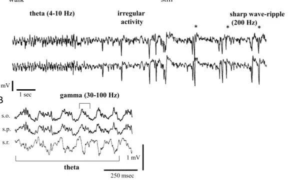
theta rhythm (occurring at 4-10 Hz); beta waves (between 12 and 30 Hz); gamma oscillations (in the range of 30-100 Hz); and sharp wave (SPW) associated ripple oscillations (from 100 up to 300 Hz) (Andersen et al., 2007) (Fig. 3).
Figure 3. Behavior-dependent rhythmic activity patterns of the rodent hippocampus. (A) Field recordings from the stratum radiatum of the left (upper trace) and the right (lower trace) CA1 region of the hippocampus of a rat during walk-immobility (still) transition. Note regular theta waves during walk and large monophasic SPWs during immobility. (B) Hippocampal field activity in the mouse recorded during wheel-running. Note that high frequency (30-100 Hz) gamma oscillation is embedded in the 4-10 Hz theta oscillation. (Adapted from Buzsáki, 1989.
and Buzsáki et al., 2003)
Theta oscillation occurs primarily during translational movements that change the location of the animal’s head with respect to the environment. For instance: walking,
21
running, exploratory head movements or struggling. Theta also occurs during rapid eye movement sleep (REM) and occasionally during immobile attention or arousal (Vanderwolf, 1969; Buzsáki et al., 1983; Andersen et al., 2007).
The full range of behavioral correlates of gamma and beta waves are less well characterized. Gamma oscillations are often embedded in theta rhythms, but can also be observed during non-theta epochs, during behaviors, such as immobility and slow wave sleep (Csicsvári et al., 2003; Senior et al., 2008). In rodents beta waves have been observed for instance during sniffing of odors associated with predators (Vanderwolf, 2001).
Sharp waves are large amplitude population events, lasting between 40-100 ms.
Sharp waves are associated with high-frequency oscillations of 100-300 Hz (ripples) and can be seen during drinking, eating, grooming, drowsiness, and slow wave sleep (Buzsáki et al., 1983; Ylinen et al., 1995b).
Although the behavioral correlates of certain oscillatory patterns have been known for a long time, it is still unresolved what the exact function of oscillatory activity is in different cognitive operations. There are many theories regarding the computational roles of rhythmic activity patterns in different brain operations.
Network oscillations have been suggested to provide a clock signal against which action potential of the individual cells can be timed. This feature of oscillations has been proposed to play a crucial role in the representation of sensory signals. For instance it can synchronize the firing of many cells and therefore can determine how the different sensory inputs can be bound into a coherent cognitive percept (Gray and Singer, 1989; Sejnowski and Paulsen, 2006). The information could also be represented by the firing phase of the cells compared to the ongoing oscillation. Phase-precession of hippocampal place cells is a well-known phenomenon that could be a good example for phase-encoding of the information (O'Keefe and Recce, 1993). Place cells are hippocampal pyramidal cells that fire preferentially, when an animal is in a specific location in an environment, also referred to as the cell's place field. When the animal enters the cell’s place field, the given place cell increases its firing rate and as the animal traverses the place field, the cell fires progressively earlier relative to the theta oscillation.
22
An important requirement for both synchronization and phase codes to work is the reliable spike timing of the cells. Synchronized membrane potential oscillations of the cells provided either by intrinsic membrane properties or rhythmic synaptic inputs could promote the temporal precision of spike generation of the cells (Kamondi et al., 1998; Penttonen et al., 1998; Pike et al., 2000).
Another possible function of oscillations could be to regulate the flow of information in neural circuits. By changing the balance between the different excitatory and inhibitory inputs, oscillations could work as a filter capable of selecting the relevant and important inputs to the network (Salinas and Sejnowski, 2000; Sejnowski and Paulsen, 2006).
By regulating the strength and the exact timing of the input signals, oscillations could set the optimal circumstances for the induction of synaptic plasticity (Bliss and Lomo, 1973; Markram et al., 1997; Song et al., 2000), and so they can assist in the storage and retrieval of information in neural circuits.
Within the hippocampus theta and gamma oscillations have been proposed to work together to form a neural code based on the EC inputs (memory acquisition) (Jensen and Lisman, 2000; Hasselmo, 2005), while sharp waves have been suggested to play a crucial role in memory consolidation and in the transfer of information from hippocampus to neocortex (Skaggs and McNaughton, 1996; Siapas and Wilson, 1998;
Nádasdy et al., 1999).
Since the mechanisms investigated in this thesis are more likely to contribute to the genesis of theta and gamma frequency oscillations, in the next sections I will review the in vivo data according to these two types of rhythmic activity patterns of the hippocampus. I will focus primarily on the potential rhythm-generating mechanisms involved in these oscillations.
3.1.2.1. Theta oscillations in the hippocampus
Besides hippocampus, theta oscillations can be observed also in other brain areas, such as subiculum, entorhinal, perirhinal, cingulated cortices and amygdala (Adey, 1967; Buzsáki, 2002; Leung and Borst, 1987; Pare and Collins, 2000). In addition, several subcortical structures show theta frequency activity. The MS-DBB
23
and the supramammillary nucleus exhibit prominent theta oscillations. These areas are reciprocally connected to each other and are thought to play a critical role in the generation of theta oscillation in cortical regions (Petsche et al., 1962; Kocsis and Vertes, 1994; Borhegyi and Freund, 1998; Borhegyi et al., 1998). Neurons of other subcortical structures, such as the dorsal raphe nucleus, the ventral tegmental nucleus of Gudden and the anterior thalamic nuclei also tend to show phase-locked firing to the hippocampal theta (Buzsáki, 2002; Kocsis et al., 2001; Kocsis and Vertes, 1992; Vertes et al., 2001).
Within the hippocampus the largest theta activity can be recorded in the stratum lacunosum moleculare of CA1, but it can be seen in areas CA3 and DG as well (Bragin et al., 1995; Buzsáki, 2002). The phase of the oscillation shifts gradually between stratum oriens and stratum lacunosum moleculare (Winson, 1974; Bragin et al., 1995).
Phase is further shifted by approximately 90° in the granule cell layer (Buzsáki et al., 1983). The current-source density analysis suggests that during theta oscillation there are rhythmic sources in the pyramidal cell layer that couple with sinks in the stratum lacunosum moleculare representing a putative inhibitory source and excitation by the perforant path input. In the stratum radiatum, an additional sink can be observed presumably mediated by the Schaffer collaterals of the CA3 pyramidal cells (Kamondi et al., 1998; Buzsáki, 2002).
On the basis of pharmacological sensitivity, two components of theta oscillation can be distinguished: an atropine-sensitive and an atropine-resistant component (Kramis et al., 1975). Theta oscillations in the anesthetized animal can be abolished by muscarinic receptor blockers (atropine sensitive theta), while in the awake walking animal muscarinic receptor antagonists change only the shape and depth profile of theta oscillation, whereas the amplitude and the frequency remains stable (atropine resistant theta) (Buzsáki et al., 1986). The pharmacological characteristics of atropine resistant theta have not been precisely identified, but it appears to depend on glutamate and serotonin (Leung et al., 1994; Buzsáki, 2002) and probably involves the activation of NMDA receptors (Soltész and Deschenes, 1993). Different types of surgical lesions and inactivation experiments showed that both components of theta activity are dependent on MS-DBB input, but only the atropine resistant theta is dependent on the EC input (Petsche et al., 1962; Buzsáki et al., 1983).
24
The septo-hippocampal projection has both cholinergic and GABAergic component. Selective lesion of cholinergic cells only reduces, but does not abolish hippocampal theta oscillations (Lee et al., 1994). Hence, GABAergic input from the MS-DBB may be sufficient for the generation of hippocampal theta rhythms. The GABAergic septo-hippocampal connections have been shown to innervate selectively the interneurons of the hippocampus; therefore this input might control pyramidal cell firing through disinhibition (Freund and Antal, 1988; Tóth et al., 1997). Moreover, the majority of the hippocampal output to the neurons of the MS-DBB is also GABAergic (Tóth et al., 1993).
Combining these data proposes a model for hippocampal theta generation, whereby cholinergic neurons of the MS-DBB provide slow depolarization of their target pyramidal cells and basket interneurons. Meanwhile the GABAergic neurons of the MS-DBB rhythmically hyperpolarize the hippocampal interneurons, which evoke rhythmic inhibitory postsynaptic potentials (IPSPs) in pyramidal cells (sources).
Rhythmic excitatory postsynaptic potentials (EPSPs) evoked by the EC input are responsible for the sink in the stratum lacunosum moleculare (Buzsáki et al., 1983;
Leung, 1984; Stewart and Fox, 1990;Lee et al., 1994).
This model gives back many features of hippocampal theta oscillations, but there are still many unresolved details according to the underlying cellular mechanisms.
We still do not know what determines the firing phase of hippocampal pyramidal cells.
If their firing was driven by the EC input, they would be expected to fire at the peak of the theta oscillation recorded in the stratum pyramidale. However, they fire with the highest probability at the trough of the oscillation (Buzsáki and Eidelberg, 1983; Fox et al., 1986; Csicsvári et al., 1999). Although, we have to be aware of that in the behaving animal the theta phase relationship of PCs is not fixed, but changes as a function of behavior (O'Keefe and Recce, 1993, Skaggs et al., 1996).
The involvement of different types of interneurons also has to be revealed.
Extracellular recordings from freely moving animals suggest that the activity of most of the hippocampal interneurons is phase locked to the ongoing theta oscillation (Klausberger et al., 2003, 2004, 2005; Czurkó et al., 2011). Klausberger and colleagues recorded the firing activity of various types of interneurons of the CA1 during theta oscillations under urethane-ketamine-xylazine anesthesia by using the so called
25
juxtacellular method (Pinault 1996.). This technique allows labeling and post-hoc identification of the in vivo extracellularly recorded cells. The authors found that different classes of interneurons tend to fire at different phases of theta oscillation.
Among perisomatic region targeting interneurons PV-expressing basket cells fired mostly at the descending phase of theta, while CCK-immunopositive basket cells fired rather slightly before the peak. Axo-axonic cells also fired close to the peak, but slightly later than CCK-containing basket cells. The dendritic targeting OLM and bistratified cells were most active at the trough of the oscillation, in the same phase as pyramidal cells (Klausberger et al., 2003, 2004, 2005).
In vivo intracellular recordings showed that pyramidal cells display prominent membrane potential oscillations during theta activity of the network (Ylinen et al., 1995a; Kamondi et al., 1998) and some studies suggest that intrinsic resonance properties of these cells might have an important role in producing these membrane potential oscillations (Hu et al., 2002). Some interneurons might also be capable of resonating at theta frequencies (Ylinen et al., 1995a; Pike et al., 2000). The exact role of intrinsic membrane oscillations in the generation or maintenance of theta has to be revealed.
Computational modeling and in vitro data suggest that reciprocal connections between MS-DBB and the hippocampus/EC also play a critical role in the generation of hippocampal theta (Wang, 2002; Manseau et al., 2008). Moreover, the hippocampus was also suggested to function as a theta oscillator in itself (Gillies et al., 2002; Gloveli et al., 2005a; Rotstein et al., 2005; Manseau et al., 2008; Goutagny et al., 2009).
3.1.2.2. Gamma oscillations in the hippocampus
Gamma oscillations are most commonly seen during epochs of theta oscillation, but can also be observed in the absence of theta activity in both the awake and anesthetized animal (Bragin et al., 1995, Penttonen et al., 1998; Csicsvári et al., 2003).
When gamma rhythm is embedded in the theta activity, its frequency strongly correlates with the frequency of the ongoing theta oscillation (Bragin et al., 1995), and also its amplitude is modulated by the theta rhythm, with the largest amplitude gamma activity occurring around the positive peak of the theta cycle (Csicsvári et al., 2003).
26
Gamma oscillations in the hippocampus are thought to be generated in two largely independent ways. Gamma oscillations occurring in the DG are mainly driven by the EC input. Lesion of the ECpractically abolishes this type of gamma oscillation (Bragin et al., 1995). The other gamma generator is thought to be intrinsic to the hippocampus and involves the CA3-CA1 system. A recent study by Colgin et al.
suggests that these two types of gamma oscillation are likely to occur at different frequencies and at different defined phases of theta oscillation (Colgin et al., 2009).
Current source density analysis of the intrinsically generated gamma rhythm shows alternating pairs of sinks and sources in the stratum pyramidale and the stratum radiatum of both CA3 and CA1 (Csicsvári et al., 2003). The appearance of sink-source pairs in the CA3 precedes that in the CA1. These data suggest that this type of gamma oscillation is generated in CA3, and propagates somehow to CA1. A current source in the soma associated with a corresponding current sink in the dendrites, could reflect either somatic inhibition or dendritic excitation or both. Single unit recordings in the behaving animal from putative pyramidal cells and perisomatic region targeting interneurons suggest that in the CA3 region the recurrent excitation of perisomatic region targeting interneurons by CA3 pyramidal cells could give rise to synchronization of the local neuronal network (Csicsvári et al., 2003). This hypothesis was also strengthened by in vitro models of gamma oscillation (Hájos et al., 2004; Mann et al., 2005; Oren et al., 2006; see also later in this chapter).
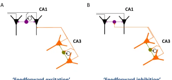
The gamma oscillation generated in CA3 entrains the CA1 circuitry, but the way of propagation is still unclear. Both CA1 pyramidal cells and interneurons could be activated by the Schaffer collaterals or commissural projections of CA3 pyramidal cells, therefore the oscillation could propagate from CA3 to CA1 region via either feed- forward excitation or feed-forward inhibition. The “feed-forward excitation” would mean that CA3 pyramidal cells directly discharge CA1 pyramidal cells and then a recurrent network between CA1 pyramidal cells and local interneurons produce the local field potential oscillation in CA1, similarly to CA3. In the “feed-forward inhibition” scenario CA3 pyramidal cells excite predominantly CA1 interneurons instead of CA1 pyramidal cells, and the local field potential oscillation arise from the rhythmic activity of interneurons driven by the excitation via the Schaffer- collaterals/commissural projections of CA3 pyramidal cells (see also Fig. 4).
27
In the above mentioned study of Csicsvári et al. (2003) the authors found that during CA3 driven gamma oscillation CA1 interneurons fired at the same time or somewhat later than CA3 interneurons, while CA1 pyramidal cells fired earlier than CA3 pyramidal cells within a gamma cycle. The time lag between the discharge of CA1 pyramidal cells and CA1 interneurons was too long to be taken as a monosynaptic excitation, which suggests that CA1 interneurons were discharged rather by CA3 pyramidal cells than CA1 pyramidal cells. Hence, these results propose that the feed- forward inhibition model is more likely to be valid.
Nevertheless, it is difficult to create a detailed model of gamma oscillations, since we know very little about the pattern and kinetics of synaptic currents in the different cell types during these network states. Another limitation of the in vivo multielectrode techniques is that the identification of different cell types is rather difficult under these circumstances and therefore, the technique does not allow studying the differential contribution of distinct interneuronal subclasses to the network oscillations.
In vivo intracellular recordings under urethane anesthesia revealed gamma- frequency membrane potential oscillations and phase-coupled firing of CA1 pyramidal cells during gamma oscillations. The phase and amplitude of intracellular membrane potential oscillations were voltage dependent with a phase reversal at the Cl- equilibrium potential (Penttonen et al., 1998), suggesting that they were produced by rhythmically generated IPSPs.
Juxtacellular recordings of various types of interneurons of the CA1 during spontaneously occurring gamma oscillations under anesthesia revealed that the firing activity of many different interneuron types is modulated by the ongoing gamma oscillation. PV expressing interneurons, and especially bistratified cells showed strong gamma-modulation and tended to fire at the ascending phase of the field potential oscillation recorded in the stratum pyramidale. CCK-containing basket cells also tended to fire phase locked, however, these cells preferred firing at the trough of the oscillation.
In contrast no phase-preference of OLM cells could be detected in this study (Tukker et al., 2007).
The above mentioned techniques are undisputedly helpful in understanding the functional connectivity behind certain activity patterns of the circuitry. However, we
28
still do not know how the different cell types are recruited to the network; what are the cellular and synaptic mechanisms that synchronize their activity during special behavioral or cognitive states.
Although in vitro techniques obviously have their own limitations, they allow recording synaptic currents from identified cell types in parallel with pharmacological modification of the network. Therefore, physiologically relevant in vitro models of network oscillations could definitely help understanding at least some aspects of rhythm generating mechanisms of the brain.
3.1.3. Possible models of rhythm generation within a network
Theoretically there are three principal ways in which oscillatory activity could be generated in a neuronal network (Mann and Paulsen, 2005): (1) The simplest way is the so-called “extrinsic pacemaker model” in which the network activity is driven by a rhythmic external input (Green and Arduini, 1954; Stewart and Fox, 1990). (2) The second model is the intrinsic model, where individual elements of the network have the capability of firing at certain frequencies, and the rhythmic firing of a subset of the neuronal population could entrain the network intrinsically (McCormick and Pape, 1990). The synchronized firing of rhythm-generating cells might be promoted by electrical coupling (Draguhn et al., 1998). (3) The “synaptic loop model” actually includes three scenarios. In the “synaptic feedback model” the oscillation emerges from synaptic recurrent feedback loops between excitatory and inhibitory cells (Freeman, 1968). Synaptic synchronization is, however, possible also through “mutual inhibitory”
as well as “mutual excitatory loops” (Whittington et al., 1995; Wilson and Bower, 1992). Yet, during mutual inhibition the network probably requires a tonic excitatory drive, while during mutual excitation an inhibitory coupling is needed to prevent runaway excitation.
Network oscillations occurring at the same frequencies might be generated by multifarious ways, and certainly the above mentioned models are not mutually exclusive. These scenarios might occur at the same time and can reinforce each other’s effect.
29
In order to understand the cellular and synaptic mechanisms that may underlie rhythmic activity patterns of a network, we have to investigate both the intrinsic properties of the single elements of the network and the possible synaptic synchronization mechanisms that are encoded in the connectivity of the network.
3.1.4. In vitro methods to study rhythm-generating mechanisms of the brain
3.1.4.1. Investigating the resonance properties of single cells
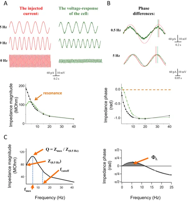
Intrinsic membrane potential oscillations in single neurons can serve as a basis for oscillatory behavior of the network (Lampl and Yarom, 1997). The ability of neurons to generate intrinsic membrane potential oscillations strongly correlates with their resonance properties (Lampl and Yarom, 1997). The easiest way to reveal the resonance properties of the neuronal membrane is to record the so-called impedance magnitude profile of the cell. To measure the impedance properties of the cells, we have to inject sinusoidal current inputs to the cell at different frequencies and record its voltage responses. The impedance values at each frequency (Z) can be calculated by dividing the Fast Fourier Transform (FFT) of the voltage response of the cell by that of the input current. The impedance magnitude profile is obtained by plotting the impedance values against the frequency of the input current (see Methods and Fig. 5A).
In modeling studies the neuronal membrane is represented by electrical equivalent circuits. The simplest circuitry that can be used to describe the behavior of a membrane is composed of a resistor and a capacitor (RC circuit). In response to sinusoidal current injections a simple passive neuronal membrane would behave as an RC circuit: the higher the input frequency is, the lower voltage response of the cell can be detected. Thus, the “passive cell” acts as a low-pass filter. However, if a voltage- gated conductance is activated in the cellular membrane (which can be modeled by introducing an inductive element to the original RC circuit, i.e. creating an RLC circuit), the cell starts to behave as a band-pass filter. Depending on the properties of the voltage-gated conductance, the cell will produce larger amplitude responses to inputs at defined frequencies. This phenomenon is called resonance and will appear as a “hump”
in the impedance magnitude profile of the neuron (Fig. 5A).
30
Besides the magnitude profile, impedance can also be characterized by the so- called impedance phase profile. The impedance phase profile illustrates the temporal relationship between the injected current and the steady-state voltage response of the cell. In the case of an RC circuit (and a passive cell membrane) the phase of the impedance is always negative at any frequencies, which means that there is a lag in the voltage response with respect to the current injected. However, the presence of an inductive element in the circuit (an active conductance in the cell membrane) causes a positive shift in the impedance phase profile. Depending on the interaction of the passive membrane properties and the active conductance the impedance phase can be positive at certain frequencies even in real neurons, which means that the voltage response of the cell could lead the current input (Fig. 5B). Therefore, the operation of voltage-gated ion channels in the cell membrane may change both the amplitude and the timing of the neuronal response in a frequency-dependent manner.
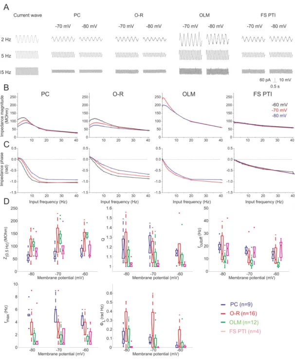
In the hippocampus, pyramidal cells are known to express subthreshold resonance at frequencies within the theta range (4-7 Hz) (Leung and Yu, 1998; Pike et al., 2000; Hu et al., 2002; Narayanan and Johnston, 2007), which might contribute to their membrane potential oscillations in vivo (Ylinen et al., 1995a; Kamondi et al., 1998) as well as to their discharge properties (Pike et al., 2000). Recent studies have revealed that subthreshold resonance in pyramidal cells is predominantly mediated by the hyperpolarization-activated cyclic nucleotide-gated channels (HCN channels), which generate a non-selective cation current – termed Ih (Hu et al., 2002). Besides Ih the so-called M-current, a muscarinic receptor controlled K+ current was also shown to create resonance in pyramidal cells at membrane potentials closer to the firing threshold. (Hu et al 2002).
Some hippocampal interneurons also have been shown to have frequency tuning properties (Gloveli et al., 2005b; Lawrence et al., 2006a) and can also resonate at certain frequencies (Pike et al., 2000). However, it is still unclear which GABAergic cell types show resonance at which frequencies, and what cellular mechanisms are involved.
3.1.4.1.1. Ih (h-current)
The hyperpolarization-activated cation current (Ih, but also know as If or IQ) was initially discovered in the sinoatrial node of the heart over 30 years ago (Noma and
31
Irisawa, 1976). So far Ih has also been described in many types of neurons and gained enormous attention in the recent years, because of its potential contribution to rhythmic activity patterns of the brain (Pape, 1996; Lüthi and McCormick, 1998). In addition to having a key role in producing resonance in distinct types of neurons (Hu et al., 2002;
Heys et al., 2010) and its vital function in pacemaker activities as well as in network oscillations (Lüthi and McCormick, 1998; Kocsis and Li, 2004), this conductance has been suggested to contribute to synaptic waveform normalization (Magee, 1999) and even to learning processes (Nolan et al., 2003).
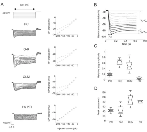
HCN channels mediating Ih can be activated by hyperpolarizing the cell to membrane potentials negative to -50 mV. Ih is a mixed monovalent cation current. HCN channels are permeable for both K+ and Na+ ions. The ratio of K+ to Na+ permeability ranges from 3:1 to 5:1, which will result in a reversal potential of the current between - 25 and -40 mV. Therefore, activation of the channel at typical resting potentials results in a net inward current largely carried by Na+ ions (Robinson and Siegelbaum, 2003). In current clamp experiments, in the voltage response of the HCN-channel expressing cells to negative current steps of appropriate magnitude, a depolarizing sag can be seen due to the activation of Ih (Fig. 6, 7). Following the end of the current pulse, there is a rebound depolarization caused by the extra inward Na+ flux through HCN channels that were activated by hyperpolarization. In some cells this rebound depolarization can even trigger an action potential.
The activation kinetics of Ih are complex. The onset of Ih is usually preceded by a significant delay. After this delay, channel opening can be described by either a single or double exponential function, depending on the cell type. The time course of activation varies widely among the different cell types in the range of few ten milliseconds to several seconds (Robinson and Siegelbaum, 2003).
Another interesting feature of HCN channels is that they can be directly modulated by cyclic nucleotides. The C-terminal fragment of each subunit contains a so-called cyclic-nucleotide binding domain (CNBD), which can bind cyclic nucleotides.
CNBD has the highest affinity to bind cAMP (Wainger et al., 2001; Wang et al., 2001), but cGMP has also been reported as an effective modulator of HCN channel activity (Pape and Mager, 1992). Binding of cyclic nucleotides usually shift the activation curve of the channel to a more depolarized range.