Contents lists available atScienceDirect
Developmental and Comparative Immunology
journal homepage:www.elsevier.com/locate/devcompimm
Short communication
Identi fi cation of reference markers for characterizing honey bee (Apis mellifera) hemocyte classes
Erika Gábor
a, Gyöngyi Cinege
a, Gábor Csordás
a,1, Miklós Rusvai
b, Viktor Honti
a, Balázs Kolics
c, Tibor Török
d, Michael J. Williams
e, Éva Kurucz
a,∗∗, István Andó
a,∗aImmunology Unit, Institute of Genetics, Biological Research Centre, P.O.Box 521, Szeged, H-6701, Hungary
bUniversity of Veterinary Medicine, 1078, Budapest, István u. 2., Hungary
cDepartment of Plant Science and Biotechnology, University of Pannonia, Georgikon Faculty, Deák F. u. 16., 8360, Keszthely, Hungary
dDepartment of Genetics, University of Szeged, Közép Fasor 52, 6726, Szeged, Hungary
eFunctional Pharmacology, Department of Neuroscience, Uppsala University, Husargatan 3, Box 593, 751 24, Uppsala, Sweden
A R T I C L E I N F O
Keywords:
Apis mellifera Monoclonal antibody Immunity Hemocyte Honey bee Insect immunity
A B S T R A C T
Cell mediated immunity of the honey bee (Apis mellifera) involves the activity of several hemocyte populations, currently defined by morphological features and lectin binding characteristics. The objective of the present study was to identify molecular markers capable of characterizing subsets of honey bee hemocytes. We developed and employed monoclonal antibodies with restricted reactions to functionally distinct hemocyte subpopulations.
Melanizing cells, known as oenocytoids, were defined by an antibody to prophenoloxidase, aggregating cells were identified by the expression of Hemolectin, and phagocytic cells were identified by a marker expressed on granulocytes. We anticipate that this combination of antibodies not only allows for the detection of functionally distinct hemocyte subtypes, but will help to further the exploration of hematopoietic compartments, as well as reveal details of the honey bee cellular immune defense against parasites and microbes.
1. Introduction
The honey bee,Apis mellifera,is a social insect that lives in highly structured colonies composed of three castes: the worker, the drone and the queen. Its larval development consists offive stages (L1-L5), which, depending on the cast, takes between 6 and 9 days.A. melliferadevelops with complete metamorphosis. The pupal stage begins when a cell is capped by worker bees, and after 16–24 days an adult emerges from the cell (Winston, 1991).
Similar to other social insects, the honey bee has both communal barriers and individual protection against parasites and pathogenic microbes (Evans et al., 2006). Hygienic behavior, including grooming and hive fever, is a good example of communal defense (Alaux et al., 2012;Evans and Spivak, 2010;Richard et al., 2008). The honey bee and other social insects have fewer canonical immunity-related genes re- lative to solitary insects, which may be a consequence of their
communal defense systems (Barribeau et al., 2015; Doublet et al., 2017). Individual defense in the honey bee shares many similarities with solitary insects, including a mechanical barrier (the cuticle), as well as humoral and cell-mediated immune responses. Humoral im- munity is manifested through the generation of antimicrobial peptides (Cerenius and Söderhäll, 2011;Hoffmann et al., 1999;Hultmark, 2003;
Vilmos and Kurucz, 1998). Cell mediated responses, which are exerted by blood cells known as hemocytes, involve phagocytosis of micro- organisms, encapsulation of larger invaders, coagulation, clotting of the hemolymph after wounding, and melanization of the cuticle at the site of physical injury (Dudzic et al., 2015; Hoffmann et al., 1999; Honti et al., 2014;Vilmos and Kurucz, 1998).
In the honey bee, hemocytes were characterized on the basis of their morphological features, adherence, lectin binding properties, granu- larity and movement (Supplementary Table 1), which led to uncertainty within the field. For instance, based on morphological analyzes,
https://doi.org/10.1016/j.dci.2020.103701
Received 25 February 2020; Received in revised form 8 April 2020; Accepted 8 April 2020
∗Corresponding author. Immunology Unit, Institute of Genetics, Biological Research Centre, P.O.Box 521, Szeged, H-6701, Hungary.
∗∗Corresponding author. Immunology Unit, Institute of Genetics, Biological Research Centre, P.O.Box 521, Szeged, H-6701, Hungary.
E-mail addresses:gabor.erika@brc.hu(E. Gábor),cinege.gyongyi@brc.hu(G. Cinege),cgabor@uni-koeln.de(G. Csordás),Rusvai.Miklos@univet.hu(M. Rusvai), honti.viktor@brc.hu(V. Honti),bkolics@gmail.com(B. Kolics),torokt@bio.u-szeged.hu(T. Török),michael.williams@neuro.uu.se(M.J. Williams),kurucz@brc.hu, kurucz.eva@brc.hu(É. Kurucz),ando@brc.hu(I. Andó).
1Present address: Institute for Genetics and Cologne Excellence Cluster on Cellular Stress Responses in Aging-Associated Diseases (CECAD), University of Cologne, 50931 Cologne, Germany.
Available online 19 April 2020
0145-305X/ © 2020 The Authors. Published by Elsevier Ltd. This is an open access article under the CC BY license (http://creativecommons.org/licenses/BY/4.0/).
T
including spreading and adherence of the cells, Negri et al. (2014) described two hemocyte subpopulations in 5th stage larvae, which they termed rounded cells, showing no locomotion (L5-1), and rounded-oval cells, having extreme pseudopodia development during spreading (L5- 2). In newly emerged workers, the authors distinguished granular cells showing extreme spreading behavior (W-1), rounded smooth mem- braned cells without granules (W-2), as well as small rounded (W-3) and spindle shaped (W-4) cells thatflowed in suspension. On the other hand, El-Mohandes et al. (2010) described prohemocytes, various subtypes of plasmatocytes, granulocytes, coagulocytes and oenocytes with immunohistochemical staining and morphological analysis.
Richardson et al. (2018)distinguished only two major populations in larvae: granulocytes and rare larval hemocytes (presumably L5-2 in Negri et al., 2014).In the adult,Richardson et al. (2018)distinguished plasmatocytes and granulocytes. Additionally, using a combination of microscopic andflow-cytometric studies,deGraaf et al. (2002)identi- fied non-fluorescening prohemocytes, oenocytes and coagulocytes, granulocytes with lowfluorescent intensity, and two highfluorescent intensity plasmatocyte populations amongfluorescently labeled lectin- binding hemocytes. With a similar approach,Marringa et al. (2014) identified permeabilized cells, plasmatocytes and microparticles.
Due to the complexity of hemocyte typing in the honey bee, the immunological compartments and mechanisms of cellular immunity have not been explored in any detail. Therefore, the development of standard reference markers is essential. Recently, Hemolectin was found as a molecular marker (Gábor et al., 2017) for non-phagocytic hemocytes, which are cells that show the characteristic morphological features of plasmatocytes. This observation opened the way for the development of hemocyte markers. Here we describe the identification of additional immunological markers able to define hemocyte sub- populations. We then use these markers to characterize hemocyte subsets in the three honey bee castes, as well as during various cellular immune reactions.
2. Materials and methods
2.1. Laboratory animals, collection of the hemocytes and the hemolymph A. melliferalarvae and adults of the worker cast were collected from an apiary in the Szeged-region (Hungary).
For the preparation ofDrosophilahemocytes, larvae from the wild- typew1118and the prophenoloxidase deficientPPO1Δ,2Δ,31triple mu- tant stocks (Binggeli et al., 2014; Dudzic et al., 2015) of Drosophila melanogaster were used. Flies were propagated at 25 °C in standard Drosophila medium. For each sample, hemocytes of six larvae were pooled and analyzed by indirect immunofluorescence as described for honey bee hemocytes (Gábor et al., 2017).
2.2. Production and screening of monoclonal antibodies
Monoclonal antibodies (mAb) (Köhler and Milstein, 1975, 1976) were raised againstA. melliferahemocytes as described previously for Drosophilahemocytes (Cinege et al., 2019;Kurucz et al., 2003, 2007b;
Márkus et al., 2015) with slight modifications forA. melliferablood cells (Gábor et al., 2017). A previously described antibody, 4E1, toA. mel- liferaHemolectin (Gábor et al., 2017) was used as a standard reagent against the plasmatocyte cell population.
2.3. Immunohistochemistry (IH) and indirect immunofluorescence (IIF) Hybridoma culture supernatants were used throughout the experi- ments as described previously (Gábor et al., 2017).
2.4. Phagocytosis, bacterial induction
The animals were injected with 50 μl fluorescein isothiocyanate
conjugated Gram negative Escherichia coli (SzMC 0582) (Szeged Microbial Collection, University of Szeged, Hungary) (FITC-E. coli) bacteria as described in Kurucz et al. (2007a). The phagocytosis was scored as described previously (Gábor et al., 2017). A 1% suspension of E. colibacteria were injected into the abdomen of young adults, the hemocytes were collected 45 min later and typed for the expression of the antigens.
2.5. Western blot analysis
Western blot analysis of the proteins was carried out as described previously (Gábor et al., 2017).
2.6. Statistical analysis
The measurement of the proportional changes between the blood cell populations was compared between each developmental stage (L1, L3, L5 larval stages, newly emerged and old adults) and castes (newly emerged workers, newly emerged queens, newly emerged drones).
Significance was determined by unpaired Student's t-test. The groups which are significantly different (p < 0.05) from each other are marked with italic letters above the columns.
3. Results and discussion
To characterize honey bee hemocytes, we produced monoclonal antibodies (mAbs) specific for molecular markers expressed by different subpopulations. Antibodies that reacted with plasmatocytes (4E1,Fig. 1 a, b) or oenocytoids (2.28,Fig. 1c, d), as well as an antibody that reacted with both granulocytes and oenocytoids (4.70,Fig. 1e, f), were selected. The 4E1 antibody reacted with approximately 20% of the spherical hemocytes and with the large adhered cells, the larval plas- matocytes (termed as L5-2 byNegri et al., 2014) in the L5 develop- mental stage (Fig. 1 a, a’). It also reacted with approximately 80% of the circulating blood cells in adults, including the small spherical and oval cells; the adult plasmatocytes (Fig. 1 b, b') (Gábor et al., 2017). In larvae and adults, the 2.28 antibody reacted with an antigen expressed by melanizing hemocytes known as oenocytoids (Fig. 1 c-d'). The 4.70 antibody defined an antigen expressed by all larval hemocytes (Fig. 1 e, e’), as well as adult granulocytes and oenocytoids (Fig. 1 f, f’). This expression pattern of the antigen, shared by both phagocytic and mel- anizing cells, may suggest a common origin in the ontogeny of the phagocytic and melanizing cells types, as found inDrosophila(Gold and Brückner, 2015).
We then used the antibodies (4E1, 2.28 and 4.70) to investigate the proportion of hemocytes belonging to the different subpopulations of newly emerged queens and drones (Suppl. Fig. 1.). In both the queen and the drone castes, plasmatocytes were the most abundant, re- presented by small round 4E1 positive Hemolectin expressing cells.
Both oenocytoid (2.28 antibody) and granulocyte-oenocytoid (4.70 antibody) cells were present, and the hemocyte marker expression pattern was similar in both castes. Moreover, we obtained similar re- sults when studying the marker expression pattern of hemocytes from adult workers.
Next, we employed the newly raised antibodies to explore the proportional variation of blood cell subpopulations throughout devel- opment in the different castes. To do this, we analyzed the composition of the worker cast circulating hemocyte population during development in L1, L3 and L5 larvae, newly emerged adults (NW) and older adults (OW) (Fig. 1g). We used individual samples to detect the plasmatocytes by Hemolectin expression, oenocytoids by melanization, and the rest of the blood cells were defined as granulocytes. The plasmatocyte ratio increased slightly throughout larval development (12%–23%). In newly emerged adults, their proportion increased sharply (77%), then di- minished in older adults (51%). On the other hand, we found that the proportion of oenocytoids remained constant (1%) in all stages of
development. The rest of the blood cells, which we deemed granulo- cytes, decreased from 87% in L1 larvae to 76% in L5 larvae. Moreover, their ratio was dramatically decreased in newly emerged adults (22%), then increased in older adults (48%). We also analyzed the proportion of the blood cell subpopulations in newly emerged queens (NQ) and drones (ND). The proportion of the hemocyte subpopulations was si- milar to that of the newly emerged workers. These results show that our
plasmatocyte specific marker, together with the monitoring of oeno- cytoid melanization activity is a valuable tool to analyze the changes in the proportion of blood cell subpopulations throughout development.
The 2.28 antibody was identified as reacting with an antigen ex- pressed specifically by oenocytoids, the honey bee melanizing hemo- cytes. Therefore, we wanted to know if it was recognizing AmPPO protein(Suppl. Fig. 2). AmPPO was identified in the hemolymph as a Fig. 1. Detection of hemocyte subpopulations in the honey bee, based on their reaction pattern with mAbs.Acetonefixed larval and adult hemocytes were stained with 4E1 (a, b), 2.28 (c, d), and 4.70 (e, f) mAb-s and anti-mouse Alexa Fluor 568 (red). Asterisks on‘a’mark the larval plasmatocytes. The nuclei were visualized with DAPI (blue). The scale bars represent 20μm. Detection was done with a Zeiss Axioskope 2 MOT epifluorescence microscope. The proportion of the circulating blood cell subpopulations changes during ontogeny (g). The ratio of the plasmatocytes stained by anti-Hemolectin antibody (4E1) and anti-mouse Alexa 568 secondary antibody (black) in L1 (12%), L3 (14%), L5 (23%) stage larvae, in newly emerged workers (NW) (77%), in newly emerged queens (NQ) (82%), in newly emerged drones (ND) (84%) and in older workers (OW) (51%). Oenocytoids were detected according to their melanization (white) as 1% in all castes and developmental stages. The rest of blood cells supposedly granulocytes (grey) were assigned as 87% in L1 larvae, 85% in L3 larvae 76% in L5 larvae, 22% in newly emerged workers, 17% in newly emerged queens, 15% in newly emerged drones and 48% in older workers. Significance was determined by unpaired Student's t-test.
Significant differences (p < 0.05) are indicated with italic letters above the columns. Values with the same italic letter are not significantly different. The numbers at the bottom indicates the number of tested individuals. (For interpretation of the references to colour in thisfigure legend, the reader is referred to the Web version of this article.)
74.4 kDa protein (Lourenço et al., 2005;Zufelato et al., 2004). Inter- estingly, the 2.28 antibody gave a double band corresponding to 70 and 75 kDa in Western blot analysis, corresponding to the zymogen and the activated forms of AmPPO (Suppl. Fig. 2 a). TheD. melanogastergenome contains three prophenoloxydase (PPO) encoding genes (PPO1,PPO2 andPPO3) (Binggeli et al., 2014;Dudzic et al., 2015). PPO1 and PPO2 are produced by the crystal cells, while PPO3 is synthesized by the lamellocytes. The PPOs are the initiators of the PPO-cascade, which causes melanization, the rapid synthesis of melanin, which is a major immune response of insects to infection and injury (Biedermann and Moritz, 1898;Cerenius et al., 2008; Kanost and Gorman, 2008). We observed that the 2.28 oenocytoid specific antibody cross reacted with crystal cells and a subset of lamellocytes in Drosophila melanogaster w1118larvae (Suppl. Fig. 2 b) but did not stain hemocytes from pro- phenoloxidase deficient PPO1Δ,2Δ,31 (Suppl. Fig. 2 c) triple mutant larvae. From these results we suggest that the 2.28A. melliferaoeno- cytoid specific antibody reacts with PPO.
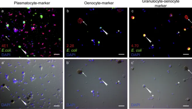
To reveal the functional role of the adult hemocyte subpopulations in the phagocytosis of microbes FITC-labeledE. colibacteria were in- jected into adults, after which hemocytes were isolated and an indirect immunofluorescence assay was carried out. We found that 4E1 positive plasmatocytes (Fig. 2a) and 2.28 positive oenocytoids (Fig. 2b) did not take up bacteria, while 4.70 positive granulocytes (Fig. 2 c) were phagocytic. No alteration of hemocyte population composition was observed after immune induction with E. coli bacteria, compared to naïve controls (data not shown).
We observed that the proportion of phagocytic cells in the social honey bee was much lower than in the solitaryD. melanogaster(over 95%), which has only individual immunity (Rizki and Rizki., 1984).
Considering the alternative defense strategies of social insects, it is possible that fewer microorganisms reach the body cavity of individual bees. In fact, only a few parasites and microbes are described that affect the honey bee cellular immune response.Spiroplasma melliferuminfec- tion results in a change in the proportion of plasmatocytes and granu- locytes, as detected by Wright staining; whileSerratia marcescens sicaria
(Ss1) infection caused a decrease in the total hemocyte number com- pared to uninfected animals (Burritt et al., 2016;Yang et al., 2017).
In conclusion, to avoid the crisis caused by the loss of honey bees, it is important to have a better understanding of their immune response.
Due to its social nature, a honey bee colony is often regarded as a complex living individual, and hygienic behavior is believed to be an important factor of its immunity. However, individual honey bees, si- milar to other insects, defend their integrity with the help of immune cells, therefore it is equally important to gain knowledge of their cel- lular immune response. By employing our newly raised antibodies, we were not only able to characterize the hemocyte subpopulations of different castes throughout development, but also defined which sub- populations are involved in the response against various immune threats. Furthermore, our results help to clarify the similarities and differences between the cellular immune responses of social and soli- tary insects. Contrary to what was found in D. melanogaster, a much smaller proportion of honey bee circulating hemocytes are phagocytic, which implies that, due to its social immunity, the honey bee is less reliant on individual immunity. Also, dissimilar toD. melanogaster, in which special hemocytes (the lamellocytes) differentiate to isolate larger invaders, encapsulation in the honey bee is performed by blood cells already present in circulation (Gábor et al., 2017). According to ourfindings, prophenoloxidase function is present in the honey bee, and similar toD. melanogaster, it plays an indispensable role in mela- nization. We believe that our newly identified markers will help to further identify the components of cellular immunity, as well as analyze the composition of the honey bee immune compartments.
Acknowledgements
We acknowledge to Professor Bruno Lemaitre and Jan Paul Dudzic for providing the PPO mutant Drosophila stock. We are grateful to Olga Kovalcsik, Anita Balázs, Anikó Képíró and Mónika Ilyés for the tech- nical help and Mr. Vince Bengyák of Miháld, Hungary, for providing experimental specimens from his apiary. We would like to extend our
Fig. 2. Phagocytic capacity of the adult hemocyte subsets.Following the injection of FITC conjugatedE. colibacteria, hemocytes were isolated and indirect immunofluorescence analysis was carried out using the 4E1 (a), 2.28 (b) and 4.70 (c) mAb-s and as secondary antibodies anti-mouse Alexa Fluor 568 (red). White arrows point to the non-phagocytic oenocytoids. White arrowheads show phagocytic granulocytes. Asterisks mark non-phagocytic plasmatocytes. Cell nuclei were stained with DAPI (blue). The scale bars represent 20μm. Immunofluorescence was analyzed with a Zeiss Axioskope 2 MOT epifluorescence microscope. (For interpretation of the references to colour in thisfigure legend, the reader is referred to the web version of this article.)
appreciation to Professor László Békési and Edit Zajácz for their com- ments and support. This research was supported by grants NKFI K 120140 (É.K.), NKFI K 120142 (I.A.), GINOP-2.3.2-15-2016-00001 (I.A.), GINOP-2.3.2-15-2016-00035 (É.K.), NKFI K 128762 (G.C.), NKFI K 131484 (V.H), EFOP-3.6.3-VEKOP-16-2017-00008 (B.K.), the Swedish Research Council (M.J.W.) and by the Hungarian National Beekeepers Association.
Appendix A. Supplementary data
Supplementary data to this article can be found online athttps://
doi.org/10.1016/j.dci.2020.103701.
References
Alaux, C., Kemper, N., Kretzschmar, A., Le Conte, Y., 2012. Brain, physiological and behavioral modulation induced by immune stimulation in honeybees (Apis mellifera):
a potential mediator of social immunity? Brain Behav. Immun. 26, 1057–1060.
https://doi.org/10.1016/j.bbi.2012.04.004.
Barribeau, S.M., Sadd, B.M., du Plessis, L., Brown, M.J.F., Buechel, S.D., Cappelle, K., Carolan, J.C., Christiaens, O., Colgan, T.J., Erler, S., Evans, J., Helbing, S., Karaus, E., Lattorff, H.M.G., Marxer, M., Meeus, I., Näpflin, K., Niu, J., Schmid-Hempel, R., Smagghe, G., Waterhouse, R.M., Yu, N., Zdobnov, E.M., Schmid-Hempel, P., 2015. A depauperate immune repertoire precedes evolution of sociality in bees. Genome Biol.
16, 83.https://doi.org/10.1186/s13059-015-0628-y.
Biedermann, W., Moritz, P., 1898. Beiträge zur vergleichenden Physiologie der Verdauung. Pflüger, Arch. 73, 219–287.https://doi.org/10.1007/BF01796256.
Binggeli, O., Neyen, C., Poidevin, M., Lemaitre, B., 2014. Prophenoloxidase activation is required for survival to microbial infections inDrosophila. PLoS Pathog. 10, e1004067.https://doi.org/10.1371/journal.ppat.1004067.
Burritt, N.L., Foss, N.J., Neeno-Eckwall, E.C., Church, J.O., Hilger, A.M., Hildebrand, J.A., Warshauer, D.M., Perna, N.T., Burritt, J.B., 2016. Sepsis and hemocyte loss in honey bees (Apis mellifera) infected withSerratia marcescensstrainsicaria. PLoS One 11, e0167752.https://doi.org/10.1371/journal.pone.0167752.
Cerenius, L., Lee, B.L., Söderhäll, K., 2008. The proPO-system: pros and cons for its role in invertebrate immunity. Trends Immunol. 29, 263–271.https://doi.org/10.1016/j.it.
2008.02.009.
Cerenius, L., Söderhäll, K., 2011. Coagulation in invertebrates. J. Innate Immun. 3, 3–8.
https://doi.org/10.1159/000322066.
Cinege, G., Lerner, Z., Magyar, L.B., Soós, B., Tóth, R., Kristó, I., Vilmos, P., Juhász, G., Kovács, A.L., Hegedűs, Z., Sensen, C.W., Kurucz, É., Andó, I., 2019. Cellular immune response involving multinucleated giant hemocytes with two-step genome amplifi- cation in thedrosophilid Zaprionus indianus. J. Innate Immun. 1–16.https://doi.org/
10.1159/000502646.
Doublet, V., Poeschl, Y., Gogol-Döring, A., Alaux, C., Annoscia, D., Aurori, C., Barribeau, S.M., Bedoya-Reina, O.C., Brown, M.J.F., Bull, J.C., Flenniken, M.L., Galbraith, D.A., Genersch, E., Gisder, S., Grosse, I., Holt, H.L., Hultmark, D., Lattorff, H.M.G., Le Conte, Y., Manfredini, F., McMahon, D.P., Moritz, R.F.A., Nazzi, F., Niño, E.L., Nowick, K., van Rij, R.P., Paxton, R.J., Grozinger, C.M., 2017. Unity in defence:
honeybee workers exhibit conserved molecular responses to diverse pathogens. BMC Genom. 18, 207.https://doi.org/10.1186/s12864-017-3597-6.
Dudzic, J.P., Kondo, S., Ueda, R., Bergman, C.M., Lemaitre, B., 2015.Drosophilainnate immunity: regional and functional specialization of prophenoloxidases. BMC Biol. 13, 81.https://doi.org/10.1186/s12915-015-0193-6.
El-Mohandes, S.S., Nafea, E.A., Fawzi, A.M., 2010. Effect of different feeding diets on the haemolymph of the newly emerged honeybee workersApis melliferaL. Egypt. Acad. J.
Biol. Sci. 3 (1), 213–220.
Evans, J.D., Aronstein, K., Chen, Y.P., Hetru, C., Imler, J.-L., Jiang, H., Kanost, M., Thompson, G.J., Zou, Z., Hultmark, D., 2006. Immune pathways and defence me- chanisms in honey beesApis mellifera. Insect Mol. Biol. 15, 645–656.https://doi.org/
10.1111/j.1365-2583.2006.00682.x.
Evans, J.D., Spivak, M., 2010. Socialized medicine: individual and communal disease barriers in honey bees. J. Invertebr. Pathol. 103 (Suppl. 1), S62–S72.https://doi.org/
10.1016/j.jip.2009.06.019.
Gábor, E., Cinege, G., Csordás, G., Török, T., Folkl-Medzihradszky, K., Darula, Z., Andó, I., Kurucz, É., 2017. Hemolectin expression reveals functional heterogeneity in honey bee (Apis mellifera) hemocytes. Dev. Comp. Immunol. 76, 403–411.https://doi.org/
10.1016/j.dci.2017.07.013.
Gold, K.S., Brückner, K., 2015. Macrophages and cellular immunity inDrosophila mela- nogaster. Semin. Immunol. 27, 357–368.https://doi.org/10.1016/j.smim.2016.03.
010.
de Graaf, D.C., Dauwe, R., Walravens, K., Jacobs, F.J., 2002. Flow cytometric analysis of lectin-stained haemocytes of the honeybee (Apis mellifera). Apidologie 33, 571–579.
https://doi.org/10.1051/apido:2002041.
Hoffmann, J.A., Kafatos, F.C., Janeway, C.A., Ezekowitz, R.A., 1999. Phylogenetic per- spectives in innate immunity. Science 284, 1313–1318.
Honti, V., Csordás, G., Kurucz, É., Márkus, R., Andó, I., 2014. The cell-mediated immunity ofDrosophila melanogaster: hemocyte lineages, immune compartments, micro- anatomy and regulation. Dev. Comp. Immunol. 42, 47–56.
Hultmark, D., 2003.Drosophilaimmunity: paths and patterns. Curr. Opin. Immunol. 15, 12–19.
Kanost, M.R., Gorman, M.J., 2008. Phenoloxidases in insect immunity. Insect Immunol. 1, 69–96.
Köhler, G., Milstein, C., 1976. Derivation of specific antibody-producing tissue culture and tumor lines by cell fusion. Eur. J. Immunol. 6, 511–519.https://doi.org/10.
1002/eji.1830060713.
Köhler, G., Milstein, C., 1975. Continuous cultures of fused cells secreting antibody of predefined specificity. Nature 256, 495–497.
Kurucz, E., Váczi, B., Márkus, R., Laurinyecz, B., Vilmos, P., Zsámboki, J., Csorba, K., Gateff, E., Hultmark, D., Andó, I., 2007b. Definition ofDrosophilahemocyte subsets by cell-type specific antigens. Acta Biol. Hung. 58 (Suppl. l), 95–111.https://doi.org/
10.1556/ABiol.58.2007.Suppl.8.
Kurucz, E., Zettervall, C.-J., Sinka, R., Vilmos, P., Pivarcsi, A., Ekengren, S., Hegedüs, Z., Ando, I., Hultmark, D., 2003. Hemese, a hemocyte-specific transmembrane protein, affects the cellular immune response inDrosophila. Proc. Natl. Acad. Sci. U.S.A. 100, 2622–2627.https://doi.org/10.1073/pnas.0436940100.
Lourenço, A.P., Zufelato, M.S., Bitondi, M.M.G., Simões, Z.L.P., 2005. Molecular char- acterization of a cDNA encoding prophenoloxidase and its expression inApis melli- fera. Insect Biochem. Mol. Biol. 35, 541–552.https://doi.org/10.1016/j.ibmb.2005.
01.013.
Márkus, R., Lerner, Z., Honti, V., Csordás, G., Zsámboki, J., Cinege, G., Párducz, Á., Lukacsovich, T., Kurucz, É., Andó, I., 2015. Multinucleated giant hemocytes are ef- fector cells in cell-mediated immune responses ofDrosophila. J. Innate Immun. 7, 340–353.https://doi.org/10.1159/000369618.
Marringa, W.J., Krueger, M.J., Burritt, N.L., Burritt, J.B., 2014. Honey bee hemocyte profiling byflow cytometry. PLoS One 9.https://doi.org/10.1371/journal.pone.
0108486.
Negri, P., Maggi, M., Szawarski, N., Lamattina, L., Eguaras, M., 2014.Apis mellifera haemocytes in-vitro, What type of cells are they? Functional analysis before and after pupal metamorphosis. J. Apicult. Res. 53, 576–589.https://doi.org/10.3896/IBRA.1.
53.5.11.
Richard, F.-J., Aubert, A., Grozinger, C.M., 2008. Modulation of social interactions by immune stimulation in honey bee,Apis mellifera, workers. BMC Biol. 6, 50.https://
doi.org/10.1186/1741-7007-6-50.
Richardson, R.T., Ballinger, M.N., Qian, F., Christman, J.W., Johnson, R.M., 2018.
Morphological and functional characterization of honey bee,Apis mellifera, hemocyte cell communities. Apidologie 1–14.https://doi.org/10.1007/s13592-018-0566-2.
Rizki, R.M., Rizki, T.M., 1984. Selective destruction of a host blood cell type by a para- sitoid wasp. Proc. Natl. Acad. Sci. U.S.A. 81, 6154–6158.
Vilmos, P., Kurucz, E., 1998. Insect immunity: evolutionary roots of the mammalian in- nate immune system. Immunol. Lett. 62, 59–66.
Winston, M.L., 1991. The Biology of the Honey Bee. Harvard University Press, Cambridge, Mass.
Yang, D., Zha, G., Li, X., Gao, H., Yu, H., 2017. Immune responses in the haemolymph and antimicrobial peptide expression in the abdomen ofApis melliferachallenged with Spiroplasma melliferumCH-1. Microb. Pathog. 112, 279–287.https://doi.org/10.
1016/j.micpath.2017.10.006.
Zufelato, M.S., Lourenço, A.P., Simões, Z.L.P., Jorge, J.A., Bitondi, M.M.G., 2004.
Phenoloxidase activity inApis melliferahoney bee pupae, and ecdysteroid-dependent expression of the prophenoloxidase mRNA. Insect Biochem. Mol. Biol. 34, 1257–1268.https://doi.org/10.1016/j.ibmb.2004.08.005.