Poly(aspartic acid) hydrogels showing reversible volume change upon redox stimulus
Enikő Krisch1, Benjámin Gyarmati1, Dóra Barczikai1, Véronique Lapeyre2, Barnabás Áron Szilágyi1, Valérie Ravaine2, András Szilágyi1*
1Soft Matters Group, Department of Physical Chemistry and Materials Science, Budapest University of Technology and Economics, Műegyetem rkp. 3. H-1111 Budapest, Hungary E-mail: aszilagyi@mail.bme.hu
2Université de Bordeaux, ISM, CNRS UMR 5255, Bordeaux INP, Site ENSCBP, 16 Avenue Pey Berland, 33607 Pessac Cedex, France
*corresponding author
GRAPHICAL ABSTRACT
HIGHLIGHTS
Redox- and pH-responsive poly(aspartic acid) (PASP) hydrogels are synthesized in aqueous solution
The degree of swelling and the stiffness of the gels can be controlled by using reducing and oxidising media
PASP nanogels are stable in reducing media and swell similarly to their macroscopic counterparts
Drug release measurements show considerably faster drug release in reducing than in oxidising environment
ABSTRACT
Redox and pH responsive hydrogels were synthesized, which can serve as matrices for smart drug delivery systems exploiting the redox and pH gradients in the human body. Water soluble thiolated poly(aspartic acid), a biocompatible synthetic polymer, enables us to cross- link in water with a non-cleavable cross-linker, poly(ethylene glycol) diglycidyl ether. The permanent cross-linker establishes stable polymer hydrogels regardless of the redox environment, thus the gels swell but do not dissolve upon redox stimuli. The reversible response upon redox stimulus was induced by thiol-disulphide transformation inside the hydrogel. The degree of swelling and the stiffness of the macroscopic hydrogels were controlled by their chemical composition including thiol content and cross-linking ratio as well as the redox state of the hydrogels. The degree of swelling of the hydrogels showed strong pH dependence due to the polyelectrolyte nature of the polymer network. Release of a macromolecular model drug was considerably faster in reducing than in oxidising environment, which indicates the potential use of the synthesized hydrogels as redox responsive drug delivery systems. Nanogels were prepared in water-in-oil emulsion and displayed redox-responsive properties. The hydrodynamic diameter of the nanogels strongly increased upon the cleavage of the disulphide linkages in reducing solution without the disruption of the gels.
KEYWORDS
biopolymer; hydrogel; nanogel; redox-responsive; reversible swelling; drug delivery
1 Introduction
Macromolecular therapeutic agents including proteins, peptides, hormones or vaccines are an emerging class of drugs. Their effect is highly specific because of their selective adsorption on receptor binding sites [1–3]. Mass production of these therapeutic agents has been already resolved but their medical use is limited. Their therapeutic effect is associated with their 3D structure, the stability of which is very poor against physical and chemical effects.
Consequently, macromolecular drugs cannot be administrated directly by the oral route and their bioavailability is also poor in the case of parenteral administration. In order to enhance the therapeutic efficiency, their stability must be preserved. Special drug formulations can protect the sensitive compounds and release them at a controlled rate, which maintain the therapeutic level of the drug for a long period. Temporal and spatial modulation of the drug release by the physiological state of the body is preferred [4].
Hydrogels are a particularly promising class of such drug formulations. The large amount of water adsorbed by their 3D network improves their biocompatibility and ensures large capacity for the entrapment of bioactive molecules [5,6]. They can adapt to the biological environment by their controllable response to various stimuli, such as the change in pH, temperature, redox potential or the presence of enzymes [7]. This responsive behaviour allows for the treatment of various diseases by providing the on-demand release of the therapeutic agent from the carrier. For instance, diabetes can be treated with glucose sensitive hydrogels developed for controlled delivery of insulin [8]. Similarly, pH-responsive hydrogels with glucose oxidase entrapped can be used for the glucose modulated release of insulin [9,10].
Some hydrogel drug formulations are already commercially available or reached the phase of clinical trials [11–13], for instance hormone-releasing poly(hydroxyalkyl methacrylate) hydrogel implants for the treatment of prostate cancer or injectable hydrogel formulation for the sustained release of protein-type drugs present in the market [14]. These formulations enhance the stability of the drugs and provides a sustained drug release, but do not adapt to the pathological state of the patient, furthermore, most of them are not biodegradable and the surgical removal of the exhausted device is needed.
The redox state of body sites is intensively studied and there are some evidences on the difference in the glutathione level of the cytosol and the extracellular milieu [15–18]. This redox gradient can be exploited to modulate the drug release from the formulation. Various
the cytosol [19–22], but there are quite few examples of hydrogels that preserve the gel state in both the oxidised and the reduced states. These hydrogels would have the large benefit of providing modulated release depending on the redox state of the body site, if long circulating times in blood can be achieved [23,24]. The rational design of such redox-responsive hydrogels used in biological environments requires a biocompatible polymer network with redox-sensitive functional groups incorporated [6]. Therefore, we chose poly(aspartic acid) (PASP), a synthetic poly(amino acid), which is a well-known representative of biocompatible and biodegradable, cross-linkable materials. The pre-cursor polymer of PASP, polysuccinimide (PSI) is prepared from aspartic acid, a natural amino acid. The repeating unit of PSI easily reacts with nucleophiles, thus PASP derivatives and cross-linked PASP can be synthesized in various ways [25–28]. Redox responsive properties can be incorporated into the polymer network via the thiolation of the polymer, and responsive behaviour is based on the reversible thiol-disulphide conversion, which resembles the redox processes of biological systems, e.g. disulphide formation during protein folding. The intermolecular oxidation of the thiol groups results in a dual responsive PASP hydrogel showing gel-sol transition upon reduction [29] also in the nano size range [30]. PASP gels showing volume change upon redox stimulus were synthesized by using a non-redox sensitive cross-linker molecule together with disulphides [31,32]. Although the latter type of gels showed reversible redox- response, their use as drug formulation is limited as the synthesis was done in non-aqueous medium which makes the encapsulation of drugs and the preparation of nanogels difficult [33]. Drug encapsulation and release were not studied in those papers.
The aim of this study is to present a new synthesis route for PASP hydrogels showing reversible swelling upon redox stimulus. The synthesis of the macroscopic hydrogels was carried out in aqueous medium, which enabled us to synthesize nanogels of the same composition in water-in-oil emulsion. The swelling and the mechanical properties of the macroscopic hydrogels were studied to choose the composition which had remarkable redox response and good mechanical stability to encapsulate a model macromolecular drug, fluorescein labelled dextran. The drug release profile was determined in both oxidising and reducing medium to prove the existence of redox-modulated release kinetics. Following the experiments on macroscopic hydrogels, PASP nanogels were prepared in water-in-oil emulsion to study the redox-dependent properties of the nanogels.
2 Materials and methods
2.1 Materials
L-aspartic acid (99%), cysteamine (98%), potassium chloride (>99.5%), potassium dihydrogen phosphate (>99.5%), sodium bromate (99%), dithiothreitol (DTT, for biochem.) were purchased from Merck. Phosphoric acid (cc. 85%) and dimethylformamide (DMF, for analysis) were bought from Lach-Ner. Poly(ethylene glycol) diglycidyl ether (PEGDGE, Mn= 500), fluorescein isothiocyanate-dextran (FITC-dextran, Mw= 70 kDa) sodium tetraborate (for analysis) and disodium hydrogen phosphate (>99.5%) were purchased from Sigma-Aldrich. Imidazole (>99%) was obtained from Acros Organics. Citric acid (puriss) was purchased from Molar Chemicals. All reagents and solvents were used without further purification.
Deionised water (Milli-Q reagent grade, ρ > 18.2 MΩcm, Millipore, USA) was used for the preparation of aqueous solutions. Buffer solutions were prepared from citric acid (c = 0.033 M, pH = 2 to 6), imidazole (c = 0.1 M, pH = 6 to 8) and sodium tetraborate (c = 0.025 M, pH = 8 to 12). The pH of the buffer solutions was adjusted by adding 1 M HCl or 1 M NaOH. Phosphate buffered saline (PBS) solution of pH = 7.4 was prepared by dissolving 8.00 g NaCl, 0.20 g KCl, 1.44 g Na2HPO4·2H2O and 0.12 g KH2PO4 in 1 dm3 water, the pH was adjusted with 0.1 M HCl. The ionic strength of the solutions was adjusted to 0.15 M by the addition of KCl. The pH of the buffer solutions was checked with a pH/ion analyser (Radelkis OP-271/1, Hungary). The synthesis and all experiments were carried out at 25 °C unless otherwise indicated.
2.2 Synthesis of thiolated poly(aspartic acid)(PASP-SH)
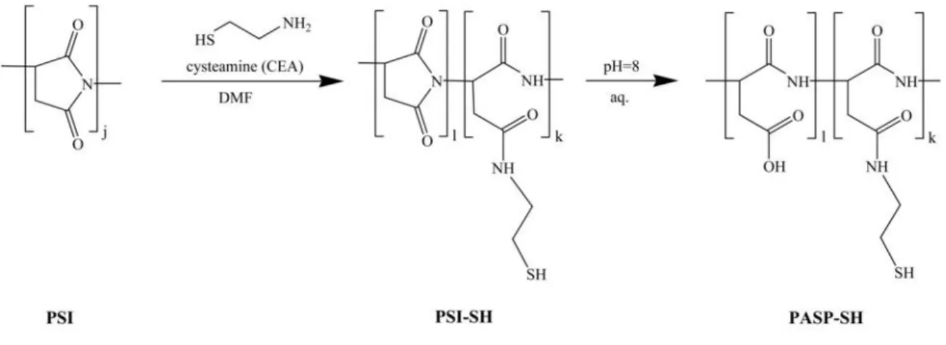
The precursor polymer of thiolated poly(aspartic acid) (PASP-SH), polysuccinimide (PSI) was prepared by thermal polycondensation of L-aspartic acid, as reported earlier [30]. 20 g of L-aspartic acid and 20 g of phosphoric acid was mixed and heated up to 200 °C on a rotary evaporator, and rotated at 180 rpm for 7 hours at a pressure of 10 mbar. The resulting PSI was dissolved in 250 ml of DMF, then precipitated and washed with a large amount of water, finally dried at 40 °C for 2 days (the yield was 95%). Chemical structure was confirmed by1H NMR (300 MHz, DMSO-d6, δ: 5.12 ppm (1H, CO-CH-CH2-CO); 3.22 ppm and 2.74 ppm (2H, CO-CH-CH2-CO)). The molecular weight of the obtained PSI was 31.5 kDa measured
by a rolling ball viscometer (Anton Paar Lovis 2000 ME, solvent: 0.1 M LiCl in DMF, Mark- Houwink constants [34]: a = 0.76; K = 1.32·10-2).
For the preparation of PASP-SH, PSI was reacted with cysteamine (CEA) in DMF under nitrogen atmosphere for 3 days (Fig. 1). The feed ratio of the cysteamine to the repeating units of PSI (thiol content) was changed from 10 to 50 n/n%. Cysteamine modified poly(aspartic acid) (PASP-SH) was obtained by the hydrolysis of the remaining succinimide rings of the PSI derivative (PSI-SH). To do this, the reaction mixture was poured into a large excess (10- fold) buffer solution (imidazole) of pH = 8 and stirred for 3 days (Fig. 1). To prevent the oxidation of thiol groups, DTT (1/2 equivalent to thiol groups) was added to the solution.
PASP-SH was purified with dialysis against deionised water (cut off: ~13000 Da, pH was around 5.5) until the specific conductivity of water decreased below 10 µScm-1, and then lyophilized. The resulting dry polymer was kept under argon atmosphere and stored at 4 °C.
Chemical structure was confirmed by 1H NMR (300 MHz, D2O, δ: 4.58 ppm (1H, CO-CH- CH2-CO), 3.36 ppm (2H, NH-CH2-CH2-SH), 2.81 ppm (2H, CO-CH-CH2-CO), 2.64 ppm (2H, NH-CH2-CH2-SH). The degree of modification was determined by NMR spectroscopy (Fig. S1-S2, Table S1, [35]).
Fig. 1Synthesis of thiolated poly(aspartic acid) (PASP-SH) by the modification of polysuccinimide (PSI) with cysteamine followed by the hydrolysis of PSI-SH
2.3 Synthesis of macroscopic hydrogels
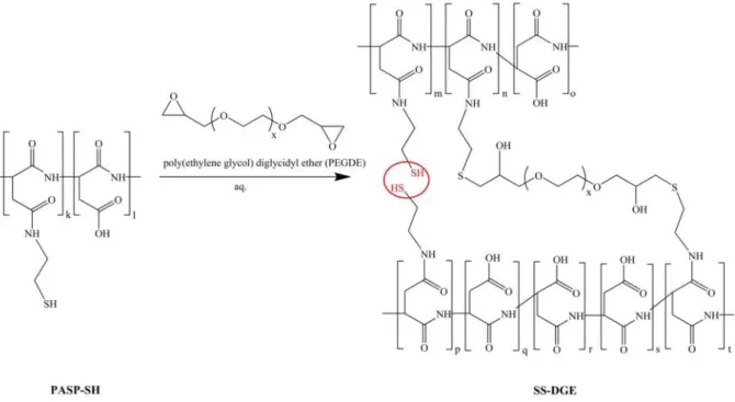
PASP-SH was dissolved in 0.25 M NaOH at a concentration of 15 g polymer/100 ml solution. An aqueous solution of poly(ethylene glycol) diglycidyl ether (PEGDGE) (1 M) was
added to the PASP-SH solution to obtain poly(aspartic acid) (SS-DGE) hydrogels cross- linked with PEGDGE (Fig. 2). The permanent cross-linking ratio, which is the molar feed ratio of the permanent cross-linker, PEGDGE to the repeating units of PASP-SH varied between 5 and 15 n/n% (Table S2). Hydrogels were named according to their composition, for instance in the name of SS50-DGE5 hydrogel the first number refers to the thiol content of PASP-SH (50 n/n%) and the second number refers to the molar feed ratio of the permanent cross-linker to the repeating units (5 n/n%). As an example, the SS50-DGE5 hydrogel was prepared by dissolving 0.15 g PASP-SH50 (1.038 mol repeating unit) in 948 µl 0.25 M NaOH, then permanently cross-linked with 52 µl 1 M PEGDGE solution (0.052 mol). In this case 20% of the thiol-groups were reacted with PEGDGE to establish a permanent network and 80% of the thiol groups are able to form reversible disulphide linkages. To determine the effect of pH on gelation time, the same synthesis was done with replacing a certain amount (0-200 μl) of 0.25 M NaOH with 1 M HCl, and accordingly, the pH of the polymer solution varied between 6.0 and 10.5. Gelation time was determined by visual observation using the tilting method. Hydrogels were cast in different shapes: discs (diameter ≈ 10 mm, thickness ≈ 2 mm) were prepared for swelling experiments, while cylinder shaped hydrogels (diameter ≈ height ≈ 1 cm) were prepared for mechanical testing. The chemical structure of SS50-DGE5 hydrogel was studied by means of FTIR spectroscopy (Fig. S3).
Fig. 2Cross-linking of PASP-SH with poly(ethylene glycol) diglycidyl ether (x = 7-8)
2.4 Synthesis of nanogels
PASP nanogels were prepared using nanosized aqueous droplets in a water-in-oil (W/O or inverse) miniemulsion [36]. 0.2250 g PASP-SH50 was dissolved in 1.200 ml of 0.25 M NaOH, then the pH was adjusted to 8 by the addition of 0.222 ml of 1 M HCl. Prior to emulsification 78 µl of 1 M PEGDGE solution was added to the PASP-SH50 solution (like in the case of SS50-DGE5 gel) and shaken vigorously for 30 seconds. Then 1.5 ml of the solution was added to 13.5 ml ofn-octane containing sorbitan monooleate (Span 80, 8 w/w%) as surfactant. The mixture was emulsified by high pressure homogenization (M-110S Microfluidizer® Processor, operating at 600 kPa). The emulsion was homogenized for 50 passes and cooled to 8 °C between passes. This procedure took less than one minute. After that, the emulsion was stirred on a magnetic stirrer for 24 hours to complete the cross-linking reaction within the droplets. Thereafter, 50 ml THF was added to the dispersion and the sample was centrifuged for 1 h at 16000 g. Supernatant THF containing the unnecessary surfactant andn-octane was removed. The washing procedure was repeated four times and the dispersion centrifuged for 30 min each time at 16000 g. Afterwards, the sample was dried overnight at room temperature under vacuum. Finally, nanogels were re-dispersed in 30 ml of water (polymer concentration ~0.87 w/w%).
2.5 Degree of swelling
The degree of swelling ( ) is defined as the mass ratio of the swollen ( ) to the dried hydrogel ( ):
= (1)
The weight of the swollen hydrogel ( ) was measured after the removal of excess solution from the surface of the gel. The weight of the dried polymer gel ( ) was measured after removing the buffer components by immersing the gels into a large amount of deionised water for three days with daily change of the water, finally drying them for two days in vacuum at ambient temperature. To determine the pH-dependence of the degree of swelling, PASP hydrogels were swollen for two days in large excess of aqueous buffer solutions. The degree of swelling was also measured in phosphate buffer (PBS, pH = 7.4) in the presence of the reducing agent DTT (c = 10 mM) or the oxidising agent, sodium bromate (c = 10 mM).
2.6 Mechanical properties of PASP hydrogels
The compressive elastic modulus ( ) of the hydrogels was determined with uniaxial compression tests using an Instron 5543 mechanical tester (USA) with a cell load of 5 N. The force ( ) and the height (ℎ) of the cylindrical samples were recorded during the measurements, and the nominal stress ( ) and compression ratio( ) were calculated using the following equations: = / and = ℎ/ℎ , where( ) is the cross-sectional area of the undeformed hydrogel and(ℎ )is the original height. The maximum deformation was 10%
of the original height and the samples were compressed stepwise with 0.1 mm deformation in each step followed by a relaxation time of 8 s. The compressive elastic modulus was calculated by Equation 2. Moduli were determined as the slope of the straight line fitted to the stress-strain function between 0 and 10% strain.
= ( − ) (2)
2.7 Drug loading and release
SS50-DGE5 hydrogels containing fluorescein isothiocyanate-dextran (FITC-dextran, Mw= 70 kDa) were prepared to study redox induced drug release. FITC-dextran in the concentration of 1.7 mg/ml was added to the polymer solution of PASP-SH50 before the addition of PEGDGE as permanent cross-linker. Hydrogel discs with the diameter of 10 mm and height of 2 mm were prepared. The hydrogels containing FITC-dextran were placed into 2 ml of sodium bromate solution (c = 1 M, prepared with PBS) for 3 hours to obtain the oxidised form. The hydrogels were then leached twice with 20 ml of PBS for 1 hour each. The FITC-dextran content of the sodium bromate solution and the PBS used for washing was determined by a fluorimeter (PerkinElmer LS 50 B, = 490 , = 518 , calibration curves can be found in the Supplementary Information, Fig. S4 and Table S3), the drug loading efficiency ( ) was also calculated.
= , ,
, ∗ 100% (3)
where , is the total amount of FITC-dextran in the precursor solution and
, is the amount of FITC-dextran removed by washing steps in PBS prior to drug release experiments.
For the drug release measurements, FITC-dextran containing gel discs were placed into 20 ml of PBS or 20 ml of PBS containing 10 mM DTT and at certain time intervals 2 ml of the medium was removed and replaced with fresh solution (either PBS or PBS containing 10 mM DTT). After 92 hours, DTT was added to the PBS as well, to reach the concentration of 10 mM. Three parallel measurements were carried out in both media. The concentration of FITC-dextran in the removed solutions was determined by fluorimetry.
2.8 Characterization of PASP nanogels
PASP nanogels were studied by transmission electron microscopy (TEM). One drop of the aqueous dispersion of PASP nanogel was deposited onto a copper grid coated with carbon membrane (Formvar/carbon 200 Mesh Cu(50), Agar Scientific). Phospotungstic acid was stained to increase the contrast after the PASP nanogels were deposited on the TEM grid. The grid was observed with a FEI Tecnai BioTwin (120 kV) instrument.
The re-dispersed nanogel samples were diluted 25-fold in water or in 0.1 M DTT aqueous solution (polymer concentration: ~0.035 w/w%). Particle sizes were determined by dynamic light scattering (DLS) using a Zetasizer Nano ZS (Malvern Instruments) apparatus operating with a HeNe laser at 173° (back scatter method). Measurements took 2 minutes and were carried out in a square glass cuvette. The hydrodynamic diameters, dH, were calculated from diffusion coefficients using the Stokes–Einstein equation. All correlograms were analysed with the software supplied by the manufacturer using the non-negative least squares (NNLS) analysis. The Z-average hydrodynamic diameter and polydispersity index (PDI) were calculated by the cumulant analysis of the software.
The zeta potential of PASP nanogels was measured with Zetasizer Nano ZS (Malvern Instruments). Re-dispersed samples were diluted 5-fold in 1 mM KCl aqueous solution and the pH was adjusted with 1 M HCl or NaOH. Measurements took 2 minutes and were carried out in a disposable, solvent resistant micro cuvette. Zeta potential was calculated from the electrophoretic mobility of the nanogels using Smoluchowski equation.
3 Results and discussion
3.1 Synthesis of the hydrogels
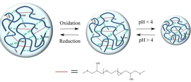
pH responsive PASP hydrogels with reversible response to redox stimuli were designed by using thiol groups as biologically relevant redox-susceptible moiety. Thiolated PASP as a water-soluble, pH and redox-responsive polymer [29] was cross-linked with PEGDGE in aqueous solution to obtain hydrogels providing redox modulated delivery of encapsulated macromolecular compounds. Because of the good reactivity of epoxy groups toward thiol groups, poly(ethylene glycol) diglycidyl ether was used to form redox-inert cross-links, these ensure the chemical stability of the gels both in the reduced and the oxidised state (Fig. 3).
Mechanically robust hydrogels were obtained in a wide range of permanent cross-linking ratio (from 5 to 15 n/n%) independently of the thiol content of PASP-SH.
Fig. 3Scheme of poly(aspartic acid) (PASP) hydrogel with a reversible volume response to external redox or pH stimuli without the dissolution of the gel, the presence of non-cleavable cross-linker, poly(ethylene glycol) diglycidyl ether, ensures the chemical stability of the gel both in the reduced and
the oxidised state
The effect of pH on gelation time was investigated in detail because controllable gelation is a requirement for the subsequent preparation of nanogels in water-in-oil emulsion. At pH = 10.5 the gelation time of SS50-DGE5 hydrogels was faster than 1 min. The concentration of
would increase the gelation time of PASP hydrogels. Accordingly, the gelation time increased to more than one hour upon decreasing the pH to 7 (Fig. 4). The degree of swelling in PBS measured after 24 hours of gelation (Fig. 4) was constant at pH values of preparation between 7.5 and 10.5, but increased sharply at smaller pH values indicating that gelation is complete within 24 hours only between pH 7.5 and 10.5. As a result, it is recommended to keep the pH above 7.5 during the cross-linking reaction and further adjustment of pH depends on the gelation time needed for the method of gel preparation. During the preparation of bulk hydrogels, the pH was kept at 10.5.
Fig. 4The gelation time (red circles) and the degree of swelling (blue squares) of SS50-DGE5 gels in PBS as a function of pH of preparation
3.2 Reversible redox-response
PASP hydrogels were designed to exploit thein vivoredox gradient available in the human body. As a simple approach, intracellular milieu is generally more reductive than the extracellular environment because of the large concentration difference of glutathione between the two sites, but it should be added that further redox differences exist in subcellular parts, which must also be considered in future application [37]. Contrary to the vast variety of bioreducible drug carriers already reported [19,38], we prepared hydrogels with reversible
response to redox stimuli [32], which allows for drug release in multiple “on-off” cycles. The thiol-disulphide exchange reaction is utilized in the PASP gel synthesized (Fig. 5). The pH was kept at 7.4 in each experiment which is biologically relevant and ensures that the majority of thiol groups are in their active, thiolate form (the pKavalue of cysteamine is approximately 8.35 [39]). The hydrogels remained stable both in reducing and oxidising solution because of the presence of non-cleavable cross-linkers (Fig. 6). The thiol-disulphide transformation was proven by FTIR spectroscopy. A peak at 2550 cm-1 corresponding to thiol groups (S−H stretching) appears in the spectrum of the reduced hydrogel, but disappears in oxidised state (Fig. S3).
Fig. 5Reversible reduction/oxidation of SS-DGE hydrogels
Fig. 6Reduced and oxidised forms of the SS50-DGE5 hydrogels
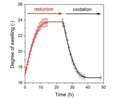
The thiol-disulphide transformation in the PASP hydrogels strongly affects the concentration of cross-links in the reduced and oxidised states, which was expected to control the swelling and mechanical properties as well as the release of encapsulated molecules. To demonstrate the redox-responsive character of the hydrogels, the degree of swelling of the SS50-DGE5 gels was determined in reducing (10 mM DTT) and then oxidising (10 mM NaBrO3) solutions. The degree of swelling increased in reducing medium because of the cleavage of the disulphide linkages, while decreased in oxidising medium due to the formation of disulphide linkages from thiol groups (Fig. 7). The hydrogels discs (with a thickness of 2 mm) reached their equilibrium degree of swelling within half a day thus in further experiments the equilibrium degree of swelling was determined after 24 hours.
Fig. 7Degree of swelling of the SS50-DGE5 hydrogel upon reduction/oxidation
The reversibility of the response is required to obtain hydrogels for on-demand, on-off type drug release. The rate of drug release is generally controlled by the mesh size available for the molecular payload, which correlates to degree of swelling. Accordingly, the swelling properties of PASP hydrogels were studied in repeating oxidising and reducing steps. Redox induced volume change was proven to be reversible in a wide composition range (thiol content or cross-linking ratio) and it has no effect on swelling, whether the starting step is oxidation or reduction (Fig. 8a and b). The extent of redox sensitivity was characterised by the relative degree of swelling ( , ) which is defined as the ratio of the degree of swelling in the reduced state to the oxidised state in the first redox cycle. The relative degree of swelling
increased with increasing thiol content (the permanent cross-linking ratio was fixed at 5%) as seen in Fig. 8c, and decreased with increasing permanent cross-linking ratio (Fig. 8d). We can conclude that the magnitude of the redox response could be controlled with the composition in a reversible manner, which enables us to use the gels for controlled release of encapsulated materials in the response to the change in the redox environment.
Fig. 8 Degree of swelling of a) SS30-DGE5 and b) SS50-DGE5 hydrogels, respectively, upon alternating reduction/oxidation starting with a reduction step (red circles) or an oxidation step (black squares); c) relative degree of swelling of SS-DGE5 hydrogels as a function of thiol content and d) relative degree of swelling of SS30-DGE and SS50-DGE hydrogels as a function of permanent cross- linking ratio
a) b)
c) d)
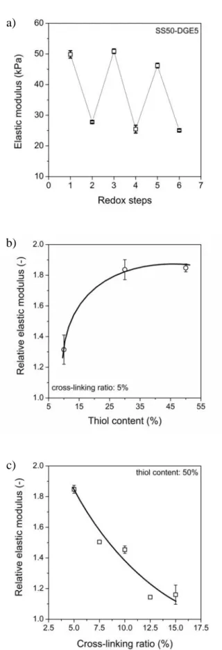
Fig. 9a) Elastic modulus of SS50-DGE5 hydrogel responding to alternating oxidation/reduction.
Relative elastic moduli of SS-DGE hydrogels as a function of b) thiol content and c) permanent cross- linking ratio
a)
b)
c)
Changes in the redox state of the environment affected the stiffness of the hydrogels. The stiffness of the hydrogels decreases in the reduced state due to the cleavage of disulphide linkages, while it increases in the oxidised state because of the re-formation of chemical cross-links. In accordance with the results of swelling experiments, repeated redox cycles showed reversible response and the gels preserved their mechanical stability (Fig. 9a). The relative elastic modulus ( / ), which was defined as the ratio of the elastic modulus in the oxidised state to the elastic modulus in the reduced state in the first redox cycle, thus the redox sensitivity of elastic modulus increased with increasing thiol content (Fig. 9b) and decreased with increasing permanent cross-linking ratio (Fig. 9c).
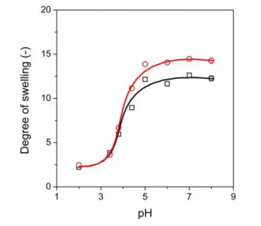
3.3 pH responsive swelling
A precise control of drug release can be achieved by the design of dual or multi responsive hydrogels, which can simultaneously respond to different external stimuli. As we have shown, the synthesized hydrogels respond to redox stimuli, but PASP also has an inherent pH- responsive character, because of its polyelectrolyte chain. A large number of carboxyl groups, i.e. at least 50% of the repeating units, are present in the SS-DGE hydrogels, which promote the pH-sensitive swelling of the gels. The environmental pH determines the ionisation degree of these dissociative groups, and the presence of ionised groups affects the degree of swelling of the hydrogels. Accordingly, a significant response in the degree of swelling was observed as a function of pH (Fig. 10). The swelling curves of both the reduced and oxidised form of SS30-DGE5 gels showed that the degree of swelling is about 3 at pH < 3. The small degree of swelling can be explained by the fact that the limited solubility of the polymer determines the degree of swelling in acidic medium, and the effect of disulphide content is not significant. A sharp increase is observed at approximately pH = 4 as a result of the deprotonation of the carboxyl groups on the PASP chains in both the reduced and the oxidised state. The degree of swelling reaches a constant value at about pH = 5 in both cases. A significant difference exists in the degree of swelling of the hydrogels in the reduced and the oxidised state at pH 6-8. A larger degree of swelling was achieved in the reduced form caused by the decreased number of chemical cross-links. The dual – pH- and redox – responsive behaviour of the these hydrogels may find various biomedical applications, and further adjustment of pH-dependent swelling by the introduction of other functional groups into the polymer and the control of
redox-dependent properties enable us to design hydrogels with a highly selective response to the local environment of the hydrogels.
Fig. 10Degree of swelling of SS30-DGE5 hydrogels as a function of pH in the reduced (red circles) and in the oxidised (black squares) state
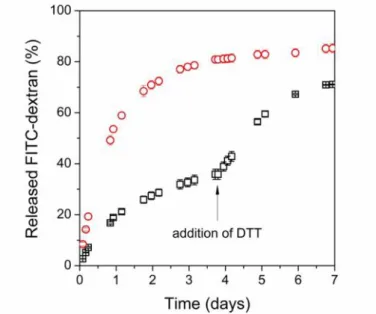
3.4 Redox-dependent drug release
As a proof of concept on redox-modulated drug release of PASP hydrogels, we demonstrate the release of a macromolecular model drug, fluorescein isothiocyanate-dextran (FITC-dextran, Mw= 70 kDa) from SS50-DGE5 gels. The encapsulation was done during the cross-linking reaction with an efficiency as high as 96%. Drug-loaded, oxidised hydrogel discs were placed into reducing (10 mM DTT in PBS) and redox inert (PBS) medium, and the amount of released drug was followed over time with fluorimetry (Fig. 11, for calibration curves please see Fig. S4 and Table S3). Reducing media imitates the reducing intracellular environment, while PBS models the physiological state of the human blood (pH = 7.4 [40]).
Approximately 80% of the entrapped drug is released within 3 days in the reducing solution, while only 33% of the drug was released from gels in the oxidised state. We added the reducing agent (DTT) to the PBS solution after 4 days to mimic the intracellular pathway, and an additional drug release was observed, which prove the redox-switchable drug release from the gels. We verified that a reducing environment promotes the drug release from the PASP hydrogels and the release of the encapsulated molecules is controlled by the swelling of the
hydrogels. In the further work, the selectivity of drug release needs to be improved, i.e. the leakage must be stopped in the oxidised state. We demonstrated the responsive properties and the drug release on bulk hydrogels, but future use may demand the formulation of gels on smaller length scales. The nanogels have potential use in intracellular drug delivery.
Fig. 11Amount of releasedFITC-dextranfrom SS50-DGE5 hydrogels in reducing medium (10 mM DTT in PBS, red circles), and at first in redox inert (PBS) medium, after 4 days in reducing medium
(10 mM DTT in PBS, black squares)
3.5 Redox-response of nanogels
Responsive nanogels gained a considerable attention in the last decade in the field of targeted drug delivery, since these matrices possess all the beneficial properties of hydrogels – e.g. biocompatibility, responsiveness and high drug-loading capacity – and due to their small size they can be injected directly to the blood-stream [41]. In order to prepare nanogels from linear polymers they have to be cross-linked in small, defined and discrete volumes [33]. Thus the cross-linking of thiolated poly(aspartic acid) (PASP-SH) took place in the nanosized droplets of an inverse miniemulsion [30]. While the dispersed aqueous phase contains the aqueous solution of PASP-SH and poly(ethylene glycol) diglycidyl ether as the cross-linker, the continuous phase was n-octane, which contained the surfactant. To prepare a stable emulsion and to reduce the coalescence of the droplets, the right choice of surfactant is very
the concentration of 8 w/w %). The pH of the precursor polymer solution was adjusted to pH = 8 to increase gelation time to more than 15 min, which is sufficient for emulsification.
The technique was published earlier for the preparation of redox PASP nanogels showing gel- sol transition upon reduction [30].
PASP nanogels were successfully prepared using the composition of the SS50-DGE5 hydrogel, which showed the strongest redox sensitivity in bulk experiments (Fig. 8). A TEM study confirmed the preparation of spherical nanogels with diameter between 130 and 150 nm in the dried state (Fig. 12 a). This size range was also confirmed by DLS measurements showing an average hydrodynamic diameter (dH ) of 141 nm with moderate width of size distribution (PDI = ~0.5).
Fig. 12a) TEM image of SS50-DGE5 nanogels and b) size distribution of SS50-DGE5 nanogels in water and in 0.1 M DTT solution
The volume response of the nanogels was investigated in reducing medium to confirm that they swell, but do not dissolve due to the presence of non-cleavable cross-links, and to demonstrate their redox-dependent swelling. The average hydrodynamic diameter increased to 216 nm in a reducing solution (0.1 M DTT) with a similar size distribution (PDI = ~0.5) to that of gels prior to reduction (Fig. 12 b). The zeta potential was negative throughout the studied pH scale as shown in Fig. 13. It predicts protein resistance and prolonged circulation time of PASP nanogels when used in future in vivo applications. Around the pKa values of aspartic acid repeating units (3.25 and 4.35 [42]) the zeta potential decreased strongly with
a) b)
increasing pH, while above pH ~5 the zeta potential became independent of pH and showed a strongly negative value (–45 mV). The pH interval of the abrupt change in the zeta potential is in good correlation with that of the pH dependent degree of swelling of SS50-DGE5 hydrogel (Fig. 10). All these characteristics suggest that PASP nanogels are promising for use in redox- controlled, systemic delivery of macromolecular compounds to the targeted site of the human body.
Fig. 13Zeta potential of SS50-DGE5 nanogels as a function of pH
4 Conclusions
Dual, redox- and pH-responsive hydrogels can be prepared in aqueous solution by cross- linking thiolated poly(aspartic acid) (PASP) with a non-cleavable cross-linker, poly(ethylene glycol) diglycidyl ether, through the thiol groups of the polymer. After the cross-linking reaction thiol groups remaining in the polymer network could be converted reversibly into disulphide linkages upon oxidation. The presence of cleavable linkages enabled us to control the degree of swelling and the stiffness of bulk gels using reducing and oxidising media, while the permanent cross-links maintained the gels as cohesive materials. A chosen macromolecular model drug was entrapped into the gel with high encapsulation efficiency and was released in reducing medium at considerably faster rate than in oxidising medium.
The combination of the redox-responsive character of the gels and the inherent pH-dependent
drug release as implants or nanogels. Spherical PASP nanogels were also prepared successfully in a water-in-oil emulsion as proven by TEM and DLS. PASP nanogels were stable in reducing media and swelled similarly to their macroscopic counterparts, which makes them promising for use in redox-controlled, systemic delivery of macromolecular compounds to the targeted site of the human body.
Acknowledgements
This research was supported by the National Research, Development and Innovation Office – NKFIH (FK 125074). Benjámin Gyarmati acknowledges the János Bolyai Research Scholarship of the Hungarian Academy of Sciences for financial support. A. Szilágyi is grateful to the support of the ÚNKP-17-4-III New National Excellence Program of the Ministry of Human Capacities. The authors also thank the Bordeaux Imaging Center facility (Bordeaux, France) for technical assistance in TEM imaging.
5 References
[1] H. Maeda, L.W. Seymour, Y. Miyamoto, Conjugates of anticancer agents and polymers: advantages of macromolecular therapeutics in vivo, Bioconjug. Chem. 3 (1992) 351–362. doi:10.1021/bc00017a001.
[2] Y. Lu, P.S. Low, Folate-mediated delivery of macromolecular anticancer therapeutic agents, Adv. Drug Deliv. Rev. 64 (2012) 342–352. doi:10.1016/j.addr.2012.09.020.
[3] H. Nakamura, F. Jun, H. Maeda, Development of next-generation macromolecular drugs based on the EPR effect: challenges and pitfalls., Expert Opin. Drug Deliv. 12 (2015) 53–64. doi:10.1517/17425247.2014.955011.
[4] M. Belting, A. Wittrup, Developments in macromolecular drug delivery, Drug Deliv.
480 (2009) 1–10. doi:10.1007/978-1-59745-429-2.
[5] N.A. Peppas, J.Z. Hilt, A. Khademhosseini, R. Langer, Hydrogels in Biology and Medicine: From Molecular Principles to Bionanotechnology, Adv. Mater. 18 (2006) 1345–1360. doi:10.1002/adma.200501612.
[6] B. Gyarmati, Á. Némethy, A. Szilágyi, Reversible disulphide formation in polymer networks: A versatile functional group from synthesis to applications, Eur. Polym. J. 49 (2013) 1268–1286. doi:10.1016/j.eurpolymj.2013.03.001.
[7] M.C. Koetting, J.T. Peters, S.D. Steichen, N.A. Peppas, Stimulus-responsive hydrogels: theory, modern advances, and applications, Mater. Sci. Eng. R Reports. 93 (2015) 1–49. doi:10.1016/j.mser.2015.04.001.
[8] V. Lapeyre, C. Ancla, B. Catargi, V. Ravaine, Glucose-responsive microgels with a core-shell structure, J. Colloid Interface Sci. 327 (2008) 316–323.
doi:10.1016/j.jcis.2008.08.039.
[9] L. Klouda, A.G. Mikos, Thermoresponsive hydrogels in biomedical applications: a seven-year update, Eur. J. Pharm. Biopharm. 97 (2015) 338–349.
doi:10.1016/j.ejpb.2015.05.017.
[10] J. Liu, C. Detrembleur, M. Hurtgen, A. Debuigne, M.-C. De Pauw-Gillet, S. Mornet, et al., Reversibly crosslinked thermo- and redox-responsive nanogels for controlled drug release, Polym. Chem. 5 (2014) 77–88. doi:10.1039/c3py00839h.
[11] A.S. Hoffman, Hydrogels for biomedical applications, Adv. Drug Deliv. Rev. 54 (2002) 3–12. doi:10.1016/j.addr.2012.09.010.
[12] T. Vermonden, R. Censi, W.E. Hennink, Hydrogels for protein delivery, Chem. Rev.
112 (2012) 2853–2888. doi:10.1021/cr200157d.
[13] E. Calo, V. V. Khutoryanskiy, Biomedical applications of hydrogels: A review of patents and commercial products, Eur. Polym. J. 65 (2014) 252–267.
doi:10.1016/j.eurpolymj.2014.11.024.
[14] J. Li, D.J. Mooney, Designing hydrogels for controlled drug delivery, Nat. Rev. Mater.
1 (2016) Article number: 16071. doi:10.1038/natrevmats.2016.71.
[15] F.Q. Schafer, G.R. Buettner, Redox environment of the cell as viewed through the redox state of the glutathione disulfide/glutathione couple, Free Radic. Biol. Med. 30 (2001) 1191–1212. doi:10.1016/S0891-5849(01)00480-4.
[16] D.P. Jones, J.L. Carlson, P.S. Samiec, P. Sternberg, V.C. Mody, R.L. Reed, et al.,
and devatization conditions for analysis of densyl derivatives by HPLC, Clin. Chim.
Acta. 275 (1998) 175–184. doi:10.1016/S0009-8981(98)00089-8.
[17] X. Zhang, L. Han, M. Liu, K. Wang, L. Tao, Q. Wan, et al., Recent progress and advances in redox-responsive polymers as controlled delivery nanoplatforms, Mater.
Chem. Front. 1 (2017) 807–822. doi:10.1039/C6QM00135A.
[18] V.I. Lushchak, Glutathione Homeostasis and Functions: Potential Targets for Medical Interventions, J. Amino Acids. 2012 (2012) Article ID 736837.
doi:10.1155/2012/736837.
[19] F. Meng, W.E. Hennink, Z. Zhong, Reduction-sensitive polymers and bioconjugates for biomedical applications, Biomaterials. 30 (2009) 2180–2198.
doi:10.1016/j.biomaterials.2009.01.026.
[20] R. Cheng, F. Feng, F. Meng, C. Deng, J. Feijen, Z. Zhong, Glutathione-responsive nano-vehicles as a promising platform for targeted intracellular drug and gene delivery., J. Control. Release. 152 (2011) 2–12. doi:10.1016/j.jconrel.2011.01.030.
[21] S. Singh, I. Zilkowski, A. Ewald, T. Maurell-Lopez, K. Albrecht, M. Möller, et al., Mild oxidation of thiofunctional polymers to cytocompatible and stimuli-sensitive hydrogels and nanogels, Macromol. Biosci. 13 (2013) 470–82.
doi:10.1002/mabi.201200389.
[22] Y. Wen, J.K. Oh, Dual-stimuli reduction and acidic pH-responsive bionanogels:
intracellular delivery nanocarriers with enhanced release, RSC Adv. 4 (2014) 229–237.
doi:10.1039/c3ra46072j.
[23] G. Saito, J.A. Swanson, K.-D. Lee, Drug delivery strategy utilizing conjugation via reversible disulfide linkages: role and site of cellular reducing activities, Adv. Drug Deliv. Rev. 55 (2003) 199–215. doi:10.1016/S0169-409X(02)00179-5.
[24] D. Jańczewski, J. Song, E. Csányi, L. Kiss, P. Blazsó, R.L. Katona, et al., Organometallic polymeric carriers for redox triggered release of molecular payloads, J.
Mater. Chem. 22 (2012) 6429–6435. doi:10.1039/c2jm15755a.
[25] C. Németh, B. Gyarmati, T. Abdullin, K. László, A. Szilágyi, Poly(aspartic acid) with adjustable pH-dependent solubility, Acta Biomater. 49 (2017) 486–494.
doi:10.1016/j.actbio.2016.11.065.
[26] Á. Némethy, K. Solti, L. Kiss, B. Gyarmati, M.A. Deli, E. Csányi, et al., pH- and temperature-responsive poly(aspartic acid)-l-poly(N-isopropylacrylamide) conetwork hydrogel, Eur. Polym. J. 49 (2013) 2392–2403. doi:10.1016/j.eurpolymj.2013.02.015.
[27] K. Molnár, D. Juriga, P.M. Nagy, K. Sinkó, A. Jedlovszky-Hajdú, M. Zrínyi, Electrospun poly(aspartic acid) gel scaffolds for artificial extracellular matrix, Polym.
Int. 63 (2014) 1608–1615. doi:10.1002/pi.4720.
[28] B. Gyarmati, E.Z. Mészár, L. Kiss, M.A. Deli, K. László, A. Szilágyi, Supermacroporous chemically cross-linked poly(aspartic acid) hydrogels, Acta Biomater. 22 (2015) 32–38. doi:10.1016/j.actbio.2015.04.033.
[29] B. Gyarmati, B. Vajna, Á. Némethy, K. László, A. Szilágyi, Redox- and pH-responsive cysteamine-modified poly(aspartic acid) showing a reversible sol-gel transition, Macromol. Biosci. 13 (2013) 633–640. doi:10.1002/mabi.201200420.
[30] E. Krisch, L. Messager, B. Gyarmati, V. Ravaine, A. Szilágyi, Redox- and pH- responsive nanogels based on thiolated poly(aspartic acid), Macromol. Mater. Eng. 301 (2016) 260–266. doi:10.1002/mame.201500119.
[31] M. Zrínyi, T. Gyenes, D. Juriga, J.-H. Kim, Volume change of double cross-linked poly(aspartic acid) hydrogels induced by cleavage of one of the crosslinks., Acta Biomater. 9 (2013) 5122–5131. doi:10.1016/j.actbio.2012.08.046.
[32] B. Gyarmati, Á. Némethy, A. Szilágyi, Reversible response of poly(aspartic acid) hydrogels to external redox and pH stimuli, RSC Adv. 4 (2014) 8764–8771.
doi:10.1039/c3ra47530a.
[33] E. Krisch, B. Gyarmati, A. Szilágyi, Preparation of pH-responsive poly(aspartic acid) nanogels in inverse emulsion, Period. Polytech. Chem. Eng. 61 (2017) 19–26.
doi:10.3311/PPch.9788.
[34] J. Vlasák, F. Rypáček, J. Drobník, V. Saudek, Properties and reactivity of polysuccinimide, J. Polym. Sci. Polym. Symp. 66 (1979) 59–64.
doi:10.1002/polc.5070660109.
[35] B.Á. Szilágyi, B. Gyarmati, G. Horvát, Á. Laki, M. Budai-Szűcs, E. Csányi, et al., The effect of thiol content on the gelation and mucoadhesion of thiolated poly(aspartic acid), Polym. Int. 66 (2017) 1538–1545. doi:10.1002/pi.5411.
[36] L.L. Messager, N.N. Portecop, E. Hachet, V.V. Lapeyre, I. Pignot-Paintrand, B.
Catargi, et al., Photochemical crosslinking of hyaluronic acid confined in nanoemulsions: towards nanogels with a controlled structure., J. Mater. Chem. B. 1 (2013) 3369. doi:10.1039/c3tb20300j.
[37] D.J. Phillips, M.I. Gibson, Redox-sensitive materials for drug delivery: targeting the correct intracellular environment, tuning release rates, and appropriate predictive systems., Antioxid. Redox Signal. 21 (2014) 786–803. doi:10.1089/ars.2013.5728.
[38] T. Kim, S.W. Kim, Bioreducible polymers for gene delivery, React. Funct. Polym. 71 (2011) 344–349. doi:10.1016/J.REACTFUNCTPOLYM.2010.11.016.
[39] I.H. Pitman, I.J. Morris, Covalent additions of glutathione, cysteine, homocysteine, cysteamine and thioglycolic acid to quinazoline cation, Aust. J. Chem. 134 (1979) 1567–1573. doi:10.1071/CH9791567.
[40] J.A. Kellum, Determinants of blood pH in health and disease, Crit Care. 4 (2000) 6–14.
doi:10.1186/cc644.
[41] K. Raemdonck, J. Demeester, S. De Smedt, Advanced nanogel engineering for drug delivery, Soft Matter. 5 (2009) 707–715. doi:10.1039/b811923f.
[42] S. Roweton, S.J. Huang, G. Swift, Poly(aspartic acid): synthesis, biodegradation, and current applications, J. Environ. Polym. Degrad. 5 (1997) 175–181.
doi:10.1007/BF02763661.