Alterations in the neuropeptide galanin system in
major depressive disorder involve levels of transcripts, methylation, and peptide
Swapnali Bardea,1, Joelle Rüeggb,c,d, Josée Prud’hommee, Tomas J. Ekströmb,c, Miklos Palkovitsf,g,h, Gustavo Tureckie,i, Gyorgy Bagdyj,k,l, Robert Ihnatkom,n, Elvar Theodorssonm,n, Gabriella Juhaszj,o,p, Rochellys Diaz-Heijtza,
Naguib Mechaware,i, and Tomas G. M. Hökfelta,1
aDepartment of Neuroscience, Karolinska Institutet, 17 177 Stockholm, Sweden;bDepartment of Clinical Neuroscience, Karolinska Institutet, 17 177 Stockholm, Sweden;cThe Center for Molecular Medicine, 17 177 Stockholm, Sweden;dSwedish Toxicology Science Research Center Swetox, 151 36 Södertälje, Sweden;
eMcGill Group for Suicide Studies, Douglas Mental Health University Institute, Verdun, QC H4H 1R3, Canada;fNeuromorphological and Neuroendocrine Research Laboratory, Hungarian Academy of Sciences, Semmelweis University, H-1085, Budapest, Hungary;gDepartment of Anatomy, Histology and Embryology, Semmelweis University, H-1085, Budapest, Hungary;hHuman Brain Tissue Bank and Laboratory, Semmelweis University, H-1085, Budapest, Hungary;iDepartment of Psychiatry, McGill University, Montreal, QC, H3A 0G4, Canada;jDepartment of Pharmacodynamics, Semmelweis University, H-1089, Budapest, Hungary;kMagyar Tudományos Akadémia-Semmelweis Egyetem (MTA-SE)-Neuropsychopharmacology and Neurochemistry Research Group, Hungarian Academy of Sciences, Semmelweis University, H-1089, Budapest, Hungary;lNemzeti Agykutatási Program-A alprogram-Semmelweis Egyetem (NAP- A-SE) Research Group, Semmelweis University, H-1089, Budapest, Hungary;mDepartment of Clinical Chemistry, Linköping University, 581 83 Linköping, Sweden;
nDepartment of Clinical and Experimental Medicine, Linköping University, 581 83 Linköping, Sweden;oMagyar Tudományos Akadémia, Semmelweis Egyetem, Nemzeti Agykutatási Program B alprogram (MTA-SE-NAP B) Genetic Brain Imaging Migraine Research Group, Hungarian Academy of Sciences, Semmelweis University, H-1089, Budapest, Hungary; andpNeuroscience and Psychiatry Unit, University of Manchester, Manchester M13 9PT, United Kingdom
Contributed by Tomas G. M. Hökfelt, October 28, 2016 (sent for review September 7, 2016; reviewed by Francesc Artigas, Jacqueline N. Crawley, Ranga Krishnan, and Anantha Shekhar)
Major depressive disorder (MDD) is a substantial burden to patients, families, and society, but many patients cannot be treated adequately.
Rodent experiments suggest that the neuropeptide galanin (GAL) and its three G protein-coupled receptors, GAL1–3, are involved in mood regulation. To explore the translational potential of these results, we assessed the transcript levels (by quantitative PCR), DNA methyl- ation status (by bisulfite pyrosequencing), and GAL peptide by RIA of the GAL system in postmortem brains from depressed persons who had committed suicide and controls. Transcripts for all four members were detected and showed marked regional variations,GALand galanin re- ceptor 1 (GALR1) being most abundant. Striking increases inGALand GALR3mRNA levels, especially in the noradrenergic locus coeruleus and the dorsal raphe nucleus, in parallel with decreased DNA methylation, were found in both male and female suicide subjects as compared with controls. In contrast,GALandGALR3transcript levels were decreased, GALR1was increased, and DNA methylation was increased in the dor- solateral prefrontal cortex of male suicide subjects, however, there were no changes in the anterior cingulate cortex. Thus,GALand its receptorGALR3are differentially methylated and expressed in brains of MDD subjects in a region- and sex-specific manner. Such an epigenetic modification inGALR3, a hyperpolarizing receptor, might contribute to the dysregulation of noradrenergic and serotonergic neurons impli- cated in the pathogenesis of MDD. Thus, one may speculate that a GAL3antagonist could have antidepressant properties by disinhibiting the firing of these neurons, resulting in increased release of noradren- aline and serotonin in forebrain areas involved in mood regulation.
epigenetics
|
human postmortem brain|
neuropeptides|
stress|
transmitter coexistence
M
ajor depressive disorder (MDD) is a serious mental illness affecting up to 20% of the population at some point during their lives, women more frequently than men, and representing a major burden to patients, their families, and society (1, 2). MDD is thought to arise from the interaction of genetic and environmental factors, with stressful life events representing an important predis- posing factor (3–5). Growing evidence suggests that epigenetic mechanisms mediate such interactions, namely through altered DNA methylation, thus leading to stable changes in brain function that may underlie psychopathology (6, 7).Pharmacological management of depression currently involves drugs that often target the monoamine transporters, which include selective reuptake inhibitors for serotonin (5-hydroxytryptamine,
5-HT) (SSRIs), noradrenaline (NA) inhibitors (NRIs), or a com- bination of both (SNRIs) (8–10), as well as a number of other medications (11). However, the therapeutic efficacy of these anti- depressants is hampered by a slow onset of action, a limited re- sponse rate, and considerable side effects (12, 13). These issues have led to intensive search for novel therapeutic approaches for MDD (14), including targeting receptors for neuropeptides (15–19), the most diverse family of brain messenger molecules (20).
In this context, the 29/30 amino acid neuropeptide galanin (GAL) (21), which is widely distributed in the rat (22–25) and human (26) brain, may be of special interest. In particular, it co- exists with NA in the locus coeruleus in both rat (27–29) and human (26, 30–32) and in rat with 5-HT in the dorsal raphe nu- cleus (28, 33, 34).
Significance
Depression is a seriously disabling disorder, twice as common in women as in men. Lack of efficacy of existing pharmacotherapies in subsets of patients has led to an intensive search for new targets for antidepressant development, including receptors for neuropeptides such as galanin (GAL). In this study, we explore GAL and its three receptors, GAL1–3, comparing postmortem brain regions from depressed suicide patients and controls. Using quantitative PCR and bisulfite pyrosequencing, we report sig- nificant changes in the transcript and DNA methylation levels ofGALand galanin receptor 3 (GALR3) in the locus coeruleus and dorsal raphe nucleus, two regions important for mood regulation. Our findings suggest GAL3involvement in depressive disorder, making it a possible drug target for this disease.
Author contributions: S.B., J.R., T.J.E., R.D.-H., N.M., and T.G.M.H. designed research; S.B., J.R., J.P., R.I., and E.T. performed research; M.P., G.T., and N.M. contributed new reagents/analytic tools; S.B., J.R., T.J.E., E.T., G.J., R.D.-H., N.M., and T.G.M.H. analyzed data; and S.B., J.R., G.B., G.J., R.D.-H., N.M., and T.G.M.H. wrote the paper.
Reviewers: F.A., Institut d’Investigacions Biomèdiques August Pi i Sunyer; J.N.C., University of California Davis School of Medicine; R.K., Rush Medical College; and A.S., Indiana University.
The authors declare no conflict of interest.
Freely available online through the PNAS open access option.
1To whom correspondence may be addressed. Email: tomas.hokfelt@ki.se or Swapnali.
Barde@ki.se.
This article contains supporting information online atwww.pnas.org/lookup/suppl/doi:10.
1073/pnas.1617824113/-/DCSupplemental.
GAL exerts its physiological actions via three subtypes of G protein-coupled receptors, GAL1–3(35, 36). The distribution of these receptors has been mapped previously with ligand-binding autoradiography in rat (37, 38), monkey, and human (39–41) brain. More recently the receptor transcripts have been localized with in situ hybridization in rat brain (42–44) and in some regions of the human brain (32). It should be noted that GAL receptor subtypes can form dimers and heterodimers, a mechanism that can profoundly change GAL signaling (45).
Interestingly, certain differences between species exist in regions of potential importance for mood-related disorders. For example, galanin receptor 3 (GalR3) mRNA has a limited distribution in the rat brain (44), where it could not even be detected in the first publication on the cloning of this receptor (46). However, it appears to be expressed in human noradrenergic locus coeruleus (NA-locus coeruleus) neurons (32). In addition GAL itself has a different profile: it is expressed in 5-HT neurons in the dorsal raphe nucleus in the rat but not in humans, but its expression in the locus coeruleus is conserved in the species investigated to date.
Animal studies have provided strong evidence that the GAL system is involved in anxiety- and depression-like behavior (33, 47–
55). A recent genetic association study supports a possible role of the GAL system in mood disorders, pointing to involvement of epige- netic processes and a strong association with high levels of stress (56). Moreover, there is an interaction between GAL and the 5-HT1A receptor (47), a receptor that plays an important role as autoreceptor in depression, as shown in experimental studies (57, 58) and in studies of suicide victims (59). In agreement with many ani- mal experimental studies, PET imaging also has indicated a role for postsynaptic 5-HT1Areceptors in depression (60).
The aim of the present study was to identify possible changes in the GAL system in MDD by analyzing postmortem brains from depressed subjects who committed suicide (hereafter“DS”subjects) and matched subjects without psychiatric symptoms (hereafter, simply“controls”). We used quantitative PCR (qPCR) to monitor transcript levels, bisulfite pyrosequencing to study DNA methyl- ation (GAL andGALR1-3), and RIA to establish GAL peptide levels. All three analyses were carried out on the same samples from five different, relevant brain regions: dorsolateral prefrontal cortex [Brodmann area (BA) 8/9], anterior cingulate cortex (BA 24), locus coeruleus, dorsal raphe nucleus, and the medullary raphe nuclei.
Results
Cohort Demographics.There were no significant differences be- tween DS subjects and their matched controls in age (DS subjects vs. controls: 51.6±15.4 vs. 57.5±15.4 y,P=0.11); postmortem interval (PMI) (DS subjects vs. controls: 43.56±24.16 vs. 49.21± 34.44 h,P =0.94); brain pH (DS subjects vs. controls: 6.61 ± 0.29 vs. 6.46±0.34;P=0.13); or RNA integrity number (RIN) (DS subjects vs. controls: 6.49±1.70 vs. 6.68±1.62;P<0.49).
The details of the demographic characteristics of DS subjects and
controls for each of the regions analyzed are provided in Table 1, and detailed information on each individual subject is provided inTable S1.
Sample Anatomy. The five brain regions analyzed encompass a heterogeneous collection of neurons. It is important to note that in the samples labeled locus coeruleus and especially in those labeled dorsal raphe and medullary raphe only a part of these neurons are monoaminergic (SI Materials and Methods,Brain Samples).
The Effects of Medication.An inherent problem of autopsy analysis is the effect of medication, particularly when looking at a dynamic parameter, such as transcripts. For example, it has been reported that an antidepressant can exert epigenetic changes (61, 62). We have analyzed the data using ANOVA followed by Fisher’s least significant difference post hoc test and ANCOVA by treating an- tidepressants as a confounding factor to compare the effects in male and female DS subjects. The samples were divided into four groups based on the different medications, namely (i) SSRIs, (ii) SSRI+benzodiazepines, (iii) SSRI+others (including SNRIs, serotonin antagonist and reuptake inhibitors, serotonin-norepinephrine- dopamine reuptake inhibitors, tricyclic antidepressants, and noradrenergic and specific serotonergic antidepressants; details are given inTable S1), and (iv) none. Based on the analysis, we conclude that in our study the various psychiatric medications show no significant effect on the gene expression of GAL and GalR3 in the five regions analyzed. The longer imprints of DNA methylation would require analysis of the complete anamnesis.
Transcript Levels Vary Across Brain Regions.The overall distribution and levels of the transcripts of the galanin system are summarized in Table 2. Briefly, transcripts forGALandGAL1–3are differentially expressed in the five brain regions, with the difference reaching statistical significance for all the markers studied (seeTables S2and S3 for P values). GAL is expressed at high levels in the lower brainstem and at three- to-fourfold lower levels in the two cortical Table 1. Demographic characteristics of the cohort
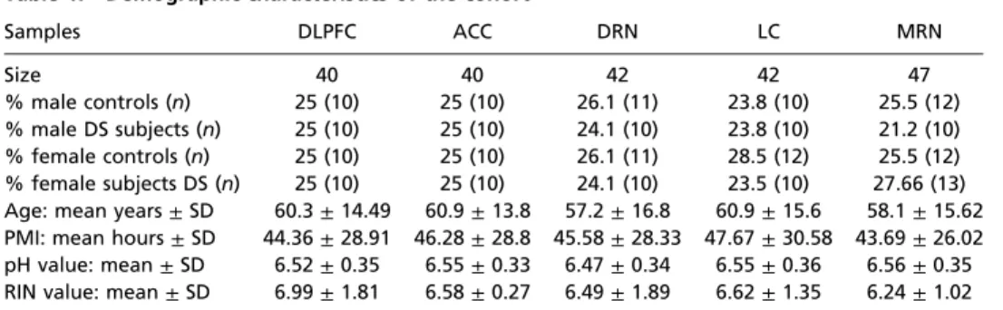
Samples DLPFC ACC DRN LC MRN
Size 40 40 42 42 47
% male controls (n) 25 (10) 25 (10) 26.1 (11) 23.8 (10) 25.5 (12)
% male DS subjects (n) 25 (10) 25 (10) 24.1 (10) 23.8 (10) 21.2 (10)
% female controls (n) 25 (10) 25 (10) 26.1 (11) 28.5 (12) 25.5 (12)
% female subjects DS (n) 25 (10) 25 (10) 24.1 (10) 23.5 (10) 27.66 (13) Age: mean years±SD 60.3±14.49 60.9±13.8 57.2±16.8 60.9±15.6 58.1±15.62 PMI: mean hours±SD 44.36±28.91 46.28±28.8 45.58±28.33 47.67±30.58 43.69±26.02 pH value: mean±SD 6.52±0.35 6.55±0.33 6.47±0.34 6.55±0.36 6.56±0.35 RIN value: mean±SD 6.99±1.81 6.58±0.27 6.49±1.89 6.62±1.35 6.24±1.02 ACC, anterior cingulate cortex; DLPFC, dorsolateral prefrontal cortex; DRN, dorsal raphe nucleus; LC, locus coeruleus; MRN, medullary raphe nucleus; PMI, postmortem interval; RIN, RNA integrity number.
Table 2. Raw cycle threshold (Ct) values±SEM for samples from the regions analyzed in male and female controls and DS subjects
Region GAL GalR1 GalR2 GalR3
DLPFC 27.1±0.2 25.7±0.2 31.4±0.2 33.2±0.1 ACC 27.5±0.1 28.6±0.1 31.6±0.2 33.5±0.2 DRN 26.2±0.4 27±0.4 29.3±0.2 31.5±0.4 LC 24.6±0.5 26.4±0.6 32.3±0.1 31.4±0.6 MRN 25.2±0.4 26.9±0.4 34.6±0.2 31.9±0.4 ACC, anterior cingulate cortex; DLPFC, dorsolateral prefrontal cortex;
DRN, dorsal raphe nucleus; LC, locus coeruleus; MRN, medullary raphe nucleus.
NEUROSCIENCEPNASPLUS
regions. The most prominent receptor transcript overall isGALR1, with up to eightfold differences in regional expression (dorsolateral prefrontal cortex>anterior cingulate cortex). TheGALR2mRNA level is high in the dorsal raphe nucleus and low in medullary raphe nuclei.GALR3mRNA follows the pattern ofGALwith high levels in the lower brainstem and low levels in cortex.
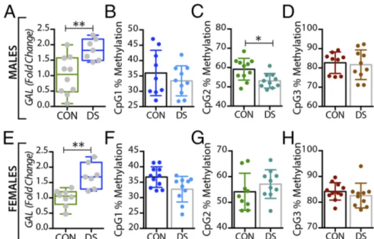
Expression ofGALand Its Receptors Is Altered in the Brains of DS Subjects.In the dorsolateral prefrontal cortex,GALmRNA levels were significantly decreased in male DS subjects compared with controls (P<0.01) (Fig. 1A) and were significantly increased in fe- male DS subjects (P < 0.05) (Fig. 1E), a distinct sex difference.
GALR1mRNA levels were significantly increased in both male and female DS subjects (P<0.05) (Fig. S1AandC).GALR3mRNA levels were significantly decreased in male (P<0.01) (Fig. 2A) but not in female (Fig. 2E) DS subjects. However, there were no sig- nificant changes inGALR2mRNA levels in either sex (Fig. S1E and G). In the anterior cingulate cortex, no significant changes in GALor its receptors were found (Fig. S2).
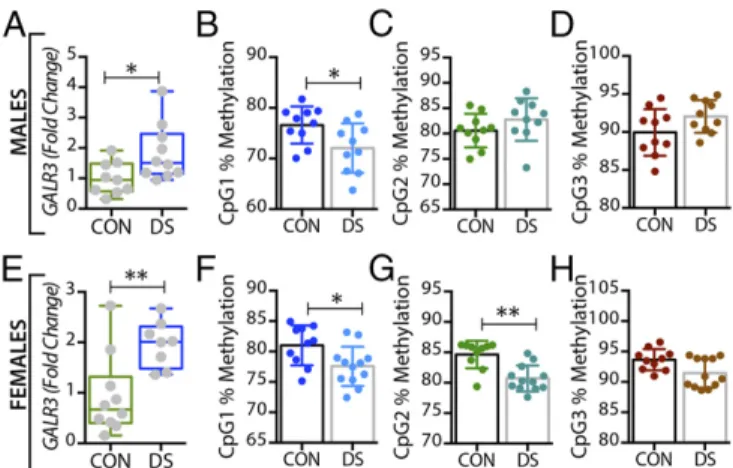
In the dorsal raphe nucleus and locus coeruleus,GALmRNA levels were significantly increased in both male (P<0.01) (Figs. 3A and 4A) and female (P<0.01) (Figs. 3Eand 4E) DS subjects. In- terestingly, higher levels of tryptophan hydroxylase-2 (TPH2) mRNA and immunoreactive neurons have previously been shown in the dorsal raphe nucleus of suicides relative to normal controls (63, 64).GALR1mRNA levels were significantly increased only in the dorsal raphe nucleus of male DS subjects (P<0.05,Fig. S3 AandC) but were not increased in the locus coeruleus of these subjects (Fig. S4AandC). The expression levels ofGALR3were significantly increased in both the dorsal raphe nucleus (Fig. 5A andE) and locus coeruleus (Fig. 6AandE) in both sexes, more robustly in females (female vs. males:P<0.01 vs.P<0.05). No significant changes were found inGALR2mRNA levels in these two regions (Figs. S3EandGandS4EandG).
In the medullary raphe nuclei,GALmRNA levels were significantly increased in both male and female DS subjects (P<0.05) (Fig. S5A andC). In male DS subjects, theGALR1expression levels were sig- nificantly increased (P<0.05) (Fig. S5E), whereasGALR2mRNA levels were significantly decreased (P< 0.01) (Fig. S5I). However,
hand, the expression levels ofGALR3were significantly increased in both sexes (P<0.05) (Fig. S5MandO). All mRNA changes are summarized in Table 3.
The DNA Methylation Status ofGALandGALR3Changes in the Brain of DS Patients.In the dorsolateral prefrontal cortex, At the first three CpG sites analyzed (CpG1–3, Fig. 1B–D), the percentage of methylated DNA was significantly higher in male DS subjects than in controls (P<0.05,P<0.01, andP<0.01, respectively). In female DS subjects, the methylation status was different among the CpG sites, with a significant increase at CpG1 and a significant decrease at CPG2 (P <0.05) (Fig. 1 FandG, respectively), and with no changes at CpG3 (Fig. 1H). No significant changes were found for any of the receptors in this brain region (Fig. 2B–HandFig. S1) or for any of the four markers in the anterior cingulate cortex (Fig. S2).
In the dorsal raphe nucleus, a significant decrease was seen in the percentage of methylated DNA in theGALpromoter of male DS subjects, but only at the CpG1 site (P< 0.05) (Fig. 3B), with no changes in female DS subjects (Fig. 3F–H). Interestingly,GALR3 was the only receptor that was significantly altered in the brains of DS subjects in a sex-dependent manner. Thus, only female DS subjects showed a significant decrease in the percentage of methylated DNA at CpG1 and CpG2 sites (P<0.01) (Fig. 5FandG, respectively).
In the locus coeruleus the percentage of methylated DNA in the GALpromoter at CpG1 and CPG2 was significantly decreased in DS subjects (male:P<0.01 andP<0.05; female:P<0.05 andP<0.01) (Fig. 4BandCandFandG, respectively). There was a signif- icant decrease in the percentage of methylated DNA at the CpG1 site of theGALR3gene in DS subjects of both sexes (P<0.05) (Fig. 6 B and F), whereas methylation at the CpG2 site was significantly decreased only in female DS subjects (P < 0.01) (Fig. 6G). No changes were found at the CpG3 site (Fig. 6DandH).
There were no significant changes in the methylation status of GALor its receptors in either the anterior cingulate cortex or the medullary raphe nuclei (Figs. S2andS5). All methylation changes are summarized in Table 3. For the region including differentially methylated CpG sites inGALandGALR3, a number of potential transcription factor-binding sites were identified using the JASPER database (Fig. S6C) (65, 66).
Fig. 1. Alterations inGALgene expression and DNA methylation in postmor- tem dorsolateral prefrontal cortex (BA 8/9) of male and female DS subjects.
(AandE) Quantitative RT-PCR was used to examine the mRNA expression levels ofGALin the dorsolateral prefrontal cortex of male (A) and female (E) controls and DS subjects. The expression level ofGALwas normalized to HPRT levels and expressed relative to their respective control levels. (B–Dand F–H) Bisulfite pyrosequencing was used to quantify percentage of DNA methylation levels at individual CpG sites of theGALgene in male (B–D) and female (F–H) controls and DS subjects. All data are presented as mean±SEM;n=10 per group. Significant differences between DS subjects and controls are indicated: *P<0.05, **P<0.01.
CON, controls; DS, depressed suicide.
Fig. 2. Alterations inGALR3mRNA and DNA methylation in the dorsolateral prefrontal cortex (BA 8/9) of male and female DS subjects. (AandE) Gene- expression levels ofGALR3in the dorsolateral prefrontal cortex of male (A) and female (E) controls and DS subjects. (B–DandF–H) Percentage of DNA methylation levels at individual CpG sites of theGALR3gene in male (B–D) and female (F–H) controls and DS subjects. All data are presented as mean± SEM;n=10 per group. Significant differences between DS subjects and controls are indicated: **P<0.01. CON, controls; DS, depressed suicide.
Increased Concentrations of GAL Protein in the Locus Coeruleus in Female DS Subjects.The RIA monitors intracellular, vesicle-stored GAL concentrations and not extracellular peptide, which is rapidly degraded (67). The concentrations varied considerably among the regions analyzed, with low and intermediate levels in the forebrain and medullary raphe nuclei, respectively, and high and very high levels in locus coeruleus and dorsal raphe nucleus, respectively (Table 4). These results are reflected in the transcript levels. With regard to differences between DS subjects and controls, significant changes were only found in the female locus coeruleus (P=0.025) (Fig. S7I), where the mean concentration was 56% higher in DS subjects than in controls (67.7 vs. 43.4 pmol/mg) (Table 4). In the dorsolateral prefrontal cortex there was an apparent difference between male and female controls, on the one hand, and male and female DS subjects, on the other. However, the differences in the mean and median concentrations of immunoreactive GAL did not reach statistical significance (P<0.05) using either logarithmically transformed data in a parametric ANOVA or in a Kruskal–Wallis nonparametric test on nontransformed data.
Discussion
The present study provides a comprehensive set of results suggesting that the GAL system has an important role in MDD. The analysis of postmortem brain samples from DS subjects and matched controls reveals significant increases in transcription levels for GAL and GALR3with parallel decreases in DNA methylation in the locus coeruleus in DS subjects of both sexes. A similar profile was seen in the dorsal raphe nucleus samples, although the methylation changes were found mainly for the femaleGALR3transcript. In contrast, in the forebrainGALmRNA levels were decreased in the male but were increased in the female, dorsolateral prefrontal cortex, with a wide increase in methylation in the maleGALgene. Some ways that these changes in the locus coeruleus might influence the develop- ment of MDD are illustrated in Fig. 7.
In addition, the present study reports markedly different tran- script levels for the four GAL family members with individual, re- gional distribution patterns.GALandGALR1mRNAs are the most abundant transcripts,GALR1mRNA being expressed at eightfold higher levels in dorsolateral prefrontal cortex than in anterior cin- gulate cortex (Table 2). Finally, the RIA shows major differences in GAL peptide levels among the brain regions analyzed, paralleling
transcript levels and evidencing translation of the transcript. How- ever, a sex difference could be detected, but only in the locus coeruleus, where levels were higher in females.
These results are discussed in relation to our previous in situ hybridization analysis describing the cellular localization of the galanin system in the lower brainstem (locus coeruleus and dorsal raphe nucleus) of the normal human postmortem brain (32). This approach can suggest which neuron populations express the transcripts and are the site of the methylations, here reported based on qPCR and bisulfite pyrosequencing, i.e., biochemical analyses (see below). For a comparison of the results in Le Maître, et al. (32) and the present study, seeSI Discussion,Comparison of in Situ Hybridization and RT-qPCR Results.
GAL and MDD: Genome-Wide Association Studies.Two recent large genome-wide association studies (GWAS) (68, 69) have analyzed in-depth cohorts of MDD patients and controls. The first study, investigating 2,431 cases and 3,673 controls, found a suggestive association ofGAL with MDD using a gene-based test, which retained low-associationPvalues in two additional independent cohorts (68). However, in the second GWAS involving 9,240 MDD cases and 9,519 controls (at that time the largest GWAS conducted), no SNP achieved genome-wide significance in either the MDD discovery or the replication phase (69).
In addition, two recent huge GWAS analyzing depressive symptoms (70) and self-reported depression (71), with tens of thou- sands participants, could not support a role of the galanin system genes within their top results. However, these GWAS did not control for the well-known environmental risk factors of de- pression (72). In fact, in the study by Juhasz, et al. (56) galanin system gene variants altered the development of depression only in people who also were exposed to strong stress, and no significant effect of GAL and its receptors could be seen without taking stress into account (SI Discussion,GAL and MDD: GWAS).
GAL and MDD: Candidate Gene and Gene-Environment Approach.A recent candidate gene analysis based on self-reported questionnaires from 2,361 individuals (∼70% females;∼30% males) measuring lifetime depression, depressive and anxiety symptoms, and life stressors showed that gene variants forGALand all three of its receptors are associated with increased risk of lifetime and current
Fig. 4. Alterations inGALgene expression and DNA methylation in the locus coeruleus of male and female DS subjects. (AandE) Gene-expression levels ofGALin the locus coeruleus of male (A) and female (E) controls and DS subjects. (B–DandF–H) Percentage of DNA methylation levels at indi- vidual CpG sites of theGALgene in male (B–D) and female (F–H) controls and DS subjects. All data are presented as mean±SEM; males:n=10 controls, 10 DS subjects; females:n=12 controls, 10 DS subjects. Significant differences between DS subjects and controls are indicated: *P<0.05, **P<0.01. CON, controls; DS, depressed suicide.
Fig. 3. Alterations inGALgene expression and DNA methylation in the dorsal raphe nucleus of male and female DS subjects. (AandE) Gene- expression levels ofGALin the dorsal raphe nucleus of male (A) and female (E) controls and DS subjects. (B–DandF–H) Percentage of DNA methylation levels at individual CpG sites of theGALgene in male (B–D) and female (F–H) controls and DS subjects. All data are presented as mean±SEM; males:n= 11 controls, 10 DS subjects; females:n=11 controls, 10 DS subjects. Signif- icant differences between DS subjects and controls are indicated: *P<0.05,
**P<0.01. CON, controls; DS, depressed suicide.
NEUROSCIENCEPNASPLUS
depression and anxiety, but only after childhood adversity or recent negative life events, suggesting that the galanin system genes alter the development of depression through epigenetic mechanisms (56). In agreement with this study, the present report reveals cor- relations, mainly for GALandGALR3, between transcript levels and promoter methylation in several brain regions of both sexes (SI Discussion,GAL and MDD: Candidate Gene Approach).
DNA Methylation. Methylation changes for the galanin system have been described previously, mainly in studies on various types of cancer, and were associated with tumor suppression (73, 74).
Here we report that DNA methylation of members of the galanin system also may play a role in MDD. Analyzing three or four se- lected CpG sites, we observed significant changes in the degree of methylation at several sites in the dorsolateral prefrontal cortex, dorsal raphe nucleus, and locus coeruleus of DS subjects compared with controls. The changes in receptor methylation were associated withGALR3in the locus coeruleus and dorsal raphe nucleus, mainly in female subjects. With some exceptions, there was an inverse
with the view that methylation represses gene transcription (75) (SI Discussion,DNA Methylation).
Transcription Factor-Binding Analysis.In many cases, DNA meth- ylation of specific CpGs influences transcription factor binding.
Using the JASPER database (65, 66), we identified a number of potential transcription factor-binding sites in the regions including the differentially methylated CpG sites in GAL and GALR3.
Two of the transcription factors, specificity protein 1 (SP1) and NEUROD2, regulate GAL expression in mouse (76, 77) and potentially also in human (78). Further, SP1 binding is known to be affected by DNA methylation (79). Both transcription factors potentially bind to CpG2, which was hypermethylated in male and hypomethylated in female DS subjects, corresponding to decreased and increased mRNA expression, respectively. An additional SP1-binding site was identified in the region including CpG1. For GALR3, the transcription factors binding to the identified sites have not been shown to be involved in the regula- tion of this gene. Taken together, these findings strengthen the notion that the observed epigenetic and gene-expression changes are connected and open the way for mechanistic studies to un- derstand the regulation of these genes in depth.
Locus Coeruleus and Dorsal Raphe Nucleus.The locus coeruleus and dorsal raphe nucleus regions in the rat lower brainstem harbor NA and 5-HT cell bodies, respectively (80) and are key nodes in the mood circuitry. Both theGALandGALR3transcripts in the locus coeruleus samples are expressed mainly in the noradren- ergic neurons (32); thus they are likely to be the sites for the regulation of transcripts and the methylation shown in the present study.
The dorsal raphe nucleus, where 50–70% of all neurons in rodents are nonserotonergic (81–83), can be discussed in a similar way: The monitoredGALandGALR1mRNAs may not be present in 5-HT neurons, but GALR3likely is present. However, bothGALandGALR1transcripts have been observed in the close vicinity of the 5-HT neurons (32). Thus the changes inGALmRNA and methylation likely occur in nonserotonin cells. Interestingly, the up-regulation ofGALR1mRNA in the ventral periaqueductal gray is in agreement with two recent experimental studies on male rats that showed an increase inGALR1 mRNA levels (likely not in 5-HT neurons) after mild, blast-induced traumatic brain injury (84) and chronic mild stress (85). For a discussion of medullary raphe nuclei, seeSI Discussion,Medullary Raphe Nuclei. However, we note here that in this region there is an increase inGALandGALR3transcript
Fig. 5. Alterations inGALR3gene expression and DNA methylation in the dorsal raphe nucleus of male and female DS subjects. (AandE) Gene-expression levels ofGALR3in the dorsal raphe nucleus of male (A) and female (E) controls and DS subjects. (B–DandF–H) Percentage of DNA methylation levels at indi- vidual CpG sites of theGALR3gene in male (B–D) and female (F–H) controls and DS subjects. All data are presented as mean±SEM; males:n=11 controls, 10 DS subjects; females:n=11 controls, 10 DS subjects. Significant differences be- tween DS subjects and controls are indicated: *P<0.05, **P<0.01. CON, controls; DS, depressed suicide.
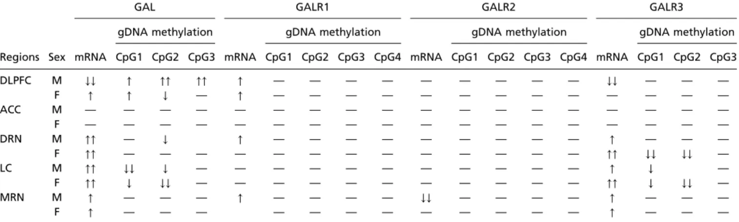
Table 3. Overview of mRNA and DNA methylation changes
GAL GALR1 GALR2 GALR3
gDNA methylation gDNA methylation gDNA methylation gDNA methylation
Regions Sex mRNA CpG1 CpG2 CpG3 mRNA CpG1 CpG2 CpG3 CpG4 mRNA CpG1 CpG2 CpG3 CpG4 mRNA CpG1 CpG2 CpG3
DLPFC M ↓↓ ↑ ↑↑ ↑↑ ↑ — — — — — — — — — ↓↓ — — —
F ↑ ↑ ↓ — ↑ — — — — — — — — — — — — —
ACC M — — — — — — — — — — — — — — — — — —
F — — — — — — — — — — — — — — — — — —
DRN M ↑↑ — ↓ ↑ — — — — — — — — — ↑ — — —
F ↑↑ — — — — — — — — — — — — — ↑↑ ↓↓ ↓↓ —
LC M ↑↑ ↓↓ ↓ — — — — — — — — — — — ↑ ↓ —
F ↑↑ ↓ ↓↓ — — — — — — — — — — — ↑↑ ↓ ↓↓ —
MRN M ↑ — — — ↑ — — — — ↓↓ — — — — ↑ — — —
F ↑ — — — — — — — — — — — — ↑ — — —
The arrows represent statistical significance; upward arrows signify increased in gene expression and methylation status, and downward arrows signify decreased gene expression and methylation status. A single arrow representsP<0.05, and two arrows representP<0.01. ACC, anterior cingulate cortex;
DLPFC, dorsolateral prefrontal cortex; DRN, dorsal raphe nucleus; LC, locus coeruleus; MRN, medullary raphe nucleus.
levels in males and females and inGALR1transcript levels in males only, as well as a robust decrease inGALR2mRNA in males only; in fact, this is the only region showing a change in this receptor tran- script. No effects on DNA methylation were recorded.
Prefrontal Cortex.A number of symptoms and diagnostic markers of MDD have been associated with prefrontal cortex, including decreased gray matter volume, hypermetabolism, and spine morphology (86). We observed distinct decreases inGAL and GALR3mRNA levels in the dorsolateral prefrontal cortex of DS males, with increased methylation at three sites of theGALgene, the reverse of the effects seen in locus coeruleus and dorsal ra- phe nucleus. In contrast, GAL mRNA is up-regulated in this region in female DS subjects; this up-regulation is the only sex difference for theGALtranscript seen in the qPCR analysis and requires further analysis. However, no changes were detected in another, closely related cortical area, the anterior cingulate cortex, which also has been consistently implicated in depression and suicide (87). Interestingly, although the average levels for GAL,GALR2, andGALR3transcripts are approximately similar in the cortical regions,GALR1mRNA levels are around eight- fold higher in dorsolateral prefrontal cortex than in anterior cingulate cortex.
The findings in the prefrontal cortex are difficult to interpret, because we have no information on the cells expressing GAL system members in human dorsolateral prefrontal and anterior cingulate cortices. In principle, however, the mechanistic effects of GAL release should be equally mediated by GAL1 and GAL3, because both exhibit a similar (nanomolar) affinity for GAL (88).
Therefore the differences will depend on which neuron subtype expresses the receptor and on the coupling to intracellular signaling cascades.
In general, peptides in the cortical region are detected in in- terneurons (89), but many pyramidal neurons express the transcript for cholecystokinin (90) and the peptide itself (91). Quantitative autoradiographic125I-GAL–binding studies showed a strong corti- cal binding in the human brain (39, 41), in sharp contrast with the rat brain, which lacks 125I-GAL binding in dorsal cortical and hippocampal areas (37, 38). However,125I-GAL(1-15ol) binds to these regions (92), and GAL(1-15) hyperpolarizes hippocampal C3 neurons (93). Investigation of these findings is continuing (SI Discussion,Dorsolateral Prefrontal Cortex) (94).
As for the mechanism(s) involved, one may speculate that the increasedGALmRNA levels in the locus coeruleus result in up- regulated GAL synthesis, enhanced centrifugal, axonal GAL transport, and increased GAL release in dorsolateral prefrontal cortex (Fig. 7). Here, in a possibly desensitizing process, GAL may down-regulate GALR3 expression in cortical projections and/or interneurons.
Functional Significance. It has been suggested that epigenetic mechanisms, such as DNA methylation, may be involved in psychiatric disorders (95, 96) and play an important role in stress and depression (7, 97). Here CpG methylation, catalyzed by DNA methyltransferases, in general represses gene transcription (75). We focus the discussion on the NA/locus coeruleus system and the prefrontal cortex. The role of NA vs. GAL has been discussed by Kuteeva, et al. (54).
Animal Studies in the Locus Coeruleus.Early experimental evidence suggested that locus coeruleus neurons in rats are activated by stress, resulting in increased NA synthesis and NA release in the forebrain (98–100). Stress also increases GAL expression in this nucleus (101, 102). In general terms neuropeptides are released following increased/burst firing (103, 104), also in the forebrain (105, 106). Thus, stress and increased/burst firing likely causes GAL release from nerve terminals in the forebrain as well as from soma/dendrites of the locus coeruleus neurons. Peptide release from soma/dendrites was first shown for oxytocin/vasopressin in magnocellular neurons in the hypothalamic paraventricular nu- cleus (107) and may be relevant for GAL in the locus coeruleus also (108).
Rat locus coeruleus neurons expressGALR1mRNA (42, 43), and, as evidenced by electrophysiological studies (109), this re- ceptor is likely functional in this site, mediating hyperpolarization (46, 110). Thus, somato-dendritically released GAL may inhibit firing via a GAL1 autoreceptor, hypothetically to prevent over- excitation (54). In agreement, results from a study on a transgenic mouse overexpressing GAL under the dopamine β-hydroxylase promoter with a fivefold increase in GAL mRNA in the locus coeruleus (111) suggest that GAL is important for modulating anxiety states driven by high noradrenergic signaling (112).
The Human Locus Coeruleus.In trying to translate the animal ex- perimental scenario to humans and MDD, one could argue that
Fig. 6. Alterations inGALR3gene expression and DNA methylation in the locus coeruleus of male and female DS subjects. (AandE) Gene expression levels ofGALR3in the locus coeruleus of male (A) and female (E) controls and DS subjects. (B–DandF–H) Percentage of DNA methylation levels at individual CpG sites of theGALR3gene in male (B–D) and female (F–H) controls and DS subjects. All data are presented as mean±SEM; males:n= 10 controls, 10 DS subjects; females:n=12 controls, 10 DS subjects. Signif- icant differences between DS subjects and controls are indicated: *P<0.05,
**P<0.01. CON, controls; DS, depressed suicide.
Table 4. Concentration of GAL (picomoles per milligram±SEM) in the different regions analyzed by RIA
Subject DLPFC ACC DRN LC MRN
Male controls 3.89±1.21 5.35±1.15 61.58±13.74 31.80±7.46 15.65±2.32 Male DS subjects 2.68±0.52 4.94±0.96 68.14±11.6 37.39±10.71 18.73±4.76 Female controls 1.70±0.38 6.37±1.79 80.62±10.24 43.35±6.33 15.45±3.53 Female DS subjects 1.33±0.29 5.97±1.32 81.61±5.19 67.68±7.47 13.55±2.95 ACC, anterior cingulate cortex; DLPFC, dorsolateral prefrontal cortex; DRN, dorsal raphe nucleus; LC, locus coeruleus; MRN, medullary raphe nucleus.
NEUROSCIENCEPNASPLUS
(i) GAL3seems to have replaced GAR1in human locus coeruleus neurons (32); (ii) hyperpolarization is one transduction mechanism for GAL1and also may be the transduction mechanism for GAL3
(46, 88, 110); and (iii)GALandGALR3transcripts are increased in the locus coeruleus of DS subjects’brains. It may be speculated that chronic stress and, as a consequence, constant and persistent inhibition of the NA-locus coeruleus neurons by GAL may result in reduced NA levels in the forebrain regions involved in mood control, e.g., the dorsolateral prefrontal cortex and anterior cin- gulate cortex; reduced NA levels are a characteristic feature of mood-related disorders. Together with a genetic vulnerability (lack of resilience), this reduction in NA levels may contribute to the symptoms encountered in such disorders (Fig. 7).
Prefrontal Cortex.In the prefrontal cortex, reciprocal interactions between the dorsolateral prefrontal cortex and anterior cingulate cortex, as well as between the anterior cingulate cortex and amyg- dala, have been described, whereby MDD subjects show reduced functional connectivity between the dorsolateral prefrontal cor- tex and anterior cingulate cortex (87). The transcript levels of all four GAL members are very similar in the two regions, except that (i) GALR1 mRNA levels are eightfold higher in the dorsolateral prefrontal cortex than in the anterior cingulate cortex and (ii) the differences between MDD and controls are mainly for GAL and are found only in male dorsolateral prefrontal cortex, indicating that any prefrontal cortical GALergic mecha- nisms in MDD are associated with GALandGALR3in this region and sex (SI Discussion,Dorsolateral Prefrontal Cortex). Of note, the apparent differences in GAL concentrations in this region between normal male and female controls did not reach significance but may still be taken as support for sex having an influence on information processing in this cortical region.
Treatment of MDD with Neuropeptides.Neuropeptide receptors may be promising targets for drug development in general because, as
mentioned previously, they are primarily released upon high- frequency/burst firing (103, 104) that may occur in a selectively chal- lenged, pathologically afflicted system (16, 113). Therefore, the antagonist will block only an overactivated signaling pathway, not affecting other silent or moderately active (no peptide release) systems producing the same ligand, and thus resulting in fewer side effects.
Treatment of depression with neuropeptide antagonists has been considered previously (15–19, 114). In fact, an initial report on the administration of a substance P/neurokinin 1 antagonist showed a significant efficacy vs. placebo, without side effects (115). This result could not be reproduced in an expanded phase 3 study (15, 116); however, clinical work on neurokinin antago- nists is ongoing (117, 118).
Treatment of MDD with GAL Ligands.Both GAL and GAL3are up- regulated in the locus coeruleus and dorsal raphe nucleus of MDD patients, possibly resulting in attenuated NA and 5-HT release in the forebrain. Treatment with a GAL3antagonist could, by disinhibition, have antidepressant activity, restoring NA and 5-HT levels in these brain regions, just as SNRIs do. Moreover, it may be speculated that the well-known delay in the onset of SSRIs’ effect caused by the activation of somatic, inhibitory 5-HT1A receptors (8, 58) may not occur with GAL3antagonists. Also, GAL3 signaling in some other brain regions, e.g., the dorsolateral prefrontal cortex, likely will not be affected, because our study shows thatGALandGALR3mRNA are unchanged (in females) or even down-regulated (in males), possibly excluding GAL system-related side effects. Moreover, the regionally selective effect of a GAL3 antagonist should compare favorably with SSRIs, SNRIs, and NRIs, which increase extracellular monoamine levels in all regions that monoamine neurons innervate, i.e., essentially throughout the entire nervous system. In fact, small- molecule, blood–brain barrier-penetrating GalR3antagonists have been generated (119, 120). The extent to which such an antagonist would act via mechanisms different from those of SNRIs and
circumstances these neurons fire spontaneously in a slow and regular fashion (124), but in response to stress they react with increased activity and burst firing (100).
NA is stored primarily in synaptic vesicles and is released at low activity and more so after stress (98, 99), acting on adrenoreceptors (ARs). GAL is stored only in large, dense core (LDC) vesicles and is released extrasynaptically (125) in response to increased/burst firing (106) from these vesicles in nerve endings in the forebrain. In humans (this study), GAL may, e.g., activate inhibitory postsynaptic GAL1receptors (GalR1) on neurons in the dorsolateral prefrontal cortex (DLPFC) and anterior cingulate cortex (ACC). Presumably, release also can occur from dendrites/soma in the locus coeruleus, activating inhibitory GAL3autoreceptors (GalR3). It is hy- pothesized that under excessive/chronic stress GAL induces a long-lasting inhibition of NA neurons and thus of NA release in forebrain regions such as the dor- solateral prefrontal cortex and anterior cingulate cortex, contributing to the development of MDD. Not included are changes inGAL,GALR1, andGALR3mRNA levels in the dorsolateral prefrontal cortex.
whether the currently used antidepressant drugs act via the GAL system has been discussed (121).
Limitations.Several limitations are associated with the present study. They are discussed in some detail inSI Discussion,Limi- tationsand encompass (i) concerns about GAL3, including its transduction mechanism(s) [this receptor has emerged as a complex receptor lacking well-defined signaling properties, contrasting GAL1 and GAL2 (36); so far there is no evidence that GAL3 is involved in receptor di- or heterodimerization (45)]; (ii) the lack of information about the protein, because only the transcripts for the receptors have been studied; (iii) the modest, albeit significant, changes in transcript mRNA levels;
(iv) the relatively low number of postmortem brains analyzed;
(v) the incomplete knowledge (in some instances) of the identity of the neurons expressing the transcripts (and thus of the site where methylation occurs); for example, the study by Le Maître, et al. (32) was not systematic, and galanin–5-HT coexistence in regions not analyzed cannot be excluded; and (vi) the general low sensitivity of histochemical techniques; this low sensitivity may have been an issue when Le Maître, et al. (32) could not detect theGAL1transcript in the locus coeruleus with in situ hybridization, whereas the present qPCR analysis showed high levels of this transcript in this region; (vii) the superficial in- vestigation of methylation sites and their functional significance;
and (viii) the influence of treatment with various drugs (Table S1), which requires further investigation (also see ref. 121).
Concluding Remarks.The present results, based on the analysis of five human brain regions, suggest the GAL system is involved in MDD. Interestingly, exactly 40 y ago Asberg and colleagues (122) reported that significantly lower concentrations of the 5-HT me- tabolite 5-hydroxyindoleacetic acid (5-HIAA) in the cerebrospinal fluid are associated with a very high rate of serious suicidal attempts, opening the way for the development of SSRIs aimed at elevating extraneuronal 5-HT levels. This class of drugs then replaced the monoamine oxidase inhibitors and the tricyclic antidepressants.
It is important to note that many overlapping transmitter systems exist and, in a way similar to the GAL family, may be in- volved in MDD psychopathology. The available detailed knowledge of the cellular organization and transmitter receptor architecture of the human prefrontal cortex, two subregions of which are studied here, provides a broad perspective on the present work and also shows how much research still needs to be carried out (123).
Materials and Methods
Brain Samples.Postmortem brain tissue was obtained from the Douglas-Bell Canada Brain Bank. A total of 212 punched samples from five different brain regions was included (Table 1). Ethical approval for this study was obtained from the Institutional Review Board of the Douglas Mental Health University Institute, with written informed consent from the families. The Regional Ethical Board in Stockholm has granted The Karolinska Institutet group permission no. 2013/474-31/2 for processing postmortem brain samples (SI Materials and Methods,Brain SamplesandTable S1).
RNA Isolation and Integrity Analysis.Total RNA was isolated using the RNeasy Plus Mini Kit. RNA quantity and quality were determined spectrophotometrically by using a ND1000 nanodrop. RNA integrity was checked using Experion au-
tomated electrophoresis system (Bio-Rad Laboratories). Total RNA was reverse transcribed to generate cDNA using a High-Capacity Reverse Transcription Kit (Life Technologies) (SI Materials and Methods).
RT-qPCR. qPCR was performed as described previously (SI Materials and Methods) (32).
Genomic DNA Extraction and PyroMark PCR.Genomic DNA (gDNA) was isolated by using the DNeasy Blood and Tissue Kit (Qiagen), checked spectrophoto- metrically, and bisulfite-converted by using the EZ DNA Methylation-Gold Kit (Zymo Research). Converted DNA was subject to 40 cycles of amplification by using gene-specific primers and the pyroPCR PyroMark (Qiagen) kit (SI Materials and Methods).
Bisulfite Pyrosequencing. Bisulfite pyrosequencing was performed using PyroMark Q96 ID (Qiagen) according to the manufacturer’s protocol and as previously described (122). Primer sequences are listed inFig. S6B, and the ge- nomic locations of the CpGs analyzed are shown inFig. S6A; also seeSI Ma- terials and Methods.
RIA.Human GAL was measured using antiserum G-026-01 (Phoenix Europe GmbH) raised in a rabbit against human GAL (SI Materials and Methods).
Statistical Analysis.Statistical analysis was performed with GraphPad Prism 6 (GraphPad Software), StatView (SAS Institute Inc.), and Systat 11 (Systat Software, Inc.). RIN, age, PMI, and tissue pH values for the five brain regions in controls and DS subjects were analyzed by multivariate ANOVA and two-tailedt test for independent groups. The Shapiro–Wilks test was used to test for Gaussian distribution. Significant outliers for qPCR fold change and percent methylation were analyzed by the ROUT (robust regression followed by outlier identification) method and were excluded ifP<0.05. qPCR data were analyzed by using the nonparametric Mann–WhitneyUtest. Differences in DNA meth- ylation between controls and suicides were analyzed using the Mann–Whitney Utest.Pvalues below 0.05 were considered significant, andPvalues between 0.05 and 0.1 were considered to represent a trend.
ACKNOWLEDGMENTS.We thank Drs. Tamas Bartfai (Stockholm University and Oxford University), Jean-Pierre Changeux (Institut Pasteur), Wayne C. Drevets (Janssen Research and Development), Eric Kandel (Columbia Uni- versity), Diego Pizzagalli (McLean Hospital), and Sol Snyder (Johns Hopkins University) for valuable comments and suggestions. This project presently is supported by Swedish Research Council Grant 04X-2887 (to T.G.M.H.); the Swedish Brain Foundation (T.G.M.H.); and by grants from Karolinska Institutet (to T.G.M.H.) and the Swedish Research Council for Environment, Agricultural Sciences, and Spatial Planning (Formas) (to J.R.). Earlier phases of this project were supported by a National Association for Research on Schizophrenia and Depression Distinguished Investigator Award (2009) (to T.G.M.H.), the Euro- pean Union Framework 6 Integrated Project New-Mood LSHM-CT-2004- 503474 (2004–2008) (to G.B., G.J., and T.G.M.H.), AFA Insurance (2008) (T.G.M.H.), and by a 5-y unrestricted Bristol-Myers-Squibb Grant in Neuroscience (to T.G.M.H.). Of particular importance were generous grants over a longer pe- riod from the Marianne and Marcus Wallenberg Foundation (1998–2009) and from the Knut and Alice Wallenberg Foundation (to T.G.M.H.). N.M. is a Cana- dian Institute of Health Research New Investigator and Fonds de la Recherche en Santé du Québec (FRQ-S) Chercheur-boursier. The Douglas–Bell Canada Brain Bank is supported by the Réseau Québécois sur le Suicide, les Troubles de l’Humeur et les Troubles Associés (FRQ-S) and by a Platform Support Grant from Brain Canada (to N.M.). Support was provided by the Magyar Tudományos Akadémia (MTA)–Semmelweis Egyetem (SE)–Nemzeti Agykutatási Program (NAP) B alprogram Genetic Brain Imaging Migraine Research Group through Kutatási és Technológiai Innovációs Alap (KTIA) (Grant KTIA_NAP_13-2-2015- 0001, to G.J.); by the NAP A-SE Research Group (Grant KTIA_NAP_13-1-2013- 0001, to G.B.) and (Grant KTIA_13_NAP-A-II/14, to G.B.); and by the MTA-SE Neuropsychopharmacology and Neurochemistry Research Group (G.B.).
1. Kessler RC, et al.; National Comorbidity Survey Replication (2003) The epidemiology of major depressive disorder: Results from the National Comorbidity Survey Repli- cation (NCS-R).JAMA289(23):3095–3105.
2. Wittchen HU, et al. (2011) The size and burden of mental disorders and other disorders of the brain in Europe 2010. Eur Neuropsychopharmacol 21(9):
655–679.
3. de Kloet ER, Joëls M, Holsboer F (2005) Stress and the brain: From adaptation to disease.Nat Rev Neurosci6(6):463–475.
4. Nestler EJ, et al. (2002) Neurobiology of depression.Neuron34(1):13–25.
5. McEwen BS (2008) Central effects of stress hormones in health and disease: Un- derstanding the protective and damaging effects of stress and stress mediators.Eur J Pharmacol583(2-3):174–185.
6. Labonté B, et al. (2013) Genome-wide methylation changes in the brains of suicide completers.Am J Psychiatry170(5):511–520.
7. Vialou V, Feng J, Robison AJ, Nestler EJ (2013) Epigenetic mechanisms of depression and antidepressant action.Annu Rev Pharmacol Toxicol53:59–87.
8. Blier P, de Montigny C (1994) Current advances and trends in the treatment of de- pression.Trends Pharmacol Sci15(7):220–226.
9. Millan MJ (2006) Multi-target strategies for the improved treatment of depressive states: Conceptual foundations and neuronal substrates, drug discovery and thera- peutic application.Pharmacol Ther110(2):135–370.
10. Gardier AM, Malagié I, Trillat AC, Jacquot C, Artigas F (1996) Role of 5-HT1A autor- eceptors in the mechanism of action of serotoninergic antidepressant drugs: Recent findings from in vivo microdialysis studies.Fundam Clin Pharmacol10(1):16–27.
NEUROSCIENCEPNASPLUS