International Journal of
Molecular Sciences
Article
The Neosartorya fischeri Antifungal Protein 2 (NFAP2):
A New Potential Weapon against Multidrug-Resistant Candida auris Biofilms
RenátóKovács1,2,3,* , Fruzsina Nagy1,4, Zoltán Tóth1,4,5, Lajos Forgács1,4, Liliána Tóth6,7, Györgyi Váradi8, Gábor K. Tóth8,9, Karina Vadászi1, Andrew M. Borman10,11 , LászlóMajoros1and LászlóGalgóczy6,7
Citation: Kovács, R.; Nagy, F.;
Tóth, Z.; Forgács, L.; Tóth, L.;
Váradi, G.; Tóth, G.K.; Vadászi, K.;
Borman, A.M.; Majoros, L.; et al. The Neosartorya fischeriAntifungal Protein 2 (NFAP2): A New Potential Weapon against Multidrug-ResistantCandida aurisBiofilms.Int. J. Mol. Sci.2021,22, 771. https://doi.org/10.3390/ijms 22020771
Received: 30 November 2020 Accepted: 11 January 2021 Published: 14 January 2021
Publisher’s Note: MDPI stays neu- tral with regard to jurisdictional clai- ms in published maps and institutio- nal affiliations.
Copyright:© 2021 by the authors. Li- censee MDPI, Basel, Switzerland.
This article is an open access article distributed under the terms and con- ditions of the Creative Commons At- tribution (CC BY) license (https://
creativecommons.org/licenses/by/
4.0/).
1 Department of Medical Microbiology, Faculty of Medicine, University of Debrecen, Nagyerdei krt. 98, 4032 Debrecen, Hungary; nagyfruzsina0429@gmail.com (F.N.); toth.zoltan@med.unideb.hu (Z.T.);
forgacs.lajos.89@gmail.com (L.F.); vadaszikarina13@freemail.hu (K.V.); major@med.unideb.hu (L.M.)
2 Faculty of Pharmacy, University of Debrecen, Nagyerdei krt. 98, 4032 Debrecen, Hungary
3 Department of Metagenomics, University of Debrecen, Nagyerdei krt. 98, 4032 Debrecen, Hungary
4 Doctoral School of Pharmaceutical Sciences, University of Debrecen, Nagyerdei krt. 98, 4032 Debrecen, Hungary
5 Department of Pharmacology and Pharmacotherapy, Faculty of Medicine, University of Debrecen, Nagyerdei krt. 98, 4032 Debrecen, Hungary
6 Institute of Plant Biology, Biological Research Centre, Temesvári krt. 62, 6726 Szeged, Hungary;
toth.liliana@brc.hu (L.T.); galgoczi.laszlo@brc.hu (L.G.)
7 Department of Biotechnology, Faculty of Science and Informatics, University of Szeged, Közép fasor 52, 6726 Szeged, Hungary
8 Department of Medical Chemistry, Faculty of Medicine, University of Szeged, Dóm tér 8,
6720 Szeged, Hungary; varadi.gyorgyi@med.u-szeged.hu (G.V.); toth.gabor@med.u-szeged.hu (G.K.T.)
9 MTA-SZTE Biomimetic Systems Research Group, University of Szeged, Dóm tér 8, 6720 Szeged, Hungary
10 UK National Mycology Reference Laboratory, Public Health England, Science Quarter, Southmead Hospital, Bristol BS10 5NB, UK; andy.borman@nbt.nhs.uk
11 Medical Research Council Centre for Medical Mycology (MRC CMM), University of Exeter, Exeter EX4 4QD, UK
* Correspondence: kovacs.renato@med.unideb.hu; Tel.: +36-52-255-425
Abstract: Candida aurisis a potential multidrug-resistant pathogen able to persist on indwelling devices as a biofilm, which serve as a source of catheter-associated infections. Neosartorya fischeri antifungal protein 2 (NFAP2) is a cysteine-rich, cationic protein with potent anti-Candidaactivity. We studied the in vitro activity of NFAP2 alone and in combination with fluconazole, amphotericin B, anidulafungin, caspofungin, and micafungin againstC. aurisbiofilms. The nature of interactions was assessed utilizing the fractional inhibitory concentration index (FICI), a Bliss independence model, and LIVE/DEAD viability assay. NFAP2 exerted synergy with all tested antifungals with FICIs ranging between 0.312–0.5, 0.155–0.5, 0.037–0.375, 0.064–0.375, and 0.064–0.375 for flucona- zole, amphotericin B, anidulafungin, caspofungin, and micafungin, respectively. These results were confirmed using a Bliss model, where NFAP2 produced 17.54µM2%, 2.16µM2%, 33.31µM2%, 10.72µM2%, and 111.19µM2% cumulative synergy log volume in combination with fluconazole, am- photericin B, anidulafungin, caspofungin, and micafungin, respectively. In addition, biofilms exposed to echinocandins (32 mg/L) showed significant cell death in the presence of NFAP2 (128 mg/L). Our study shows that NFAP2 displays strong potential as a novel antifungal compound in alternative therapies to combatC. aurisbiofilms.
Keywords:antifungal lock therapy;Candida auris; biofilm; NFAP2; drug–drug interaction; antifungal susceptibility testing
1. Introduction
Candida aurisis the first fungal pathogen to be announced as a global public health threat due to its ability to spread from patient-to-patient and cause invasive infections
Int. J. Mol. Sci.2021,22, 771. https://doi.org/10.3390/ijms22020771 https://www.mdpi.com/journal/ijms
Int. J. Mol. Sci.2021,22, 771 2 of 14
with high mortality [1–3]. Although, the majority ofC. aurisisolates have been recovered from patients with candidemia; several cases have also been observed from catheter- associated infections becauseC. aurisshows a potent capacity to develop biofilms on medical devices [4–6]. Clinical surveys indicated that catheters were the predominant source of infection in 89% ofC. auriscandidaemia cases while this ratio was only 46%
in non-C. aurisbloodstream infections [7,8]. Recently, it was reported that nearly 40% of clinicalC. aurisisolates exhibit a multidrug-resistant phenotype, which is more pronounced in sessile communities. In addition, the ratio of pan-resistant isolates to all three commonly prescribed antifungal drugs is increasing in multiple countries [9,10].
In the last decade, alternative antifungal strategies, such as antifungal lock therapy- has received more attention as an alternative salvage therapy to eradicate intraluminal Candidabiofilms [11,12]. To date, there is no officially approved antifungal lock strategy, such an approach would be particularly important in certain populations, such as patients with coagulopathies [13]. Echinocandins are promising targets for several potential lock solutions; however, the minimum inhibitory concentration (MIC90) values of C. auris biofilms ranged from 0.25 to >32 mg/L for caspofungin and micafungin, which represents a 2- to >512-fold increase in resistance when compared to planktonic cells, a difference that is likely to negatively impact clinical outcome [10,14].
A new potential lock strategy may be the antifungal protein-based lock solution againstC. aurisbiofilms; although, to date, there are no data regarding the susceptibility of C. aurisbiofilms to antifungal proteins.Neosartorya fischeriantifungal protein 2 (NFAP2) is a novel member of small cysteine-rich and cationic antifungal proteins from filamentous ascomycetes (crAFPs) [15]. Previous studies demonstrated that this protein has a potential applicability in the treatment ofCandidainfections. NFAP2 inhibited the growth of clinically relevantCandidaspecies. Furthermore, it interacted synergistically in combination with fluconazole against planktonic and sessileC. albicanscells in vitro and in vivo [16,17]. In light of these promising findings, the present study aimed to examine the in vitro efficacy of NFAP2 alone and in combination with traditional antifungal agents againstC. auris biofilms to evaluate a new potential therapeutic approach against this fungal superbug.
2. Results
2.1. In Vitro Susceptibility of Planktonic Cells
The median MICs against planktonicC. auriscells (pMIC) ranged from 4 to > 32 mg/L, 0.25 to 1 mg/L, 0.06 to 0.5 mg/L, 0.5 to 1 mg/L, and 0.12 to 2 mg/L for fluconazole, amphotericin B, anidulafungin, caspofungin, and micafungin, respectively. The median pMIC to fluconazole of strains 12 and 27 was 4 mg/L, which correspond to the susceptible tentative breakpoint;
while isolates 10, 20, and 82 were considered fluconazole-resistant based on the tentative MIC breakpoints recommended by the Centers for Disease Control and Prevention (≥32 mg/L for fluconazole) [18]. Regarding amphotericin B and the tested echinocandins, all isolates were susceptible according to tentative MIC breakpoints (≥2 mg/L for amphotericin B,≥4 mg/L for anidulafungin,≥2 mg/L for caspofungin, and≥4 mg/L for micafungin) [18]. The median pMICs for NFAP2 ranged from 32 to 512 mg/L.
2.2. In Vitro Susceptibility of Sessile Biofilm Cells
The MIC results (medians and ranges) onC. aurisbiofilms (sMIC) are shown in Table1.
Most of the isolates in biofilm form proved to be resistant to fluconazole, anidulafungin, caspofungin, micafungin, and NFAP2; while amphotericin B effectively inhibited the via- bility of sessileC. auriscells (sMIC: 1–2 mg/L) (Table1). The median sMICs observed for fluconazole, amphotericin B, anidulafungin, caspofungin, and micafungin in combination with NFAP2 were reduced by 32- to 128-fold, 4- to 64-fold, 16- to 128-fold, 4- to 128-fold, and 64- to 128-fold, respectively (Table1). The median sMICs for NFAP2 exhibited a 2- to 4-fold, 4- to 128-fold, 8- to 128-fold, 8- to 16-fold, and 4- to 256-fold decrease combined with fluconazole, amphotericin B, anidulafungin, caspofungin, and micafungin, respectively (Table1). These results indicated that the combinatorial application of the tested conven-
Int. J. Mol. Sci.2021,22, 771 3 of 14
tional antifungal agents with NFAP2 results in a significant reduction in the viability of sessile cells.
Table 1.Sessile minimum inhibitory concentrations (sMICs) of fluconazole (FLU), amphotericin B (AMB), anidulafungin (ANI), caspofungin (CAS) and micafungin (MICA) alone and in combination with NFAP2 againstC. aurisbiofilms.
Drug Isolate
Median MIC (Range) of Drug Used (50% OD492Reduction in Metabolic Activity)
Alone In Combination
Drug (mg/L) NFAP2 (mg/L) Drug (mg/L) NFAP2 (mg/L)
FLU
10 >512a >512a 32 256 (256–512)
12 >512a >512a 16 (16–32) 256
20 >512a >512a 32 (32–64) 256 (256–512)
27 >512a >512a 8 (8–16) 512 (256–512)
82 >512a >512a 32 (32–128) 512
AMB
10 1 512 (256–512) 0.25 128
12 1 >512a 0.03 (0.03–0.06) 64 (64–128)
20 1 >512a 0.03 (0.03–0.06) 16 (16–64)
27 1 512 0.25 64
82 2 512 0.03 (0.03–0.06) 4 (4–16)
ANI
10 16 (16–32) >512a 1 128 (128–256)
12 >64b >512a 1 32
20 >64b >512a 1 128 (128–256)
27 16 512 1 4 (4–8)
82 >64b 512 1 64 (64–128)
CAS
10 >64b 512 (128–512) 32 32 (32–64)
12 >64b >512a(512–>512) 1 (1–2) 64
20 >64b 512 (256–512) 1 32 (32–64)
27 >64b 512 (128–512) 32 32 (32–64)
82 >64b 512 (256–512) 1 (1–4) 32
MICA
10 >64b >512a 2 (2–4) 256
12 >64b >512a 1 4
20 >64b >512a 1 256 (128–256)
27 >64b >512a 1 4
82 >64b >512a 1 32 (32–64)
aMIC is offscale at >512 mg/L, 1024 mg/L (one dilution higher than the highest tested concentration) was used for analysis;bMIC is offscale at >64 mg/L, 128 mg/L (one dilution higher than the highest tested concentration) was used for analysis.
2.3. Nature of the NFAP2-Antifungal Drugs Interactions
Table2summarises the nature of in vitro interactions between NFAP2 and the five tested antifungal drugs based on the calculated fractional inhibitory concentration index (FICI). Synergy was observed for all antifungals and all isolates. Median FICI values ranged from 0.312 to 0.5, 0.155 to 0.5, 0.037 to 0.375, 0.064 to 0.375, and 0.064 to 0.375 for fluconazole, amphotericin B, anidulafungin, caspofungin, and micafungin, respectively (Table2). The results obtained by FICI were partly confirmed using a Bliss independence model. It is noteworthy, that strain dependency of the nature of the drug interaction was observed when the strains were tested individually using MacSynergy II analysis (Table2), which was prominent at lower concentrations of the combined antifungal drugs. However, this
Int. J. Mol. Sci.2021,22, 771 4 of 14
strain dependency disappeared when the five strains were analysed simultaneously. This cumulative analysis indicated that NFAP2 exerts 17.54µM2%, 2.16µM2%, 33.31µM2%, 10.72µM2%, and 111.19µM2% cumulative synergy volume in combination with flucona- zole, amphotericin B, anidulafungin, caspofungin and micafungin, respectively (Table2 and Figure1).
Table 2.In vitro interactions by Fractional Inhibitory Concentration Indexes (FICI) and MacSynergy II analysis of fluconazole (FLU), amphotericin B (AMB), anidulafungin (ANI), caspofungin (CAS) and micafungin (MICA) in combination with Neosartorya fischeriantifungal protein 2 (NFAP2) againstC. aurisbiofilms.
Drug Isolate
FICI MacSynergy II Analysis
Median (Range) of
FICI
Interaction
Individual Log Volume of Isolates, (Syn- ergy/Antagonism
µM2%)
Interaction Based on Individual Log
Volume
Cumulative Log Volume of Five
Isolates, (Syn- ergy/Antagonism
µM2%)
Interaction Based on Cumulative Log Volume
FLU
10 0.5 (0.5–0.53) Synergy 72.43/−126.35 Antagonism for most combinations
17.54/0 Synergy
12 0.312 Synergy 12.31/0 Synergy
20 0.312
(0.312–0.375) Synergy 210.17/−3.67 Synergy for most combinations
27 0.375
(0.375–0.625) Synergy 70/−78.26 Antagonism for most combinations
82 0.375
(0.375–0.625) Synergy 119.31/−11.47 Synergy for most combinations
AMB
10 0.5 (0.5–0.75) Synergy 53.9/−42.3 Synergy for most combinations
2.16/0 Synergy
12 0.312
(0.312–0.375) Synergy 196.99/−31.39 Synergy for most combinations
20 0.155
(0.155–0.185) Synergy 126.38/−4.52 Synergy for most combinations
27 0.375 Synergy 46.51/−27.26 Synergy for most
combinations
82 0.25 Synergy 75.39/−12.25 Synergy for most
combinations
ANI
10 0.375 (0.25–0.5) Synergy 127.59/−18.52 Synergy for most combinations
33.31/0 Synergy
12 0.037 Synergy 371.84/−2.43 Synergy for most
combinations
20 0.185
(0.185–0.312) Synergy 154.33/−40.86 Synergy for most combinations
27 0.069
(0.069–0.09) Synergy 71.52/−10.83 Synergy for most combinations
82 0.312
(0.185–0.312) Synergy 148.15/−7.3 Synergy for most combinations
CAS
10 0.375
(0.375–0.75) Synergy 8.64/−33.23 Antagonism for most combinations
10.72/0 Synergy
12 0.067
(0.067–0.14) Synergy 235.24/0 Synergy
20 0.075
(0.075–0.257) Synergy 22.23/−17.48 Synergy for most combinations
27 0.375
(0.375–0.75) Synergy 14.76/−44.63 Antagonism for most combinations
82 0.122 Synergy 20.2/−11.59 Synergy for most
combinations
Int. J. Mol. Sci.2021,22, 771 5 of 14
Table 2.Cont.
Drug Isolate
FICI MacSynergy II Analysis
Median (Range) of
FICI
Interaction
Individual Log Volume of Isolates, (Syn- ergy/Antagonism
µM2%)
Interaction Based on Individual Log
Volume
Cumulative Log Volume of Five
Isolates, (Syn- ergy/Antagonism
µM2%)
Interaction Based on Cumulative Log Volume
MICA
10 0.375 Synergy 164.37/−99.58 Synergy for most
combinations
111.19/0 Synergy
12 0.064 Synergy 277.54/0 Synergy
20 0.375 Synergy 378.15/−17.51 Synergy for most
combinations
27 0.253 Synergy 100.94/−41.11 Synergy for most
combinations
82 0.132 Synergy 212.19/−18.37 Synergy for most
combinations
Int. J. Mol. Sci. 2021, 22, x FOR PEER REVIEW 5 of 15
12 0.067 (0.067–0.14) Synergy 235.24/0 Synergy 20 0.075 (0.075–0.257) Synergy 22.23/−17.48 Synergy for most
combinations
27 0.375 (0.375–0.75) Synergy 14.76/−44.63
Antagonism for most combinations 82 0.122 Synergy 20.2/−11.59 Synergy for most
combinations
MICA
10 0.375 Synergy 164.37/−99.58 Synergy for most combinations
111.19/0 Synergy 12 0.064 Synergy 277.54/0 Synergy
20 0.375 Synergy 378.15/−17.51 Synergy for most combinations 27 0.253 Synergy 100.94/−41.11 Synergy for most
combinations 82 0.132 Synergy 212.19/−18.37 Synergy for most
combinations
Figure 1.Cont.
Int. J. Mol. Sci.2021,22, 771 6 of 14
Int. J. Mol. Sci. 2021, 22, x FOR PEER REVIEW 6 of 15
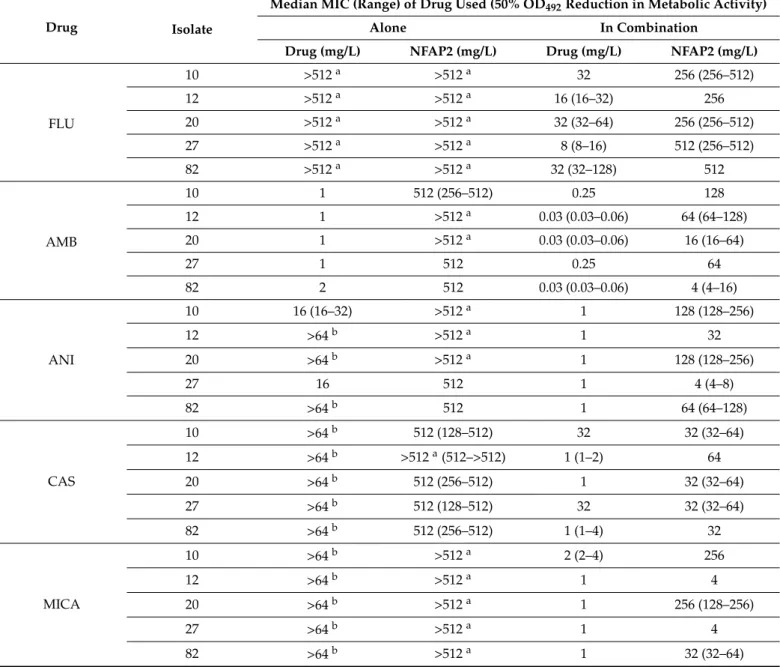
Figure 1. Effect of NFAP2 in combination with fluconazole (A), amphotericin B (B), anidulafungin (C), caspofungin (D) and micafungin (E) against C. auris isolates using MacSynergy II analysis. Additive interactions appear as a horizontal plane at 0% inhibition. The interaction is defined as synergistic if the observed surface is greater compared to the predicted additive surface. The volumes are calculated at the 95% confidence interval. The figures represent the cumulative synergy volume of five tested isolates. In panel E, higher synergy was observed; therefore, the scale of the z axis is different than in panels (A–D). Each figure presents the cumulative values of the five tested C. auris isolates.
2.4. Fluorescence Viability Assay
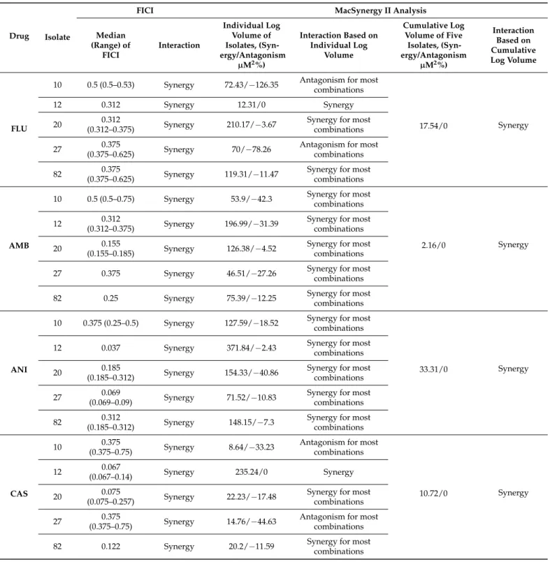
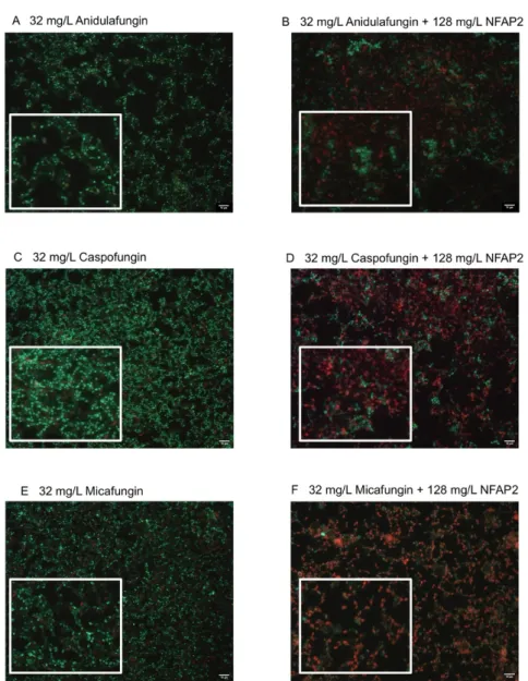
The LIVE/DEAD viability assay focused primarily on the echinocandins because this antifungal group is considered in alternative anti-biofilm therapeutic strategies, such as antifungal lock therapy [11]. For the NFAP2 concentrations tested (Figure 2A–E), an extensive anti-biofilm effect was only observed after exposure to 512 mg/L NFAP2 (Figure 2E), when compared to untreated one-day old biofilms (Figure 2A). The NFAP2 treatment alone showed a concentration-dependent activity, where the ratio of dead cells was 9%, 43%, 67% and 90% after 32 mg/L, 128 mg/L, 256 mg/L and 512 mg/L NFAP2 exposure, respectively (Figure 2A–E). It is noteworthy that the 128 mg/L NFAP2 (Figure 2C) and 32 mg/L echinocandin treatments alone did not produce remarkable cell death. The ratio of dead cells was 28%, 16% and 24% in the samples following the 32 mg/L anidulafungin, caspofungin and micafungin treatments, respectively (Figure 3A,C,E). However, their combined application with 128 mg/L NFAP2 resulted in a significant total cell number reduction. The cell number decreased with 44%, 34% and 41% after co-application of 128 mg/L NFAP2 with 32 mg/L anidulafungin, caspofungin and micafungin, respectively (Figure 3B,D,F). In addition, the percentage of dead cells was 74%, 72% and 60% in these samples, respectively (Figure 3B,D,F). This observation further strengthened the results of the previous XTT assay, and clearly demonstrates that co-administration of NFAP2 with echinocandins on potentially echinocandin-resistant biofilm rendered them susceptible to these antifungal agents.
Figure 1.Effect of NFAP2 in combination with fluconazole (A), amphotericin B (B), anidulafungin (C), caspofungin (D) and micafungin (E) againstC. aurisisolates using MacSynergy II analysis. Additive interactions appear as a horizontal plane at 0% inhibition. The interaction is defined as synergistic if the observed surface is greater compared to the predicted additive surface. The volumes are calculated at the 95% confidence interval. The figures represent the cumulative synergy volume of five tested isolates. In panel E, higher synergy was observed; therefore, the scale of thezaxis is different than in panels (A–D). Each figure presents the cumulative values of the five testedC. aurisisolates.
2.4. Fluorescence Viability Assay
The LIVE/DEAD viability assay focused primarily on the echinocandins because this antifungal group is considered in alternative anti-biofilm therapeutic strategies, such as an- tifungal lock therapy [11]. For the NFAP2 concentrations tested (Figure2A–E), an extensive anti-biofilm effect was only observed after exposure to 512 mg/L NFAP2 (Figure2E), when compared to untreated one-day old biofilms (Figure2A). The NFAP2 treatment alone showed a concentration-dependent activity, where the ratio of dead cells was 9%, 43%, 67% and 90% after 32 mg/L, 128 mg/L, 256 mg/L and 512 mg/L NFAP2 exposure, respectively (Figure2A–E). It is noteworthy that the 128 mg/L NFAP2 (Figure2C) and 32 mg/L echinocandin treatments alone did not produce remarkable cell death. The ratio of dead cells was 28%, 16% and 24% in the samples following the 32 mg/L anidulafungin, caspofungin and micafungin treatments, respectively (Figure3A,C,E). However, their combined application with 128 mg/L NFAP2 resulted in a significant total cell number reduction. The cell number decreased with 44%, 34%
and 41% after co-application of 128 mg/L NFAP2 with 32 mg/L anidulafungin, caspofungin and micafungin, respectively (Figure3B,D,F). In addition, the percentage of dead cells was 74%, 72% and 60% in these samples, respectively (Figure3B,D,F). This observation further strength- ened the results of the previous XTT assay, and clearly demonstrates that co-administration of NFAP2 with echinocandins on potentially echinocandin-resistant biofilm rendered them susceptible to these antifungal agents.
Int. J. Mol. Sci.2021,22, 771 7 of 14
Int. J. Mol. Sci. 2021, 22, x FOR PEER REVIEW 7 of 15
Figure 2. LIVE/DEAD fluorescence imaging of one representative C. auris isolate (isolate 10).
Image (A) shows the untreated biofilm, while images (B–E) present the NFAP-exposed biofilms at 32 mg/L, 128 mg/L, 256 mg/L and 512 mg/L NFAP2 concentrations, respectively. Live cells (green) and nonviable cells (red) were stained with Syto9 and propidium iodide, respectively. All images show typical fields of view. Scale bars represent 10 μm.
Figure 2.LIVE/DEAD fluorescence imaging of one representativeC. aurisisolate (isolate 10). Image (A) shows the untreated biofilm, while images (B–E) present the NFAP-exposed biofilms at 32 mg/L, 128 mg/L, 256 mg/L and 512 mg/L NFAP2 concentrations, respectively. Live cells (green) and nonviable cells (red) were stained with Syto9 and propidium iodide, respectively. All images show typical fields of view. Scale bars represent 10µm.
Int. J. Mol. Sci.2021,22, 771 8 of 14
Int. J. Mol. Sci. 2021, 22, x FOR PEER REVIEW 8 of 15
Figure 3. LIVE/DEAD fluorescence imaging of one representative C. auris isolate (isolate 10).
Images (A,C,E) demonstrate the anidulafungin-, caspofungin- and micafungin-exposed biofilms (32 mg/L), respectively, while images (B,D,F) show the anti-biofilm effect of anidulafungin, caspofungin and micafungin (32 mg/L for each drug alone) in the presence of NFAP2 (128 mg/L), respectively. Live cells (green) and nonviable cells (red) were stained with Syto9 and propidium iodide, respectively. All images show typical fields of view. Scale bars represent 10 μm.
3. Discussion
The eradication of C. auris biofilms from medical indwelling devices (e.g., catheters and cannulas) still remains a big challenge in the nosocomial environment because these sessile communities can withstand exposure to the most frequently administered antifungal agents. Thus, biofilms serve as a continuous source of C. auris-related candidaemia [8]. Currently, several novel antifungal drugs are under development against C. auris, including ibrexafungerp [19], manogepix [20], VT-1598 [21], and rezafungin [22] and they may represent potential treatment options in the near future.
However, considering the increasing number of multidrug-resistant C. auris isolates, new and alternative therapeutic strategies are needed to prevent and eliminate the growth of C. auris biofilms from indwelling devices.
Although there is increasing recognition that antibiotic lock solutions can reduce the risk of catheter-related bacterial infections, there is no approved antifungal lock Figure 3.LIVE/DEAD fluorescence imaging of one representativeC. aurisisolate (isolate 10). Images (A,C,E) demonstrate the anidulafungin-, caspofungin- and micafungin-exposed biofilms (32 mg/L), respectively, while images (B,D,F) show the anti-biofilm effect of anidulafungin, caspofungin and micafungin (32 mg/L for each drug alone) in the presence of NFAP2 (128 mg/L), respectively. Live cells (green) and nonviable cells (red) were stained with Syto9 and propidium iodide, respectively.
All images show typical fields of view. Scale bars represent 10µm.
3. Discussion
The eradication ofC. aurisbiofilms from medical indwelling devices (e.g., catheters and cannulas) still remains a big challenge in the nosocomial environment because these sessile communities can withstand exposure to the most frequently administered antifungal agents. Thus, biofilms serve as a continuous source ofC. auris-related candidaemia [8].
Currently, several novel antifungal drugs are under development againstC. auris, including ibrexafungerp [19], manogepix [20], VT-1598 [21], and rezafungin [22] and they may repre- sent potential treatment options in the near future. However, considering the increasing number of multidrug-resistantC. aurisisolates, new and alternative therapeutic strate- gies are needed to prevent and eliminate the growth ofC. aurisbiofilms from indwelling devices.
Although there is increasing recognition that antibiotic lock solutions can reduce the risk of catheter-related bacterial infections, there is no approved antifungal lock therapeu- tic protocol in clinical practice so far. Vargas-Cruz et al. (2019) reported that liposomal
Int. J. Mol. Sci.2021,22, 771 9 of 14
amphotericin B, amphotericin B deoxycholate, fluconazole, voriconazole, micafungin, caspofungin, and anidulafungin failed to completely eradicateC. aurisbiofilms, contraindi- cating their mono-therapeutic use in lock therapy [23]. Therefore, certain non-antifungal agents alone or in combination with traditional antifungals have been investigated as a potential approach to overcomeC. auris-related catheter-associated infections. These include ebselen [24], miltefosine [25], farnesol [26], and silver or bismuth nanoantibi- otics [27,28]. To date, three conventional potential line lock solutions have been tested againstC. aurisbiofilms. Taurolidine showed moderate activity against biofilms and only partially eradicated sessile populations. Conversely, the minocycline-EDTA-ethanol lock so- lution and nitroglycerin-citrate-ethanol combination completely eradicatedC. aurisbiofilms in vitro [23,29].
The environmentally highly stable crAFPs represent promising bioactive natural compounds in anti-Candidatherapy [30], and they can provide potential bases to develop new lock solutions againstCandidabiofilms. Although the number of these molecules is steadily increasing, only a few of them have been well-characterised so far [16,17,30]. It is noteworthy that several cell-culture-based cytotoxicity assays proved that crAFPs have no remarkable cytotoxic effects on mammalian cells in vitro and in vivo. Additionally, physiologically compatible solutions can be prepared from the majority of these proteins, which is a basic requirement for a lock solution [30]. Although novel therapeutic approaches focusing on the activity of short antifungal peptides from other origins againstC. auris have previously been published, the potential effects of crAFPs remained unknown.
Del Mas et al. (2019) demonstrated the effect of crotamine, the venom of South American rattlesnake, which exerted 50% inhibition ofC. aurisplanktonic growth at a concentration of 160µM [31]. Van Eijk et al. (2020) described that two cathelicidin-inspired antimicrobial peptides (CT172 and CR 184) strongly interfered with metabolic activity, growth, and viability at sub-micromolar levels (≤1µM) againstC. aurisplanktonic cells [32].
Kubiczek et al. (2020) reported that antifungal peptides might have a promising anti-biofilm activity: derivates of the antifungal peptide Cm-p5 exhibited a semi-inhibitory effect at concentrations ranging from 10 to 21 mg/L againstC. auris biofilms. In addition, the mature sessile populations were also inhibited by 71–97% [33].
NFAP2 represents a novel, phylogenetically distinct group of crAFPs [15]. In silico analysis predicted that NFAP2 has a strong ability to bind human serum albumin, question- ing the systemic application of this compound [17]. Nevertheless, it may be a promising target in the above-mentioned newly defined antifungal lock strategies. The previously well-documented membrane disrupting effect renders NFAP2 suitable as very potent anti- Candidacompound [17]. Before the present study, an in vitro synergistic interaction was already documented between NFAP2 and fluconazole againstC. albicansandC. parapsilosis, suggesting the justification of NFAP2 in combination-based therapies [16]. Furthermore, the in vivo therapeutic potency of NFAP2 as a topical agent was proven for the treatment of vulvovaginal candidiasis caused by fluconazole-resistantC. albicansin a murine model system [17]. Kovács et al. (2019) reported that 800 mg/L daily NFAP2 together with 5 mg/kg daily fluconazole treatment was superior compared to 5 mg/kg daily fluconazole treatment. In addition, this NFAP2 concentration did not cause significant morphological alterations in the vaginal and vulvar tissues and did not show a cytotoxic effect on human keratinocytes and dermal fibroblasts in vitro [17]. Synergistic interaction between NFAP2 and fluconazole were already reported againstC. albicans[16]. In the present study, NFAP2 alone showed a concentration-dependent activity againstC. aurisbiofilms (Figure2). Based on FICI calculation, synergism was detected for NFAP2 in the presence of fluconazole, amphotericin B, anidulafungin, caspofungin, and micafungin againstC. aurisbiofilms (Table2). It is noteworthy that the synergistic interaction was observed primarily at high NFAP2 concentrations ranging between 4–512 mg/L (Table1). The interaction between NFAP2 and the tested antifungals was variable based on the Bliss independence model (Table2). This strain dependency was observed primarily at lower concentrations of the combined drugs and was not observed at their higher concentrations. Furthermore, this
Int. J. Mol. Sci.2021,22, 771 10 of 14
variability disappeared when the five strains were analysed simultaneously by MacSynergy II algorithm. Existence of such variability points to the necessity to use multiple analytic approaches in parallel when examining drug–drug interactions.
The mechanism underlying the synergy observed between NFAP2 and the antifungals tested here might result from the fact that NFAP2 has a pore-forming effect in the cell membrane [17], which would exacerbate the osmotic stress derived from echinocandin- related cell wall damage and from membrane-active antifungals, such as fluconazole and amphotericin B. Our study had a limitation: we examined strains derived from only oneC. auris clade (South Asian/Indian lineage). However, despite this limitation, the potentiator effect of NFAP2 in combination with traditional antifungals againstC. auris one-day-old biofilms is unquestionable.
In summary, improvements and clinical verifications in alternative combination-based antifungal therapies can help the development of new treatment strategies againstC. auris biofilms. Based on our in vitro findings, combined application of NFAP2 with widely used traditional antifungals may provide a potential novel approach in the antifungal armoury ofC. auris-specific alternative treatments as lock therapy. In the future, further animal experiments of these new combinations are warranted.
4. Materials and Methods 4.1. Isolates
FiveC. aurisisolates (isolate 10, 12, 20, 27, and 82) derived from the South Asian/Indian lineage were obtained from the National Mycology Reference Laboratory, United Kingdom.
Strain 10 (NCPF 8971) and 20 (NCPF 8985) were isolated from wound swabs. Isolate 27 (NCPF 89891) and 82 (NCPF 13013) were obtained from pleural fluid and urine, respectively, while the source of strain 12 (NCPF 8973) was not stated [34,35]. Each strain derived from different patients. All isolates were identified to species level by Matrix-Assisted Laser Desorption-Ionisation-Time of Flight Mass Spectrometry [34,35]. Clade delineation was conducted by PCR amplification and sequencing of the 28S rDNA gene and the internal transcribed spacer region 1, as described previously [34,35].
4.2. Recombinant NFAP2 Production and Purification
Recombinant NFAP2 was produced in aPenicillium chrysogenum-based expression sys- tem and purified to 100% homogeneity, as described previously by Kovács et al. (2019) [17]).
4.3. In Vitro Susceptibility Testing of Planktonic Cells
pMIC were determined in line with the protocol M27-A3 of the Clinical Laboratory Standards Institute [36]. pMICs of fluconazole (cat. # J62015, VWR, Debrecen, Hungary), amphotericin B (cat. # Y0001361, Merck, Budapest, Hungary) anidulafungin (cat. # ADF00- 100, Molcan Corporation, Toronto, ON, Canada), caspofungin (cat. # CSF00A-100, Molcan Corporation, Toronto, ON, Canada), micafungin (cat. # MCF00N-100, Molcan Corporation, Toronto, ON, Canada) and NFAP2 were determined in RPMI 1640 (with L-glutamine and without bicarbonate, pH 7.0 with MOPS; Merck, Budapest, Hungary). The drug concentrations tested ranged from 0.06 to 32 mg/L, 0.008 to 4 mg/L, and 0.008 to 4 mg/L for fluconazole, amphotericin B, and echinocandins, respectively; while NFAP2 concentrations ranged from 1 to 512 mg/L (corresponding to 0.2–92µM). For fluconazole, echinocandins and NFAP2, pMICs were determined as the lowest drug concentration that produces at least 50% growth reduction compared to the growth of the control. For amphotericin B, pMIC was considered the first concentration exerting 100% growth inhibition compared to the growth of the drug-free control. pMICs represent three independent experiments for each isolate and are presented as the median.
4.4. Biofilm Development
One-day-old biofilms were prepared as described in our previous studies [26,37,38].
Briefly,C. aurisisolates were suspended in RPMI 1640 liquid medium to a final concentration
Int. J. Mol. Sci.2021,22, 771 11 of 14
of 1×10⁶cells/mL, and aliquots of 0.1 mL were pipetted onto flat-bottom 96-well sterile microtiter plates (TPP, Trasadingen, Switzerland) and then incubated statically at 37◦C for 24 h. After the incubation time, plates were washed three times with physiological saline to remove unattached cells.
4.5. Antifungal Susceptibility Testing of Biofilms
The concentrations tested for sMIC determination ranged from 8 to 512 mg/L, 0.03 to 2 mg/L, and 1 to 64 mg/L for fluconazole, amphotericin B, and echinocandins, re- spectively. Meanwhile, the examined NFAP2 concentrations ranged from 2 to 512 mg/L.
The prepared one-day-old biofilms were washed three times with sterile physiological saline. Different drug concentrations in RPMI 1640 were added to one-day-old pre-formed biofilms and then the plates were incubated for an additional 24 h at 37◦C. Afterwards, sMIC determinations were carried out using the metabolic activity change-based XTT [2,3-bis(2-methoxy-4-nitro-5-sulfophenyl)-2H-tetrazolium-5-carboxanilide] reduction as- say. The prepared XTT working solution (Merck, Budapest, Hungary) (0.5 g/L) was supplemented with menadione (Merck, Budapest, Hungary) to a final concentration of 1µM [26,37–39]. Drugs were removed prior to assay of metabolic activity by washing three times with sterile physiological saline. Afterwards, a 100µL aliquot of XTT/menadione solution was added to each well containing the preformed biofilms as well as to negative control wells. Plates were incubated in darkness for 2 h at 37◦C. Following incubation, 80µL of supernatant from each well was measured spectrophotometrically at 492/620 nm (Multiskan Sky Microplate Spectrophotometer, Thermo-ScientificTM, Waltham, MA, USA).
sMICs were considered as the lowest drug concentration exerting at least a 50% reduction in metabolic activity compared to the untreated biofilms [26,37–39]. sMICs represent three independent experiments for each isolate and are expressed as the median value.
4.6. Evaluation of Interactions by Fractional Inhibitory Concentration Index (FICI) and Bliss Independence Model
Interactions between tested antifungal agents and NFAP2 were evaluated using a previously well-documented two-dimensional broth microdilution checkerboard as- say [26,37,40]. Antifungal drug-NFAP2 interactions were then analyzed using FICI deter- mination and a Bliss independence model-based MacSynergy II analysis [26,37–42]. The tested concentration ranges were the same as those described in the previous section for sMIC determination. FICIs were calculated using the following formula: ΣFIC = FICA
+ FICB= MICAcomb/MICAalone+ MICBcomb/MICBalone, where MICAaloneand MICBalone represent the MICs of drugs A and B when used alone, and MICAcomband MICBcombare the MIC of drugs A and B in combination at isoeffective combination, respectively [40].
FICI was determined as the lowestΣFIC. sMIC values of the tested antifungals and NFAP2 alone and of all isoeffective combinations were determined as the lowest concentration, resulting in at least a 50% decrease in metabolic activity compared to the growth control ses- sile cells. If the obtained MIC value was higher than the highest tested drug concentration, the next highest two-fold concentration was considered as the MIC. The obtained FICIs were interpreted based on the following algorithm: synergistic interaction was defined as FICI≤0.5, an indifferent interaction as a FICI between >0.5 and 4, and antagonistic interaction as FICI >4 [26,37,40]. FICIs were determined in three independent experiments, and their median values were presented with ranges.
To further evaluate antifungal drugs–NFAP2 interactions, MacSynergy II analysis was used in case of all isolates, employing the Bliss independence algorithm in a Microsoft Excel-based interface to determine synergy. MacSynergy-based analysis was performed as previously described [26,37,41,42]. Briefly, synergy and antagonistic volumes were calculated by adding all of the positive values and all of the negative values for each drug combination, respectively [26,37,41,42]. These volumes were then statistically evaluated using the 95% confidence level and expressed in units ofµM2%, which are analogous to the units for area under a dose–response curve in the two-dimensional graph [26,37,41,42].
Synergy or antagonism is significant if the interaction volumes are >25µM2% or <25µM2%,
Int. J. Mol. Sci.2021,22, 771 12 of 14
respectively (corresponding to log volumes > 2 and log volumes < 2, respectively). Values between 25µM2% and 50µM2% (values in log volume between >2 to 5) should be con- sidered as minor synergy. Values between 50µM2% and 100µM2% (values in log volume between >5 to 9) indicate moderate synergy or antagonism, while values over 100µM2% (values in log volume between >9) represent strong synergy [26,37,41,42]. When a small number of drug concentration combinations results in antagonistic interaction in a gener- ally synergistic combination, the applied terminology is ‘synergy for most combinations’.
While a small number of drug concentration combinations results in synergistic interaction in a generally antagonistic combination, the applied terminology is ‘antagonism for most combinations’ [41].
4.7. Biofilm Viability Assay in the Presence or Absence of NFAP2
C. aurisbiofilms were grown on the surface of 8-well Permanex slides statically at 37◦C for 24 h (Lab-Tek®Chamber Slide™ System, VWR, Debrecen, Hungary) [26,43]. The one-day-old biofilms were washed three times with physiological saline. After the washing step, the antifungal effect of NFAP2 (32 mg/L, 128 mg/L, 256 mg/L, and 512 mg/L) and echinocandins (32 mg/L) alone, and echinocandins (32 mg/L)-NFAP2 (128 mg/L) combinations were tested on the sessile biofilm cells. These concentrations were chosen based on our previous antifungal susceptibility test results on biofilms.
Following 24 h of drug exposure statically at 37◦C, biofilms were washed with sterile physiological saline, then the ratio of viable and dead cells was evaluated using the fluorescent LIVE/DEAD®BacLight™ viability kit (ThermoFisher Scientific, Waltham, MA, USA). Biofilms were stained for 15 min in darkness at 37◦C using Syto 9 (3.34 mM solution in DMSO) and propidium iodide (20 mM solution in DMSO) to visualize viable and non-viableC. auriscells, respectively [26,43]. Fluorescent cells were examined with a Zeiss AxioSkop 2 mot microscope (Jena, Germany) coupled with a Zeiss AxioCam HRc camera (Jena, Germany). Axiovision 4.8.2 software was used to analyze images (Jena, Germany). Further picture analysis and calculation of the percentage of the dead cells was performed using ImageJ software (version: 2.1.0/1.53c) (Fiji, ImageJ, Wayne Rasband National Institutes of Health). All images were changed to 8-bit grayscale with background noise subtracted, afterwhich the threshold was defined [44,45].
Author Contributions:Conceptualization, R.K. and L.G.; methodology, R.K., F.N., Z.T., L.F., L.T., K.V.; validation, R.K. and L.G.; formal analysis, R.K., L.G., G.K.T., G.V., A.M.B.; investigation, R.K., F.N., Z.T., L.F., L.T., G.V., K.V.; resources, R.K., G.K.T., G.V., A.M.B., L.M.; writing—original draft preparation, R.K. and L.G.; writing—review and editing, R.K., L.G., L.M., A.M.B., G.K.T., G.V.; project administration, R.K.; funding acquisition, R.K. All authors have read and agreed to the published version of the manuscript.
Funding:RenátóKovács was supported by the EFOP-3.6.3-VEKOP-16-2017-00009 program, OTKA Bridging Fund and FEMS Research and Training Grant (FEMS-GO-2019-502). Zoltán Tóth and Fruzsina Nagy were supported by theÚNKP-19-3 andÚNKP-20-3 New National Excellence Pro- gram of the Ministry of Human Capacities. LászlóGalgóczy is financed by the bilateral Austrian- Hungarian Joint Research Project (ANN 131341) of the Hungarian National Research, Development and Innovation (NKFIH) Office. The research of LászlóGalgóczy was supported by the János Bolyai Research Scholarship of the Hungarian Academy of Sciences. The current work of LászlóGalgóczy was supported by theÚNKP-20-5-New National Excellence Program of the Ministry for Innovation and Technology from the source of the National Research, Development and Innovation Fund. Györ- gyi Váradi and Gábor K. Tóth were supported by the NKFIH Office (GINOP-2.3.2-15-2016-00014 and the Ministry of Human Capacities, Hungary grant, TKP-2020. The publication is supported by the GINOP-2.3.4-15-2020-00008 project. The project is co-financed by the European Union and the European Regonal Development Fund.
Institutional Review Board Statement:Not applicable.
Informed Consent Statement:Not applicable.
Data Availability Statement:Not applicable.
Int. J. Mol. Sci.2021,22, 771 13 of 14
Conflicts of Interest:LászlóMajoros received conference travel grants from MSD, Astellas, Pfizer and Cidara. All other authors report no conflicts of interest. The funders had no role in the design of the study; in the collection, analyses, or interpretation of data; in the writing of the manuscript, or in the decision to publish the results.
References
1. Lockhart, S.R.; Etienne, K.A.; Vallabhaneni, S.; Farooqi, J.; Chowdhary, A.; Govender, N.P.; Colombo, A.L.; Calvo, B.; Cuomo, C.A.;
Desjardins, C.A.; et al. Simultaneous Emergence of Multidrug-ResistantCandida aurison 3 Continents Confirmed by Whole- Genome Sequencing and Epidemiological Analyses.Clin. Infect. Dis.2017,64, 134–140. [CrossRef] [PubMed]
2. Clancy, C.J.; Nguyen, M.H. Emergence ofCandida auris: An International Call to Arms. Clin. Infect. Dis. 2017,64, 141–143.
[CrossRef] [PubMed]
3. Kean, R.; Brown, J.L.; Gülmez, D.; Ware, A.; Ramage, G.Candida auris: A Decade of Understanding of an Enigmatic Pathogenic Yeast.J. Fungi2020,6, 30. [CrossRef] [PubMed]
4. Lee, W.G.; Shin, J.H.; Uh, Y.; Kang, M.G.; Kim, S.H.; Park, K.H.; Jang, H.-C. First Three Reported Cases of Nosocomial Fungemia Caused byCandida auris.J. Clin. Microbiol.2011,49, 3139–3142. [CrossRef] [PubMed]
5. Schelenz, S.; Hagen, F.; Rhodes, J.; Abdolrasouli, A.; Chowdhary, A.; Hall, A.; Ryan, L.; Shackleton, J.; Trimlett, R.; Meis, J.F.; et al.
First Hospital Outbreak of the Globally EmergingCandida aurisin a European Hospital.Antimicrob. Resist. Infect. Control.2016, 5, 35. [CrossRef]
6. Taori, S.; Khonyongwa, K.; Hayden, I.; Athukorala, G.D.A.; Letters, A.; Fife, A.; Desai, N.; Borman, A.M.Candida aurisOutbreak:
Mortality, Interventions and Cost of Sustaining Control.J. Infect.2019,79, 601–611. [CrossRef]
7. Sayeed, M.A.; Farooqi, J.; Jabeen, K.; Mahmood, S.F. Comparison of Risk Factors and Outcomes ofCandida aurisCandidemia with Non-Candida aurisCandidemia: A Retrospective Study from Pakistan.Med. Mycol.2020,58, 721–729. [CrossRef]
8. Horton, M.V.; Nett, J.E.Candida aurisInfection and Biofilm Formation: Going Beyond the Surface.Curr. Clin. Microbiol. Rep.2020, 7, 51–56. [CrossRef]
9. Arensman, K.; Miller, J.L.; Chiang, A.; Mai, N.; Levato, J.; Lachance, E.; Anderson, M.; Beganovic, M.; Pena, J.D. Clinical Outcomes of Patients Treated forCandida aurisInfections in a Multisite Health System, Illinois, USA.Emerg. Infect. Dis.2020,26, 876–880.
[CrossRef]
10. Sherry, L.; Ramage, G.; Kean, R.; Borman, A.; Johnson, E.M.; Richardson, M.D.; Rautemaa-Richardson, R. Biofilm-Forming Capability of Highly Virulent, Multidrug-ResistantCandida auris.Emerg. Infect. Dis.2017,23, 328–331. [CrossRef]
11. Imbert, C.; Rammaert, B. What Could Be the Role of Antifungal Lock-Solutions? From Bench to Bedside.Pathogens2018,7, 6.
[CrossRef] [PubMed]
12. Chandra, J.; Long, L.; Isham, N.; Mukherjee, P.K.; Disciullo, G.; Appelt, K.; Ghannoum, M.A. In Vitro and In Vivo Activity of a Novel Catheter Lock Solution against Bacterial and Fungal Biofilms.Antimicrob. Agents Chemother.2018,62, e00722-18. [CrossRef]
[PubMed]
13. Mermel, L.A.; Allon, M.; Bouza, E.; Craven, D.E.; Flynn, P.; O’Grady, N.P.; Raad, I.I.; Rijnders, B.J.; Sherertz, R.J.; Warren, D.K.
Clinical Practice Guidelines for the Diagnosis and Management of Intravascular Catheter-Related Infection: 2009 Update by the Infectious Diseases Society of America.Clin. Infect. Dis.2009,49, 1–45. [CrossRef] [PubMed]
14. Romera, D.; Aguilera-Correa, J.J.; Gadea, I.; Viñuela-Sandoval, L.; García-Rodríguez, J.; Esteband, J.Candida auris: A Comparison between Planktonic and Biofilm Susceptibility to Antifungal Drugs.J. Med. Microbiol.2019,68, 1353–1358. [CrossRef] [PubMed]
15. Tóth, L.; Kele, Z.; Borics, A.; Nagy, L.G.; Váradi, G.; Virágh, M.; Takó, M.; Vágvölgyi, C.; Galgóczy, L. NFAP2, a Novel Cysteine- Rich Anti-Yeast Protein fromNeosartorya fischeriNRRL 181: Isolation and Characterization.AMB Express2016,6, 75. [CrossRef]
[PubMed]
16. Tóth, L.; Váradi, G.; Borics, A.; Batta, G.; Kele, Z.; Vendrinszky,Á.; Tóth, R.; Ficze, H.; Tóth, G.K.; Vágvölgyi, C.; et al. Anti- Candidal Activity and Functional Mapping of Recombinant and SyntheticNeosartorya fischeriAntifungal Protein 2 (NFAP2).
Front. Microbiol.2018,9, 393. [CrossRef] [PubMed]
17. Kovács, R.; Holzknecht, J.; Hargitai, Z.; Papp, C.; Farkas, A.; Borics, A.; Tóth, L.; Váradi, G.; Tóth, G.K.; Kovács, I.; et al.
In Vivo Applicability of Neosartorya fischeri Antifungal Protein 2 (NFAP2) in Treatment of Vulvovaginal Candidiasis.
Antimicrob. Agents Chemother.2019,63, e01777-18. [CrossRef]
18. Centers for Disease Control and Prevention. Antifungal Susceptibility Testing and Interpretation. 2020. Available online:
https://www.cdc.gov/fungal/candida-auris/c-auris-antifungal.html(accessed on 29 May 2020).
19. Ghannoum, M.A.; Isham, N.; Angulo, D.; Borroto-Esoda, K.; Barat, S.; Long, L. Efficacy of Ibrexafungerp (SCY-078) against Candida aurisin an In Vivo Guinea Pig Cutaneous Infection Model.Antimicrob. Agents Chemother.2020. [CrossRef]
20. Zhu, Y.; Kilburn, S.; Kapoor, M.; Chaturvedi, S.; Shaw, K.J.; Chaturvedi, V. In Vitro Activity of Manogepix against Multidrug- Resistant and PanresistantCandida aurisfrom the New York Outbreak.Antimicrob. Agents Chemother.2020,64. [CrossRef]
21. Wiederhold, N.P.; Lockhart, S.R.; Najvar, L.K.; Berkow, E.L.; Jaramillo, R.; Olivo, M.; Garvey, E.P.; Yates, C.M.; Schotzinger, R.J.;
Catano, G.; et al. The Fungal Cyp51-Specific Inhibitor VT-1598 Demonstrates In Vitro and In Vivo Activity againstCandida auris.
Antimicrob. Agents Chemother.2019,63, e02233-18. [CrossRef]
Int. J. Mol. Sci.2021,22, 771 14 of 14
22. Tóth, Z.; Forgács, L.; Locke, J.B.; Kardos, G.; Nagy, F.; Kovács, R.; Szekely, A.; Borman, A.M.; Majoros, L. In Vitro Activity of Rezafungin against Common and RareCandidaSpecies andSaccharomyces cerevisiae.J. Antimicrob. Chemother.2019,74, 3505–3510.
[CrossRef] [PubMed]
23. Vargas-Cruz, N.; Reitzel, R.A.; Rosenblatt, J.; Chaftari, A.-M.; Dib, R.W.; Hachem, R.; Kontoyiannis, D.P.; Raad, I.I.
Nitroglycerin-Citrate-Ethanol Catheter Lock Solution Is Highly Effective for In Vitro Eradication of Candida aurisBiofilm.
Antimicrob. Agents Chemother.2019,63, e00299-19. [CrossRef] [PubMed]
24. Wall, G.; Chaturvedi, A.K.; Wormley, F.L., Jr.; Wiederhold, N.P.; Patterson, H.P.; Patterson, T.F.; Lopez-Ribot, J.L. Screening a Repurposing Library for Inhibitors of Multidrug-ResistantCandida aurisIdentifies Ebselen as a Repositionable Candidate for Antifungal Drug Development.Antimicrob. Agents Chemother.2018,62, e01084-18. [CrossRef] [PubMed]
25. Barreto, T.L.; Rossato, L.; De Freitas, A.L.D.; Meis, J.F.; Lopes, L.B.; Colombo, A.L.; Ishida, K. Miltefosine as an Alternative Strategy in the Treatment of the Emerging FungusCandida auris.Int. J. Antimicrob. Agents2020,56, 106049. [CrossRef] [PubMed]
26. Nagy, F.; Tóth, Z.; Daróczi, L.; Székely, A.; Borman, A.M.; Majoros, L.; Kovács, R. Farnesol Increases the Activity of Echinocandins againstCandida aurisBiofilms.Med. Mycol.2019,58, 404–407. [CrossRef] [PubMed]
27. Vazquez-Muñoz, R.; Lopez, F.D.; Lopez-Ribot, J.L. Silver Nanoantibiotics Display Strong Antifungal Activity against the Emergent Multidrug-Resistant YeastCandida aurisunder Both Planktonic and Biofilm Growing Conditions.Front. Microbiol.2020,11, 1673.
[CrossRef]
28. Vazquez-Muñoz, R.; Lopez, F.D.; Lopez-Ribot, J.L. Bismuth Nanoantibiotics Display Anticandidal Activity and Disrupt the Biofilm and Cell Morphology of the Emergent Pathogenic YeastCandida auris.Antibiotics2020,9, 461. [CrossRef]
29. Reitzel, R.A.; Rosenblatt, J.; Gerges, B.Z.; Vargas-Cruz, N.; Raad, I.I. Minocycline-EDTA-Ethanol Antimicrobial Catheter Lock Solution Is Highly Effective In Vitro for Eradication ofCandida aurisBiofilms.Antimicrob. Agents Chemother.2020,64, e02146-19.
[CrossRef]
30. Galgóczy, L.; Yap, A.; Marx, F. Cysteine-Rich Antifungal Proteins from Filamentous Fungi Are Promising Bioactive Natural Compounds in Anti-CandidaTherapy.Isr. J. Chem.2019,59, 360–370. [CrossRef]
31. Mas, C.D.; Rossato, L.; Shimizu, T.; Oliveira, E.B.; Da Silva, J.P.I.; Meis, J.F.; Colombo, A.L.; Hayashi, M.A.F. Effects of the Natural Peptide Crotamine from a South American Rattlesnake onCandida auris, an Emergent Multidrug Antifungal Resistant Human Pathogen.Biomolecules2019,9, 205. [CrossRef]
32. Van Eijk, M.; Boerefijn, S.; Cen, L.; Rosa, M.; Morren, M.J.H.; Van Der Ent, C.K.; Kraak, B.; Dijksterhuis, J.; Valdes, I.D.;
Haagsman, H.P.; et al. Cathelicidin-Inspired Antimicrobial Peptides as Novel Antifungal Compounds. Med. Mycol. 2020, 58, 1073–1084. [CrossRef] [PubMed]
33. Kubiczek, D.; Raber, H.; García, M.G.; Vicente, F.E.M.; Ständker, L.; Otero-González, A.; Rosenau, F. Derivates of the Antifungal Peptide Cm-p5 Inhibit Development ofCandida aurisBiofilms In Vitro.Antibiotics2020,9, 363. [CrossRef] [PubMed]
34. Borman, A.M.; Szekely, A.; Johnson, E.M. Comparative Pathogenicity of United Kingdom Isolates of the Emerging Pathogen Candida aurisand Other Key PathogenicCandidaSpecies.mSphere2016,1, e00189-16. [CrossRef] [PubMed]
35. Borman, A.M.; Szekely, A.; Johnson, E.M. Isolates of the Emerging PathogenCandida aurisPresent in the UK Have Several Geographic Origins.Med. Mycol.2017,55, 563–567. [CrossRef] [PubMed]
36. Clinical and Laboratory Standards Institute.Reference Method for Broth Dilution Antifungal Susceptibility Testing of Yeasts. Approved Standard, 3rd ed.; M27-A3; CLSI: Wayne, PA, USA, 2008.
37. Kovács, R.; Bozó, A.; Gesztelyi, R.; Domán, M.; Kardos, G.; Nagy, F.; Tóth, Z.; Majoros, L. Effect of Caspofungin and Micafungin in Combination with Farnesol againstCandida parapsilosisBiofilms.Int. J. Antimicrob. Agents2016,47, 304–310. [CrossRef] [PubMed]
38. Nagy, F.; Vitális, E.; Jakab,Á.; Borman, A.M.; Forgács, L.; Tóth, Z.; Majoros, L.; Kovács, R. In Vitro and In Vivo Effect of Exogenous Farnesol Exposure againstCandida auris.Front. Microbiol.2020,11, 957. [CrossRef]
39. Hawser, S. Adhesion of DifferentCandidaspp. to Plastic: XTT Formazan Determinations.J. Med. Vet. Mycol.1996,34, 407–410.
[CrossRef]
40. Meletiadis, J.; Verweij, P.E.; Dorsthorst, D.T.A.T.; Meis, J.F.; Mouton, J.W. Assessing In Vitro Combinations of Antifungal Drugs against Yeasts and Filamentous Fungi: Comparison of Different Drug Interaction Models. Med. Mycol. 2005, 43, 133–152.
[CrossRef]
41. Prichard, M.N.; Shipman, C.J. A Three-Dimensional Model to Analyze Drug-Drug Interactions.Antivir. Res.1990,14, 181–205.
[CrossRef]
42. Rhoden, E.; Ng, T.F.F.; Campagnoli, R.; Nix, W.A.; Konopka-Anstadt, J.; Selvarangan, R.; Briesach, L.; Oberste, M.S.; Weldon, W.C.
Antifungal Triazole Posaconazole Targets an Early Stage of the Parechovirus A3 Life Cycle.Antimicrob. Agents Chemother.2020, 64, e02372-19. [CrossRef]
43. Basas, J.; Morer, A.; Ratia, C.; Martín, M.T.; Del Pozo, J.L.; Gomis, X.; Rojo-Molinero, E.; Torrents, E.; Almirante, B.; Gavaldà, J.
Efficacy of Anidulafungin in the Treatment of ExperimentalCandida parapsilosisCatheter Infection Using an Antifungal-Lock Technique.J. Antimicrob. Chemother.2016,71, 2895–2901. [CrossRef] [PubMed]
44. Drago, L.; Agrappi, S.; Bortolin, M.; Toscano, M.; Romanò, C.L.; De Vecchi, E. How to Study Biofilms after Microbial Colonization of Materials Used in Orthopaedic Implants.Int. J. Mol. Sci.2016,17, 293. [CrossRef] [PubMed]
45. Kagan, S.; Jabbour, A.; Sionov, E.; Alquntar, A.A.; Steinberg, D.; Srebnik, M.; Nir-Paz, R.; Weiss, A.; Polacheck, I. Anti-Candida albicansbiofilm effect of novel heterocyclic compounds.J. Antimicrob. Chemother.2014,69, 416–427. [CrossRef] [PubMed]