Multi-omics Signature of Candida auris, an Emerging and Multidrug-Resistant Pathogen
Daniel Zamith-Miranda,a,b Heino M. Heyman,c*Levi G. Cleare,a,b Sneha P. Couvillion,c Geremy C. Clair,c Erin L. Bredeweg,d Attila Gacser,e,f Leonardo Nimrichter,g Ernesto S. Nakayasu,c Joshua D. Nosanchuka
aDepartment of Microbiology and Immunology, Albert Einstein College of Medicine, Bronx, New York, USA
bDivision of Infectious Diseases, Department of Medicine, Albert Einstein College of Medicine, Bronx, New York, USA
cBiological Sciences Division, Pacific Northwest National Laboratory, Richland, Washington, USA
dEnvironmental and Molecular Sciences Laboratory, Pacific Northwest National Laboratory, Richland, Washington, USA
eDepartment of Microbiology, Interdisciplinary Excellence Centre, University of Szeged, Szeged, Hungary
fMTA-SZTE “Lendület” Mycobiome Research Group, University of Szeged, Szeged, Hungary
gInstituto de Microbiologia Paulo de Goes, Universidade Federal do Rio de Janeiro, Rio de Janeiro, Brazil
ABSTRACT Candida aurisis a recently described pathogenic fungus that is causing invasive outbreaks on all continents. The fungus is of high concern given the num- bers of multidrug-resistant strains that have been isolated in distinct sites across the globe. The fact that its diagnosis is still problematic suggests that the spreading of the pathogen remains underestimated. Notably, the molecular mechanisms of viru- lence and antifungal resistance employed by this new species are largely unknown.
In the present work, we compared two clinical isolates ofC. auriswith distinct drug susceptibility profiles and aCandida albicansreference strain using a multi-omics ap- proach. Our results show that, despite the distinct drug resistance profile, both C.
aurisisolates appear to be very similar, albeit with a few notable differences. How- ever, compared toC. albicans bothC. aurisisolates have major differences regarding their carbon utilization and downstream lipid and protein content, suggesting a multifactorial mechanism of drug resistance. The molecular profile displayed by C.
aurishelps to explain the antifungal resistance and virulence phenotypes of this new emerging pathogen.
IMPORTANCE Candida auriswas first described in Japan in 2009 and has now been the cause of significant outbreaks across the globe. The high number of isolates that are resistant to one or more antifungals, as well as the high mortality rates from pa- tients with bloodstream infections, has attracted the attention of the medical mycol- ogy, infectious disease, and public health communities to this pathogenic fungus. In the current work, we performed a broad multi-omics approach on two clinical iso- lates isolated in New York, the most affected area in the United States and found that the omic profile of C. aurisdiffers significantly fromC. albicans. In addition to our insights intoC. auriscarbon utilization and lipid and protein content, we believe that the availability of these data will enhance our ability to combat this rapidly emerging pathogenic yeast.
KEYWORDS Candida auris, antifungal resistance, fluconazole, multi-omics
C
andida auris is an emerging pathogenic fungus that was first described in 2009 after being isolated from the ear discharge of a patient in Tokyo, Japan (1). After the new species identification, a study in South Korea reported a misidentified C. auris strain isolated in 1996, which then became the first known case of human C. auris infection (2). Despite the fact that bloodstream infections are the main cause of mortality amongCandidaspp. infections,C. aurishas been isolated from various sites,CitationZamith-Miranda D, Heyman HM, Cleare LG, Couvillion SP, Clair GC, Bredeweg EL, Gacser A, Nimrichter L, Nakayasu ES, Nosanchuk JD. 2019. Multi-omics signature of Candida auris, an emerging and multidrug- resistant pathogen. mSystems 4:e00257-19.
https://doi.org/10.1128/mSystems.00257-19.
EditorJoshua Elias, Stanford University Copyright© 2019 Zamith-Miranda et al. This is an open-access article distributed under the terms of theCreative Commons Attribution 4.0 International license.
Address correspondence to Joshua D.
Nosanchuk, josh.nosanchuk@einstein.yu.edu.
*Present address: Heino M. Heyman, Bruker Daltonics, Inc., Billerica, Massachusetts, USA.
Received24 April 2019 Accepted22 May 2019 Published
RESEARCH ARTICLE Molecular Biology and Physiology
11 June 2019
on November 11, 2019 by guest http://msystems.asm.org/ Downloaded from
such as the respiratory tract, bones, and central nervous system (3), as well as on a variety of abiotic surfaces (4), which suggests a metabolic plasticity to survive in distinct environments. The reports of C. auris outbreaks in all continents suggest that this pathogen is spreading rapidly across the globe, and many of the isolates are resistant to at least one class of antifungals or even multidrug resistant (5–11).C. aurisproduces biofilms and can be very resilient in substrates commonly used in hospitals, features that are correlated with the frequency of reported hospital-associated infections, as well as its increased resistance against antifungals (4, 9, 12–15). In addition, its prob- lematic identification suggests that reports regarding infection might be underesti- mated (16–18).
To understand the molecular mechanisms of infection, antifungal resistance, and disease employed by this new pathogen, we performed a multi-omics approach using two clinical isolates ofC. aurisand compared to a standardC. albicansstrain. The tested C. aurisisolates presented different levels of antifungal resistance, since one of them is highly resistant to fluconazole and slightly resistant to caspofungin. Both C. auris isolates had very similar metabolic, lipid, and protein profiles. However, both isolates were significantly distinct compared to C. albicans. Taken together, our data show metabolic, lipidomic, and proteomic similarities and differences between C. auris isolates, as well as in comparison toC. albicans, and our findings provide interesting insights into metabolic features, with some correlating with antifungal resistance.
RESULTS
Antifungal resistance. SinceC. auris is a recently identified pathogen, its break- points for resistance to different antifungals have not been formally established. Given the lack of information, our results were interpreted based on the Centers for Disease Control and Prevention (CDC) breakpoint suggestions (https://www.cdc.gov/fungal/
candida-auris/recommendations.html). The MICs for the tested organisms against am- photericin B were similar, and all of them had an MIC below 2g/ml and were thus susceptible to this antifungal. MMC2 was consider susceptible since the MIC to caspo- fungin was⬍2g/ml. MMC1 had an MIC of 2g/ml for caspofungin, which qualifies as resistance to this drug. Notably,C. aurisisolates were able to grow when exposed to caspofungin concentrations above their MIC, a phenomenon known as “paradoxical effect” or “Eagle effect” (19). This effect was previously reported for Aspergillusand Candidaspecies (19) and was very recently described forC. auris(20).C. aurisMMC2 was susceptible to fluconazole, presenting an MIC at 8g/ml. In contrast,C. aurisMMC1 isolate was highly resistant since it was able to grow at concentrations of 1,000g/ml of fluconazole (Table 1). As a reference, we also examined a standardC. albicansstrain (ATCC 90028), which is susceptible to all the three drugs used in this work.
Proteomic profiling ofC. aurisversusC. albicans.The proteomic analysis resulted in the identification of 1,869 and 2,317 proteins inC. aurisandC. albicans, respectively.
To compare the data from these two species, we performed BLAST searches and considered orthologous proteins with⬎40% similarity. Of the 1,869 identifiedC. auris proteins, 1,726 (92%) had orthologues in theC. albicansgenome, whereas 1,954 of the 2,317 (84%)C. albicansproteins had orthologues in theC. aurisgenome. In all, 2,323 orthologues were detected in the proteomic analysis. However, only 1,357 (58% of total) orthologues were consistently abundant in bothCandidaspecies (see Tables S1 to S3 in the supplemental material). This indicates that despite the sequence similarity TABLE 1Antifungal susceptibility test using the broth microdilution
Organism/isolate
MIC (g/ml)
Amphotericin B Caspofungin Fluconazole
Candida aurisMMC1 1.6 2 ⬎256a
Candida aurisMMC2 0.8 1.6 8
Candida albicans90028 1.3 0.3 0.75
aMMC1 was resistant to fluconazole concentrations of 1,000g/ml.
on November 11, 2019 by guest http://msystems.asm.org/ Downloaded from
between these two species, their gene expression regulation is much more divergent even under identical culture conditions.
It is noteworthy that the peptides were not identical between the two species;
therefore, a quantitative proteomic analysis comparison cannot be directly achieved across the different samples. To circumvent these issues, we performed an absolute quantification of each protein using the intensity-based absolute quantification (iBAQ) method and normalized each protein by the relative number of copies in the cells. The heatmap shown in Fig. 1 depicts the orthologues that were differentially abundant between bothCandidaspecies. Clustering these proteins using the k-means method showed a striking similarity between the two C. aurisisolates but strong differences between the different species. To better understand the differences betweenC. auris isolates and also between theCandidaspecies, we performed a function-enrichment analysis, which revealed that pathways such as glycolysis/gluconeogenesis, ribosomes, FIG 1 Abundance of proteins inC. aurisandC. albicans. Proteins are listed in the heatmap with enriched KEGG pathways separated into two clusters based on the protein abundance between the twoCandida species. For a complete list of proteins, their relative abundances, andPvalues (determined byttest), see Table S3 in the supplemental material.
Multi-omics Signature ofCandida auris
on November 11, 2019 by guest http://msystems.asm.org/ Downloaded from
and phagosomes were more abundant in C. albicans. On the other hand, C. auris seemed to have a more active tricarboxylic acid (TCA) cycle, along with lipid and amino acid metabolism.
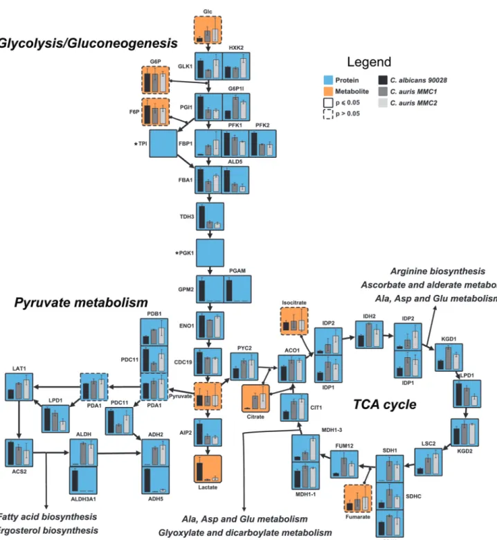
Central carbon metabolism in C. auris and C. albicans.The pathway analysis showed that the glycolytic pathway was enriched in proteins with higher abundance in C. albicans, whereas the TCA cycle proteins were enriched with proteins more abundant inC. auris. Different proteins of the pyruvate metabolism were more abundant in one of the otherCandidaspecies (Fig. 1). To validate these observations and to correlate with downstream metabolic pathways, we integrated the proteomics data with a metabolite analysis into a map of central carbon metabolism. Ten of the fifteen glycolysis/gluconeogenesis proteins were more abundant inC. albicansthan inC. auris, whereas only two proteins were consistently more abundant in C. auris (Fig. 2). In agreement with these observations, lactate, one of the end products of this pathway, was 16-fold more abundant in C. albicans than C. auris MMC1 and 6-fold more abundant inC. aurisMMC2 (Fig. 2). On the other hand, 14 of 15 TCA cycle proteins were more abundant inC. auris isolates than inC. albicans (Fig. 2 and Table S4). Further validating these observations, citrate had similar abundance profiles (Fig. 2). In the pyruvate metabolism, proteins were not consistently more abundant in one or the other species. Some differentially abundant proteins seemed to be due to gene isoforms that were preferentially expressed between the species. For example,C. auris produces alcohol dehydrogenase Adh2, while C. albicans produces Adh5 (Fig. 2).
Unfortunately, the metabolites of this pathway, such as acetate, acetaldehyde, and ethanol, are small and not detectable in our gas chromatography-mass spectrometry (GC-MS) analysis. The fact that different proteins of this pathway were not uniformly more abundant in one of the species makes it more difficult to predict whether the downstream metabolic pathways would be affected. We decided to investigate the ergosterol and glycerolipid biosynthesis pathways in more detail.
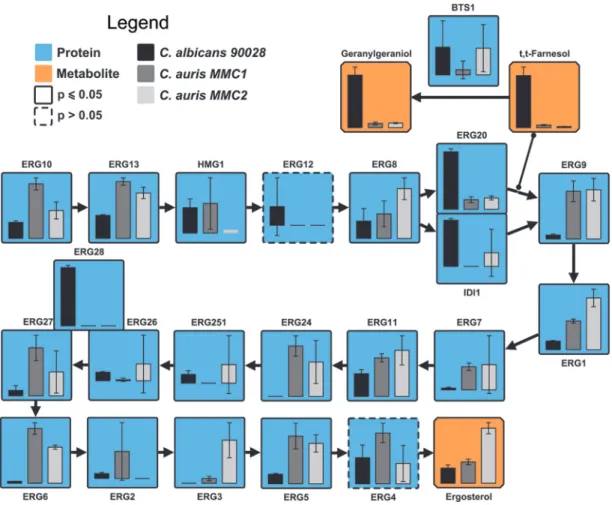
Ergosterol biosynthesis pathway in C. auris versus C. albicans. Fluconazole inhibits the activity of Erg11 (lanosterol 14-␣-demethylase) and consequently ergos- terol biosynthesis. Due to the remarkable resistance displayed by MMC1 against fluconazole, we performed a comparative analysis of the enzymes and some of the metabolites present in the ergosterol synthesis pathway. Eleven (Erg10, Erg13, Erg9, Erg1, Erg7, Erg11, Erg24, Erg27, Erg6, Erg3, and Erg5) of nineteen of the ergosterol synthesis enzymes were significantly more abundant in C. auris MMC1 than in C.
albicans, including Erg11 (Fig. 3). Similarly, 7 (Erg13, Erg8, Erg9, Erg1Erg6, Erg3, and Erg5) of 13 of the ergosterol synthesis enzymes were significantly more abundant inC.
aurisMMC2 than inC. albicans, including Erg11 (Fig. 3). Ergosterol itself was four times more abundant in MMC2 compared to MMC1 andC. albicansand had similar abun- dances in MMC1 and C. albicans (Fig. 3). There were, however, a few exceptions of proteins from the ergosterol pathway that were more abundant inC. albicansthan in C. auris, which was the case for Idi1, Erg20, and Erg28. Idi1 and Erg20 seem to diverge from the pathway to produce farnesol, a quorum-sensing molecule involved in C.
albicansdimorphism and its downstream product geranylgeraniol. Farnesol was 20.7- and 51.8-fold more abundant inC. albicanscompared toC. aurisMMC1 and MMC2, respectively (Fig. 3). Similarly, geranylgeraniol was 12.9- and 11.6-fold times more abundant inC. albicanscompared to C. aurisMMC1 and MMC2, respectively (Fig. 3).
Erg28 is a scaffold protein that docs Erg26 and Erg27 close together (21), but how its abundance affect enzymatic reaction still needs be to investigated.
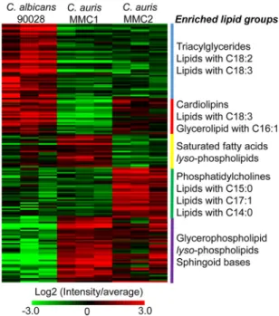
Lipid profile of C. auris and C. albicans. The differential abundance of carbon metabolism, especially in the pyruvate metabolism, is indicative that the fatty acid (FA) biosynthesis and consequently the lipid structures could be altered. Considering that lipids are major targets of antifungal drugs (22) and part of resistance mechanisms (23, 24), we analyzed this category of biomolecules. A total of 169 lipids from 10 different classes were identified and quantified. The most diverse lipid class was triacylglycerol (TG), with 38 distinct species, followed by phosphatidylcholine (PC) with 28 (Table S5).
on November 11, 2019 by guest http://msystems.asm.org/ Downloaded from
To compare groups of lipids from differentCandidaspecies/isolates, we clustered lipids based on their abundance and performed an enrichment analysis using a recently developed tool named Lipid Mini-On (described in Materials and Methods). This analysis is analogous to pathway enrichment and determines whether groups of lipids are significantly enriched based on their intrinsic features (class, head group, FA length, and unsaturation, etc.). The results showed that TG and lipids carrying polyunsaturated FA were enriched inC. albicans. Cardiolipins, lipids containing C18:3FA and glycerolipids
FIG 2 Central carbon metabolism ofC. aurisandC. albicans. The figure shows the relative abundance of proteins (blue boxes) and the production of metabolites (orange boxes) involved in the central carbon metabolism in bothC. albicansandC. auris. Paralog proteins were grouped and posted side by side in the map.*, Genes that were only annotated in theC. albicansgenome.Pⱕ0.05 indicates statistically significant hits determined byttest in any of the three comparisons. For complete comparisons between the different samples and abundances of each analyte, see Table S4 in the supplemental material.
Multi-omics Signature ofCandida auris
on November 11, 2019 by guest http://msystems.asm.org/ Downloaded from
carrying C16:1 FA were significantly reduced in the resistant isolate MMC1 (Fig. 4).
Lysophospholipids were enhanced inC. aurisMMC1 and to a lesser extent inC. auris MMC2 compared to C. albicans. The enriched amount of lysophospholipids is an indication of a higher phospholipase activity. We investigated the abundance profiles of enzymes with phospholipase activity in the proteomics data (Table 2). Our analysis detected seven phospholipases in C. auris and only five in C. albicans. Excepting Pld1 (A0A0L0P056), all of them were significantly byt test more abundant in MMC1 than in C. albicans. Remarkably, lysophospholipases Plb3 (A0A0L0NWB3) and Plb5 (A0A0L0P465) were not detected inC. albicans.
C. auris MMC2 produced more phosphatidylcholines and lipids containing odd- chain FA compared toC. aurisMMC1 andC. albicans(Fig. 4). A GC-MS analysis of the lipid fraction indeed confirmed that C17:0and C17:1FA were more abundant inC. auris MMC2 (Fig. 5). C17:0was 23.3- and 28.9-fold more abundant inC. aurisMMC2 compared toC. albicansandC. aurisMMC1, respectively. Similarly, C17:1was 22.3- and 10.5-fold more abundant inC. aurisMMC2 compared toC. albicansandC. aurisMMC1, respec- tively (Fig. 5). Both isolates ofC. auriswere enriched in sphingoid bases (Fig. 4), which was also validated by the detection of phytosphingosine in the GC-MS analysis. This sphingolipid was 3.3-fold more abundant inC. aurisMMC1 compared toC. albicansand 10.3- and 3.2-fold more abundant inC. aurisMMC2 compared toC. albicansandC. auris MMC1, respectively (Fig. 5). In addition to the sphingoid bases, other sphingolipids such as ceramides, hexosylceramides, and inositolphosphoceramides were also more abun- dant inC. aurisMMC1 (Table S5).
FIG 3 Ergosterol biosynthesis pathway inC. aurisandC. albicans. The bar graphs represent the relative abundances of proteins (blue boxes) and metabolites (orange boxes) of the pathway. Note that Erg28 is not an enzyme but a scaffold protein that docs Erg26 and Erg27 close together.Pⱕ0.05 indicates statistically significant hits determined byttest in any of the three comparisons.
For complete comparisons between the different samples and abundances of each analyte, see Table S4 in the supplemental material.
on November 11, 2019 by guest http://msystems.asm.org/ Downloaded from
Cell wall integrity pathway and major structural components. The proteomic analysis showed that proteins involved in the cell wall integrity (CWI) pathway dis- played a significant difference betweenC. albicansandC. auris. Rom2, Tpk2, and the mitogen-activated protein kinase Mck1 were higher in MMC1 compared toC. albicans and the fluconazole-susceptible MMC2 (Fig. 6), suggesting that the MMC1 isolate is better suited to respond to this antifungal. Notably, the protein Pkc1 was detected only inC. albicans, suggesting thatC. aurismay have an alternative pathway to control CWI (Fig. 6).
The enzymes involved in the synthesis and degradation of the major cell wall polysaccharides (glucans and chitin) and mannoproteins were particularly distinct when C. albicans and C. auris were compared. Remarkably, the chitin remodeling enzymes,1,3-glucan synthase, and most of the mannoprotein remodeling enzymes were higher inC. albicanscompared to bothC. aurisisolates. The only exceptions were glucan 1,3--glucosidase Xog1 and␣-1,2-mannosyltransferase MN21, which were both more abundant inC. aurisisolates compared toC. albicans(Fig. 6).
Biofilm transcription factors and proteins.Fungal biofilms are highly resistant to drug treatment due to a combination of factors, including cell density and matrix content (25). We compared the abundance of transcription factors and proteins pre- FIG 4 Lipid species found inC. aurisandC. albicans. The abundance of all detected lipids is shown above in the heatmap. Lipids were grouped in clusters based on their abundance between different species/
isolates. The enrichment of lipid intrinsic features (head group, fatty acid length, fatty acid unsaturation, etc.) is listed by the side of each cluster. For a complete list of proteins, their relative abundances, and Pvalues (determined byttest), see Table S5 in the supplemental material.
TABLE 2Proteins with phospholipase activity inC. aurisandC. albicans
Protein C. aurisUniProt no.
Relative abundancea
C. albicans90028 C. aurisMMC1 C. aurisMMC2
Plc2p A0A0L0P5S6 ⫺ ⫹ ⫹ ⫹
Patatin-like phospholipase domain-containing protein A0A0L0NS42 ⫺ ⫺ ⫹ ⫹ ⫺
Lysophospholipase A0A0L0NWB3 ND ⫹ ⫹ ⫹ ⫹
A0A0L0P465 ND ⫹ ⫹ ⫹
Doa1p A0A0L0NP71 ⫹ ⫹ ⫹ ⫹ ⫹
Phospholipase A0A0L0P056 ⫹ ⫹ ⫹
Lysophospholipase Nte1 (intracellular phospholipase B) A0A0L0P1C1 ⫹ ⫹ ⫺ ⫺
a⫹,⬍0.5 and⬎⫺1;⫹ ⫹,⬎0.5; –,⬎⫺3 and⬍⫺1; – –,⬍⫺3; ND, not determined.
Multi-omics Signature ofCandida auris
on November 11, 2019 by guest http://msystems.asm.org/ Downloaded from
viously reported in biofilm formation and proteins found in the biofilm matrix. Six transcription factors were reported as biofilm regulators in C. albicans(26–29). Our results showed that Efg1 and Ndt80 were more abundant inC. albicansunder plank- tonic growth conditions with almost no abundance inC. auris. Remarkably, only Rob1 was more abundant in C. auris, specifically in the resistant isolate MMC1. A list of proteins upregulated inC. albicansbiofilms and biofilm matrix was also investigated (Table S6). Of 24 proteins previously reported upregulated in biofilm (30), 8 were detected at higher levels in theC. aurisisolates than inC. albicans.
Transporters. The proteomic analysis identified six transporters related to drug resistance. Notably, the ABC transporter efflux pump Cdr1 and orf19.4780, an unchar- acterized member of the Dha1 family of drug:proton drug antiporter, were significantly higher in the azole-resistant isolate MMC1 (Fig. 7). The other four transporters showed greater abundance in either MMC2 orC. albicans(Fig. 7); therefore, they are less likely to be involved in the fluconazole resistance of MMC1.
DISCUSSION
C. auris is an emerging pathogen that is causing extremely worrisome outbreaks across the globe. One remarkable feature of this fungus is the frequency of resistance FIG 5 Fatty acids and sphingoid bases analyzed by GC-MS. The graph indicates the abundance of lipids containing odd-chain fatty acids and phytosphingosine for bothCandidaspecies/isolates.ttest deter- minations:**,Pⱕ0.01; ns,P⬎0.05.
FIG 6 Cell wall integrity pathway. The heatmap includes signaling and major cell wall polysaccharides synthesis/degradation enzymes found inC. aurisandC. albicans. For a complete list of proteins, their relative abundances, andPvalues (determined byttest), see Table S6 in the supplemental material.
on November 11, 2019 by guest http://msystems.asm.org/ Downloaded from
against at least one class of antifungals. In addition, multidrug-resistant strains have been isolated from all continents. The search for a new class of antifungal drug has been a major challenge in the medical mycology community, and this quest becomes even more urgent with the spread of a multidrug-resistant fungal organism such asC.
auris. In the present study, two clinical isolates ofC. aurisisolated in the Bronx, NY, were analyzed by a multi-omics approach to better understand the molecular repertoire employed by this pathogen. In parallel toC. auris, we also performed the same analyses with a reference strain ofC. albicans.C. aurishas been classified into four distinct clades according to their biogeography (31). A recent epidemiological study by the CDC (32) has shown that the vast majority ofC. aurisisolates from New York City belong to the South Asian clade (98% of all analyzed isolates). Even though there are obvious limitations about studying a small number of isolates, our study was nevertheless able to delve deep into theC. aurisbiology and provides a platform for future analyses of additional isolates.
We found that MMC2 and theC. albicansstrain were susceptible to amphotericin B, caspofungin, and fluconazole, but MMC1 was resistant to both caspofungin and fluconazole. TheC. aurisMMC2 MIC value of fluconazole was approximately 8g/ml, which, based on the CDC report, would make it a susceptible isolate, even though the MIC was about 10 times higher than forC. albicans. Although MMC1 just met resistance criteria to caspofungin, its resistance to fluconazole was impressive, as even 1 mg/ml was not able to totally inhibit growth. The “Eagle effect,” also known as “paradoxical effect” was observed in bothC. aurisisolates after treatment with caspofungin, since growth occurred at concentrations higher than the MIC.
The protein profiles fromC. aurisandC. albicanswere qualitatively and quantita- tively distinct, and both isolates ofC. aurispresented very few differences from one another (Table S3). The major observed difference betweenC. aurisandC. albicanswas in their central carbon metabolism. While proteins in the glycolysis pathway were upregulated inC. albicans,C. aurisshowed an enrichment of proteins in the TCA cycle.
These results show thatC. auris favors respiration, which is already known to be an important mechanism of fluconazole resistance in C. albicans, by increasing ATP production and reducing oxidative stress, resulting in better overall fitness of the cell (33).
In Saccharomyces cerevisiae, overexpression of HMG1 or deletion of ERG2, can significantly increase susceptibility to fluconazole, whereas the deletion of HMG1, ERG6, and ERG3, as well as the overexpression of ERG11, is associated with fluconazole resistance (34). Therefore, we integrated the data of proteins and metabolites of the ergosterol biosynthesis pathway. Despite the extreme resistance of MMC1 against fluconazole, the abundance of Erg11 in this isolate was similar to that observed for MMC2. On the other hand, the higher abundance of Erg2 and lower abundance of Erg3 FIG 7 Protein abundance profile of drug resistance-related transporters. The heatmap shows the detected transporters involved with drug resistance and their abundances in both Candidaspecies/
isolates. For a complete list of proteins, their relative abundances, andPvalues (determined byttest), see Table S3 in the supplemental material.
Multi-omics Signature ofCandida auris
on November 11, 2019 by guest http://msystems.asm.org/ Downloaded from
of MMC1 compared to the MMC2 isolate are in agreement with drug resistance phenotype of MMC1. The higher abundance of Idi1 and Erg20 inC. albicansdiverges part of the pathway to produce more isoprenoids, whileC. auris has a more robust production of ergosterol, which is possibly involved in fluconazole resistance. Recently, sequence divergences/mutations on ERG11 inC. aurishave been shown to be associ- ated with resistance to azoles (35). However, the ERG11 mutations by themselves cannot explain why the level of fluconazole resistance was lower (up to 128g/ml) when theC. aurisgene was expressed inS. cerevisiae(36). Therefore, our data combined with reports from the literature suggest that the fluconazole resistance inC. aurisis due to modifications of multiple steps in the ergosterol biosynthesis pathway.
The lipids detected inC. auriswere qualitatively similar to those found inC. albicans.
However, a quantitative analysis showed thatC. albicanshas more lipids involved with energy storage, whileC. aurishas more structural glycerophospholipids and lysophos- pholipids. The resistant isolate (MMC1) has a remarkable abundance of lysophospho- lipids, suggesting intense phospholipase activity. Phospholipases are virulence factors in a variety of pathogenic fungi where their activity is important for invasiveness, morphology, and persistence of infection (37–39). Phospholipase activity was recently described inC. auris isolates (40). In the present study, the evaluatedC. aurisisolates were found to produce seven enzymes with phospholipase activity, whileC. albicans had five of them. In addition, most of these enzymes were more abundant inC. auris, particularly in the resistance isolate (MMC1). Corroborating these findings, an increased content of lysophospholipids was previously reported in aC. albicansstrain adaptedin vitro to higher concentrations of fluconazole (41). It is possible that this class of enzymes is more finely employed byC. auristhan byC. albicansto promote survival and environmental adaptation for the fungus. Regarding its biological role during the host-pathogen interaction, lysophosphatidylcholine is a “find me” signal released by apoptotic cells to induce the recruitment of phagocytes to remove apoptotic bodies before an episode of secondary necrosis and enhanced inflammation (42). The MMC1 isolate also had a higher abundance of sphingolipids, which can also be correlated with resistance to antifungals. These lipids are important for the assembly of membrane platforms where proteins such as drug efflux pumps are present in membrane mi- croenvironments responsible for the export of drugs (23).
The response orchestrated by the CWI signaling pathway is central during cell wall and membrane perturbation (43). Sensors at fungal cell surface initiate a downstream cascade in order to adapt the cells under stress conditions controlling cell wall biogenesis and cell integrity (43). Remarkably, we observed that the enzymes involved with cell wall remodeling were reduced in bothC. aurisisolates. However, some CWI proteins were specifically higher in the resistant isolate, suggesting that the response to external signals, such as drug treatment, could be promptly controlled by the cell wall metabolism and help to explain the resistant phenotype in the MMC1 isolate.
The efflux of drugs mediated by efflux pumps is an important mechanism of antifungal resistance employed byCandidaspp. (23, 44, 45). Of six distinct drug efflux transporters produced by the analyzed organisms, two (CDR1 and orf19.4780) were more abundant in the fluconazole-resistantC. aurisisolate (MMC1) than in MMC2 orC.
albicans. Previous publications showed thatC. aurisyeast cells, organized in a biofilm, are more resistant to antifungals than planktonic cells and correlated this phenotype with the increased expression of CDR1 (13). The impact of these efflux pumps is important during early stages of biofilm formation but decreases when it becomes mature. In mature biofilms, resistance is increased by the ability of matrix components to limit drug diffusion, along with the presence of persistent cells (46). Notably, theC.
auris isolate MMC1 has a significant increase in proteins associated with biofilm formation and a higher abundance of superoxide dismutase, an enzyme involved with reactive oxygen species (ROS) detoxification and overexpressed in miconazole-tolerant persisters (46). Furthermore, several proteins characterized in the biofilm matrix were also higher in the resistantC. aurisisolate.
The comprehensive multi-omics approach used in this study has enabled us to
on November 11, 2019 by guest http://msystems.asm.org/ Downloaded from
begin to uncover and characterize the molecular profile of the emerging pathogenC.
auris, which suggest a multifactorial mechanism of drug resistance in MMC1, including major differences in carbon utilization, sphingolipids, glycerolipids, sterols, the cell wall, and efflux pumps. Further functional omic studies that include larger numbers of C.
aurisisolates will likely have significant impact on our understanding of the biology of this remarkable fungus and may facilitate the development of new therapeutic ap- proaches to combat this frequently multidrug-resistant yeast.
MATERIALS AND METHODS
Cell lines.Two clinical isolates (MMC1 and MMC2) were acquired from Montefiore Medical Center (Bronx, NY) under approved protocols in the Nosanchuk laboratory, and a standardC. albicans(ATCC 90028) strain was purchased from the American Type Culture Collection (ATCC). The cells were stored at – 80°C. Prior to use in experiments, cells were cultivated in yeast extract-peptone-dextrose (YPD) broth and seeded onto Sabouraud agar plates. For each experiment, one colony was inoculated in 10 ml of Sabouraud broth overnight at 30°C before use. Cells were transferred to 200 ml of fresh Sabouraud medium and incubated for an additional 24 h. After being extensively washed with phosphate-buffered saline (PBS), the cell pellets were frozen until the protein, metabolite, and lipid extractions.
Antifungal susceptibility.The antifungal susceptibility tests were carried out based on the CLSI protocol with modifications (47, 48). Yeast cells were inoculated in Sabouraud agar for 48 h at 30°C and then stored at 4°C up to 1 month for experimentation. One colony from each organism was inoculated in Sabouraud broth and kept for 24 h at 30°C under constant shaking. Cells were then washed in PBS and plated (2.5⫻103cells/ml) in 96-well plates containing serial dilutions of amphotericin B, caspofungin, and fluconazole. After 48 h of incubation in Sabouraud broth in the presence or absence of antifungals, cells were visually analyzed, and the MIC was determined as the lowest concentration of a given drug that showed no apparent growth within all replicates.
Proteomic analysis.Three independent cell cultures were submitted to metabolite, protein, and lipid extraction (MPLEx) according to the protocol by Nakayasu et al. (49). Extracted proteins were digested with trypsin, and the resulting peptides were extracted with 1 ml of Discovery C18SPE columns (Supelco, Bellefonte, PA) as previously described (50). Digested peptides were suspended in water, quantified by BCA assay and 0.5g of peptides were loaded into trap column (4 cm by 100m inner diameter [ID], packed in-house with 5m C18; Jupiter). Peptide separation was carried out an analytical column (70 cm x 75m ID packed with C18, 3-m particles) using a gradient of acetonitrile– 0.1% formic acid (solvent B) in water– 0.1% formic acid (solvent A). The flow was set to 300 nl/min with 1% solvent B and kept for 15 min. Then, the concentration of solvent B was increased linearly as follows: 19 min, 8%
B; 60 min, 12% B; 155 min, 35% B; 203 min, 60% B; 210 min, 75% B; 215 min, 95% B; 220 min, 95% B.
Eluting peptides were directly analyzed by electrospray in an orbitrap mass spectrometer (Q-Exactive Plus; Thermo Fisher Scientific) by scanning a window of 400 to 2,000m/zwith a resolution of 70,000 at m/z400. Tandem mass spectra were collected using high-energy collision dissociation (32% normalized collision energy) on the 12 most intense multiple-charged parent ions at a resolution of 17,500.
Mass spectrometry data were analyzed using MaxQuant software (v.1.5.5.1) (51). Peptide identifica- tion was performed by searching against theC. albicansSC5314 andC. aurissequences from Uniprot Knowledge Base (downloaded 6 December 2017). The search parameters included the variable modifi- cations protein N-terminal acetylation and oxidation of methionine, in addition to carbamidomethylation of cysteine residues. Parent and fragment mass tolerance were kept as the default setting of the software.
Only fully tryptic digested peptides were considered, allowing up to two missed cleaved sites per peptide. Quantification of proteins was done using the intensity-based absolute quantification (iBAQ) method (52). Intensities of each protein were normalized by the total iBAQ sum of each sample to obtain a relative protein copy number (percentage from total). The comparison between the two species was performed by blast searches and considering a cutoff of 40% of sequence similarity to consider a protein orthologous.
Lipid analysis. Extracted lipids were suspended in 100% methanol and analyzed by liquid chromatography-tandem mass spectrometry (LC-MS/MS) as described elsewhere (53). The identification of the species was done using LIQUID software and manually inspected for validation (54). Peak intensities of each identified lipid species were extracted with MZmine v2.0 (55).
Gas chromatography-mass spectrometry analysis. Extracted hydrophilic metabolite and lipid fractions were derivatized as described previously (56) and analyzed in an Agilent GC 7890A using an HP-5MS column (30 m⫻0.25 mm⫻0.25m; Agilent Technologies, Santa Clara, CA) coupled with a single quadrupole MSD 5975C (Agilent Technologies). The GC was set to splitless mode with the port temperature at 250°C. Samples were injected with the oven temperature equilibrated at 60°C. The same temperature was kept for 1 min and then raised at a 10°C/min rate to a final temperature of 325°C for a 5-min hold. A standard mixture of fatty acid methyl ester (Sigma-Aldrich) was used for calibrating the retention time. Retention time calibration, spectral deconvolution, and peak alignment were done with MetaboliteDetector (57). Metabolites were identified by matching against FiehnLib library (58) containing additional metabolites entered in-house and/or the NIST14 GC-MS library. All identified metabolites were manually inspected.
Quantitative analysis and data integration. Protein orthologues, lipids, or metabolites were considered significantly different with aPvalue ofⱕ0.05 usingttest considering equal variance and two-tailed distribution. For comparative analyses, missing values were zero-filled with half of the smallest
Multi-omics Signature ofCandida auris
on November 11, 2019 by guest http://msystems.asm.org/ Downloaded from
value of the data set. Proteins were clustered by the k-means method using Multi-Experiment Viewer (MeV, v4.9.0) (59), which was also used to build the heatmaps. Pathway analysis on different protein clusters was performed with DAVID (60), and specific pathways of interested were manually inspected with Vanted v2.1.1 (61). We have recently developed an R package called Rodin (https://github.com/
PNNL-Comp-Mass-Spec/Rodin), to perform structural “lipid ontology” (LO) enrichment analysis. A web interface, Lipid Mini-On, was developed for non-R users (https://omicstools.pnnl.gov/shiny/lipid-mini -on/) (62). Briefly, this tool creates automatically LO bins based on the lipids naming and their inferred structure, and then it performs enrichment analysis using enrichment statistics to compare a query list to a Universe (Fisher exact test, EASE score, binomial test, or hypergeometric tests). In this study, a Fisher exact test was used to perform the enrichment analysis, and only the enrichmentPvalues below 0.05 were considered significant.
Data availability.Proteomics data were deposited into Pride repository (www.ebi.ac.uk/pride) under accession numbersPXD013456andPXD013457.
SUPPLEMENTAL MATERIAL
Supplemental material for this article may be found at https://doi.org/10.1128/
mSystems.00257-19.
TABLE S1, XLSX file, 0.2 MB.
TABLE S2, XLSX file, 0.2 MB.
TABLE S3, XLSX file, 0.5 MB.
TABLE S4, XLSX file, 0.02 MB.
TABLE S5, XLSX file, 0.1 MB.
TABLE S6, XLSX file, 0.01 MB.
ACKNOWLEDGMENTS
We thank Erika Zink, Jeremy Teuton, and Jeremy Zucker for technical assistance.
J.D.N. and E.S.N. were partially supported by NIH R21 AI124797. A.G. was supported by NKFIH K 123952, GINOP 2-3-2-15-2016-00015, and GINOP 2.3.3-15-2016-00006.
Ministry of Human Capacities, Hungary, grant 20391-3/2018/FEKUSTRAT is also ac- knowledged. Parts of this work were performed in the Environmental Molecular Science Laboratory, a U.S. Department of Energy (DOE) national scientific user facility at Pacific Northwest National Laboratory (PNNL) in Richland, WA.
We declare that there are no conflicts of interest.
REFERENCES
1. Satoh K, Makimura K, Hasumi Y, Nishiyama Y, Uchida K, Yamaguchi H.
2009.Candida aurissp. nov., a novel ascomycetous yeast isolated from the external ear canal of an inpatient in a Japanese hospital. Microbiol Immunol 53:41– 44.https://doi.org/10.1111/j.1348-0421.2008.00083.x.
2. Lee WG, Shin JH, Uh Y, Kang MG, Kim SH, Park KH, Jang H-C. 2011. First three reported cases of nosocomial fungemia caused byCandida auris.
J Clin Microbiol 49:3139 –3142.https://doi.org/10.1128/JCM.00319-11.
3. Jeffery-Smith A, Taori SK, Schelenz S, Jeffery K, Johnson EM, Borman A, Manuel R, Brown CS. 2018.Candida auris: a review of the literature. Clin Microbiol Rev 31:e00029-17.
4. Kean R, Sherry L, Townsend E, McKloud E, Short B, Akinbobola A, Mackay WG, Williams C, Jones BL, Ramage G. 2018. Surface disinfection chal- lenges forCandida auris: an in-vitro study. J Hosp Infect 98:433– 436.
https://doi.org/10.1016/j.jhin.2017.11.015.
5. Sarma S, Upadhyay S. 2017. Current perspective on emergence, diag- nosis, and drug resistance in Candida auris. Infect Drug Resist 10:
155–165.https://doi.org/10.2147/IDR.S116229.
6. Prakash A, Sharma C, Singh A, Kumar Singh P, Kumar A, Hagen F, Govender NP, Colombo AL, Meis JF, Chowdhary A. 2016. Evidence of genotypic diversity among Candida auris isolates by multilocus se- quence typing, matrix-assisted laser desorption ionization time-of-flight mass spectrometry and amplified fragment length polymorphism. Clin Microbiol Infect 22:277 e1–9.https://doi.org/10.1016/j.cmi.2015.10.022.
7. Oh BJ, Shin JH, Kim MN, Sung H, Lee K, Joo MY, Shin MG, Suh SP, Ryang DW. 2011. Biofilm formation and genotyping ofCandida haemulonii, Candida pseudohaemulonii, and a proposed new species (Candida auris) isolates from Korea. Med Mycol 49:98 –102. https://doi.org/10.3109/
13693786.2010.493563.
8. Chowdhary A, Anil Kumar V, Sharma C, Prakash A, Agarwal K, Babu R, Dinesh KR, Karim S, Singh SK, Hagen F, Meis JF. 2014. Multidrug-resistant
endemic clonal strain ofCandida aurisin India. Eur J Clin Microbiol Infect Dis 33:919 –926.https://doi.org/10.1007/s10096-013-2027-1.
9. Calvo B, Melo AS, Perozo-Mena A, Hernandez M, Francisco EC, Hagen F, Meis JF, Colombo AL. 2016. First report ofCandida auris in America:
clinical and microbiological aspects of 18 episodes of candidemia. J Infect 73:369 –374.https://doi.org/10.1016/j.jinf.2016.07.008.
10. Schelenz S, Hagen F, Rhodes JL, Abdolrasouli A, Chowdhary A, Hall A, Ryan L, Shackleton J, Trimlett R, Meis JF, Armstrong-James D, Fisher MC.
2016. First hospital outbreak of the globally emergingCandida aurisin a European hospital. Antimicrob Resist Infect Control 5:35.https://doi .org/10.1186/s13756-016-0132-5.
11. Chowdhary A, Sharma C, Meis JF. 2017.Candida auris: a rapidly emerg- ing cause of hospital-acquired multidrug-resistant fungal infections globally. PLoS Pathog 13:e1006290.https://doi.org/10.1371/journal.ppat .1006290.
12. Welsh RM, Bentz ML, Shams A, Houston H, Lyons A, Rose LJ, Litvintseva AP. 2017. Survival, persistence, and isolation of the emerging multidrug- resistant pathogenic yeastCandida aurison a plastic health care surface.
J Clin Microbiol 55:2996 –3005.https://doi.org/10.1128/JCM.00921-17.
13. Kean R, Delaney C, Sherry L, Borman A, Johnson EM, Richardson MD, Rautemaa-Richardson R, Williams C, Ramage G. 2018. Transcriptome assembly and profiling of Candida auris reveals novel insights into biofilm-mediated resistance. mSphere 3:e00334-18.
14. Sherry L, Ramage G, Kean R, Borman A, Johnson EM, Richardson MD, Rautemaa-Richardson R. 2017. Biofilm-forming capability of highly viru- lent, multidrug-resistantCandida auris. Emerg Infect Dis 23:328 –331.
https://doi.org/10.3201/eid2302.161320.
15. Vallabhaneni S, Kallen A, Tsay S, Chow N, Welsh R, Kerins J, Kemble SK, Pacilli M, Black SR, Landon E, Ridgway J, Palmore TN, Zelzany A, Adams EH, Quinn M, Chaturvedi S, Greenko J, Fernandez R, Southwick K, Furuya
on November 11, 2019 by guest http://msystems.asm.org/ Downloaded from
EY, Calfee DP, Hamula C, Patel G, Barrett P, Lafaro P, Berkow EL, Moulton- Meissner H, Noble-Wang J, Fagan RP, Jackson BR, Lockhart SR, Lit- vintseva AP, Chiller TM. 2016. Investigation of the first seven reported cases ofCandida auris, a globally emerging invasive, multidrug-resistant fungus—United States, May 2013–August 2016. MMWR Morb Mortal Wkly Rep 65:1234 –1237.https://doi.org/10.15585/mmwr.mm6544e1.
16. Ghosh AK, Paul S, Sood P, Rudramurthy SM, Rajbanshi A, Jillwin TJ, Chakrabarti A. 2015. Matrix-assisted laser desorption ionization time-of- flight mass spectrometry for the rapid identification of yeasts causing bloodstream infections. Clin Microbiol Infect 21:372–378. https://doi .org/10.1016/j.cmi.2014.11.009.
17. Mizusawa M, Miller H, Green R, Lee R, Durante M, Perkins R, Hewitt C, Simner PJ, Carroll KC, Hayden RT, Zhang SX. 2017. Can multidrug- resistantCandida auris be reliably identified in clinical microbiology laboratories? J Clin Microbiol 55:638 – 640.https://doi.org/10.1128/JCM .02202-16.
18. Leach L, Zhu Y, Chaturvedi S. 2018. Development and validation of a real-time PCR assay for rapid detection ofCandida aurisfrom surveil- lance samples. J Clin Microbiol 56:e01223-17.
19. Wagener J, Loiko V. 2017. Recent insights into the paradoxical effect of echinocandins. J Fungi 4:5.https://doi.org/10.3390/jof4010005.
20. Kordalewska M, Lee A, Park S, Berrio I, Chowdhary A, Zhao Y, Perlin DS.
2018. Understanding echinocandin resistance in the emerging pathogen Candida auris. Antimicrob Agents Chemother 62:e00238-18.https://doi .org/10.1128/aac.00238-18.
21. Mo C, Valachovic M, Randall SK, Nickels JT, Bard M. 2002. Protein-protein interactions among C-4 demethylation enzymes involved in yeast sterol biosynthesis. Proc Natl Acad Sci U S A 99:9739 –9744.https://doi.org/10 .1073/pnas.112202799.
22. Pan J, Hu C, Yu JH. 2018. Lipid biosynthesis as an antifungal target. J Fungi (Basel) 4:E50.
23. Mukhopadhyay K, Prasad T, Saini P, Pucadyil TJ, Chattopadhyay A, Prasad R. 2004. Membrane sphingolipid-ergosterol interactions are im- portant determinants of multidrug resistance inCandida albicans. Anti- microb Agents Chemother 48:1778 –1787.https://doi.org/10.1128/AAC .48.5.1778-1787.2004.
24. Mukhopadhyay K, Kohli A, Prasad R. 2002. Drug susceptibilities of yeast cells are affected by membrane lipid composition. Antimicrob Agents Chemother 46:3695–3705.https://doi.org/10.1128/aac.46.12.3695-3705 .2002.
25. Silva S, Rodrigues C, Araújo D, Rodrigues M, Henriques M. 2017.Candida species biofilms’ antifungal resistance. J Fungi 3:8.https://doi.org/10 .3390/jof3010008.
26. Nobile CJ, Fox EP, Nett JE, Sorrells TR, Mitrovich QM, Hernday AD, Tuch BB, Andes DR, Johnson AD. 2012. A recently evolved transcriptional network controls biofilm development inCandida albicans. Cell 148:
126 –138.https://doi.org/10.1016/j.cell.2011.10.048.
27. Vandeputte P, Pradervand S, Ischer F, Coste AT, Ferrari S, Harshman K, Sanglard D. 2012. Identification and functional characterization of Rca1, a transcription factor involved in both antifungal susceptibility and host response inCandida albicans. Eukaryot Cell 11:916 –931.https://doi.org/
10.1128/EC.00134-12.
28. Maiti P, Ghorai P, Ghosh S, Kamthan M, Tyagi RK, Datta A. 2015. Mapping of functional domains and characterization of the transcription factor Cph1 that mediate morphogenesis inCandida albicans. Fungal Genet Biol 83:45–57.https://doi.org/10.1016/j.fgb.2015.08.004.
29. Khalaf RA, Zitomer RS. 2001. The DNA binding protein Rfg1 is a repressor of filamentation inCandida albicans. Genetics 157:1503–1512.
30. Seneviratne CJ, Wang Y, Jin L, Abiko Y, Samaranayake LP. 2008.
Candida albicansbiofilm formation is associated with increased anti- oxidative capacities. Proteomics 8:2936 –2947. https://doi.org/10 .1002/pmic.200701097.
31. Lockhart SR, Etienne KA, Vallabhaneni S, Farooqi J, Chowdhary A, Gov- ender NP, Colombo AL, Calvo B, Cuomo CA, Desjardins CA, Berkow EL, Castanheira M, Magobo RE, Jabeen K, Asghar RJ, Meis JF, Jackson B, Chiller T, Litvintseva AP. 2017. Simultaneous emergence of multidrug- resistantCandida aurison three continents confirmed by whole-genome sequencing and epidemiological analyses. Clin Infect Dis 64:134 –140.
https://doi.org/10.1093/cid/ciw691.
32. Adams E, Quinn M, Tsay S, Poirot E, Chaturvedi S, Southwick K, Greenko J, Fernandez R, Kallen A, Vallabhaneni S, Haley V, Hutton B, Blog D, Lutterloh E, Zucker H. 2018.Candida aurisin healthcare facilities, New York, USA, 2013-2017. Emerg Infect Dis 24:1816 –1824.https://doi.org/
10.3201/eid2410.180649.
33. Guo H, Xie SM, Li SX, Song YJ, Zhong XY, Zhang H. 2017. Involvement of mitochondrial aerobic respiratory activity in efflux-mediated resistance ofCandida albicansto fluconazole. J Mycol Med 27:339 –344.https://doi .org/10.1016/j.mycmed.2017.04.004.
34. Bhattacharya S, Esquivel BD, White TC. 2018. Overexpression or dele- tion of ergosterol biosynthesis genes alters doubling time, response to stress agents, and drug susceptibility inSaccharomyces cerevisiae.
mBio 9:e01291-18.
35. Hou X, Lee A, Jimenez-Ortigosa C, Kordalewska M, Perlin DS, Zhao Y.
2018. Rapid detection of ERG11-associated azole resistance and FKS- associated echinocandin resistance inCandida auris. Antimicrob Agents Chemotherhttps://doi.org/10.1128/AAC.01811-18.
36. Healey KR, Kordalewska M, Jimenez Ortigosa C, Singh A, Berrio I, Chow- dhary A, Perlin DS. 2018. Limited ERG11 mutations identified in isolates of Candida auris directly contribute to reduced azole susceptibility.
Antimicrob Agents Chemother 62:e01427-18.
37. Leidich SD, Ibrahim AS, Fu Y, Koul A, Jessup C, Vitullo J, Fonzi W, Mirbod F, Nakashima S, Nozawa Y, Ghannoum MA. 1998. Cloning and disrup- tion ofCaPLB1, a phospholipase B gene involved in the pathogenicity ofCandida albicans. J Biol Chem 273:26078 –26086.https://doi.org/
10.1074/jbc.273.40.26078.
38. Santangelo R, Zoellner H, Sorrell T, Wilson C, Donald C, Djordjevic J, Shounan Y, Wright L. 2004. Role of extracellular phospholipases and mononuclear phagocytes in dissemination of cryptococcosis in a murine model. Infect Immun 72:2229 –2239. https://doi.org/10.1128/IAI.72.4 .2229-2239.2004.
39. Evans RJ, Li Z, Hughes WS, Djordjevic JT, Nielsen K, May RC. 2015.
Cryptococcal phospholipase B1 is required for intracellular proliferation and control of titan cell morphology during macrophage infection.
Infect Immun 83:1296 –1304.https://doi.org/10.1128/IAI.03104-14.
40. Larkin E, Hager C, Chandra J, Mukherjee PK, Retuerto M, Salem I, Long L, Isham N, Kovanda L, Borroto-Esoda K, Wring S, Angulo D, Ghannoum M.
2017. The emerging pathogenCandida auris: growth phenotype, viru- lence factors, activity of antifungals, and effect of SCY-078, a novel glucan synthesis inhibitor, on growth morphology and biofilm forma- tion. Antimicrob Agents Chemother 61:e02396-16.
41. Singh A, Mahto KK, Prasad R. 2013. Lipidomics andin vitroazole resis- tance inCandida albicans. Omics 17:84 –93.https://doi.org/10.1089/omi .2012.0075.
42. Peter C, Waibel M, Radu CG, Yang LV, Witte ON, Schulze-Osthoff K, Wesselborg S, Lauber K. 2008. Migration to apoptotic “find-me” signals is mediated via the phagocyte receptor G2A. J Biol Chem 283:
5296 –5305.https://doi.org/10.1074/jbc.M706586200.
43. Dichtl K, Samantaray S, Wagener J. 2016. Cell wall integrity signaling in human pathogenic fungi. Cell Microbiol 18:1228 –1238.https://doi.org/
10.1111/cmi.12612.
44. Fonseca E, Silva S, Rodrigues CF, Alves CT, Azeredo J, Henriques M. 2014.
Effects of fluconazole onCandida glabratabiofilms and its relationship with ABC transporter gene expression. Biofouling 30:447– 457.https://
doi.org/10.1080/08927014.2014.886108.
45. Rocha MFG, Bandeira SP, Alencar LP, Melo LM, Sales JA, Paiva MAN, Teixeira CEC, Castelo-Branco D, Pereira-Neto WA, Cordeiro RA, Sidrim JJC, Brilhante R. 2017. Azole resistance inCandida albicansfrom animals:
highlights on efflux pump activity and gene overexpression. Mycoses 60:462– 468.https://doi.org/10.1111/myc.12611.
46. Bink A, Vandenbosch D, Coenye T, Nelis H, Cammue BPA, Thevissen K.
2011. Superoxide dismutases are involved inCandida albicansbiofilm persistence against miconazole. Antimicrob Agents Chemother 55:
4033– 4037.https://doi.org/10.1128/AAC.00280-11.
47. de-Souza-Silva CM, Guilhelmelli F, Zamith-Miranda D, de Oliveira MA, Nosanchuk JD, Silva-Pereira I, Albuquerque P. 2018. Broth microdilution in vitroscreening: an easy and fast method to detect new antifungal compounds. J Vis Exp 132:e57127.https://doi.org/10.3791/57127.
48. CLSI. 2016. Reference method for broth dilution antifungal susceptibility testing of yeast, 4th ed. CLSI standard M27. Clinical and Laboratory Standards Institute, Wayne, PA.
49. Nakayasu ES, Nicora CD, Sims AC, Burnum-Johnson KE, Kim YM, Kyle JE, Matzke MM, Shukla AK, Chu RK, Schepmoes AA, Jacobs JM, Baric RS, Webb-Robertson BJ, Smith RD, Metz TO. 2016. MPLEx: a robust and universal protocol for single-sample integrative proteomic, metabolo- mic, and lipidomic analyses. mSystems 1:e00043-16.
50. Matos Baltazar L, Nakayasu ES, Sobreira TJ, Choi H, Casadevall A, Nim- richter L, Nosanchuk JD. 2016. Antibody binding alters the characteris- Multi-omics Signature ofCandida auris
on November 11, 2019 by guest http://msystems.asm.org/ Downloaded from
tics and contents of extracellular vesicles released byHistoplasma cap- sulatum. mSphere 1:e00085-15.
51. Cox J, Mann M. 2008. MaxQuant enables high peptide identification rates, individualized p.p.b.-range mass accuracies and proteome-wide protein quantification. Nat Biotechnol 26:1367–1372.https://doi.org/10 .1038/nbt.1511.
52. Schwanhausser B, Busse D, Li N, Dittmar G, Schuchhardt J, Wolf J, Chen W, Selbach M. 2011. Global quantification of mammalian gene expres- sion control. Nature 473:337–342.https://doi.org/10.1038/nature10098.
53. Dautel SE, Kyle JE, Clair G, Sontag RL, Weitz KK, Shukla AK, Nguyen SN, Kim Y-M, Zink EM, Luders T, Frevert CW, Gharib SA, Laskin J, Carson JP, Metz TO, Corley RA, Ansong C. 2017. Lipidomics reveals dramatic lipid compositional changes in the maturing postnatal lung. Sci Rep 7:40555.
https://doi.org/10.1038/srep40555.
54. Kyle JE, Crowell KL, Casey CP, Fujimoto GM, Kim S, Dautel SE, Smith RD, Payne SH, Metz TO. 2017. LIQUID: an open source software for identi- fying lipids in LC-MS/MS-based lipidomics data. Bioinformatics 33:
1744 –1746.https://doi.org/10.1093/bioinformatics/btx046.
55. Pluskal T, Castillo S, Villar-Briones A, Oresic M. 2010. MZmine 2: modular framework for processing, visualizing, and analyzing mass spectrometry- based molecular profile data. BMC Bioinformatics 11:395. https://doi .org/10.1186/1471-2105-11-395.
56. Kim YM, Schmidt BJ, Kidwai AS, Jones MB, Deatherage Kaiser BL, Brewer HM, Mitchell HD, Palsson BO, McDermott JE, Heffron F, Smith RD, Peterson SN, Ansong C, Hyduke DR, Metz TO, Adkins JN. 2013.Salmo- nellamodulates metabolism during growth under conditions that in-
duce expression of virulence genes. Mol Biosyst 9:1522–1534.https://
doi.org/10.1039/c3mb25598k.
57. Hiller K, Hangebrauk J, Jager C, Spura J, Schreiber K, Schomburg D. 2009.
MetaboliteDetector: a comprehensive analysis tool for targeted and nontargeted GC/MS-based metabolome analysis. Anal Chem 81:
3429 –3439.https://doi.org/10.1021/ac802689c.
58. Kind T, Wohlgemuth G, Lee DY, Lu Y, Palazoglu M, Shahbaz S, Fiehn O.
2009. FiehnLib: mass spectral and retention index libraries for metabo- lomics based on quadrupole and time-of-flight gas chromatography/
mass spectrometry. Anal Chem 81:10038 –10048. https://doi.org/10 .1021/ac9019522.
59. Howe EA, Sinha R, Schlauch D, Quackenbush J. 2011. RNA-Seq analysis in MeV. Bioinformatics 27:3209 –3210. https://doi.org/10.1093/bio informatics/btr490.
60. Huang da W, Sherman BT, Lempicki RA. 2009. Systematic and integrative analysis of large gene lists using DAVID bioinformatics resources. Nat Protoc 4:44 –57.https://doi.org/10.1038/nprot.2008.211.
61. Rohn H, Junker A, Hartmann A, Grafahrend-Belau E, Treutler H, Klapper- stuck M, Czauderna T, Klukas C, Schreiber F. 2012. VANTED v2: a frame- work for systems biology applications. BMC Syst Biol 6:139.https://doi .org/10.1186/1752-0509-6-139.
62. Clair G, Reehl S, Stratton KG, Monroe ME, Tfaily MM, Ansong C, Kyle JE.
2019. Lipid Mini-On: mining and ontology tool for enrichment analysis of lipidomic data. Bioinformatics pii:btz250. https://doi.org/10.1093/
bioinformatics/btz250.