1
Role of intrathoracic airways in the pathomechanism of obstructive sleep apnoea
Doctoral theses
László Kunos
Semmelweis University PhD School of Clinical Medicine
Supervisor: Dr. András Bikov Ph.D.
Opponents: Dr. György Purebl Ph.D.
Dr. Éva Vizi Ph.D.
Head of doctoral committee: Dr. András Arató D.Sc.
Members of doctoral committtee: Dr. Kálmán Tory Ph.D.
Dr. László Szabó Ph.D.
Budapest
2018
2
1. Introduction
Obstructive sleep apnoea (OSA) is characterised by repetitive collapses of the pharynx during sleep with consequential development of intermittent hypoxia and arousals. The disorder is highly prevalent, moderate or severe OSA affects 13% of male and 6% of female population between 30 and 70 years of age. Without effective therapy patients’ quality of life and survival can be significantly impaired due to consequential daytime sleepiness, cognitive dysfunction and higher risk of metabolic and cardio- or cerebrovascular disorders.
The root cause of consequential symptoms and disorders most likely to be the repetitive hypoxia and sleep fragmentation which generate systemic and airway oxidative stress and inflammation cascades.
Intrathoracal airways play significant role in OSA pathomechanism due to local inflammation and volume changes.
It was confirmed by several studies that elevated levels of citokines and markers of oxidative and nitrozative stress can be detected in exhaled breath samples confirming intensified inflammation processes in the airways.
Volatile organic compounds (VOCs) are important markers of the inflammative and metabolic processes of the airways. The electronic nose is a potential technique to study exhaled volatile compounds. These devices can distinguish different gas composites based on their molecule pattern. Similarly to biological olfactory receptors, the inbuild sensors are not selective to individual ligands, but upon activation by an odour the
3
sensor array gives a signal pattern. Data reduction with principal component analysis must be performed for further statistical analysis of detected signal patterns.
Beside the inflammatory processes in the airways, lung volumes may also play role in the pathomechanism of OSA. It has been recognized that reduced lung volumes are related to increased OSA severity, regardless of whether the patients have airway or parenchymal lung disease. In particular, negative relationships were reported between the severity of OSA, determined using the apnoea/ hypopnea index (AHI) or respiratory disturbance index, and expiratory reserve volume, functional residual capacity, forced expiratory volume in one second (FEV1), and forced vital capacity (FVC). In line with this, a significant association between lung function loss and all-cause mortality was reported in patients with OSA. The association between lung volumes and OSA can be explained by the longitudinal stretch of airways which can modify the collapsibility of pharyngeal airway. Furthermore, larger lung volumes are associated with higher buffer-capacity.
The hereby presented research was dedicated to study the effects of various methodological factors on exhaled volatile compounds pattern and on the discrimination capabilities of electronic nose device. Furthermore we investigated the diurnal changes of exhaled volatile compounds pattern and lung functions in obstructive sleep apnoea patients.
4
2. Objectives
1. Examine the effects of methodological factors which may modify exhaled breath volatile compound pattern.
2. To study the changes of exhaled breath volatile compounds pattern occur during sleep in patients suffering from symptoms of sleep related breathing disorders.
3. To study the diurnal changes of lung volumes in OSA patients.
5
3. Methods
3.1. Study subjects
In total, 241 volunteers participated in the studies.
The subjects suffering from sleep related breathing disorders or lung cancer were recruited at the sleep laboratory and at oncology ward of the Department of Pulmonology, Semmelweis University respectively.
The healthy controls were recruited among students and workers of Semmelweis University.
All subjects enrolled signed an informed consent prior to the experiments.
3.2. Study designs
37 healthy individuals and 27 lung cancer patients participated in the study for analysing the effects of various breath collection methods on exhaled volatile compounds pattern. Exhaled volatile compounds pattern was measured with Cyranose 320 electronic nose with 4 different breath collection procedures in every patients.
Sampling methods differed in expiratory flow rate, time of breath hold and inclusion of anatomic dead space.
The effect of sleep on exhaled breath volatile compounds pattern was studied with participation of 26 patients suffering from symptoms of sleep related breathing disorders and 36 control subjects. Sleep related breathing disorder was confirmed or excluded with attended polysomnography (Somnoscreen PSG, Somnomedics GmbH, Germany). Exhaled breath samples were collected in the evening before sleep and in the morning after polysomnography. Exhaled breath volatile
6
compounds pattern was detected with Cyranose 320 electronic nose.
The effect of sleep on lung volumes was studied with participation of 42 OSA patients and 73 non-OSA subjects. Sleep related breathing disorder was confirmed or excluded with attended polysomnography. FVC and FEV1 values were measured with spirometry in the evening and in the morning after polysomnography in all patients.
3.3. Exhaled breath tests
Participants inhaled VOC-filtered room air with a deep inspiratory capacity manoeuvre then exhaled at a controlled flow rate (50 ml/sec) assessed with a flow- meter and, to close the soft palate, against resistance (15–
20 cmH2O). The first 500 mL of exhaled air representing anatomic dead space was discarded using a small- resistance T-valve and the remaining air representing alveolar space was collected in a Teflon-coated Mylar bag.
To study the effect of expiratory flow rate, breath hold and dead space the procedure was altered. Assessing the effect of expiratory flow rate subjects exhaled at 75 ml/sec which was compared to breath samples obtained at 50 ml/sec. To study the effect of breath hold, after deep inhalation through a VOC-filter, subjects held their breath at total lung capacity for 10 seconds and exhaled at 50 ml/sec. Finally, subjects performed an expiratory manoeuvre similar to the first one, but this time the dead space air was not discarded and mixed air was collected.
7
3.4. Detection of exhaled breath volatile compounds pattern
The Cyranose 320 electronic nose (Smiths Detection, Pasadena, USA) was used for exhaled volatile compound analysis. The confounding effect of water vapour was eliminated after exclusion of 4 water-sensitive sensors (5,6,23,31). The 28 sensor data were generated by the ratio of samples responses and the background (VOC- filtered room air in our measurements). Data reduction was performed using the principal component analysis (PCA).
3.5. Sleep studies
In the studies attended polysomnography (Somnoscreen Plus Tele PSG, Somnomedics GMBH Germany) was performed for evaluation of sleep related breathing disorders and sleep characteristics. Electroencephalogram (EEG), electrooculogram (EOG) and electromyogram (EMG) were recorded according to guidelines.
Furthermore, thoracic and abdominal respiratory excursions, breath sounds, nasal pressure, electrocardiogram (ECG) and oxygen saturation were recorded. Sleep stages, movements and cardiopulmonary events were scored manually according to the American Academy of Sleep Medicine (AASM) guideline.
Hypnogram, the number of microarousals, apnoea- hypopnoea index (AHI), respiratory disturbance index (RDI) and oxygen desaturation index (ODI) were evaluated.
3.6. Spirometry
Spirometry was performed with the Otthon device (Thor Medical Systems, Budapest, Hungary) according to the
8
European Respiratory Society guidelines. At least three technically acceptable lung function measurements were performed (the difference between the two highest values for forced vital capacity (FVC) or forced expiratory volume in one second (FEV1) was\0.150 L).
The highest values of FEV1 and FVC were recorded.
3.7. Statistical analysis
We used commercially available statistical programs (Statistica 8.0, SPSS 15, Graph Pad Prism 5.03) for statistical analyses.
In electronic nose studies the sensor responses underwent data reduction (principal component analysis), principal components (PCs) were ordered by their initial Eigen values, and the highest three PCs (PC1, PC2 and PC3) were used for further analysis. To classify cases into categorical divisions, linear canonical discriminant analysis was used following a stepwise approach, where Mahalanobis-distance was applied to exclude outliers.
Leave-one-out cross validation was also performed with Mahalanobis distance followed by linear canonical discriminant analysis.
Pearson correlation, multivariate linear and logistic regression analyses were performed to detect relationships between clinical data and PCs.
Repeated-measures ANOVA on principal components followed by the Dunnett’s post hoc test was applied to analyse this aim in two groups with 4 subsequent measurements in our methodological study.
In our study on lung volumes the data distribution was assessed with Kolmogorov–Smirnov test.
Unpaired t test, Mann–Whitney and v2 tests were used to compare clinical variables between OSA and control
9
groups. Evening-to-morning changes in lung function were evaluated with Wilcoxon test and were expressed as D = morning - evening value. The relationships between lung function values and clinical variables were assessed with Spearman’s test. Multiple logistic regression was applied to analyse the effect of potential covariates for the relationship between lung function indices and OSA severity.
10
4. Results
4.1. Effects of methodological factors
Expiratory flow rate, breath hold and dead space significantly altered exhaled VOCs pattern only in healthy individuals (p < 0.05) but not in patients with lung cancer (p > 0.05, Figure 1).
Figure 1.
The effect of expiratory flow, breath hold and dead space on principal components
(*-p < 0.05, **-p < 0.01, ***-p < 0.001)
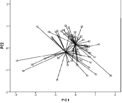
We found a significant difference between exhaled VOCs pattern in healthy subjects and patients with lung cancer when the previously standardised collection setup (50 ml/sec, no breath hold and the exclusion of dead space) was used (p = 0.02, Figure 2).
11 Figure 2.
Two dimensional PCA plot between healthy subjects (squares) and patients with lung cancer (circles).
4.2. Effect of sleep on exhaled volatile compounds pattern
Exhaled volatile compound patterns were significantly different before and after night in the SDB group (N=26, PC1, p<0.01). However, the morning and evening breath patterns could be discriminated only in patients with OSA (N=17,
PC1, p=0.01, Fig. 3) but not in the non-OSA subgroup (N= 9, p=0.49). In control volunteers,
there was no change in exhaled volatile pattern after night (p=0.23).
12
Figure 3.
Evening and morning differences in exhaled volatile compound pattern in OSA
The morning breath pattern significantly differed in the SDB group compared with control subjects (p<0.001, Figure 4A). The morning breath pattern was also different in the OSA subgroup versus control volunteers (p<0.001). The electronic nose could also discriminate morning exhaled pattern in the non-OSA SDB subgroup from that of control subjects (p<0.001, Figure 4B).
Figure 4. A, B
Comparison of exhaled volatile compound patterns between healthy (squares) and SDB (A: circles; B:
OSA-open circles, non-OSA-filled circles), patients in the morning.
13
4.3. Effect of sleep on lung volumes in OSA
Evening FVC (% predicted) was associated significantly with AHI (p = 0.04, r = -0.31; Figure 5.), ODI (p = 0.04, r = -0.31) and RDI (p\0.01, r = -0.39).
Figure 5.
Relationship between forced vital capacity (FVC) and apnoea–hypopnea index in OSA
In OSA, there was a significant increase in FEV1 from the evening to the morning (p = 0.02), while no difference in FVC (p = 0.19) or FEV1/FVC (p = 0.78) was noted. Morning and evening FEV1 (p = 0.74), FVC (p = 0.97) or FEV1/FVC (p = 0.54) were not different in the control group.
There was a tendency for a positive correlation between overnight increase of FEV1 and RDI (p = 0.05, r = 0.30).
14
5. Conclusions
5.1. Effects of methodological factors
• the expiratory flow rate, time of breath hold and the exclusion of air originated from the anatomic dead space can may significantly influence the exhaled breath volatile compounds pattern.
• The methodological factors of sample collection also influence the classification accuracy of electronic nose to separate disorders from health.
5.2. Effect of sleep on exhaled volatile compounds pattern
• the exhaled volatile compounds pattern changes during sleep in obstructive sleep apnoea patients which may refers to an alteration of airway inflammation.
• Exhaled breath analysis with electronic nose should only be used to identify OSA if sample is taken during the hours after sleep.
5.3. Effect of sleep on lung volumes in OSA
• FEV1 increases significantly from evening to morning in OSA patients
• the rate of FEV1 increase is in a tendentious relationship with the severity of the disorder (RDI)
• This change should be taken into consideration when timing and assessing spirometric results in – most importantly in case of severe disorder – sleep apnoeic patients
15
6. List of publications
6.1. Publications related to the theses
• Kunos L, Lazar Z, Martinovszky F, Tarnoki AD, Tarnoki DL, Kovacs D, Forgo B, Horvath P,
Losonczy G, Bikov A. (2017) Overnight Changes in Lung Function of Obese Patients with Obstructive Sleep Apnoea. LUNG 195(1): 127-133.
Impact factor: 1,915 (2016)
• Kunos L, Bikov A, Lazar Z, Korosi BZ, Benedek P, Losonczy G, Horvath I. (2015) Evening and morning exhaled volatile compound patterns are different in obstructive sleep apnoea assessed with electronic nose. SLEEP AND BREATHING 19:(1) pp. 247-253.
Impact factor: 2,332 (2015)
• Bikov A, Hernadi M, Korosi BZ, Kunos L, Zsamboki G, Sutto Z, Tarnoki AD, Tarnoki DL, Losonczy G, Horvath I. (2014) Expiratory flow rate, breath hold and anatomic dead space influence electronic nose ability to detect lung cancer. BMC PULMONARY MEDICINE 14:(1) 202. 9 p.
Impact factor: 2,404 (2014)
6.2. Publications not related to the theses
• Horvath P., Tarnoki D., Tarnoki A., Karlinger K., Lazar Zs., Losonczy G., Kunos L., Bikov A. (2018) Complement system activation in obstructive sleep apnoea. JOURNAL OF SLEEP RESEARCH. e12674.
Impact factor: 3,259 (2017)
• Bikov A., Losonczy G., Kunos L. (2017) Role of lung volume and airway inflammation in obstructive sleep
16
apnoea. RESPIRATORY INVESTIGATION 55:326- 333.
• Bikov A., Kunos L., Pállinger É, Lázár Zs, Kis A, Horváth G, Losonczy G, Komlósi ZI. (2017) Diurnal variation of circulating microvesicles is associated with the severity of obstructive sleep apnoea. SLEEP AND BREATHING 21(3):595-600.
Impact factor: 2,288 (2016)
• Bikov A, Hull JH, Kunos L. (2016) Exhaled breath analysis, a simple tool to study the pathophysiology of obstructive sleep apnoea. SLEEP MEDICINE
REVIEWS 27: pp. 1-8.
Impact factor: 8,958 (2016)
• Pako J, Bikov A, Karlocai K, Csosza G, Kunos L, Losonczy G, Horvath I. (2015) Plasma VEGF levels and their relation to right ventricular function in pulmonary hypertension. CLINICAL AND
EXPERIMENTAL HYPERTENSION 37:(4) pp. 340- 344.
Impact factor: 1,307 (2015)
• Benedek P, Lazar Z, Bikov A, Kunos L, Katona G, Horvath I. (2013) Exhaled biomarker pattern is altered in children with obstructive sleep apnoea syndrome.
INTERNATIONAL JOURNAL OF PEDIATRIC OTORHINOLARYNGOLOGY 77:(8) pp. 1244-1247.
Impact factor: 1,319 (2013)
• Kunos L, Kovats Z, Murakozy G, Sutto Z, Bohacs A, Czebe K, Lang G, Renyi-Vamos F, Klepetko W, Muller V. (2011) Severe mixed sleep apnoea after bilateral lung transplantation in a cystic fibrosis patient: a case report. TRANSPLANTATION PROCEEDINGS 43:(4) pp. 1292-1293.
Impact factor: 1,005 (2011)
17
• Tölgyesi G, Molnar V, Semsei AF, Kiszel P, Ungvari I, Pocza P, Wiener Z, Komlosi ZI, Kunos L, Galffy G, Losonczy G, Seres I, Falus A, Szalai CS. (2009) Gene expression profiling of experimental asztma reveals a possible role of paraoxonase-1 in the disease INTERNATIONAL IMMUNOLOGY 21:(8) pp. 967- 975.
Impact factor: 3.403 (2009)
• Kunos L, Varga J, Horváth G. (2012) Krónikus obstruktív tüdőbetegség és obstruktív alvási apnoe szindróma együttes előfordulása: az "overlap"
szindróma ORVOSI HETILAP 153:(23) pp. 892-897.
• Gyulai N, Somoskövi Á, Kunos L, Bártfai Z. (2008) Multidrog-rezisztens és extenzíven drog-rezisztens Mycobacterium tuberculosis epidemiológiája és kontrollja: I. rész MAGYAR CSALÁDORVOSOK LAPJA 2008:(Április) pp. 32-35.
• Gyulai N, Somoskövi Á, Kunos L, Bártfai Z. (2008) Multidrog-rezisztens és extenzíven drog-rezisztens Mycobacterium tuberculosis epidemiológiája és kontrollja: II. rész MAGYAR CSALÁDORVOSOK LAPJA 2008:(Május) pp. 19-22.
• Kunos L, Horváth G, Losonczy G. (2008) Az orális neurokinin-1 (NK1) receptor antagonista aprepitant szerepe az erősen emetogén kemoterápia okozta hányás és hányinger megelőzésében MEDICINA THORACALIS (BUDAPEST) 61:(6) pp. 333-338.
• Kunos L, Várdy Visi K, Bártfai Z, Losonczy G.
(2008) Az obstruktív alvási apnoe MAGYAR
CSALÁDORVOSOK LAPJA 2008:(Március) pp. 10- 16.
• Kunos L (2008) Az alvás alatti légzészavarok közlekedésbiztonsági vonatkozásai MEDICINA THORACALIS (BUDAPEST) 61:(5) pp. 265-268.
18
Kunos L. (2013) Az acetilcisztein és más mukolitikumok alkalmazási lehetőségei
GYERMEKGYÓGYÁSZAT 2013; 64. (6) pp. 308- 312.
• Kunos L, Losonczy Gy Alvási apnoe-hipopnoe
szindrómák In: Magyar Pál, Losonczy György (szerk.) A pulmonológia kézikönyve. Medicina, Budapest, 2012.: pp. 763-776. (ISBN:978 963 226 340 3)
• Kunos L. Alvás alatti légzészavarok. In: Tulassay Zsolt (szerk.). A belgyógyászat alapjai. Medicina, Budapest, 2016.: pp. 758-762. (ISBN:978 963 226 554 4).
• Losonczy G., Kunos L. Obezitás és légzőszervi megbetegedések. In: Bedros J Róbert (szerk.) Klinikai obezitológia. Semmelweis Kiadó, Budapest, 2017.: pp 381-402. (ISBN:978 963 331 343 5)
• Kunos L. Alvás alatti légzészavarok vizsgálata. In:
Tulassay Zsolt (szerk.) Klinikai belgyógyászat.
Medicina, Budapest, 2017.: pp 214-215.
• Kunos L. Elzáródásos (obstruktív) alvás apnoe- hipopnoe szindróma. In: Tulassay Zsolt (szerk.) Klinikai belgyógyászat. Medicina, Budapest, 2017.: 233-234.