S. A. V. SWANSON
Mechanical Engineering Department, Imperial College, London, England
I. Introduction . . . . . A. Necessary mechanical properties of bone B. Development of the literature
C. Objects of this paper D. Definitions and units I I . Micro-structure of Bone
A. Major constituents . B. Types of bone
C. Typical adult compact bone I I I . Experimental Conditions
A. Mechanical factors . B. Biological factors
C. Comparison of numerical results
IV. Experimental Results on Small Specimens of Compact Bone A. Selection of results of mechanical tests
B. Effects of frozen storage . C. Effects of storage in saline D. Effects of fixation or embalming E. Effects of drying
F . Effects of testing temperature G. Variations within one bone H . Variations between bones I. Variations with sex J . Variations with age K. Directional variations
L. Relationship between properties in different loading modes M. Creep and stress relaxation
N. Fatigue properties O. I m p a c t strength
P . Dependence on micro-structure
V. Properties derived from Tests on Whole Bones VI. Non-destructive Tests . . . . V I I . Summary and Discussion of Results
A. Summary of tensile properties . B. Significance for the function of bones C. Comparison with other materials D. Compact bone as a three-phase material E. Models
Acknowledgments . . . . .
References . . . . . .
137
138 S. A. V. SWANSON I. INTRODUCTION
A. Necessary Mechanical Properties of Bone
T H E bones of the human skeleton have several functions, not all of which are mechanical; to perform their mechanical functions, they must possess strength and stiffness as structures, which requires t h a t they be composed of a material which itself possesses strength and stiffness.
This is, of course, a common requirement even in components of inani
mate structures, as is the further requirement that, in order to achieve an efficiently working device (in this instance the body), the shapes of some bones should be determined by factors not all of which are mechanical.
The need for some strength, to support static loads, is obvious. For a long time, bone has been known to be weaker in tension t h a n in com
pression, and brittle lacquer studies such as those reported by Evans (1957) have shown t h a t many fractures of whole bones originate in tensile failure of the material. Both tensile and compressive strengths have been measured by many workers.
Some stiffness is also obviously needed, if adjacent structures are to be properly located. Perhaps a more critical consideration is t h a t the energy-absorbing capacity of any material is a function of its strength and, inversely, of its stiffness; because bones are likely to be subjected to some sudden loads, their energy-absorbing capacity may be impor
tant.
B. Development of the Literature
Anatomists, physiologists and orthopaedic surgeons have for a long time had obvious reasons for being interested in the mechanical properties of bones (the organs) or of bone (the tissue). More recently, engineers interested in the properties of materials have found bone worthy of study. These factors have led to the existence of a large and scattered literature which, up to the mid-nineteen-fifties, was compre
hensively surveyed by Evans (1957). By t h a t date the ranges of values of the principal mechanical properties were established; the considerable volume of work which has appeared since then has been concerned mainly to relate the variations in these properties to the factors, both biological and mechanical, which might be expected to influence them.
C. Objects of this Paper
This paper is concerned with bone (the material or tissue) and not with bones (the structures or organs). I t is concerned with the material
as it is found in the adult human skeleton, and principally as it is found in the bones of the limbs, because bone from this source has been the subject of most experiments. Other types of bone will be considered where they help the understanding of this type.
The paper will:
(a) summarize the present state of knowledge concerning those aspects of the micro-structure of bone which are likely to affect its mechanical properties;
(b) outline the major difficulties attending the production of accurate and meaningful experimental results (which difficulties seem not to have been equally appreciated by all experimenters);
(c) survey the more important and reliable published experimental results, particularly those which have appeared since Evans
(1957) including those produced by Professor Yamada's group in Kyoto, which have appeared in English only in summary form in Yamada and Evans (1970); and
(d) discuss these results in relation to the micro-structure of the material.
Some part of this paper may seem elementary to a reader with a biological background, and others to one with a physical or engineering background; this seems to be an inevitable difficulty in such a subject as this.
D. Definitions and Units
Force is t h a t interaction between bodies which tends to deform or accelerate them.
Stress is force per unit area; a tensile or compressive stress corresponds to a force acting perpendicularly to the area considered, while a shear stress corresponds to a force acting in the plane of the area considered.
Strain is relative displacement per unit length; tensile, compressive and shear strain correspond to tensile, compressive and shear stress.
Tensile strain is extension per unit length, i.e. the increase in length of a line as a fraction of its original length. Compressive strain is the decrease in length as a fraction of the original length. Shear strain is the relative movement of any two points perpendicular to the line joining them, as a fraction of the length of t h a t line.
A Stress-Strain Curve for a material shows the variation of stress with strain (tensile, compressive or shear) from no load to any desired stress
140 S. A. V. SWANSON
or strain (often to fracture). Such a curve commonly shows the following features.
The Elastic Region, within which removal of stress is accompanied by disappearance of strain. In this region the stress-strain curve is usually, but not necessarily, linear.
The Elastic Modulus is the gradient (stress/strain) of this linear part of the curve. In tension or compression it is called Young's Modulus.
When a stress-strain curve is not linear, the tangent modulus or the secant modulus at any point can be specified.
The Limit of Proportionality is the point, at which the stress-strain curve ceases to be linear.
The Yield Stress is the highest stress at which the material is elastic.
For practical purposes, in materials showing linearly elastic behaviour, it is the stress at the Limit of Proportionality.
The Plastic Region extends from the yield stress to fracture. Within this region, removal of stress is accompanied by disappearance of only part of the corresponding strain; the remainder is the plastic strain.
The Ultimate Stress (tensile, compressive or shear) is, in a relatively brittle material such as bone, the stress corresponding to the load at fracture.
Strain Energy is the energy stored in an elastic body when it is distorted by stress. The area between an elastic stress-strain curve and the strain axis represents strain energy per unit volume of the material.
In this paper, the Systeme Internationale (S.I.) of units will be used.
Forces will be expressed in Newtons, abbreviated to N. Lengths will be expressed in millimetres (mm), microns (10~6 metre, μπι), or Äng
stroms (10~1 0 metre, Ä). Stresses will be expressed in Newtons per square metre (N/m2) or in multiples such as kilonewtons or mega- newtons per square metre (kN/m2 orMN/m2, 103 and 106 N/m2 respect
ively.) Moduli of elasticity (Young's Modulus in tension or compression, Modulus of Rigidity or Shear Modulus in shear), being defined as stress/strain, has the same dimensions as stress, and will be expressed in MN/m2 or GN/m2 (Giganewtons per square metre, 109 N/m2).
Strains are dimensionless and will be expressed as ratios (not percent
ages). Temperatures will be expressed in degrees Celsius (or Centigrade);
°C.
Results quoted from other publications have been converted, where necessary, to the units listed above.
In time, it is likely t h a t all technical publications will use the S.I.,
but so long as information expressed in other systems is current, conversions such as the following will be useful.
1 MN/m2 = 0-102 kg force/mm2 (kilopond/mm2)
= 145 lb force/in2 1 GN/m2 = 145 000 lb force/in2
I I . MICRO-STRUCTURE OF B O N E
A. Major Constituents
Qualitatively, bone may be regarded as composed of organic sub
stances, inorganic substances and water.
Most of the organic component is collagen in the form of fibre bundles (different authors use the terms "fibres" and "fibre bundles", appar
ently for the same elements); Robinson (1952) gave the proportion as about 84% by weight, while Engfeldt and Strandh (1960) said "more than 9 0 % " , and Robinson (1960) gave an average of 96%. The re
mainder of the organic component is known as "cement substance"
and consists mainly of mucopolysaccharides.
Some of the inorganic constituent is present as ions in the cement substance, but most of it is present as crystals intimately associated with the collagen fibres, or as amorphous calcium phosphate. The commonest experimental method of estimating the inorganic content of bone is by ashing, and this cannot distinguish between the different components of the inorganic constituent.
Until a few years ago, the mineral phase was generally believed to be almost entirely crystalline, but recent work has shown this not to be so.
Using X-ray diffraction methods, Termine and Posner (1967) stated that, in femora from 17-day-old rats, 5 7 % of the mineral was amor
phous, falling to 3 6 % at 80 days old. Harper and Posner (1966) gave similar figures for bovine and rat bone, and stated (without giving numerical results) t h a t human bone also contained amorphous calcium phosphate. Neither pair of authors offered any evidence concerning the spatial relationship of the amorphous mineral to the rest of the micro- structure. Harper and Posner remarked t h a t the amorphous phase tends to become unstable and to be converted to a crystalline form after death. If this is so, not only is the deduction of the micro-structure in life made more difficult, if not impossible, but all mechanical tests on dead bone (i.e. virtually all mechanical tests on bone) are open to an
142 S. A. V. SWANSON
objection in principle. Posner (1969) has discussed the chemistry of bone mineral in some detail.
There is general agreement t h a t the crystals are of the apatite family, but their precise structure is still a matter of discussion. Robinson (1952) said t h a t they were hydroxyapatite, which has a composition of Cä1 0(PO4)6(OH)2. Engström (1960) suggested, on the basis of polarized light and electron microscopy studies, t h a t the crystals were hydroxy- apatite with amorphous calcium carbonate absorbed into it. Hayek (1966) synthesized a crystalline substance which X-ray and infra-red analysis showed to be very similar to the mineral constituent of bone, and in which the calcium carbonate was included in the crystal lattice in a homogeneous way. The approximate formula was given as Ca4(P02)2(HP04)o.4(C03)o_6. Robinson (1960) pointed out t h a t the composition of the crystals varies with time because ions can be ex
changed at the crystal surfaces; this is a consequence of the fact t h a t bone acts as a reservoir for various minerals.
Water is present mainly in association with the collagen and the cement substance.
B. Types of Bone
The three groups of constituents just mentioned are arranged differ
ently in different types of bone. The bone present in the adult human skeleton can be classified into a few fairly distinct types, although a wider variety is found in the human embryo and infant, and in other species of vertebrates. Only those types commonly found in adult human bones will be considered here.
1. Mature cancellous bone
I n this form, which is found in many in the vertebral bodies and in the enlarged ends of the long bones of the limbs, the bone is arranged in a three-dimensional lattice, often in the form of series of columns in regular patterns, with frequent cross-connections. These columns have typical thicknesses of about 0-5 m m ; the spaces between them are similar in size, and contain marrow, as well as providing paths for blood vessels. Some cancellous bone presents the appearance of perforated plates rather than of interconnected columns.
2. Compact bone
Lamellar. This consists of identifiable layers, the collagen fibres in each layer having a dominant direction which is different from those in adja
cent layers. Weinmann and Sicher (1955) give the thickness of lamellae as from 4 to 12 μιη.
Woven. In this form the collagen fibres are in a three-dimensional net
work instead of being confined within lamellae.
Haversian. This is the type which is laid down by the reconstructive process which is continuously in operation from before skeletal maturity until death; it is therefore the type found most commonly in the bones of adult human limbs, and will be considered in more detail below.
C. Typical Adult Compact Bone 1. General arrangement
If a cross-section is made of the shaft of a long bone, approximately the outermost 10% of the thickness of the cortex consists of roughly circumferential lamellae; the inside (endosteal) surface may tend to be cancellous, or may, like the periosteal surface, consist of roughly circumferential lamellae. Most of the thickness consists of secondary osteones (Haversian systems; hence the term "Haversian bone") and the interstitial bone between them. In three dimensions at the micro
scopic level of size, bone presents a rather untidy arrangement of branch
ing and irregularly curving secondary osteones, surrounded by the remnants of older osteones and lamellar bone, and perforated by holes containing blood vessels of a wide range of sizes.
2. Secondary osteones of Haversian systems
General arrangement and Dimensions. Each secondary osteone is roughly circular in cross-section; when non-circular, the major diameter is rarely more than twice the minor diameter. The outer diameter is usually about 0-1-0-2 mm, or occasionally larger, up to about 0-5 m m ; in the centre is a hole (Haversian canal) of about 0-02-0-1 mm diameter, and the bulk of the osteone consists of concentric lamellae of bone which have been deposited working inwards from the outermost one. One osteone contains in its cross-section typically 20 to 30 lamellae, or sometimes fewer. Scattered at various radii within the osteone are the lacunae which are occupied by the osteocytes; minute channels (canaliculi) connect these lacunae.
A secondary osteone is the result of a revascularization process, in which a path through the existing bone is first excavated to a roughly circular cross-section and then lined with the concentric lamellae, which are partly mineralized when laid down and become increasingly mineralized with increasing time (Jowsey, 1960; Smith, 1963). The life of any one osteone, from initial excavation to complete mineralization, may be many years, so a cross-section of a bone will contain cross-
144 S. A. V. SWANSON
sections of osteones of various ages, perhaps some in process of forma
tion, and highly mineralized interstitial bone between the osteones.
Because the path of a newly forming osteone is irregular (although roughly parallel to the long axis of the entire bone), the interstitial bone may contain remnants of older osteones which have been partly excavated and replaced, and also remnants of formerly circumferential lamellae which have been incorporated into the cortex as a consequence of changes in size and shape of the bone shaft.
The boundary of each osteone, called the cement line or reversal line, is a thin layer (thinner than typical lamellae) which contains no collagen; Smith (1963) refers to it as highly mineralized.
Directions of collagen fibres. Within each lamella of the osteone the collagen fibres have one dominant direction; this direction varies from one lamella to the next, giving the effect of a family of co-axial helices of different helix angles. Weinmann and Sicher (1955) state t h a t the fibres are longitudinal and circumferential in alternate lamellae (i.e.
t h a t the helix angle is alternately 90° and zero). Bloom and Fawcett (1962) say t h a t this arrangement is rare, and t h a t more frequently all the helices are at various angles, sometimes perpendicular to those in adjacent lamellae. Smith (1960) distinguished three types of arrange
ment, all of which involved longitudinal and circumferential fibres in alternate lamellae; the different types showed different relative densities of longitudinal and circumferential fibres. Ascenzi et al. (1966) describe two arrangements: one with fibres having helical paths with the helix angle changing so little t h a t fibres in adjacent lamellae were nearly parallel to each other (which they say is very infrequent in bone of old subjects), and the second with fibres in adjacent lamellae changing direction through 90°. Later observations by the same workers (Ascenzi and Bonucci, 1968), describe three arrangements: one in which the fibres in all lamellae are roughly parallel to the axis of the osteone (i.e.
the helix angle is approximately 90°); one in which fibres in alternate lamellae are longitudinal and circumferential (i.e. the helix angle alternates between approximately 90° and approximately zero); and one in which the fibres in all lamellae are inclined, but in opposite directions in successive lamellae (i.e. the helix angle alternates between plus and minus approximately 45°).
Pritchard (1956) states t h a t the dominant fibre direction changes at successive levels within a given lamella, and t h a t fibre bundles regularly leave one lamella and pass into the next. Rouiller (1956) says t h a t all lamellar systems are composed of two different types of lamellae, regularly alternating; one type is rich in collagen, while the other con-
tains more inorganic salts and cement substance, but is penetrated by collagen fibrils connecting the adjacent collagen-rich lamellae.
This last observation of Rouiller may be compared with the electron microscope findings of Ascenzi et al. (1965), who observed t h a t the inter- lamellar cementing zones were composed of irregularly oriented collagen fibres, more highly calcified than the lamellae.
In comparing these observations, some of which are apparently inconsistent, it should be remembered t h a t some of them were made on small numbers of samples of bone. If anybody had the time to make really large numbers of such observations, it seems likely t h a t all the above-mentioned patterns, and perhaps others, would be found.
Dimensions of basic components. Robinson (1952) gives the thickness of the collagen fibres as 500-1200 Ä, and says t h a t bone collagen presents the same appearance in the electron microscope as collagen from other sources. Bloom and Fawcett (1962) give the thickness of the bundles of collagen fibrils as about 3-5 μιη. Robinson (1952) gives the average size of the hydroxyapatite crystals as about 500 x 250 x 100 Ä. Molnar (1960) summarizes various workers' observations, and postulates t h a t the crystals have a well-defined width ranging from 25 to 75 Ä, with an average of 50 Ä, and are composed of chains of microcrystals end-to-end, having a minimum length of about 50 Ä and no upper limit on their length. Ascenzi et al. (1965) state t h a t the crystallites become long enough to span two or more major periods, but Glimcher (1968), also using an electron microscope, states t h a t bone crystals have a maximum length of certainly less than the periodic distance of col
lagen, and in fact about 400 Ä, with a width of 10-50 Ä.
There is perhaps scope for further co-ordinated observations, coupled with a clarification for the non-specialist of the exact meaning of such terms as " c r y s t a l s " and "chains of microcrystals".
3. Variations with age
As mentioned above, Jowsey (1960) and Smith (1963) agree that, as any one secondary osteone ages, it becomes increasingly mineralized.
Jowsey mentions less than 7 5 % mineralization (compared with t h a t of the surrounding interstitial bone) as a low density. Robinson (1960), quotes "definite evidence" that, as mineralization of the bone matrix occurs, the apatite crystals displace the water and not the organic solids. Smith (1963), however, presents evidence which suggests t h a t the progressive calcification occurs, to some extent, at the expense of the organic fraction.
As an individual progresses from skeletal maturity to old age, several changes occur in the compact bone of the shafts of long bones.
146 S. A. V. SWANSON
I t is commonly accepted t h a t the cortical thickness tends to decrease, mainly by increase of the internal diameter, although Smith and Walker (1964) found t h a t in the femora of 2030 women both the internal and the external diameters increased, with little resultant change in the cortical thickness.
In the zone initially occupied by a mixture of secondary osteones and lamellar bone, secondary osteones or their remnants occupy a larger proportion of the volume. Currey (1964b) found that, as ageing pro
gressed, intact Haversian systems became smaller and more numerous, and occupied a decreasing proportion of the cross-sectional area.
Observations by Jowsey (1960) show t h a t secondary osteones tend to invade the outer circumferential lamellae.
Jowsey showed also an increasing proportion of secondary osteones having large central canals and low relative mineralization; near the endosteal surface, the canals tended to be larger than typical osteones elsewhere, and to merge with the medullary cavity. Comparable observations were reported by Atkinson (1965).
Urist (1964) found t h a t some old secondary osteones had their canals and lacunae filled with inorganic material; this of course implies the death of the associated osteocytes.
Chatterji and Jeffery (1968), using a scanning electron microscope, found t h a t the size of the apatite crystals apparently increased with increasing age; this may be compared with Termine and Posner's finding t h a t the proportion of crystalline apatite increased with age.
4. Differences from other mammalian bone
Adult mammals of some other species, e.g. oxen and dogs, have bones of which the micro-structure is similar in kind to t h a t described above, although the relative volumes of circumferential lamellae and osteones may differ. Other species have more widely differing micro-structure;
the major types are described by Enlow (1966), but with little correla
tion between species and types of bone.
I I I . EXPERIMENTAL CONDITIONS
The conditions which must be controlled, or at least known, for a meaningful result to be obtained from an experiment can be divided into mechanical and biological factors, though the two groups overlap.
In general, the mechanical factors involved in a test must be known before the possible margin of error surrounding any numerical result can be assessed, whereas the biological factors must be known in order
to attempt to correlate the mechanical properties with the state of the specimens on which they were measured.
A. Mechanical Factors 1. All tests
Accuracy of measuring load, etc. The accurate measurement of the load exerted by a testing device (the one feature which is commonly men
tioned by authors) is the least of the problems relating to the accuracy of numerical results. Testing machines which can measure loads to within ± 1 % are common, and ± 0 - 5 % is an attainable accuracy.
Similarly, errors in the dimensions of test specimens can be made negligibly small by the use of appropriate instruments, and the accurate measurement of displacements so small as those corresponding to tensile strains in bone (about 0-03 mm extension per mm length at the elastic limit) need be no problem; the problem is to guarantee t h a t the displacement measured is t h a t of the desired section of bone.
Rate or duration of loading. This should be known because the presence of organic material and water suggests t h a t the mechanical properties may be time-dependent.
Temperature and humidity. The need to control both these is obvious, though probably the only temperature range which need be considered is from 37°C down to a typical laboratory temperature of 20°C, provided of course, t h a t precautions are taken to ensure t h a t specimens are not heated or dried sufficiently to cause irreversible changes while being machined to size.
Surface finish of specimens. In the testing of metals, particularly brittle metals, it is common for fracture to start at some minor surface defect which acts as a stress concentrator. Bone is already full of small holes and discontinuities of structure; Currey (1962) has argued t h a t many of these are so aligned as to minimize their stress concentrating effects, and Bonfield and Li (1966) found t h a t bone was notch-sensitive in impact tests, which suggests t h a t experimental results could be affected by the presence of machining marks on the specimens.
2. Tensile tests on small specimens
Axiality of load. If the load is not applied exactly along the axis of the specimen, bending stresses will be present in the specimen in addition to the intended uniform tensile stress. In a cylindrical specimen, a loading eccentricity of 1 % of the specimen diameter will give a maxi
mum stress of 8% higher than the intended uniform stress while the
148 S. A. V. SWANSON
specimen is elastic. If an averaging extensometer is used (see below), this need not cause any error in the value of Young's Modulus, and it would cause little if any error in the indicated ultimate strength of a material which deformed plastically before breaking (because the act of plastic yielding would reduce the misalignment); but an error would be more likely with a more brittle material. A misalignment of so little as 1 % of the specimen diameter is probably the best t h a t could be hoped for in the present context.
Extensometry. Because the existence of some eccentricity of loading means t h a t the stresses, and therefore the strains, are likely to be differ
ent on different sides of the specimen, it is important t h a t strains be measured on at least two sides and averaged, if a value for Young's Modulus is to be derived. For this reason, many extensometers consist in effect of two similar instruments, mounted opposite each other on the specimen, with provision for averaging their two signals. In contrast, to bond a strain gauge to one surface only of a specimen is a doubtful procedure which requires verification before the results can be accepted without question.
Measurement of the relative movement of the grips can be an accurate alternative to measuring the extension of a known length of the specimen only if it is known t h a t the grips themselves do not deflect under load, and t h a t the specimen does not move relative to the grips.
Stress concentration at grips. Particularly when testing a brittle material, the stress concentrations resulting from gripping the ends of the speci
men are likely to cause fracture in, or close to, the grips at an artificially low load. This trouble is usually avoided by using specimens having a reduced central section.
3. Compressive tests on small specimens
Axiality and extensometry. These problems are the same as for tensile specimens.
Buckling and friction. Unless special precautions are taken, the friction between the platens and the ends of the specimen will restrain the lateral expansion of the specimen to an extent which decreases towards the centre of the specimen. Thus neither the strain nor the stress system to which the specimen is subjected will be uniform. This effect is minimized by using a long, slender specimen; but such a specimen would buckle before reaching its failing stress, and the lateral supports which can be used to prevent buckling introduce the possibility of errors due to friction.
4. Bending tests on small specimens
Design of load application points. Clearly, a specimen bent by loads applied through knife-edges would be expected to fail prematurely at the loading points. If, to prevent this, these are made with a large radius, then, if the specimen bends appreciably, the effective point of contact will move, and the moment arm at which the loads are applied will have changed in length. A further difficulty arises if the loading points are not all coplanar, e.g. if, in a three-point bending test with the loads acting vertically, the central loading point is not in the same hori
zontal plane as the two end supports. Then the frictional forces at the loading points will exert on the specimen a moment which, together with the moment required to bend or break the specimen, will con
tribute to the load recorded.
Whether these errors are significant in any particular experiment can be estimated only with a detailed knowledge of the apparatus, which can virtually never be extracted from published papers.
Stress-strain curve for the material. In a bending test, the stress to which the material is subjected is not uniform. The maximum stress (at the extreme fibres) can be calculated if the stress-strain curve for the material is linear, and the same in compression as in tension. Otherwise, it cannot be calculated exactly. All workers who have used bending tests have calculated the stresses at fracture as if the stress-strain curve were linear, which is very nearly true for dry bone but not true for wet bone (see, for example, Evans and Lebow, 1951).
Non-uniform stress. When, as in a bending test, the stress varies from zero at the centre (of a symmetrical cross-section) to a maximum at the edge, the possibility exists t h a t a crack starting at the most highly stressed surface might be arrested in the less highly stressed material.
Whether this happens in fact depends on many factors, and therefore the significance of a bending test is inherently more problematical (from the point of view of the strength of the material) t h a t t h a t of a tensile test, in which a reasonable approximation to a uniform stress can be achieved. On the other hand, it can be argued t h a t a bending test is more relevant to the stresses applied to bones in life, which are more likely to result from bending them from tensile forces.
5. Shear tests on small specimens
This is an attractive form of test but, unless the specimen is restrained with extreme accuracy right up to the plane of shearing, it becomes also a bending test with an unknowable stress system acting.
150 S. A . V . S W A N S O N
6. Torsion tests on small specimens
End restraints on specimen. To apply pure torsion to the ends of the specimen, with no bending moment, can be difficult. What can be more difficult, unless the apparatus is suitably designed, is to know whether or not such unwanted bending moments have been applied.
Stress-strain curve and non-uniform stress. These present the same problems in torsion tests as were discussed above in relation to bending tests.
7. Tests on entire bones
Clearly, it is desirable to know the breaking load of, e.g. a femur in various modes, and for this purpose tests, usually in bending or torsion, are performed on entire bones. But such tests are used also as a basis from which to infer some properties of the material, and, when this is done, the relevant sources of possible errors discussed above must be considered. In addition, the irregular cross-section of the shaft of the bone introduces new problems of measurement and calculation, and of course any mechanical properties which are derived can only be some sort of average properties for the material of the particular bone, which is probably not homogeneous in any respect over the whole of the bone.
B. Biological Factors
These can be divided approximately into two groups: external factors which can be controlled, and which affect the state of the specimen, and internal factors which, if observed, describe aspects of the structure and composition of the specimen.
1. External factors
Whether a specimen was tested fresh, after frozen storage, after fixation or embalming, wet, dry (or dried and re-wetted) may be significant and should be known. Almost all authors do record this type of information.
2. Internal factors
Species. The species of origin of the bone appears to be important.
Virtually all bone on which published results have been obtained has been mammalian; certain mammals such as bovines are attractive in t h a t fresh bones can be easily obtained and have cortices thick enough to allow the extraction of specimens in various directions without the specimens being inconveniently small. Such bone is largely composed of
secondary osteones, as is adult human bone, and Ascenzi et al. (1966) found t h a t the tensile properties of a portion of a single ox osteone were similar to those measured in the same way on human bone.
Composition and micro-structure. The age and sex of the source of the bone are almost always recorded, but these can give, at the best, only indirect information about the composition or micro-structure of the specimen.
Clearly, it would be unreasonable to expect a full analysis of every specimen, but some measurement of the relative proportions of the three groups of constituents can be made by drying and ashing. This aspect of the subject has itself acquired a considerable literature (see, for example, Robinson and Elliott, 1957 and Mueller et al., 1966): but drying and ashing at appropriate temperatures can enable the weights of water and inorganic material to be obtained, leaving the weight of organic material to be found by subtraction.
Measurements of physical density are difficult to interpret, because in a material containing so many holes of such a range of sizes it is not easy to specify exactly which volume is having its density measured.
Radiography can give values for relative average densities of inorganic material, and thus some measure of the degree of mineralization of a specimen. Vose and Kubala (1959) found a linear correlation between ash content and the logarithm of the transmittance to X-rays. These results were obtained on specimens which had been dried, pulverized and compressed into standard briquettes.
Microradiography can show variations in mineralization both be
tween different osteones and within any one osteone, and is therefore more valuable t h a n ordinary large-scale radiography.
Without going to the extra complexities of electron microscopy, the directions of collagen fibres can be inferred from light micrographs using polarized light, and this technique has been used by several authors.
Death. All the specimens from which any results so far published have been obtained have been deprived of their normal blood supply, and most of them have been so treated as to ensure (incidentally) t h a t the osteocytes would be dead, even where the specimens have not been deliberately fixed. I t is conceivable t h a t a specimen might be extracted and tested in a time short enough to permit some osteocytes to survive;
but in general the question is presented: does death alter the mechanical properties of bone ? Presumably a tensile test in vivo could be devised, and followed by a corresponding test after death, but the author is aware of no results from any such test. There is no reason to suppose t h a t
6 + A.B.E.
1 5 2 S. A. V. SWANSON
the fact of death (as distinct from subsequent storage or treatment) would change the mechanical properties of collagen, of the cement substance or of hydroxyapatite, but the statement, mentioned above, of Harper and Posner (1966) t h a t the amorphous mineral phase tends to change to a crystalline form after death poses a question which at present cannot be answered. Apart from this, the necessary condition for death to affect the mechanical properties of bone would be t h a t the osteocytes or their processes should contribute significantly to these properties. Although Ascenzi et al. (1966) properly remark t h a t their tensile tests on portions of single osteones furnish no evidence to exclude the possibility t h a t the osteocytes and their processes have mechanical importance, this is no reason why other evidence should not be exam
ined. Many observations, which are summarized in standard textbooks (e.g. Weinmann and Sicher, 1955) show t h a t the osteocytes within any one osteone are in cytoplasmic continuity with each other through their processes which pass through canaliculi in the bone, and so the possi
bility exists t h a t they form a load-bearing network. But to support any significant share of an applied load, this network would have to have stiffness of the same order of magnitude as t h a t of the bone. Since the area of bone is many orders of magnitude greater than t h a t of the osteocyte processes, this would require Young's Modulus of the processes to be orders of magnitude greater than t h a t of the bone, which is hardly conceivable. Thus it seems safe to conclude t h a t any mechanical significance of the osteocytes or their processes is indirect rather than direct.
C. Comj)arison of Numerical Results 1. Strengths
Given careful measuring techniques and appropriately calibrated apparatus, values of ultimate tensile stress obtained from tensile tests ought to be accurate to within ± 1% or ± 2 % ; values derived from bending tests are potentially subject to considerably higher errors 2. Moduli of elasticity
Taking account of the difficulties of extensometry mentioned above, values of Young's Modulus in tension are likely to be surrounded by larger errors than the corresponding values of strength. Even when testing specimens of convenient size, in homogeneous metals, with no complications arising from variations in temperature or humidity, the author's experience is that, unless great care is taken, values of Young's Modulus cannot be guaranteed to closer than ± 2 - 5 % . The only paper
known to the author which contains a specific estimate of the accuracy of values for Young's Modulus of bone is t h a t by Ascenzi et dl. (1966) who state t h a t the absolute accuracy of extension measurements was 5%, with measurements on any one specimen consistent to within 1%
(which was also the best accuracy obtainable in measurements of cross- sectional dimensions). These are the only workers to state t h a t they checked the overall accuracy of their apparatus by testing a material of known Young's Modulus and comparing their result with the est
ablished one. In the absence of such external verifications, values of Young's Modulus obtained by different workers on different apparatus can be compared only with great caution.
Similar considerations apply to comparisons of Young's Modulus derived from bending experiments, or of the Modulus of Rigidity derived from torsion tests.
IV. EXPERIMENTAL R E S U L T S ON SMALL SPECIMENS OF COMPACT B O N E
A. Selection of Results of Mechanical Tests
The tensile test is probably the most useful for examining the effective strength of the material, and therefore tensile results have been preferred when they are available. Several workers have used bending tests which can, if the difficulties discussed above are properly dealt with, give accurate values for Young's Modulus. All authors deriving fracture stress values from bending tests have done so by assuming a linear stress distribution, and their results therefore do not represent the absolute strength of the material, although they may validly show the effects of different treatments on similar specimens. Bending test results have been used with this reservation.
Only those results have been used which were accompanied by sufficient description of the apparatus and methods used to permit a proper assessment of their accuracy.
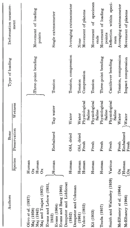
Table 1 summarizes the principal features of the techniques used by the authors whose work has been considered. Where a paper described a main series and supplementary tests, Table 1 lists the conditions of the main series. A blank indicates t h a t the information is not given explicitly in the paper.
B. Effects of Frozen Storage
I t is a common practice to store specimens at about — 18°C, and to thaw them before testing; it is therefore important to know whether
TABLE 1. Principal Authors Olivo etal. (1937) Maj (1938) Maj (1942) Maj andToajari (1937) Evans and Lebow (1951, 1952) Evans (1964) Evans and Bang (1966) Dempster and Liddicoat (1952) Dempster and Coleman (1961) Yokoo (1952) Ko (1953) Tsuda (1957) Smith and Walmsley (1959) McElhaney et al. (1964) McElhaney (1966) «j
Species Human Ox Human Ox >Human Human Human Human Human Human Various Ox 'Human .Ox
features of mechanical testing techniques on small specimens Bone Preservation Wetness Embalmed Old, dried Old, dried Fresh Fresh Fresh Fresh Fresh Embalmed Fresh
^ - Tap water Water Water Physiological Saline Physiological Saline Physiological Saline Physiological Saline Water jWater
Type of loading >Three-point bending Tension Tension, compression Tension Compression Tension Three-point bending Cantilever bending Tension, compression Impact compression
Deformation measure ment Movement of loading points Single extensometer Averaging extensometer None Movement of platens Movement of specimen ends Movement of loading points Deflection within speci men Averaging extensometer Movement of platens
Ox F° > < 3 CO O 3
Currey (1965) Hert et al. (1965) Sedlin (1965) Sedlin and Hirsch (1966) Hirsch and da Silva (1967) Ascenzi et al. (1966) BonfieldandLi (1966), (1968) Bonfield and Li (1967) Lindahl and Lindgren (1967) Lindahl and Lindgren (1968) Amtmann (1968) Piekarski (1970)
Ox Ox Tortoise Rabbit Human
Ί
vHumanJ
Human Human Ox Ox Human Ox OxFresh Formol- Alcohol Fresh Fresh Fresh Fresh Fresh Fresh Formol- Alcohol Fresh
Physiological Saline Ringer's Ringer's Distilled water, Saline Air at 65% Relative Humidity Dry
Cantilever bending Compression, impact bending Three-point bending Tension Three-point bending Tension Tension, impact Torsion Tension Compression Compression
Deflection within speci men Movement of loading points Crosshead movement Movement of loading points Crosshead movement Averaging extensometer Movement of grips Saline Notched bar bending
156 S. A. V. SWANSON
this procedure affects the mechanical properties. Sedlin (1965), using a three-point bending test on rectangular specimens extracted longi
tudinally from femoral cortices, tested some specimens about 3 hours after removal from the body, and others, from the same body, after storage for up to 4 weeks at — 20°C. With a total of 74 specimens, neither Young's Modulus, nor the stress at fracture, nor the energy absorbed to fracture, were significantly different after frozen storage.
C. Effects of Storage in Saline
Ko (1953) performed tensile tests on specimens which had been kept in physiological saline at room temperature for periods of up to one year. He reported no significant changes in the stress-strain curve, the tensile stress at fracture or the strain at fracture, compared with fresh specimens.
Tsuda (1957) used a three-point bending test on specimens stored for up to 30 days in physiological saline; both the load and the deformation at fracture were reduced after about 10 days storage.
Even allowing for possible experimental errors and the apparent disagreement between Ko's and Tsuda's findings, these results suggest t h a t the freshness of specimens is not critical.
D. Effects of Fixation or Embalming
Evans (1957) on p. 187, in surveying results obtained by many different workers, notes t h a t the average tensile stress at fracture recorded on specimens of embalmed bone is usually considerably lower than for fresh bones; but the results are from so many sources t h a t direct comparisons probably have little meaning, and Evans himself (1964) has reported t h a t embalmed bone gave a tensile stress at fracture 4 % higher than unembalmed, when both were tested wet.
Tsuda (1957) found t h a t fixation in formalin produced no significant change in the fracture load, but a reduction of about 20% in the deflection at fracture, in his three-point bending test on wet specimens.
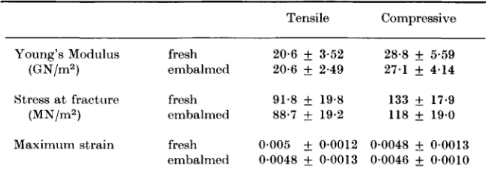
McElhaney et al. (1964) performed tensile and compressive tests (and also Rockwell ball indentation hardness tests) on beef bone. All speci
mens were extracted longitudinally from the cortices of femora;
alternate specimens (from adjacent sites) were tested within 48 hours of death, and after at least 15 hours immersed in one of four embalming fluids, all containing ethyl alcohol and formalin. Specimens were wetted with water during machining and were tested while wet; an averaging extensometer was used. The only significant change found was in the
value of the fracture stress in compression, which was 12% lower after embalming. Other compressive and tensile properties were lower after embalming by amounts (ca. 1-6%) which were not statistically signi
ficant.
Sedlin (1965) tested ten tensile specimens within the elastic region when fresh, and again after having been immersed in 10% formalin solution for 3 weeks. Values of Young's Modulus derived from cross - head movement showed no significant change.
The experimental evidence is inconclusive, but suggests t h a t the properties of bone are best measured on unfixed specimens.
E. Effects of Drying
Probably the most interesting work is t h a t of Kö (1953) and Yokoo (1952).
Ko performed tensile tests to fracture on specimens in their naturally wet state and after drying them to various extents. Values of strain and Young's Modulus were derived from measurements of the relative movement of the enlarged ends of the specimen, which is considerably better than relying on crosshead movement, although less good t h a n the ideal averaging extensometer (which would have been difficult in 1952 on the size of specimen concerned). As the water content was reduced from its natural value of 12-1%, Young's Modulus and the tensile stress at fracture increased, and the strain at fracture decreased.
The highest values of Young's Modulus and the stress at fracture occurred with a water content of 0*6%, and were each about 50%
higher than their values when naturally wet, but at water contents corresponding to air drying the increases from naturally wet values were about 20% to 30%. The final reduction in water content, from 0-6% to zero, produced a sharp drop in the tensile stress at fracture (to less than its value when naturally wet) and a drop in the strain at fracture.
The effects of reducing the water content are consistent with the idea of water acting as a lubricant in the solid matrix which is deformed under load; the embrittlement following the final removal of water could be a consequence of microscopic cracks formed during the final drying.
Such cracks would not affect Young's Modulus.
The results of Yokoo (1952) are generally similar to those of Ko for the stress and strain at fracture, except for the embrittlement at zero water content (which would be shown more clearly in Kö's tensile tests). Yokoo's stress-strain curves from these tests are too irregular to permit reliable values of Young's Modulus to be derived,
158 S. A. V. SWANSON
Other workers (Evans and Lebow, 1951; Dempster and Liddicoat, 1952; Smith and Walmsley, 1959; Evans, 1964; Ascenzi et αΖ., 1966;
Sedlin and Hirsch, 1966) have compared the properties of wet and dry bone, but because none of them have given values for the water contents of all their specimens, their results can only support, with less precision, those of Ko and of Yokoo. Some points of interest may, however, be summarized.
Dempster and Liddicoat (1952) found that the re-wetting of old, dried bone reduced both Young's Modulus and the tensile stress at fracture, which suggests that the effects of drying are reversible to some extent.
Smith and Walmsley (1959) found that the value of Young's Modulus became steady after 1 hour's air drying, at 107% of the value when wet.
These results were obtained using a carefully designed cantilever bending test from which the usual sources of error had been eliminated.
These authors found also that the dimensional changes accompanying drying were of the same order of magnitude as those produced by the application of significant stresses, which makes the control of humidity during a test seem important.
Ascenzi et at. (1966) reported no significant differences in tensile properties between specimens wetted with distilled water and with saline. Their specimens were portions of single osteones which had apparently been partly dried during machining.
Sedlin and Hirsch (1966) compared the results of bending tests on specimens of fresh bone which were (a) tested immersed in Ringer's solution, (b) allowed to dry in air for between 5 minutes and 1 hour, and (c) dried in an incubator at 105°C for 1 week. Air drying produced some increase in maximum stress after 15 minutes and a significant increase of about 4 % after 1 hour, with no change in Young's Modulus. Incuba
tion at 105°C, compared with wet testing, produced a significant increase in Young's Modulus, no significant change in maximum stress, and a significant decrease in the total deflection at failure. The authors themselves remark that some of these observations seem to be incom
patible. This is not surprising, because the bending test is unsuitable for comparing fracture stresses in materials having different stress-strain curves, as do wet and dry bone.
F. Effects of Testing Temperature
Smith and Walmsley (1959) found that the value of Young's Modulus derived from a bending test on wet bone varied from 10 GN/m2 at
4-5°C to 8-28 GN/m2 at 43°C, the variation within this range being linear.
Sedlin and Hirsch (1966), using their three-point bending test in Ringer's solution at 21°C and at 37°C, found no significant differences in maximum stress or in the energy absorbed to failure, but a 6% increase at the higher temperature in the total deflection to failure. The latter observation agrees qualitatively with the finding of Smith and Walms- ley t h a t the stiffness decreased as the testing temperature is increased, but can be reconciled with the former two observations only if the load-deflection curves were different in shape; no information is given about this.
Bonfield and Li (1966) performed tensile tests at temperatures ranging from — 196°C to +200°C. The tensile stress at fracture was highest (130-152 MN/m2) at 0°C, decreasing to about 88 MN/m2 at
- 1 9 6 ° C and to 27-5 MN/m2 at + 200°C. Whether the material which results from heating bone to 200°C can still be regarded as bone is perhaps open to doubt. The same authors (Bonfield and Li, 1968) performed similar experiments, but using an extensometer over the temperature range — 58°C to +90°C. Their specimens were extracted longitudinally from a bovine femur, and the experiments were designed to permit observation of time-dependent non-elastic deformations. The total strain at a given stress increased (but not uniformly) with increas
ing temperature. Young's Modulus, calculated after allowing for the elastic components of strain, decreased linearly from 35-2 GN/m2 at - 58°C to 26-2 GN/m2 at + 25°C. Although the rate of change of Young's Modulus with temperature in these tests ( — 0-43%/°C) is practically equal to t h a t found by Smith and Walmesley, Bonfield and Li's values of Young's Modulus are about three times higher.
G. Variations Within One Bone
Evans and Lebow (1951 and 1952), Yokoo (1952) and Ko (1953) have all found t h a t specimens from near the centre of the shaft of long bones (femur, fibula or tibia) are stronger t h a n specimens from nearer the ends, but the differences are not large in relation to the scatter in the results. Olivo et al., (1937), using a bending test, found no systematic variation in breaking strength with position along the shaft, in speci
mens taken from one 79-year-old human femur.
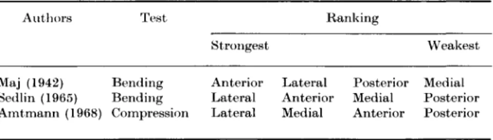
The same authors, and also Maj (1942) and Sedlin (1965), have reported results obtained from specimens extracted from different quadrants of human femora. None of the differences found is large enough in relation to the probable accuracy of the measurement, or
6*
160 S. A. V. SWANSON
the scatter in the results as given, to appear very significant, and, if the four quadrants are ranked in order of breaking strength, different authors produce different rankings. Thus, although the lateral quadrant of the femoral shaft does appear to be generally stronger than the medial, this tendency should not be regarded as having been established beyond doubt. Maj (1938) produced some more convincing results from the metatarsal and metacarpal of an ox, and also gave porosity measure
ments corresponding to the strength measurements reported by Olivo et al. (1937) for specimens from one human femur; porosity and strength were inversely related, but with large scatter.
Amtmann (1968) performed compression tests on specimens ex
tracted longitudinally from the femoral cortices of twelve humans.
Some 703 specimens were tested, and correlations sought between compressive stress at fracture and: body side, position along shaft of bone and quadrant. Careful statistical analyses were performed, but it seems unfortunate t h a t the specimens were of embalmed bone and were tested d r y ; neither Amtmann nor the present author know of any evidence to show whether embalming and drying affect all strengths in the same proportion (indeed Evans (1957) reported t h a t embalming reduced the strength of male bone significantly more, in proportion, than t h a t of female bone). With this considerable reservation, Amt- mann's results are incorporated in Table 2.
TABLE 2. Variations in strength with aspect of origin of specimens Authors
Maj (1942) Sedlin (1965) Amtmann (1968)
Test
Bending Bending Compression
Strongest Anterior Lateral Lateral
Ranking
Lateral Posterior Anterior Medial Medial Anterior
Weakest Medial Posterior Posterior
H. Variations between Bones
Most authors have extracted specimens from femora, but some have used other bones also, and have produced comparative results.
Table 3 shows the rankings produced by different authors. Maj used a bending test and Yokoo a compressive test; all the others used tensile tests.
TABLE 3. Variations in strength between small specimens extracted from different bones
Authors Ranking
Strongest Weakest (a) Tensile tests
Evans and Lebow (1952) Fibula Tibia Femur
Ko (1953) Radius Ulna Fibula Tibia Humerus Femur Evans (1964) Tibia Femur
Evans and Bang (1966) Fibula Femur Lindahl and Lindgren (1967) Humerus Femur (b) Compressive tests
Yokoo (1952): 20-39 years Femur Tibia Humerus Fibula Ulna Radius Yokoo (1952): 43 years Femur Humerus Fibula Ulna Tibia Radius Lindahl and Lindgren (1968) Femur Humerus
(c) Bending tests
Maj (1942) Ulna Tibia Humerus Femur
Not all the differences implied in Table 3 are equally significant in view of the numbers of specimens and probable experimental errors, and therefore only the most general conclusions can safely be drawn, e.g. t h a t the material of the femur is consistently weaker in tension than t h a t of other long bones.
Considering properties other than tensile strength, Lindahl and Lindgren (1967) found, using tensile tests on specimens from fresh humeri and femora, no significant difference between these two bones in respect of Young's Modulus, the Limit of Proportionality or the strain at fracture.
/ . Variations with Sex
Evans (1957), on p . 188, summarizing his own and others' results, states t h a t male femoral bone is stronger than female by 34% to 5 4 % when fresh, and by 5 % when embalmed. If this finding were consistently repeatable, it would be important to have the corresponding histo- logical observations.
Ko (1953) and Yokoo (1952) found, in tension and compression respectively, no significant differences in stress or strain at fracture in specimens from femora of the two sexes.
Lindahl and Lindgren (1967) found no significant difference between
162 S. A. V. SWANSON
the sexes in respect of any tensile properties, with the possible excep
tion that, in the youngest age group (15-19 years), female femora appeared to contain bone which was weaker than male femora or humeri of either sex.
I n compression, Lindahl and Lindgren (1968) found t h a t specimens from male femora were 8% stronger than those from female femora, but no corresponding difference was found in specimens from humeri.
J. Variations with Age
Ascenzi et al. (1966) found t h a t one type of osteone (fully calcified, helical collagen fibres) obtained from men of 30 to 80 years of age showed closely similar tensile properties. I t does not follow from this t h a t the properties of compact bone should be independent of age, because of the known variations in physical characteristics with age (see p. 145).
Evans and Lebow (1951) found no correlation of tensile or shear properties with age; neither did Sedlin (1965). Ko (1953), however, found t h a t the tensile stress at fracture fell after the age of 40, until in the interval 60-79 years it had 70% of its value in the decade 20-29.
The strain at fracture also fell, but by a smaller amount. Yokoo (1952) found similar variations in compressive properties.
The tensile tests of Lindahl and Lindgren (1967) covered the age range from 15 to 89 years. From 15 to 30 years, both the tensile stress and strain at fracture showed more scatter than at higher ages; from 30 to 89 years both properties showed a gradual decrease, the values for the group 80-89 years old being about 80% of those for the 30-39 group.
A similar fall in compressive properties with increasing age was observed by Lindahl and Lindgren (1968).
K. Directional Variations 1. Strength and stiffness
Relatively few results have been published, presumably because of the obvious experimental difficulties, which are greater for specimens extracted tangentially than for those extracted longitudinally, and greater still for those extracted radially (the terms longitudinal, tangential and radial here refer to directions in a portion of the shaft of a bone).
Maj and Toajari (1937) performed three-point bending tests on specimens of rectangular cross-section, extracted from tibiae of oxen.
The specimens were 7 mm long, and could therefore be taken with their
lengths in each of the three directions mentioned above. The testing techniques were such as to cast doubt on the absolute values of strength, but, considering only relative values, the tangential direction was consistently twice as strong as the radial, and the longitudinal direc
tion was less consistently five to six times stronger than the radial.
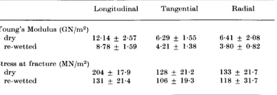
Dempster and Liddicoat (1952), using old dried bones some of which were re-wetted before testing, measured Young's Modulus and the stress at fracture in compression in each of the three directions, on cubical specimens extracted from the cortices of humeri and femora.
The results are summarized in Table 4, as means and standard devia
tions.
TABLE 4. Compressive properties in three directions
Longitudinal Tangential Radial Young's Modulus (GN/m2)
dry 1214 ± 2-57 6-29 ± 1-55 6-41 ± 2 0 8 re-wetted 8-78 ± 1-59 4-21 ± 1-38 3-80 ± 0-82 Stress at fracture (MN/m2)
dry 204 ± 17-9 128 ± 21-2 133 ± 21-7 re-wetted 131 ± 21-4 106 ± 19-3 118 ± 31-7 (From Tables 2 and 4, Dempster, W. J . and Liddicoat, R. T. (1952).
Am. J. Anat. 91, 343; 351.)
The values of Young's Modulus were derived from measurements of platen movement and are probably unreliable except as a set of relative values; see the discussion following Table 6.
Yokoo (1952), using a compressive test on specimens 8 mm long and 4 mm in diameter from 40- and 48-year-old males, found Young's Modulus and the stress at fracture to be 11 300 and 164 MN/m2 respect
ively when the load was applied parallel to the Haversian systems, compared with 6,370 and 102 MN/m2 when perpendicular to them.
These values of Young's Modulus are open, to some extent, to the same criticism as those of Dempster and Liddicoat.
Dempster and Coleman (1961), also using dried museum specimens of unknown origin, performed tensile tests on specimens extracted in the longitudinal and tangential directions. Dry specimens gave ultimate tensile strengths of about 131 MN/m2 longitudinally and 11-1 tangent- ially; the corresponding values for re-wetted specimens were 96-8 and 9-7 MN/m2. Strains were not measured.