Gomphonella olivacea (Bacillariophyceae) – a new phylogenetic position for a well-known taxon, its typification, new species and combinations
Regine Jahn
1,*, Wolf-Henning Kusber
1, Oliver Skibbe
1, Jonas Zimmermann
1, Anh Tu Van
1, Krisztina Buczkó
2,3& Nélida Abarca
11Botanischer Garten und Botanisches Museum Berlin, Freie Universität Berlin, Königin-Luise-Str. 6-8, 14195 Berlin, Germany
2Department of Botany, Hungarian Natural History Museum, Könyves Kálmán krt. 40, 1087 Budapest, Hungary
3MTA Centre for Ecological Research, Danube Research Institute, Karolina street 29, 1113 Budapest, Hungary
*Author for correspondence: r.jahn@bgbm.org
REGULAR PAPER
Background and aims – Within the project “German Barcode of Life – Diatoms” common diatoms of German waters were routinely isolated and cultivated. In order to understand the taxonomy and phylogeny of the genus Gomphonema, one of the most common taxa of Central Europe, known currently either under the name Gomphonema olivaceum in Europe or Gomphoneis olivacea in America, was studied.
Methods – Twenty unialgal strains were established from five different water bodies in Germany and one from Lake Balaton, Hungary, which supplied molecular data (18S V4 and rbcL) besides morphometric and ultrastructural data. In addition, on eight populations from different water bodies including the type from Denmark, morphometric and micromorphological studies by light and scanning electron microscopy were performed.
Key results – Molecular and micromorphological data show that the target taxon neither belongs to Gomphonema Ehrenb. nor to Gomphoneis Cleve. By reinstating the genus name Gomphonella Rabenh., the nomenclatural and taxonomic enigma of this taxon is solved, and with the presentation of the type by Hornemann the authorship of the epithet is clarified. Molecular data for the unialgal strains and several environmental clones show that there is more diversity in the Gomphonella olivacea clade than can be identified morphologically. In addition, the establishment of the new species Gomphonella coxiae and Gomphonella acsiae is supported. The molecular data classified Gomphonella species as belonging to the Cymbellales but not to the Gomphonemataceae. In addition, molecular data put Gomphoneis tegelensis R.Jahn & N.Abarca also into Gomphonella. In order to make the genera Gomphoneis and Gomphonema monophyletic, their astigmate members are transferred to Gomphonella.
Conclusions – The results clarify that the gomphonemoid outline is not restricted to the family Gomphonemataceae but seem to be distributed across the entire order Cymbellales. This is shown in this paper for the revived genus Gomphonella, which contains the astigmate group of Gomphoneis and Gomphonema besides the longly disputed G. olivacea. Only a polyphasic approach, combining molecular and micromorphological data for taxonomy, nomenclatural evaluation, and observations from clonal cultures can reveal the full intricacies of evolutionary relations.
Key words – Bacillariophyceae, diatoms, genus emendation, Gomphoneis, Gomphonema, nomenclatural types, phylogeny, polyphasic approach.
© 2019 The Authors. This article is published and distributed in Open Access under the terms of the Creative Commons Attribution License (CC BY 4.0), which permits use, distribution, and reproduction in any medium, provided the original work (author and source) is properly cited.
Plant Ecology and Evolution is published by Meise Botanic Garden and Royal Botanical Society of Belgium ISSN: 2032-3913 (print) – 2032-3921 (online)
INTRODUCTION
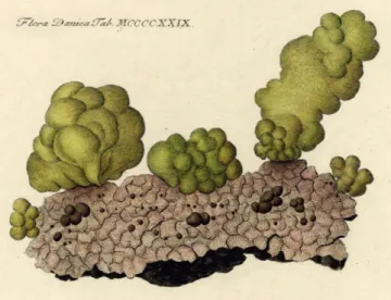
The taxon currently known under the name Gomphonema olivaceum (Hornem.) Ehrenb. or Gomphoneis olivacea (Hor- nem.) P.A.Dawson ex R.Ross & P.A.Sims is a very common diatom in fresh waters of Central Europe. Its habit of living on stalks or being free-living and producing fair amounts of mucus had originally placed it into different genera since, at the beginning of diatom research, life forms where thought to be decisive for phylogeny. In 1810 Hornemann pictured an olive coloured mass of mucus from a Danish river and named it Ulva olivacea Hornem. (fig. 1). Lyngbye (1819) used his material to describe and draw G. olivacea-like cells and recombined the name as Echinella olivacea (Hornem.) Lyngb. Kützing (1833) named it Frustulia olivacea (Hor- nem.) Kütz. and Brébisson & Godey (1835) Cymbella oliva- cea (Hornem.) Bréb. & Godey. When it was recognized that those two growth habits – attached on stalks or freely mov- ing – were just two different life forms of the same species, the species was subsumed under the genus Gomphonema Ehrenb. In 1838 Ulva olivacea was transferred twice: in July/August by Ehrenberg and in October by Brébisson. In 1853 Rabenhorst published the new genus name and com- bination Gomphonella olivacea (Hornem.) Rabenh., a name which was reduced to a section of Gomphonema by Brun (1880) and which, since then, has apparently been neglected by the diatom community; nevertheless, in the Index Nom- inum Genericorum (2018) Gomphonella Rabenh. is listed as a genus with an unassigned type of the name of the genus even though a type was given in Round et al. (1990).
In parallel to the above sketch of the nomenclatural his- tory of the epithet olivacea, there are further names which seem to refer to the same taxon. Agardh (1824) gave it the superfluous name Meridion vernale C.Agardh. In 1830 Lei- blein, using diatom material from waters near Würzburg, Germany, published a picture of an unnamed gompho- nemoid diatom and discussed the taxonomic identity of Me- ridion vernale. Leiblein sent this material among others to C. Agardh in Lund who described Gomphonema leibleinii C.Agardh validly from this material (Agardh 1830). Kützing
Figure 1 – Ulva olivacea. Reprint of Flora Danica Tab MCCCCXXIX 1429 (Hornemann 1810).
(1833) accepted Agardh’s name whereas Ehrenberg (1838) put Gomphonema leibleinii into synonymy with his Gom- phonema clavatum Ehrenb., which he had described validly in 1832 (in Ehrenberg 1830, it was a nomen nudum). Ehren- berg (1838) also used (and transferred) the taxon name Gom- phonema olivaceum, which meant that for him Gomphonema olivaceum and Gomphonema clavatum (= Gomphonema lei- bleinii) were not conspecific. In 1844 Kützing put Gompho- nema leibleinii into synonymy with Gomphonema olivaceum and also put Gomphonema clavatum into synonymy with Gomphonema subramosum C.Agardh (see also Reichardt 2015).
Despite its complex early history, this taxon has been known for 140 years as Gomphonema olivaceum. With the advent of the electron microscope it became obvious that its micromorphology was different from most Gomphone- ma. Dawson (1974) proposed that, because of its biseriate striation, it should be separated from Gomphonema and put into the genus Gomphoneis Cleve, which had been erected by Cleve (1894); this proposal was formally correctly ex- ecuted by Ross & Sims in 1978. Since that time in the USA (Kociolek 2011) this taxon has been assigned to the genus Gomphoneis as Gomphoneis olivacea (Hornem.) P.Dawson ex R.Ross & P.A.Sims (1978) whereas in Central Europe it stayed within Gomphonema (Hofmann et al. 2013, Levkov et al. 2016) because biseriate striation was seen as common in this genus also and therefore not identified as a differenti- ating feature (Reichardt 2007). In addition, since it lacks an axial plate and mantle lamella it was seen as not fitting the genus Gomphoneis (Krammer & Lange-Bertalot 1985).
The logical approach for solving nomenclatural and tax- onomic enigmas is to locate the type specimen or original material that was in the hands of the first describer. Locat- ing this material was quite challenging, since 200 years ago Prof. Hornemann had worked in Copenhagen (Denmark), and Lyngbye, who had studied this material in more detail and published figures, had gotten his education in Copen- hagen but was later based in Lund (Southern Sweden). Re- quests were made to both Herbaria, resulting in the finding of Agardh’s Gomphonema leibleinii in the Lund Herbarium (plus comments made in 1982 by an earlier researcher). The type material of Ulva olivaceum had been loaned in 1965 to the Diatom Herbarium in Philadelphia, USA, by the Copen- hagen Herbarium (C) but fortunately, an intensive search in Philadelphia resulted in the finding of this material after 52 years!
Within the framework of the project “German Barcode of Life – Diatoms” we were finally successful in isolating and cultivating several strains of this taxon. In our studies to un- derstand the taxonomy and phylogeny of the genus Gompho- nema (Abarca et al., in prep.), we also questioned its phyloge- netic position and taxonomic affiliation with morphological as well as molecular data. Since these data are very different from the core group of Gomphonema yet similar to Gompho- neis tegelensis R.Jahn & N.Abarca (Skibbe et al. 2018), we are publishing the results here separately.
MATERIAL AND METHODS
The original material from Denmark of 1810 was studied (Lectotype C-A9208; see Typification below). In addition, data from eight populations and 38 strains are included in the present study (see electronic appendices 1 & 2). Thirty- two strains were established by the authors. The sequence data for the other six strains were downloaded from ENA/
Genbank (as part of the International Nucleotide Sequence Database Collaboration, INSDC). All sequences downloaded from INSDC were BLASTed (basic local alignment search tool) against the INSDC database to test for taxonomic con- sistency.
Field collection and cultivation
Freshwater samples were collected from Germany and Hun- gary between 2004 and 2017. Twenty unialgal strains of the target taxon were isolated from nine samples of six different waters in Germany and from Lake Balaton in Hungary (for details see electronic appendices 1 & 2).
Clonal strains were established by micropipetting single cells using a stereo microscope (Olympus, Japan) and an in- verted LM (Olympus, Japan). All strains were treated accord- ing to Romero & Jahn (2013). Non-axenic unialgal cultures were maintained at room temperature (19–25°C for cultures until 2016), 10°C (in 2016) and at 20°C (in 2017) in a growth chamber. A 12:12 h light/dark photoperiod from a daylight LED light source following Jahn et al. (2017) was applied. In addition, seven populations from the original samples from which clonal cultures were established were used for sup- porting documentation of the morphologies of the clones (for details see electronic appendix 2).
Documentation and vouchering
For all newly established strains the frustule preparation and morphological documentation were executed following Zim- mermann et al. (2014). LM pictures of live cells (fig. 2) and of permanent specimens on slides were taken with a Zeiss AxioImager.M2 (Zeiss, Germany). SEM images were taken with a Hitachi FE SEM 8010 (Hitachi, Japan) of unsput- tered material. The vouchers for all new strains are depos- ited at B (Herbarium Berolinense), where long-term stable and semantic web compatible identifiers for specimens are used according to Güntsch et al. (2017). Molecular data for all isolates are deposited in INSDC (see electronic appendix 1). DNA samples are stored in the Berlin DNA Bank and are available via the Genome Biodiversity Network (GGBN, Droege et al. 2014); nomenclatural acts are registered (Tur- land et al. 2018, Art. 42) in PhycoBank (continuously up- dated). Data are available through AlgaTerra (Jahn & Kusber continuously updated).
Morphological criteria
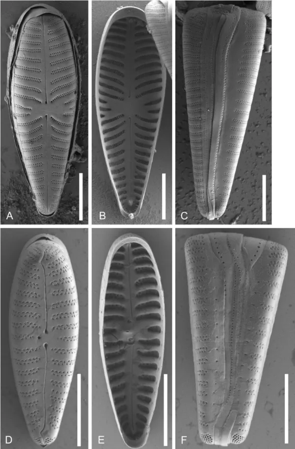
Besides valve outline and morphometric measurements of each clone (length, width, number of striae in 10 μm (elec- tronic appendix 2) valves were investigated under SEM to compare internal and external valve and girdle views (fig.
5A–C). Special attention was given to the presence or lack of
Figure 2 – Five living cells of Gomphonella olivacea (Strain D129_007): one in valve view and four in girdle view. Note the single chloroplast, consisting of two H-shaped lobes connected by a bridge which contains the pyrenoid. These cells contain abundant reserve material. Scale bar = 10 µm.
stigmata or stigmoids, the form of the striae and covering of the areolae, and the porelli of the footpole. For comparison, SEM images of the same features of Gomphonema minutum are presented (fig. 5D–F).
DNA extraction, sequencing and alignment
Cultured material was transferred to 1.5mL tubes. The DNA was isolated using the NucleoSpin® Plant II Mini Kit (Macherey and Nagel, Düren, Germany) or Qiagen®
Dneasy Plant Mini Kit (Qiagen, Valencia, CA) following the respective product instructions. The DNA fragment size and concentrations were measured via gel electrophoresis (1.5% agarose gel) and Nanodrop® (PeqLab Biotechnology, Erlangen, Germany), respectively. The DNA samples were stored at −20°C for future use and finally deposited in the Berlin collection of the DNA bank network (Droege et al.
2014). The polymerase chain reaction (PCR) for rbcL was conducted following Abarca et al. (2014). The V4 section of the 18S SSU rRNA gene locus (18S V4) was amplified and PCR performed following Zimmermann et al. (2011). PCR products were visualized in a 1.5% agarose gel and cleaned with MSB SpinPCRapace® (Invitek LLC, Berlin, Germany) following standard procedures. DNA concentrations were measured using Nanodrop® (PeqLab Biotechnology) and samples were normalized to a total DNA content >100 ng μL−1 for sequencing.
Sanger sequencing was conducted by Starseq® (GENter- prise, Mainz, Germany), rbcL gene according to Abarca et al.
(2014) and 18S V4 according to Zimmermann et al. (2011).
In both cases the same primers were used for amplification and sequencing. The editing, as well as the quality control of the pherograms for the new sequences, were done in Phyde®
(Müller et al. 2010). The evaluated sequences were aligned using MUSCLE (Edgar 2010), as implemented in MEGA6 (Tamura et al. 2013) with subsequent manual adjustments in
case of 18S V4. The lengths of the newly generated sequenc- es were 432 bp for 18S V4 and 979 bp for rbcL. For com- parison, the alignments for 18S V4 and rbcL also included other sequences of our own and some others from INSDC representing the Cymbellales D.G.Mann, as well as Ach- nanthidium saprophilum (H.Kobayasi & Mayama) Round &
Bukht., which was added as the outgroup for the phyloge- netic tree generation following Kermarrec et al. (2011). All added sequences were trimmed to fit to the newly generated sequences for 18S V4 as well as rbcL The accessions used are given in electronic appendix 1).
Phylogenetic analyses
Two different data sets (18S V4, rbcL) were used for the phy- logenetic analyses. Each dataset was analysed using Maxi- mum Likelihood (ML) as implemented in RAxML (Stama- takis 2006, 2014, Stamatakis et al. 2008) using the CIPRES platform (Miller et al. 2010) in both cases.
For the ML analysis of the molecular datasets, the opti- mal model of sequence evolution that best fits the sequence data was calculated under the hierarchical likelihood ratio test (hLRT) and the Akaike information criterion (AIC) us- ing model test 3.7 (Posada & Crandall 1998). The best fit- ting model was GTR+G+I (Tavaré 1986). A ML analysis was conducted using RAxML 8.2.8 (Stamatakis 2006, 2014, Stamatakis et al. 2008), ML search option (GTR+G+I) and 1,000 bootstrap replicates (model GTRCAT as implemented in RAxML for the rapid bootstrap algorithm). Additionally, Bayesian phylogenetic inference was conducted for both data sets using MrBayes v. 3.1.2. (Huelsenbeck & Ronquist 2001, Ronquist & Huelsenbeck 2003) with the same model. The de- fault settings were used, runs with four incrementally heated Metropolis-coupled Monte-Carlo Markow Chains and runs with 10 million generations were executed. The runs were sampled every 1000 generations, the first 25% generations being discarded as burn-in; the rest were used to calculate a 50% majority rule consensus tree. The best ML tree found by RAxML and the 50% majority rule tree of the BI analy- sis were compared for rbcL as well as 18S V4. In all cases the trees showed no different topologies and were therefore summarized in one tree for each marker, showing bootstrap statistics (> 75) for ML (LB) and (> 0.90) posterior probabili- ties (BI). Trees were drawn using FigTree v. 1.4.2 (Rambaut 2008) and Adobe Illustrator (Adobe Systems, San Jose, CA).
Genetic distances for 18S V4 and rbcL were calculated us- ing MEGA6 (Tamura et al. 2013) and the implemented p- distance option.
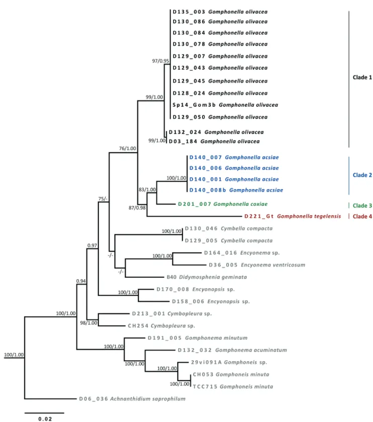
RESULTS Molecular data (figs 3, 4)
The molecular markers 18SV4 and rbcL were used and both provided similar results, clustering all target strains into one main clade (figs 3 & 4) with 99/1.00 (18SV4) and 76/1.00 (rbcL) ML bootstrap support/posterior probabilities. We will refer to this clade from now on as the Gomphonella clade, in anticipation of the conclusions drawn later. Four clades, numbered 1, 2, 3 and 4 with even better bootstrap support are separated within the Gomphonella clade (details see be-
low), which is well separated from taxa of the genera En- cyonopsis (18S V4, p-distance 10%) or Cymbella (rbcL, ca 5%). The Gomphonema clade, with G. acuminatum Ehrenb., G. minutum (C.Agardh) C.Agardh and Gomphoneis minuta (J.L.Stone) Kociolek & Stoermer, is well separated and sup- ported by 99/1.00 (18SV4) and 100/1.00 (rbcL) bootstrap value/posterior probabilities and p-distances of around 12%
(18SV4) and 6% (rbcL) from Gomphonella spp.
Clade 2 is the sister group to Clade 3 in the 18S V4 (1.00 posterior probabilities, fig. 3) as well as in the rbcL tree (83/1.00 bootstrap value/posterior probabilities, fig. 4). In the case of Clade 4 the two trees show slight differences in the topologies regarding the sister group relation. For 18SV4 (fig. 3) Clade 4, supported by 99/1.00 bootstrap value/poste- rior probabilities, is the sister group to the branch with Clad- es 1a, b, 2 and 3. Clades 2 and 3 for their part are building the sister group to Clade 1a, b supported by 75/1.00 bootstrap value/posterior probabilities. In contrast, for the rbcL tree (fig. 4) Clade 4 is the sister group to the branch with Clade 2 and 3 with a support of 87/0.98 bootstrap value/posterior probabilities and these three Clades (Clades 2, 3 and 4) are the sister group to Clade 1A, B with a support of 76/1.00 bootstrap value/posterior probabilities.
Clades 1A and 1B – From a molecular point of view the 13 clones (for details see electronic appendix 1) from Tegeler See (samples D128 and D130), Müggelsee (D129) and riv- er Main (D135_024) are the same (Clade 1A). They show well supported but small differences – p-distance = 0.7% for 18SV4 and 0.4% for rbcL – to the two clones of river Spree (D03_184), and Saale (D132_024) (Clade 1B). The molecu- lar data from the environmental sample named Cymbellales from brook Westerhöver Bach (Brinkmann et al. 2015) sits in Clade 1A.
Clade 2 – The two strains with data for 18S V4 and the four strains for which rbcL data are available, are all from Lake Balaton (D140; see electronic appendix 1). They show 0%
differences between each other in 18SV4 and rbcL. There are well supported differences from the other three clades:
For 18SV4 the p-distances are 1.6–2.3% to Clade 1, 1.6%
to Clade 3 and 5.1% to Clade 4; for rbcL they are 3.5% to Clade 1, 2.9% to Clade 3 and 5.1% to Clade 4.
Clade 3 – This clade is defined by the data of strain D201_007 for which 18S V4 and rbcL data are available.
For 18S V4 the p-distances are 2.3–2.8% to Clade 1, 1.6%
to Clade 3 and 5.3% to Clade 4; for rbcL they are 2.9% to Clade 1, 2.2% to Clade 2 and 4.6% to Clade 4.
Clade 4 – This clade is defined by the data of the isolate D221_Gt, which has been separated into a new species, Gomphoneis tegelensis R.Jahn & N.Abarca (Skibbe et al.
2018). Concerning 18S V4 data, this strain has a p-distance of 4.9–5.3% to Clade 1, 5.1% to Clade 2 and 5.3% to Clade 3; concerning rbcL data, this strain has a p-distance of 5.0%
to Clade 1, 5.1% to Clade 2 and 4.6% to Clade 3.
Morphology (figs 5–14)
The most conspicuous trait of all studied strains is their pro- nounced variability in outline: from the typical ovate-oblan- ceolate to symmetrically lanceolate, with apices that are
Figure 3 – Phylogenetic tree using maximum likelihood of the dataset of the 18SV4 molecular marker with bootstrap statistics (> 75) for ML (LB) and (> 0.90) posterior probabilties (BI). Black: Clade 1 Gomphonella olivacea (upper branch: genodeme 1 and lower branch genodeme 2); blue: Clade 2 Gomphonella acsiae; green: Clade 3 Gomphonella coxiae; purple: Clade 4 Gomphonella tegelensis.
broadly rounded (i.e. fig. 6Q) to pointed (i.e. fig. 6R & S).
They often grew fan-like (strains D129_043 and D132_024) or in lumps (strain D132_036) producing plenty of gelati- nous material. Since the strains have such variable und of- ten untypical outlines which might be due to long lasting cultivation, we also studied the populations of the samples from which the strains were isolated such as the populations from Tegeler See (figs 6AI–AO & 7A–I), Müggelsee (fig.
6I–P), river Main (fig. 7AO–AV), Saale (fig. 7S–AA), river Spree for Clade 1, Lake Balaton (fig. 11A–F) for Clade 2 and
Helenesee (fig. 13A–F) for Clade 3. Specimens of Clade 4 are selectively isolated cells of clonal origin (Skibbe et al.
2018) which were not kept in culture.
Concerning morphological synapomorphies, the most important and conspicuous feature of clades 1–4 visible in LM is that they do not have any stigmata (fig. 5A & B); occa- sionally, there are a few isolated puncta visible in the central area (e.g. figs 7AR, AS & 8K) but they look like the areolae and seem to be continuations of the striae, just separated by a gap. In SEM, these puncta have no internal structure and
Figure 4 – Phylogenetic tree using maximum likelihood of the dataset of the rbcL molecular marker with bootstrap statistics (> 75) for ML (LB) and (> 0.90) posterior probabilties (BI). Black: Clade 1 Gomphonella olivacea (upper branch: genodeme 1 and lower branch genodeme 2); blue: Clade 2 Gomphonella acsiae; green: Clade 3 Gomphonella coxiae; purple: Clade 4 Gomphonella tegelensis.
are therefore not true stigmata (see also definition in www.
diatoms.org). A further conspicuous trait visible only in SEM is their biseriate striation (in Clade 4 only, some striae are triseriate) with small round uniform areolae not occluded by siliceous flaps (see Skibbe et al. 2018: figs 17–21). The
striae are not interrupted near the valve face/mantle junction and continue onto the valve mantle (fig. 5C). These double rows of areolae can terminate either as single or as double rows along the axial area and mantle. But all striae taper into only a single row of areolae at the central area (fig. 5A). The
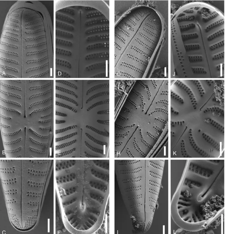
Figure 5 – SEM-comparison of autapomorphies: A–C, Gomphonella olivacea, C_A9208 lectotype population; D–F, Gomphonema minutum, strain D191_005. A & D, external valve view, note the differences in the areolae of the biseriate striae, the form of the raphe slit and the stigma in D; B & E, internal valve view, note the differences in the central raphe endings and helictoglossae as well as the stigma in E; C &
F, girdle view, note the abrupt endings of the striae in C and the tapering into a single punctum or slit in F. Scale bars = 5 µm.
foot pole is composed of a bilobed field of porelli. The out- side distal ends of the raphe extend through these porelli but not all the way to the end of the mantle. The raphe is either straight or very slightly undulated at both apices. Internally, the pseudosepta are wide, distinct and prominent at both api- ces. Also the helictoglossae are prominent at both poles and in some clones lie well away from the valve terminus.
The autapomorphies separating the clades (and species) are aspects of the striation, such as the number of striae in 10 µm, their parallel or radial direction throughout the valve face, and the form of the central area. Of special importance too are the porelli at the footpole, which are either of similar size and shape to the areolae of the striae or relatively larger and distinct; they are either arranged in double rows or with-
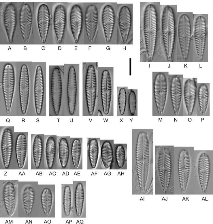
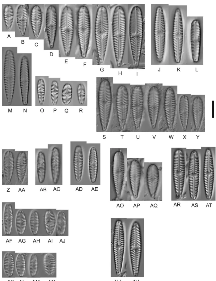
Figure 6 – Gomphonella olivacea, LM: A–H, C_A9208, lectotype population from Denmark. D represents the lectotype; I–AH, specimens from sample D129, Müggelsee, Berlin, Germany; I–P, natural population; Q–S, strain D129_43, Q represents the epitype; T & U, strain D129_050, V,W, strain D129_007; X & Y, strain D129_045; Z & AA, strain D129_065; AB–AE, strain D129_065; AF–AH, strain D129_062;
AI–AQ, specimens from sample D128, Tegeler See, Berlin, Germany; AI–AO, natural population; AP & AQ, strain D128_024. Scale bar = 10 µm.
out any order and they are located close to the striae or well separated.
Clade 1 (figs 6–10 & 14A) – The valves are heteropolar, clavate with broadly rounded headpoles and acutely round- ed footpoles (fig. 6A–P). There is wide variation within the clone cultures: the valves can be heteropolar, clavate with broadly rounded headpoles and acutely rounded footpoles (see fig. 6Q), but they can also be heteropolar, lanceolate to linear lanceolate (see fig. 6R), or even be only slightly heter- opolar valves with almost parallel to slightly convex margins and rounded headpole and footpole (see fig. 6S). The axial area is narrow, straight, expanded at the centre to form a rec- tangular, bow-tie-shaped to transversally elliptical central area bordered at the margins by 1–3 approximately equally- shortened striae (fig. 8B & E). Transapical striae are strongly radiate in the central part of the valve and towards the foot- pole (fig. 8H & K), becoming slightly radiate towards the headpole (figs 8A, G & 9A). The footpole has a large apical pore field with porelli of the same size and structure as the areolae (figs 8C, I, 9C, I & 10C). The porelli appear to be arranged in double rows, located close to the striation of the valve face (figs 8C, I, 9C, I & 10C) and therefore undifferen- tiated structurally and spatially from them.
Clade 1A and 1B – Morphological differences between the Clades 1A and 1B are not discernible. They are very similar and uniform in shape.
Clade 2 (figs 11, 12 & 14B) – The valves are slightly heter- opolar, lanceolate in larger specimens (fig. 11A–C & G–I) to clavate in smaller specimens, with narrowly rounded headpoles and acutely rounded footpoles. Wide variation of shape occurs within the clone cultures, since the valves can be slightly heteropolar, lanceolate and widest at the centre, or they can be heteropolar, clavate with broadly rounded head- poles and acutely rounded footpoles. The valves of the popu- lation of Clade 2 are on average longer and have a higher stria density than the club-shaped populations of Clade 1, but both have the same average width. Valves of Clade 2 are differentiated from valves of Clade 1 by a transapically widened central area, which is smaller and more rectangular (fig. 12B, E, H & K) than the central area of Clade 1 (which resembles a bow tie). Clade 2 possesses parallel striae which become radial at the centre (fig. 11A–Q), whereas in Clade 1 the striae are radial throughout the valve face. The foot- pole has a large apical pore field with relatively large porelli, which differ in size from the areolae of the striae and are well separated from the striation of the valve face (fig. 12C & I).
Clade 3 (figs 13A–O & 14C) – The valves of the population are slightly heteropolar, lanceolate in larger specimens to clavate in smaller specimens, with narrowly rounded head- poles and acutely rounded footpoles (fig. 13A–F). The valves of the clone cultures are linear-clavate or with a slight tumid swelling at the centre, headpoles narrowly rounded and foot- poles rounded (fig. 13G–I). The axial area is narrow, straight, expanded at the centre to form a broad, bow-tie-shaped to rectangular central area bordered at the margin by two or three approximately equally-shortened striae (fig. 13L & M).
The striae are composed of two alternating rows of areolae (fig. 13J). The footpole has a large apical pore field with rela- tively large porelli, which differ in size from the areolae of
the striae and are well separated from the striation. Valves of Clade 3 possess parallel transapical striae, which become ra- dial at the centre as in Clade 2 (fig. 13A–I), whereas in Clade 1 the striae are radial throughout the valve face.
Clade 4 (illustrated in Skibbe et al. 2018) – The most promi- nent differences from Clade 1, 2 and 3 are the striation, which is bi- to triseriate, and the small axial plate and mantle lamel- la (for details see Skibbe et al. 2018). Otherwise, there are no stigmoids and the areolae are small, round and uniform, and are not occluded by siliceous flaps. The striae are not inter- rupted near the valve face/mantle junction and continue onto the valve mantle (Skibbe et al. 2018: figs 17, 18, 23).
Typification and nomenclature
Clades 2 and 3 cannot be identified with already known taxa and need to be described as new (see below). Specimens of Clade 4 were recently described as Gomphoneis tegelensis (Skibbe et al. 2018). Specimens of Clade 1 were described more than 200 years ago as Ulva olivacea (fig. 1), which went through a number of name changes until today. We were able to study this material in LM and SEM for the first time (see figs 5A–C & 6A–H).
Since clade 1 obviously does not belong to the genus Gomphonema (fig. 5D–F) and an earlier valid genus name exists, namely Gomphonella Rabenhorst (1853), we are here reinstating this name. Rabenhorst introduced this genus name and made the combination Gomphonella olivacea (1853: 61), describing it as “Eine gestielte Gomphonema in einer gestalt- losen Gallertmasse” [a stalked Gomphonema in an amor- phous gelatinous mass].
Gomphonella Rabenh. (Rabenhorst 1853: 61, pl. IX) Original description –“bis 2/100 Mm lang, verkehrt-eiför- mig-lanzettlich; Nebenseiten breit keilförmig, am Rande mit zarten Querstreifen. Durch ganz Europa.” (Rabenhorst op.
cit.) [up to 20 µm long, ovate oblanceolate; sides broadly wedge-shaped, with delicate bars at the margin. Throughout the whole of Europe].
Type species (lectotype) – Gomphonella olivacea (Hornem.) Rabenh. (Ulva olivacea Hornem.), designated in Round et al.
(1990: 691).
Registration – https://phycobank.org/100348
Emended description – The most important and conspicu- ous feature of this genus visible in LM, separating it from other gomphonemoid taxa, is that there are no stigmoids or stigmata present on the valve face. A further conspicuous trait separating it from Gomphonema s. str. but visible only in SEM is the bi- to triseriate striation with small round uni- form areolae not occluded by siliceous flaps. The diameter of the areolae is about 100 nm. The striae sit in moderately deep alveolae between thicker vimines. The striae continue onto the valve mantle. The apical foot pole is composed of a bilobed field of porelli which are round and similar to the normal areolae in the striae (= undifferentiated AFPs). The raphe is filiform and either straight or very slightly undulate at both apices. Internally, both polar raphe endings end in prominent helictoglossae well away from the valve terminus and there is an intermissio in the centre with the raphe end-
Figure 7 – Gomphonella olivacea, LM: A–R, specimens from sample D130, Tegeler See, Berlin, Germany; A–I, natural population; J–L, strain D130_086; M & N, strain D130_078; O–R, strain D130_084; S-AE, specimens from sample D132, Saale, Germany; S–AA, natural population; AB & AC, strain D132_036; AD & AE, strain D132_024, genodeme 2; AF–AN, strain D03_184, genodeme 2, river Spree, Berlin, Germany; AO–AV, specimens from sample D135, river Main, Germany; AO–AQ, natural population; AR–AV, strain D135_003.
Scale bar = 10 µm.
Figure 8 – Gomphonella olivacea, SEM: A–F, C-A9208 lectotype population, Denmark; G–L, epitype population, specimens from sample D129; Müggelsee, Berlin, Germany; A–C & G–I, external valve views; D–F & J–L, internal valve views; A, D, G & J, headpole; B, E, H &
K, central area, note the arched striation in the centre tapering into a row of single areolae and the rectangular bow-tie to transversely elliptical form; C, F, I & L, footpole, note the porelli organized in double rows and no gap of striation between striae and porelli; Scale bars = 1 µm.
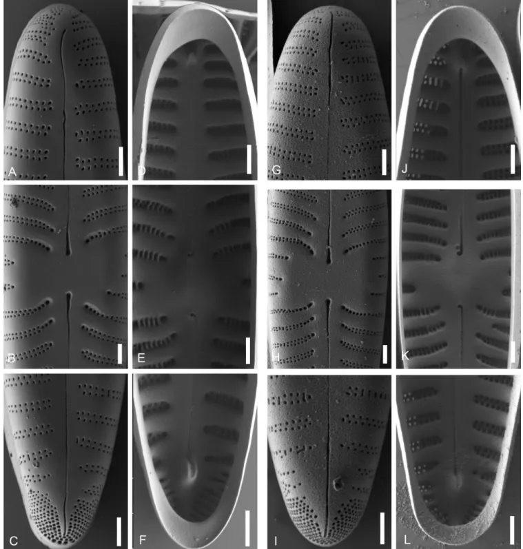
Figure 9 – Gomphonella olivacea, SEM, Müggelsee, Berlin, Germany: A–F, epitype strain D129_043; G–L, strain D129_007; A–C & G–I, external valve views; D–F & J–L, internal valve views; A, D, G & J, headpole; B, E, I & K, central area, note the arched striation in the centre tapering into a row of single areolae and the rectangular bow-tie to transversely elliptical form. C, F, I & L, footpole; note the porelli organized in double rows and no gap of striation between striae and porelli. Scale bars = 1 µm.
ings slightly bent to the same side. The pseudosepta are dis- tinct and prominent at both apices. At least two girdle bands belong to each valve and open alternately at either pole; each bears a line of pores along the junction between pars exterior and pars interior.
Gomphonella olivacea (Hornem.) Rabenh. (Rabenhorst 1853: 70).
Figs 1, 2, 5A–C, 6, 7A–AC, AO–AV, 8, 9 & 14A)
Ulva olivacea Hornem. (Hornemann 1810: 5, pl. MCCC- CXXIX (1429)).
Original description – “Ulva olivacea (mihi): frondibus cy- lindricis obtusis subrotundis v. oblongis sinuatis olivaceis, minutissime punctatis. Obs. Substantia gelatinosa lubrica su- pellucida.” (Hornemann op. cit.)
Type – Denmark, “In rivulo prope Dams Mölle Siaellad- niae inveni, saxis innascentem”, 1810 (lecto-: C, material C_A9208, designated here, represented in fig. 6D; isolecto-:
B, slide B 40 0042052, designated here, SEM and material available).
Epitype – Germany, Berlin, Müggelsee (52.443233°N, 13.676318°E), 13 Feb. 2016, R. Jahn D129 (epi-: B, slide B 40 0042144, designated here, prepared from strain D129_043 isolated by O. Skibbe, represented in fig. 6Q).
Registration – https://phycobank.org/100349
Echinella olivacea (Hornem.) Lyngb. (Lyngbye 1819:
209). 1819. – Frustulia olivacea (Hornem.) Kütz. (Kützing 1833: 556) – Cymbella olivacea (Hornem.) Bréb. & Godey (Brébisson & Godey 1835: 51) – Gomphonema olivaceum (Hornem.) Ehrenb. (Ehrenberg 1838: 218) – Gomphonema olivaceum (Hornem.) Bréb. (Brébisson 1838: 14) – Gompho- neis olivacea “olivaceum” (Hornem.) P.A.Dawson ex R.Ross
& P.A.Sims (Ross & Sims 1978: 162) – Gomphonema clava- tum sensu Reichardt (2015) non Ehrenberg (1832: 88) – Gomphonema leibleinii sensu Reichardt (2015) non Agardh (1830: 33).
Emended description – The morphometric data of the popu- lations (n = 113) are: length 14.3–42.2 μm, width 5.5–8.7 μm and 8.0–15 striae in 10 µm. Valves are heteropolar, clavate with broadly rounded headpole and acutely rounded footpole (figs 6A–P, AI–AO, 7A–I, S–Y & AO–AQ). The morphomet- ric data of the clone cultures (n = 207) are: length 9.5–33.9 μm, width 4.1–9.3 μm and 8–15 striae in 10 µm (figs 6Q–S, T–AH, AP, AQ, 7J–R, AB–AC & AR–AV). The valves have a wide variation within the clone cultures: they can be heter- opolar, clavate with broadly rounded headpoles and acutely rounded footpoles (i.e. fig. 6Q), or heteropolar and lanceolate to linear lanceolate (i.e. fig. 6R), or be slightly heteropolar with almost parallel to slightly convex margins and rounded headpole and footpole (e.g. fig. 6S). In both natural popula- tions and clone cultures, an axial plate and mantle lamella is lacking. The axial area is narrow, straight, expanded at the centre to form a rectangular, bow-tie-shaped to transversely elliptical central area bordered at the margins by 1–3 approx- imately equally-shortened striae arched around the central area (figs 5A, B, 8B, E, H, K, 9B, E, H, K, 10B, E). Except for occasional isolated puncta, which seem to be simple are- olae as continuations of the central striae (figs 7AR–AS &
Figure 10 – Gomphonella olivacea, SEM, strain D132_024, genodeme 2, Saale, Germany: A–C, external valve views; D–F, internal valve views; A & D, headpole; B & E, central area, note the arched striation in the centre tapering into a row of single areolae and the rectangular bow-tie to transversely elliptical form; C & F, footpole; note the porelli organized in double rows and no gap of striation between striae and porelli. Scale bars = 1 µm.
9K), stuctures similar to stigmoids or stigmata are lacking.
Raphe lateral, with external proximal ends dilated or drop- shaped (figs 8B, H, 9B, H & 10B), extending into the cen- tral area; the external distal raphe endings extend straight (in some valves slightly deflected) onto the valve mantle at both poles (figs 8A, C, G, I, 9A, C, G, I & 10A, C). Internal proxi- mal raphe endings curved in the same direction and located on a raised central nodule (figs 8E, 9E, K & 10E). Internal distal raphe endings - terminal nodules or helictoglossae - are distinct, positioned well before valve terminus (figs 8F, L, 9F, L & 10F). The striae are biseriate composed of small, round areolae not occluded by siliceous flaps that terminate as single arched rows around the central area (figs 8B, E, H & K). The internal structure of the single row of areolae is more pronounced in some valves, bordered by thickened vimines (fig. 8E). At the headpole, the striae are composed of two alternating rows of areolae (fig. 8A); some areolae are slit like (figs 9C, I & 10A). The striae are not interrupted near the valve face/mantle junction and continuing onto the valve mantle. (figs 5C & 14A–C). Transapical striae are strongly
radiate in the central valve and towards the footpole (fig. 8B, C, E & F), becoming slightly radiate towards the headpole (figs 8A & 10A). The footpole has a large apical pore field with porelli of the same size and structure as the areolae. The porelli appear to be arranged in double rows, located close to the striation and therefore undifferentiated structurally and spatially from them (figs 8C, I, & 9C, I). Both apices have distinct pseudosepta (figs 8D, F, J, 9D, F & 10D, F).
Gomphonella olivacea genodeme 2 Fig. 7AD–AN
Fourteen strains show the same molecular data for 18SV4 and rbcL (see electronic appendix 1), but two strains (D03_184 Spree, fig. 7AF–AN; and D132_024 Saale; figs 7AD–AE &
10A–F) are slightly different and show p-distances from the others of 0.7% (18SV4) and 0.4% (rbcL). These two strains could represent a separate variety but since no morphologi- cal differences have been found, we are refraining here from naming and ranking this taxon, just using instead the neutral
Figure 11 – Gomphonella acsiae, LM, specimens from Lake Balaton, Hungary: A–F, population from sample D140; G–I, type strain D140_006; H represents the holotype; J–L, strain D140_008b; M–Q, strain D140_001; R & S, strain 140_007. Scale bar = 10 µm.
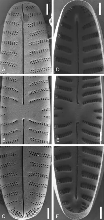
Figure 12 – Gomphonella acsiae, SEM, Lake Balaton, Hungary: A–F, epitype strain D140_006; G–L, strain D140_001; A–C & G–I, external valve views; D–F & J–L internal valve views; A, D, G & J, headpole; B, E, H & K, central area, note the slightly arched striae and the rectangular form; C, F, I & L, footpole, the porelli are larger than the areolae; note the disordered porelli and the large gap of striation between the striae and the porelli. Scale bars = 1 µm.
term genodeme to mark molecular differences from geno- deme 1 (i.e. the epitype of G. olivacea, see above).
Gomphonella acsiae R.Jahn & N.Abarca, sp. nov.
Figs 11–12 & 14B
Type – Hungary, Tihany, Lake Balaton (46.914021°N, 17.892833°E), 23 Apr. 2016, R. Jahn, K. Buczkó D140 (holo-:
B, slide B 40 0042417, prepared from strain D140_006 iso-
lated by O. Skibbe, represented in fig. 11H; iso-: BP, slide HNHM-ALG-D002300).
Description – The morphometric data of the type population (n = 37) are: length 19.2–44.5 μm, width 6.6–8.8 μm and 12–
15 striae in 10 µm (fig. 11A–F). Valves are slightly heteropo- lar, lanceolate in larger specimens to clavate in smaller speci- mens with narrowly rounded headpoles and acutely rounded footpoles. The morphometric data of the clone cultures (n
Figure 13 – Gomphonella coxiae, specimens from sample D201, Helenesee, Brandenburg, Germany: A–I, LM; J–O, SEM; A–F, population;
G–O, strain D201_007; I represents the holotype; J, L & Na, show external valve view and Nb an external girdle view of the footpole; K, M & O, show internal valve views; J & K, headpole; L & M, central area, note the bow tie and the straight striae; Na, Nb & O, footpole, the porelli are larger than the areolae. Note the densely arranged porelli and no gap of striation between the striae and the porelli (compared to Clade 1). Scale bars: A–I = 10 µm; J–O = 1 µm.
= 58) are: length 13–39.7 μm, width 4.5–6.8 μm and 11–15 striae in 10 µm. The valves have a wide variation of shape within the clone cultures: they can be slightly heteropolar, lanceolate and widest at the centre, or heteropolar, clavate with broadly rounded headpoles and acutely rounded foot- poles (figs 11G–S). In both, the natural population and clone cultures, an axial plate and mantle lamella is lacking. The axial area is narrow, straight, expanded at the centre to form a small, rectangular central area bordered at the margins by 1–3 irregularly-shortened striae slightly arched around the central area (fig. 12B, E, H & K). Stigmoids in the central area are lacking. Raphe lateral, with external proximal ends slightly dilated (fig. 12B & H), extending into the central area; external distal raphe endings extend straight (fig. 12A) (in some valves slightly deflected, fig. 12C) onto the valve mantle at both poles. Internal proximal raphe endings curved in the same direction and located on a raised central nodule (fig. 12E & K). Internal distal raphe ends -terminal nodules or helictoglossae - are distinct, in the foot pole positioned
well before valve terminus (fig. 12F & L). The striae are bi- seriate composed of small, round areolae not occluded by si- liceous flaps that terminate as single rows around the central area (fig. 12B, E, H & K). The internal structure of the single rows of areolae is more silicified in some valves, bordered by thickened vimines at the central area (fig. 12E). At the head- pole, the striae are composed of two alternating rows of are- olae (fig. 12A & G). Transapical striae are parallel, becoming radial at the centre 11–15 in 10 µm. The footpole has a large apical pore field with relatively large porelli, different in size from the areolae of the striae and well separated from the striation (fig. 12C & I). Both apices have distinct pseudosep- ta (figs 12D, F & L).
Registration – https://phycobank.org/100350
Etymology – We are dedicating this species to Dr. Éva Ács who has dedicated her scientific life to promote algae re- search in Hungary.
Figure 14 – Comparison of girdle views of Gomphonella species, SEM: A, Gomphonella olivacea, strain 129_007; B, Gomphonella acsiae, strain D140_001; C, Gomphonella coxiae, strain D201_007. Scale bars = 1 µm.
Gomphonella coxiae R.Jahn & N.Abarca, sp. nov.
Figs 13A–O & 14C
Type – Germany, Brandenburg, Helenesee (52.267597°N, 14.503299°E), 13 Jul. 2017, R. Jahn & J. Zimmermann D201 (holo-: B, slide B 40 0042914, prepared from strain D201 007 isolated by O. Skibbe, represented in fig. 13I).
Description – The morphometric data of the type population (n = 10) are: length 33.5–55.7 μm, width 6.7–8.6 μm and 9–11 striae in 10 µm. Valves are slightly heteropolar, lanceo- late in larger specimens to clavate in smaller specimens with narrowly rounded headpoles and acutely rounded footpoles (fig. 13A–F). The morphometric data of the clone culture (n
= 18) are: length 42.8–46.1 μm, width 5.8–7.4 μm and 8–10 striae in 10 µm. The valves of the clone cultures are linear- clavate or with a slight tumid swelling at the centre; head- poles narrowly rounded and footpoles rounded (fig. 13G–I).
In both the natural population and clone cultures the axial plate is lacking. The axial area is narrow, straight, expanded at the centre to form a broad, bow-tie-shaped to rectangular central area bordered at the margin by 2 or 3 approximately equally-shortened striae (fig. 13L–M). Stigmoids are lack- ing. Raphe lateral, with external proximal ends dilated, ex- tending into the central area (fig. 13L); external distal raphe slightly bent before the apical point and extending onto the valve mantle at both poles (fig. 13J & N). Internal proximal raphe endings curved in the same direction and located on a raised central nodule (fig. 13M). Internal distal raphe endings - terminal nodules or helictoglossae - are distinct, positioned well before valve terminus (fig. 13O). The striae are biseriate and composed of two small, round, and alternating but not occluded rows of areolae (fig. 13J); they terminate as single rows but are not arched around the central area. Transapi- cal striae parallel, becoming radial at the centre. The foot- pole has a large apical pore field with relatively large porelli, which differ in size from the areolae of the striae and are well separated from the striation (fig. 13Na, b). Both apices have distinct pseudosepta (fig. 13K & O).
Registration – https://phycobank.org/100352
Etymology – We are dedicating this species to Dr. Eileen Cox for her outstanding contributions to diatom research as an author, editor and colleague. In addition, she organ- ized and hosted the first German Speaking Diatom Meeting when she was doing research at the Limnological Station in Schlitz, Germany, in 1987.
New combinations
As explained further in the Discussion section, astigmate taxa of the genera Gomphoneis and Gomphonema from several localities around the world need nomenclatural transfer to the genus Gomphonella. We effect those transfers here. But we are refraining from recombining taxa where SEM data is missing for an unambiguous demonstration that there is no stigma, typical striation, areolae and porelli (see table 1).
This means that we had to rely on recent descriptions or in- terpretations. And we are refraining from recombining many of the infraspecific taxa of G. olivacea because we think that they need to be studied also molecularly in order to find out if we are dealing here with species or only outline variations.
Differentiating features are listed in table 1.
Gomphonella baicaliana (Kociolek & Kulikovskiy) R.Jahn
& N.Abarca, comb. nov.
Basionym – Gomphoneis baicaliana Kociolek &
Kulikovskiy, Phytotaxa 154: 18, figs 283–286. 2013 (Koci- olek et al. 2013). – Type: Russia, Lake Baikal, 1998 (holo-:
COLO).
Registration – https://phycobank.org/100353
Gomphonella baltica (Cleve) R.Jahn & N.Abarca, comb.
nov.
Basionym – Gomphonema balticum Cleve, Öfversigt af Förhandlingar: Kongl. Svenska Vetenskaps-Akademien 25:
231, pl. 4, figs 10–16. 1868 (Cleve 1868). – Type: Sweden, Gotland.
Registration – https://phycobank.org/100355
Gomphonella basiorobusta (Q.You & Kociolek) R.Jahn &
N.Abarca, comb. nov.
Basionym – Gomphoneis basiorobusta Q.You & Kocio- lek, Phytotaxa 103: 12, figs 55–66. 2013 (You et al. 2013).
– Type: P.R.China, Xinjiang, Kalakule Lake, 16 Jul. 2007, Wang & You 071018 (holo-: SHTU).
Registration – https://phycobank.org/100356
Gomphonella calcarea (Cleve) R.Jahn & N.Abarca, comb.
nov.
Basionym – Gomphonema calcareum Cleve, Öfversigt af Förhandlingar: Kongl. Svenska Vetenskaps-Akademien 25:
231, pl. 4, figs 7–9. 1868 (Cleve 1868).
Synonym – Gomphonema olivaceum var. calcareum (Cleve) Van Heurck, Synopsis des diatomées de Belgique: explana- tion of pl. 25, fig. 23. 1880 (Van Heurck 1880). – Type: Swe- den, Gotland.
Registration – https://phycobank.org/100357
Gomphonella densistriata (Levkov) R.Jahn & N.Abarca, comb. nov.
Basionym – Gomphonema densistriatum Levkov, Phytotaxa 30: 30, figs 210–221, 253–258. 2011 (Levkov & Williams 2011). – Type: Macedonia, Lake Ohrid, 17 Mar. 2007 (holo-:
BM).
Registration – https://phycobank.org/100358
Gomphonella distorta (Q.You & Kociolek) R.Jahn &
N.Abarca, comb. nov.
Basionym – Gomphoneis distorta Q.You & Kociolek, Phy- totaxa 103: 19, figs 97–108. 2013 (You et al. 2013). – Type:
P.R.China, Xinjiang, Little Kalakule Lake, 16 Jul. 2007, Wang & You 071015 (holo-: SHTU).
Registration – https://phycobank.org/100359
Gomphonella fourtanierae (Kociolek & Kulikovskiy) R.Jahn & N.Abarca, comb. nov.
Basionym – Gomphoneis fourtanierae Kociolek &
Kulikovskiy, Phytotaxa 154: 18, figs 287–306, 402–404.
Taxon name Corresponding referenceLength (in µm)W idth (in µm)
Striae (in 10 µm)Areolae per striae
Axial plate Mantle lamellaShape of central areaStriae in central areaPorelli at footpolePore field
close to striae
Pore field
arranged in double r
ows G. acsiae this study13.0–44.54.5–8.811.0–15.0biseriatenonorectangular
slightly arched
relatively large, distinctnono/ discernible G. baicaliana Kociolek et al. (2013: figs 283–286)35–609–1114–17???
irregularly expanded slightly arched
distinct?? G. baltica Levkov (2016: figs 189: 1–19)34–538–1010–11???small, round to irregular
slightly arched
distinct?? G. basiorobusta You et al. (2013: figs 61–66)28.5–52.05.0–8.213–15biseriatenonorectangular to bow-tie
slightly arched
distinctyesyes G. calcarea Cleve (1868: figs 4: 7–9), Levkov (2016: figs 188: 1–19).22–557.5–109–11???bow-tie to round
arched, to slightly arched
??? G. coxiae this study33.5–55.76.7–8.69–11biseriatenonobow-tiestraightrelatively large, distinctyesno G. densistriata Levkov & Williams (2011: figs 253–258)27–447.0–8.515–18biseriatenonosmall, elliptical to rhombicstraight
similar size and shape as yesdiscernible areolae G. distorta small, round to You et al. (2013: figs 30–45.25.5–7.510–14biseriatenono elliptical 102–108)
slightly arched
distinctyesyes G. fonticola Levkov et al. (2007: pl. 176, fig. 20)21–425.5–7.510–14biseriateno?rectangular to bow-tie
slightly arched
similar size and shape as yes? areolae G. fourtanierae rectangular to Kociolek et al. (2013: figs 19–555–1115–18biseriatenono bow-tie 402–404)
straight/ slightly arched
??? G. linearoides Levkov & Williams (2011: figs 246–250)24–355–6.57–10biseriatenonorectangular to bow-tie
straight/ slightly arched
similar size and shape as yesdiscernible areolae G. ohridana 7.5–9.5 at Levkov et al. (2007: pl. 64–13512–22biseriateyes?rectangular centre 178, figs 1–9)
slightly arched
??? G. olivacea this study9.5-42.24.1-9.38.0-15biseriatenono
rectangular bow-tie to transversely elliptical
arched
similar size and shape as yes/ yes discernible areolae G. olivacea 2 13.2–23.34.6–7.011.0–15biseriatenonosee abovearchedsee aboveyesdiscernible this study
Table 1 – Gomphonella taxa, their morphometrics and their ultra-structural differentiating features as documented in the corresponding reference.
Table 1 (continued) – Gomphonella taxa, their morphometrics and their ultra-structural differentiating features as documented in the corresponding reference. Taxon name Corresponding referenceLength (in µm)Width (in µm)
Striae (in 10 µm)Areolae per striae
Axial plate Mantle lamellaShape of central areaStriae in central areaPorelli at footpolePore field
close to striae
Pore field
arranged in double r
ows G. olivaceolacua Lange-Bertalot (1993: fig. 81: 3,4 & fig. 82: 1–4)
23–358.5–10.514–16biseriatenono
rectangular to elliptical- rhombic slightly arched
relatively large, distinctyesyes G. erolivaceolacua Levkov & Williams (2011: figs 241–245)31–539.5–1112–17biseriateno?
small, elliptical to ellongated slightly arched
similar size and shape as yesyes areolae G. potapovae narrow, relatively Kociolek et al. (2013: figs 13–284–616–20biseriatenonorectangular to straight?discernible large, distinct 417–419)X-shaped G. prespanensis Levkov et al. (2007: pl. 47–10512–16.5 180, figs 1–6)
9–12.5 at centre
biseriateyesyesrectangular to elliptical
slightly arched
?yes? G. pseudosubtiloides You et al. 2013: figs 19–25)45.4–518.1–8.69–12biseriatenonorectangular to bow-tie
slightly arched
relatively largeyesyes G. qii You et al. (2013: figs 31–38)39–426.510–12biseriatenonorectangular
straight/ slightly arched
similar size and shape as yesno areolae G. reediae Levkov et al. (2016: pl. 24–355.5–6.611–14biseriatenonobow-tie 187: fig. 31)
straight/ slightly arched
??? G. rostratoides You et al. (2013: figs 91–96)35.8–455.9–6.012–14.5biseriatenonosmall, round to elliptical
slightly arched
similar size and shape as yesdiscernible areolae G. russica Kociolek et al. (2013: figs 47–6811–1411–15biseriatenonorectangularstraight??? 353–359) G. stauroneiformis Van Heurck (1880: pl. 25, 421210–12?nonobow-tiestraight??? fig. 22), this study G. stoermeri You et al. (2013: figs 36.0–54.75.9–7.610–13biseriatenono 77–83)
round to elliptical slightly arched
distinctyesyes G. strelnikovae Kociolek et al. (2013: figs 412–416)12–18.53–519–22biseriatenononarrow, rectangular to X-shapedstraight
yes but absent from valve
faceyesno G. subolivacea Levkov et al.(2007: pl. 176, figs. 10–15)13–286.5–8.515–20biseriatenonosmall variable
straight/ slightly arched
similar size and shape as yesyes areolae
2013 (Kociolek et al. 2013). – Type: Russia, Lake Baikal, 1998 (holo-: COLO).
Registration – https://phycobank.org/100360
Gomphonella fonticola (Hust.) R.Jahn & N.Abarca, comb.
nov.
Basionym – Gomphonema olivaceum var. fonticola Hust., Archiv für Hydrobiologie 40: 942, pl. 40, figs 19–22. 1945 (Hustedt 1945).
Synonyms – Gomphonema fonticola ‘fonticolum’ (Hust.) Levkov & Krstic (Levkov et al. 2007). – Gomphoneis fonti- cola (Hust.) Kociolek & Kulikovskiy (Kociolek et al. 2013).
– Type: Macedonia, Lake Ohrid, St. Naum (lecto-: BRM).
Registration – https://phycobank.org/100361
Gomphonella linearoides (Levkov) R.Jahn & N.Abarca, comb. nov.
Basionym – Gomphonema linearoides Levkov, Phytotaxa 30: 30, figs 199–209, 246–250. 2011 (Levkov & Williams 2011). – Type: Macedonia, Lake Ohrid, 25 Apr. 2003 (holo-:
BM).
Registration – https://phycobank.org/100362
Gomphonella ohridana (Levkov) R.Jahn & N.Abarca, comb. nov.
Basionym – Gomphoneis ohridana Levkov, Iconographia Diatomologica 16: 60, pl. 178, figs 1–9. 2007 (Levkov et al.
2007). – Type: Macedonia, Lake Ohrid, 25 Apr. 2003 (holo-:
MKNH).
Registration – https://phycobank.org/100363
Gomphonella olivaceolacua (Lange-Bert. & E.Reichardt) R.Jahn & N.Abarca, comb. nov.
Basionym – Gomphonema olivaceum var. olivaceolacuum Lange-Bertalot & E.Reichardt, Bibliotheca Diatomologica 27: 67, pl. 80, figs 1–8; pl. 81, figs 3, 4; pl. 82, figs 1–4. 1993 (Lange-Bertalot 1993).
Synonyms – Gomphonema olivaceolacuum (Lange-Bert. &
E.Reichardt) Lange-Bert. & E.Reichardt (Werum & Lange- Bertalot 2004). – Gomphoneis olivaceolacua (Lange-Bert. &
E.Reichardt) Kociolek & Kulikovskiy (Kociolek et al. 2013).
– Type: Switzerland, Lake Geneva, Mar. 1976 (holo-: FR).
Registration – https://phycobank.org/100364
Gomphonella perolivaceolacua (Levkov) R.Jahn &
N.Abarca, comb. nov.
Basionym – Gomphonema perolivaceolacuum Levkov, Phy- totaxa 30: 28, figs 185–198, 241–245. 2011 (Levkov & Wil- liams 2011). – Type: Macedonia, Lake Ohrid, 17 Mar. 2007 (holo-: BM).
Registration – https://phycobank.org/100365
Taxon name Corresponding referenceLength (in µm)W
idth (in µm)
Striae (in 10 µm)Areolae per striae
Axial plate Mantle lamellaShape of central areaStriae in central areaPorelli at footpolePore field
close to striae
Pore field
arranged in double r
ows G. subrussica Kociolek et al. (2013:figs 360–364)29–417–911–13biseriatenonosmall irregular
slightly arched
??? G. subtiloides You et al. (2013: figs 7–14)35–636.4–9.411–13biseriatenonorectangular to bow-tie
slightly arched
similar size and shape as yesno areolae G. tegelensis Skibbe et al. (2018: figs 99.6–124.216.6–19.37–8bi-triseriateyesyesrectangular 6–10 & 13–26)
straight/ slightly arched
relatively large, distinctyesno G. transylvanica Skibbe et al. (2018: figs 11, 12)72–10014–1610–12biseriateyesyesbow-tiestraightdistinctyes? G. xinjiangiana You et al. (2013: figs 48–54)22.4–32.75.3–6.314–16biseriatenonorectangular
slightly arched
slightly largeryesyes
Table 1 (continued) – Gomphonella taxa, their morphometrics and their ultra-structural differentiating features as documented in the corresponding reference.