w w w . e l s e v i e r . e s / b s e c v
Development and characterization of multi-element doped hydroxyapatite
bioceramic coatings on metallic implants for orthopedic applications
Monika Furko
a,∗, Viktor Havasi
b, Zoltán Kónya
b, Alina Grünewald
c, Rainer Detsch
c, Aldo R. Boccaccini
c, Csaba Balázsi
aaHungarianAcademyofSciences,CentreforEnergyResearch,H-1121Konkoly-Thegestr.29-33,Budapest,Hungary
bUniversityofSzeged,DepartmentofAppliedandEnvironmentalChemistry,RerrichB.sqr1,H-6720Szeged,Hungary
cUniversityofErlangen-Nuremberg,DepartmentofMaterialsScienceandEngineering,InstituteofBiomaterials,Cauerstr.6,91058 Erlangen,Germany
a r t i c l e i n f o
Articlehistory:
Received12June2017 Accepted13September2017 Availableonline9October2017
Keywords:
Coatings Microstructure Corrosion Bioceramics
a bs t r a c t
Multi-elementmodifiedbioactivehydroxyapatitebioceramic(mHAp)coatingsweresuccess- fullydevelopedontosurgicalgradetitaniumalloymaterial(Ti6Al4V).Thecoatingswere prepared bypulsecurrentdepositionfromelectrolytecontainingadequate amountsof calciumnitrateandammoniumdihydrogenphosphateat70C.ThepureHAplayerwas doped andco-depositedwithAg,Zn,Mg,Sr ions. Thebiocompatiblepropertiesoflay- erswereinvestigatedbyseedingosteoblast-like MG-63 cellsonto thesamples’surface.
The biocompatiblemeasurementsrevealed enhancedbioactivityofmodifiedHApcom- paredtouncoatedimplantmaterialsandpurebioceramiccoating.Themorphologyand structureofcoatingsandcellswerecharacterizedbyscanningelectronmicroscopy(SEM), energy-dispersiveX-rayspectroscopy(EDX)aswellasFT-IRandXRDmeasurements.The biodegradablepropertiesofsampleswereinvestigatedbyelectrochemicalpotentiodynamic measurements.
©2017SECV.PublishedbyElsevierEspa ˜na,S.L.U.Thisisanopenaccessarticleunderthe CCBY-NC-NDlicense(http://creativecommons.org/licenses/by-nc-nd/4.0/).
∗ Correspondingauthor.
E-mailaddress:furkomonika@gmail.com(M.Furko).
https://doi.org/10.1016/j.bsecv.2017.09.003
0366-3175/©2017SECV.PublishedbyElsevier Espa ˜na,S.L.U.Thisisanopen accessarticleundertheCCBY-NC-NDlicense (http://
creativecommons.org/licenses/by-nc-nd/4.0/).
Desarrolloycaracterizaciónderecubrimientosbiocerámicos dehidroxiapatitadopadosconmulti-elementosenimplantes metálicosparaaplicacionesortopédicas
Palabrasclave:
Recubrimientos Microestructura Corrosión Biocerámicas
r e s u m en
Se handesarrolladocon éxito recubrimientosbiocerámicosde hidroxiapatitabioactiva modificada conmulti-elementos (mHAp)sobresoportes detitanio de gradoquirúrgico (Ti6Al4V). Los recubrimientos sedepositaron con la técnicade lacorriente pulsada a partirdeelectrólitosconcantidadesadecuadasdenitratodecalcioydihidrogenofosfato deamonioa70◦C.LacapadeHAppurasedopóyco-depositóconionesAg,Zn,Mg,Sr.
Labiocompatibilidadde lascapas seinvestigómediantesiembra decélulas deMG-63, similaresalososteoblastos,enlasuperficiedelasmuestras.Losresultadosdelosensayos debiocompatibilidadrevelaronunabioactividadmejoradadelaHApmodificadaencom- paraciónconmaterialesdeimplantenorevestidosyderevestimientobiocerámicopuro.La morfologíayestructuradelosrevestimientosylascélulasfueroncaracterizadasmediante microscopía electrónica de barrido(MEB), espectrometría de dispersión de energía de rayosX(EDX),asícomomediantemedicionesdeFT-IRyDRX.Labiodegradabilidaddelas muestrasseinvestigómedianteensayospotenciométricosdinámicos.
©2017SECV.PublicadoporElsevierEspa ˜na,S.L.U.Esteesunart´ıculoOpenAccessbajo lalicenciaCCBY-NC-ND(http://creativecommons.org/licenses/by-nc-nd/4.0/).
Introduction
Great efforts are made to improve the biocompatibility propertiesofcommonlyusedmetallicimplantmaterialsin orthopedicsurgery. Onesolution canbeapplying bioactive coatings such as calcium phosphates. The phase, struc- ture, compositionand morphology ofthe CaPsurfaces are important parameters that must be accurately controlled toinfluencetheirpotentialbiofunctionalitywithrespectto osteoblasts since interaction between calcium phosphate (CaP) thin layers and osteoblastscan be influenced bythe outermostsurfacepropertiesofthosematerials.Hydroxyap- atite(HAp)hasbeenextensivelystudiedduetothestructural andchemicalsimilaritiestothemaininorganicconstituentof bonetissues.However,itiswelldocumentedthatbiological hydroxyapatite, which forms the mineral phases of calci- fiedtissues(enamel,dentinandbone),differfrompureand syntheticallyproducedHAp[1–3].Biologicalapatiteconsists ofa mixture ofcalcium phosphate phases, such astrical- ciumphosphate(TCP),carbonatedhydroxyapatite(CHA)and calcium-deficienthydroxyapatite(CDHA).Inthisregard,syn- theticHApexhibitsaCa/Pratioof1.67,whilebiologicalapatite deviates significantly from this value and its Ca/P ratio is knowntobeaslowas1.5.Onepromisingwaytomodifythe osteoblasticresponseofHApcoatings, bothinvitroand in vivo,couldinvolvetheuseofsubstitutedHAp,incorporating differentions,suchassilicon[3],magnesium[5],zinc[6],sil- ver[7],strontium[8]intotheHAplattice.Numerousresearch worksontheuseofthesesubstitutedmaterialscanbefound intheliterature[3–11].Ontheotherhand,deepinfectionof megaprosthesesisstillaseriouscomplicationinorthopedic surgery.Bacterial adhesionand biofilm formationon these alloyscaneasilycausevarioushumandiseasesaftersurgery [12].Removingbacteriainabiofilmisimpossibleandalocal orsystemic antibiotictreatmentisnoteffective.Therefore,
theinhibitionofbacterialadhesionisthemostcriticalstep inpreventingimplant-associatedinfections[13].
Inviewoftheproblemofbacterialresistancetoantibiotics and antiseptics, nano-structured silver-containing coatings maybeaneffectivewaytopreventdevicerelatedinfections, becauseitshighandpermanentantimicrobialactivitycom- bines witha remarkablylowhumantoxicity[14–16]. Silver and in particular the free silver ion is well known for its broad-spectrumantimicrobialactivityanditslowtoxicityto mammalian cells, but still allowsfor the independent use oftherapeuticantibiotics[13–16].Strontiumhasbeenshown to havethe dualbenefitof promotingboneformation and reducing boneresorption. Furthermore,it has been shown that strontium has the ability to enhance pre-osteoblastic cellreplicationandcanstimulatetheformationofnewbone throughosteogenesisanddifferentiationintoosteoblastsand has theabilitytoinhibit theactivity ofosteoclasts [17–22].
Mg2+ dopingcanenhancethe osteoblastadhesionstrength ascomparedtopureHApsinceincorporationofMgintopure calcium HApmakesitclosertothenaturalbone[23]while theZncontentcanpromotethewoundhealingprocessafter implantation.
One of the most promising and cheapest methods to depositcoatingsontometallicsubstratesistheelectrodepo- sition,morespecificallypulsecurrentdeposition.Themain advantagesofapplyingpulsecurrentinsteadofdirectcurrent arethatmorehomogeneous,uniformcoatingswithsmaller grainsizecanbeachievedthusimprovingthemechanicaland chemicalpropertiesofcoatings.Sofar,manyresearchworks havebeenperformedusingthisnovelmethodforlayerdepo- sition[24–30].Gopietal.[24]havepreparedmineralsdoped hydroxyapatitecoatingbypulsecurrenton andofftimein seconds(from1sto4s)andinvestigatedtheeffectofparam- eterchange.Wangetal.[25],however,appliedpulse-reverse currentforelectrodeposition.Intheirexperimentstheposi- tiveandreversepulsedutycycleswere0.1and0.5,andthe
positiveand reverse plating times were 10 and 2ms. They foundthatwelladherentcoating couldbeachievedbythis methodwithoutanypost-treatment.Themorphologyofthe suchpreparedcoatingwasmainlyplate-likewiththicknessof around100nm.Inamorerecentstudy,Marashi-Najafietal.
[26]reportedhydroxyapatitecoatingdepositionontoNitinol superelasticalloybypulsecurrentwithdutycycleof0.2at differentcurrentdensities.Theyalsostudiedtheeffectofelec- trolyteconcentrationonthemorphologyofcoatingsandthey revealedthatthestructurechangedfromneedleliketoplate likeastheelectrolyteconcentrationdecreased.Inaddition,it isworthwhiletomentionthatinsomeresearchworksvoltage (pulsedordirect)wasusedfordepositioninsteadofcurrent, accordingtotheauthors’reports[27–30].
Inourpresentresearchworkmulti-element(Ag,Zn,Srand Mg) doped hydroxyapatite coatings havebeen prepared by combinationofpulsecurrentelectrodepositionmethodand surfacepost-treatment.Themorphologyandstructureoflay- ershavebeenstudiedwithSEM-EDXmeasurements.Layers havebeen alsocharacterizedbyFT-IRspectroscopy and X- raydiffractionmeasurements.Thebiocompatibleproperties oflayershavebeenassessedusingMG-63osteoblast-likecells andthebiodegradablecharacteristics ofsampleshavebeen testedinsimulatedbodyfluidbyelectrochemicalmethod.
Experimental
Preparationofpureandsubstitutedcalcium phosphate/hydroxyapatitecoatings
Titanium alloy (Ti6Al4V, ISO5832-3, Protetim Ltd.) discs (10mm×1mm)wereusedassubstrates.Onesideofeachdisk wasroughenedusingasandblastingprocedurewitha180-grit aluminumoxidemedia(accordingtothestandardprocedure appliedbythemanufacturersimilarlythan inthe casesof commercialimplantmaterials).Thissurfacepre-treatmentis necessarytoenhancetheadherenceoflayers.
IGTV-4i/6ttypepulsecurrentgeneratorwasusedtoprepare thedifferentbioceramiccoatings.Inthepulsecurrentwave- form ton is the time when current flows and toff is the relaxationtimewhenthecurrentiszero.Applyingtofftimein pulsecurrentdepositiongivesthesystemtimetorecoverdur- ingtherelaxationperiods.Theelectrodepositionprocesswas carriedoutinatwo-electrodecellundernormalatmospheric conditions,wherethe anodewasaplatinumsheetandthe metallicimplantdiskwasusedasacathode.Thedeposition parametersaresummarizedinTables1and2.Thethickness oflayerswasaround 1–2min allcases(Fig. 1). Themor- phologicalpropertiesofthelayerswerestudiedbySEMand FIBmeasurementswithLEO1540XBCrossbeamworkstation.
ThebeamparametersinSEMimagingmodewere5keVbeam energyand30maperturesize,Everhart-ThornleyandInLens secondaryelectrondetectorswereused.Theionbeamparam- etersinFIBmillingmodewere30kVacceleratingvoltageand 5nAbeamcurrent.ForSEM/FIBmeasurementsthesamples weretiltedat36angle.Theelectronbeamparametersforthe EDXwere8and16keVbeamenergy.ARöntecSi(Li)detector andtheBrukerEsprit1.9softwarehadbeenusedfortheEDX measurements.
Table1–Electrodepositionparametersforobtaining purehydroxyapatitelayers.
Electrochemicaldeposition
Electrolyte Concentration/gL
Ca(NO3)2 115.6
NH4H2PO4 33.30
H2O2(30%) 10ml
Depositionparameters
ton/ms 1
toff/ms 10
ip/Acm−2 5
Bathtemperature/◦C 70
pH 4.5
Depositiontime/s 3
Surfacetreatmentafterdeposition 1MNaOHsolution,70◦C,2h
Table2–Electrodepositionparametersforobtaining modifiedHAplayers.
Electrodeposition
Electrolyte Concentration/gL
Ca(NO3)2 115.6
Mg(NO3)2 2.56
Sr(NO3)2 2.10
NH4H2PO4 33.30
H2O2(30%) 10ml
Depositionparameters
ton/ms 1
toff/ms 10
ip/Acm−2 5
Bathtemperature/◦C 70
pH 4.5
Depositiontime/s 3 Surfacetreatmentafter
deposition
Soakinginsolutioncontaining 0.01MZn(NO3)2and0.0025M AgNO3for24handafterwardin 1MNaOHsolutionat70◦Cfor2h withsubsequentheattreatmentat 150◦Cfor2h.
FT-IRcharacterization
Torecord FT-IRabsorptionspectraofinvestigatedsamples, specular reflection technique was employed. All infrared spectra of the samples were recorded on a Bruker Ver- tex 70 FT-IR spectrometer coupled with Hyperion 2000 IR microscope with 15× (NA=0.4) specular reflection objec- tive.Spectrawererecordedover therangeofwave number 4000–400cm−1atroomtemperatureusing128scansat2cm−1 resolution.
X-raydiffractionmeasurements
The crystal structures of the samples were investigated using X-ray diffraction. XRD spectra were recorded at room temperature by Rigaku MiniFlex II diffractometer (Cu K␣ radiation source, 0.15418nm) equipped with a high count DTEX II detector and operated at 40kV and 40mA. The diffraction patterns were collected over a 2Â range from 10◦ to 60◦ with 1◦/min steps using flat plane geometry.
a
b
c
d
e
f
500 nm
500 nm 500 nm
500 nm
HAp
o AI
P Ca
Ti V C
0 2 4 6 8 10
500 µm
mHAp KeV
o AI
P Ca Ti
C V
0 2 4 6 8 10
500 µm KeV
1.896 µm 1.484 µm
2.028 µm
758.4 nm
Ag Zn Zn MgSr
cps / evcps / ev
Figure1–SEMandSEM/FIBmeasurementsonpureHAplayer(a,b)andonmulti-ionmodifiedHAp(c,d)aswellasEDX spectraonHAp(e)andmHAp(f).
Electrochemicalcorrosionmeasurements
Thepotentiodynamic polarizationstudies were carried out withZahnerIM6eelectrochemicalworkstation(Zahner,Ger- many). In the electrochemical measurements conventional three-electrode cell was used. The working electrode was ametallicimplant disk(19mm)withand withoutcoatings and platinumnet and Ag/AgCl/KClsat electrodes were used as counter electrode and reference electrode, respectively.
Thepotentiodynamicpolarizationcurveswererecordedwith 1mV/s scanningrate.Simulatedbody fluidwas usedasan electrolyteforalltheelectrochemicalexperiments,whichhas ion concentrations nearly equal to those of human blood plasma and isbuffered at pH7.40 with 50mM trishydrox- ymethylaminomethane and 45mM hydrochloric acid. The compositionofsimulatedbodyfluidcanbeseeninTable3.By measuringthecorrosionpropertiesofsamplesitispossibleto tracetheirbiodegradationproperties.Alltheelectrochemical characterizationswerecarriedoutattemperatureof37◦Cto simulatebodyconditions.
Biocompatiblemeasurementsonpureandmodified hydroxyapatitelayers
Cellculture
Cells used for the experiments are represented by MG-63 cellline(Sigma–Aldrich,Germany),whichisalineofhuman osteoblast-likecells.Cellsweregrownon75mlflasksandwere detachedbytripsin.MediumwasDMEM(Dulbecco’sModified EaglesMedium)with10%ofFBS(fetalbovineserum,contain- inggrowthfactorsandnutrientstosupportcellgrowth)and 100U/mlpenicillinand100g/mlstreptomycintominimize theriskofinfections.Theculturesweremaintainedat37◦C, 5%CO2inahumidifiedatmosphereinincubator(NewBran- swickGalaxy170S).Theculturemediawerechangedinevery threedays.ThecellswerecountedinaNeubauerchamber.
Table3–Compositionofsimulatedbodyfluid[31].
Reagent Amount
(g/L)
Sodiumchloride 7.996
Sodiumbicarbonate 0.350
Potassiumchloride 0.224
Potassiumphosphatetrihydrate 0.228 Magnesiumchloridehexahydrate 0.305
1Mhydrochloricacid 40mL
Calciumchloride 0.278
Sodiumsulfate 0.071
Tris(hydroxymethyl)aminomethane 6.057
CellviabilitymeasurementswithWST-8reagent
For cell viability measurementsthe sampleswere put ina 24-wellmicrotiterplateand1mlofcellsuspensionatconcen- trationof10,000cells/mLwasseededontothesurfaceofeach samples.Thesameamountofculturemediumwithcellswith- outsampleswasusedascontrol.Afteracultivationperiodof 2,7and14days,theculturemediawasremovedfromthe24 wellcultureplateandthecellswerewashedwithsterilePBS.
Afterwashing,1mLofDMEMmediumcontaining1%WST-8 reagentwereaddedtothewellsanditwasincubatedfor3.5h.
Theincubation periodwas followedbyspectrophotometric assayofcoloredproduct.Duringthisincubationperiodviable cellsconvertWST-8toawatersolubleformazandye.Thespe- cificabsorbanceofformazandye(at450nm)intheMTPcan bedonewithanELISAplatereader(PHomoAutobioAnthos MykrosystemGMbh,Germany).Theabsorbancedirectlycor- relateswiththecellnumber.
ALPactivitymeasurements
ALP enzyme activity wasmeasured after 6and 14 daysof incubation inordertocharacterizethe osteoblasticactivity of the MG-63 cells. The cells were lysed with a cell lysis bufferwhichcontains20mMTRISbufferedsolution(Merck) with0.1wt%TritonX-100(Sigma,Germany),1mMMgCl2and
Table4–ElementalanalysisofHApandmHApcoatingsderivedfromEDXmeasurements.
Atomicpercent(%)
Spectrum C O Al Ti V Ca P Ag Zn Mg Sr
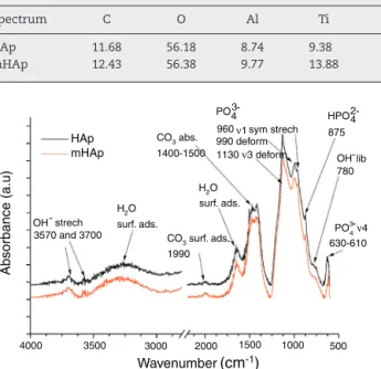
HAp 11.68 56.18 8.74 9.38 0.47 8.53 5.02 – – – –
mHAp 12.43 56.38 9.77 13.88 0.17 4.08 2.62 0.34 0.06 0.19 0.08
HAp mHAp
Absorbance (a.u)
4000 3500 3000 2000 1500 1000 500
OH-
- -
strech H2O
H2O CO3 abs.
CO3 surf. ads.
HPO
- -
3 4
2 4 960ν1
ν4 sym strech
990 deform 1130 ν3 deform 1400-1500
surf. ads.
surf. ads.
875 OH lib 780
PO PO
630-610 1990
3570 and 3700
Wavenumber(cm-1)
3 4
Figure2–FT-IRspectraofHApcoatingandmodifiedHAp coating.
0.1mMZnCl2.Thecelllysatewasincubatedwithareacting solutioncontaining0.1MTrissolution,2mMMgCl2and9mM p-Nitrophenylphosphatefor120min.Afterincubationabsorp- tionwasmeasuredat405nmusingaspectrometer(Specord 40).
Calceinstaining
Forstainingthelivecells,acetoxymethyl(AM)ester(Calcein, MolecularProbes,Germany)wasusedwhichisafluorescent indicator.Thecelldistributiongrowthonthesamplesurface wasanalyzedusingflorescentmicroscope(FM,Scope.A1,Carl Zeiss).Afterthecultivationperiodof48h,theadherentcells werefixedwith3.7vol%paraformaldehydefor10minandper- meabilisedwith0.1vol%TritonX-100(in PBS)for10minat roomtemperature.
DAPI(4,6-diamidino-2-phenylindol)staining
Thenucleioffixedcellswerestainedwiththefluorescence dye4,6-diamidino-2-phenylindol(DAPIRotiVR-MountFluor- Care).Forstainingofthesamples,thematriceswereincubated 15mininthedarkinDAPI-solution(2mLDAPI-stocksolution in1mLDAPIbuffer).Afterstainingward,thematriceswere washedthreetimesinPBStoeliminatethebackground.The nucleiwereimagedbythefluorescencemicroscopewithblue filter.
MorphologicalcharacterizationofMG-63cellsbySEM imaging
Thesamples,seededandculturedwithMG-63cellsfor2days werewashedwithPBS,fixedwithasolutioncontaining3vol%
glutaraldehyde(Sigma, Germany) and3vol%paraformalde- hyde(Sigma,Germany)in0.2Msodiumcacodylatebuffer(pH 7.4),andthoroughlyrinsedwithPBSforSEManalysis(Auriga
CrossBeam,CarlZeissMicroscopyGmbH,Germany).Allsam- plesweredehydratedinethanol,storedin99.8vol%ethanol andcritical-pointdried(EMCPD300,Leica,Germany).
Statistics
Resultsarepresentedusingthemeanvalueandstandarddevi- ationoffourreplicatesofeachsampletype.Allresultswere normalizedtoMG-63cellsgrowthonawellplate(REF=100%).
Thedifferences inanalysis parameters betweenthe differ- entsamplesinvestigatedwereevaluatedbyone-wayanalysis ofvariance(ANOVA).Thelevelofthestatisticalsignificance was definedatp<0.05(Origin 8.6,Origin LabCorporations, USA).Thesignificancelevelwassetas*p<0.05,**p<0.01and
***p<0.001.ForthecomparisonofthemeanvaluestheTukey testwasused.
Results and discussion
MorphologicalinvestigationFig.1showstheSEMandFIBmeasurementsonHAplayerand onmodifiedHApcoating.ItcanbeseeninFig.1(a)thatthe pulseelectrodepositedHApcoatingaftersurfacetreatmentin 1MNaOHsolutionhasmainlysmallneedle-like andlarger rod-likeparticleswithlengthof100–200nmandwithdiame- terof20–50nm.TheCa/Pelementalratiointhiscaseis1.78 (Table4)whichcanindicatemainlyhydroxyapatitecrystalsin thelayer.TheSEM-FIBcrosssectionalimage(Fig.1b)revealed that thelayerhasavery porous,sponge-likestructureand its thickness is notuniform. The thickness oflayervaried between700nmand2m,dependingonthesiteofsamples.
Themetalion-modifiedHAplayer(Fig.1b)showssimilar morphology,however,inthiscaseflake-likeparticleagglom- erationscan alsobeobserved.TheSEM-FIBcross sectional imageshowssimilarlyporousstructurewithlayerthickness of1–2m.OnthecorrespondingEDXspectra,weakpeaksof Ag,ZnSrandMgelementsignalsarealsovisibleprovingthe presenceandincorporationofmetallicionsandparticlesin HAplayer.TheelementalanalysisrevealstheCa/Pelemental ratiotobe1.55whichcanindicatetheHApcrystalstructure disruptionorthepresenceofotherCaPphasesasimpurities.
However,thissmallamountofothercalciumphosphatephase couldnotbedetectedbyXRDmeasurementduetothedetec- tionlimit(Fig.3).ItisvisibleonEDXspectrathatTiandAl andVpeaksalsoappearbecausetheappliedelectronbeam excitedthesubstratematerialalsoduetotheverythinand inhomogeneouscoating.TheappearingveryweaksignalofC ontheEDXspectramightindicatethepresenceofsomecar- bonateimpurities.Thisresultisingoodaccordancewiththe FT-IRmeasurementsinFig.2.
HAp
HAp
mHAp
Intensity (a.u.)
10 20 30 40 50 60
2 Θ
0 0 2 2 2 0 2 1 1 1 1 2 2 2 2 2 1 3 0 0 4
1 1 3
3 0 0 1 3 0
Figure3–XRDmeasurementsonHApcoatingandon modifiedHApcoating.
FT-IRanalysisofpureandmodifiedcalciumphosphate layers
As Fig. 2 shows, the FT-IR spectra are very identical for both coatings. On the spectraof HAp and mHAp samples peaks at627, 960, 990and 1130cm−1 are related to PO43−
anionicgroupcontent,whilethewideabsorptionpeakinthe 1400–1500cm−1regionisconnectedtoabsorbedCO32−con- tentofHApphase[32].Weakeroverlappedpeaksat875cm−1 canberelatedtoHPO42−content,suggestingthepresenceof aminorcarbonatedhydroxyapatite(cHAp)phaseincoatings.
However,theslightlyhigherabsorptionofOH−groups(OH− stretch vibration)at 3700cm−1 inthe caseof mHAp coat- ingmightbeexplainedbysomeeliminationofcHApphase fromHApowingtotheincorporationofdopingelements.In addition,slightsignsofadsorbedwaterbandsalsoappearon spectrafrom3600cm−1toaround2600cm−1andat3570cm−1. X-raydiffractionanalysis
TheXRDpatternsofpureanddopedHApsamplesareshown inFig.3.BothspectrashowscharacteristicpeaksofHApat 2=31.7◦(211),32.9◦(300),25.88◦(002)inaccordancewiththe JCPDSfile09-0432.ThebroadXRDpeaksforHApindicateits nanocrystallinity.Inthecaseofmulti-ionmodifiedHAp,very similarpeakscanbeobserved.NootherCaPphasesorphos- phateimpuritiescanbedetectedonthespectraowingtothe detectionlimitandthecomponents’verylowconcentrations.
Inourcase,there isnovisiblelineshifting, peakbroaden- ing andchanging inpeakintensity whenmetallicionsare addedtothehydroxyapatitecoating.However,severalstudies reportedlineshiftingtohigher2valuesduetothereplace- mentoflargersizedCa2+(0.099 ˚A)ionswithsmallersizedMg2+
(0.69 ˚A)ionsandZn2+(0.77 ˚A)ions[33–35].Inotherresearch work,Zianietal.foundbroadeningofthepeaksduetothe reductioninthecrystallitesizeandincreaseinthelatticedis- order,whichtheyattributedtotheMg2+ substitutioninthe HAplattice[36].Ontheotherhand,thesubstitutionofstron- tiumandsilvercancausephaseshiftingtolower2Âindicating anincreaseinthelatticeparameters,whichcanbeattributed
Ti6AI4V HAp coating mHAp coating -3
-4 -5 -6 -7 -8
-2 -1 0 1 2
E VS Ag/AgCI / V
log LjL / A
Figure4–Potentiodymanicpolarizationcurvesofuncoated Ti6Al4Valloy(blackline),ofHApcoating(blueline)andof mHAp(greenline)recordedaftertwoweeksimmersionin SBFsolutionat37◦C.Thepotentialscanningrateis1mV/s.
Table5–Electrochemicalparameters:passivecurrent density(jp),corrosionpotential(Ecorr)andcorrosion current(jcorr)valuesderivedfromthepotentiodynamic curvesinFig.4.
Sample jp/Acm−2 jcorr/Acm−2 Ecorrvs Ag/AgCl/mV
Ti6Al4V 0.91 0.26 −190
HApcoating 2.57 1.04 −295
mHApcoating 3.30 1.51 −486
tothehigherionicradiusofSr(1.13 ˚A)andAg(1.15 ˚A),ascom- paredtoCa2+[37].
Corrosioncharacterizationbyelectrochemical potentiodynamicmeasurements
Fig. 4demonstratesthepotentiodynamic curvesofimplant material(Ti6Al4V)andHApcoatingandmodifiedHApcoating.
ThecurveswererecordedaftertwoweeksimmersioninSBF solution.
As Fig. 4 reveals, large anodic passive regions can be observedontheanodicbranchesofpotentiodynamiccurves inallcaseswithsmallpassivecurrentdensities(jp)andthe shapesofpotentiodynamiccurvesofallsamplesisquitesim- ilar. Inthe caseofuncoatedimplant materialthe onsetof this passive region isaround +100mV vsAg/AgCl and the passive film breakdown potential is at +980mV. The pas- siveregiononpotentiodynamiccurves ofpureHApcoating becameslightlywideraftertwoweeksofimmersionthanthat foruncoatedsample,itstartsataround−120mVvsAg/AgCl anditsbreakdownpotentialissimilarlyataround+980mV.
Ontheotherhand,thewidestpassiveregionisobservedin thecaseofmHApcoating,spreadingfrom−280mVtoaround +1VvsAg/AgCl.Theverylargeslopesofanodicandcathodic branchesofcurvesindicatemixedkineticanddiffusioncon- trolledelectrodeprocessesforallsamples.
***
*** ***
*** ***
***
**
*
Control Ti6AI4V HAp mHAp 120
100
80
60
40
20
0
% cell viability
Time / days
2. day 6. day 14. day
p=0.94
Figure5–Cellviabilitypercentageontheinvestigated samplescomparedtopositivecontrol.Positivecontrol:
MG-63cellsweregrowninwellplateswithoutsamples.
Thelevelofthestatisticalsignificanceisgivenbyp-values ascomparedtocontrolandtitaniumsubstrate.Allsamples weremeasuredin6replicateandcalculatedthemean values±standarddeviation.
Control Ti6AIaV HAp mHAp p = 0.094
* *
*
*
120 **
100
80
60
40
20
0
ALP activity %
6. day 14. day
Time / days
Figure6–ALPexpressionpercentageontheinvestigated samplescomparedtopositivecontrol.Positivecontrol:
MG-63cellsweregrowninwellplateswithoutsamples.
Thelevelofthestatisticalsignificanceisgivenbyp-values ascomparedtocontrolandtitaniumsubstrate.Allsamples weremeasuredin6replicateandcalculatedthemean values±standarddeviation.
Theelectrochemicalparameters,suchaspassivecurrent densities,corrosioncurrentdensitiesandcorrosionpotentials ofdifferentsamplesaresummarizedinTable5.
Itis visible that the titanium alloy substrate possesses the lowest passive current density (0.91Acm−2), whilethe highestvaluebelongs tomulti-elementdopedHApcoating (3.30Acm−2). On the other hand, it can also be observed ontheanodicbranchofpotentiodynamiccurvesthatwhile thepassivecurrentsofmHApsamplesslightlydecreasewith potentialscan,thepassivecurrentsofsubstratematerialand HApcoatingarestableandhardlychangetillthebreakdown potential.
Thecorrosioncurrentdensity(jcorr)valuesandcorrosion potentials(Ecorr)canbeobtainedbytheintersectionoflines extrapolatedtothecathodicandanodicbranchofpotentio- dynamiccurves inthe Tafelregion(±50mVfrom corrosion potential).Thetitaniumalloyhasthenoblestcorrosionpoten- tialand lowestcorrosioncurrentdensity whichdenotesits highestcorrosionstability.Ontheotherhand,themostneg- ative Ecorrand the highestjcorrvaluesbelong tothemHAp samples.Thisresultcanprovethatduringimmersioninphys- iological solution, dissolutionprocesses ofdifferentdoping elementsaswellascalciumphosphatecomponentscanoccur.
Thereareseveralresearchworksinvestigatingthedegra- dation processes of hydroxyapatite coatings prepared by differentmethods. Itisreportedthat theporous character- istic (sizeand number of pores present in the coating) of calcium phosphate coatings significantlyaffects the corro- sion/dissolution rate of hydroxyapatite. The coatings with smallerandfewerporesprovedtobemorecorrosionresis- tantthancoatingswithhigherdegreeofporositybecausethe formercanprovidebetterbarrierproperty[38–40].
Zhangetal.[32]statedthatthecorrosionmechanismof HApcoatingwithporesinvolveshydrogenion(H+)generation attheinterfacewherecorrosionoccurs,thusdecreasingthe localpHvalue,andthencausessubsequentdissolutionofHAp inthehighH+concentrationarea.ThedissolutionrateofHAp increaseswithdecreasingpH.
Biocompatiblemeasurementsonsamples
CellviabilitymeasurementwithWST-8assay
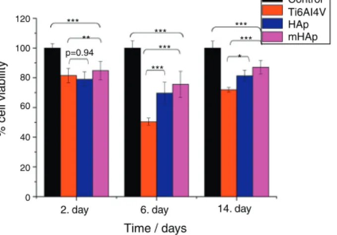
Fig.5showsthatinallcultureperiodthemHApsamplehad thehighestcellviabilityvalues,after2daysitwas85%while aftertwoweeksitincreasedtoaround90%comparedtopos- itivecontrol.
Thecell viabilitypercentageswere 78% and 85%after2 days,81%and90%after2weeksofcultureonpureHApand multi-ionmodifiedHApcoatings,respectively.Foruncoated titanium,theviabilitywas81%at2nddayanditdecreased to71% at14thday. After2daysofculture,the differences betweenthecellviabilityvalueswerenotstatisticallysignif- icant forHApcomparedtotitaniumsubstrate (pvaluewas 0.94), whilethedifference betweenTialloyandmHApwas statisticallydifferent(p<0.01).Itisvisiblethatthereisaslight decrease incell viabilityforeach sampleafteroneweekof incubation.Thisphenomenoncanbeexplainedbycelldiffer- entiation.Severalresearchersprovedthatwhencellsarein thestateofdifferentiation,theyshowlessmetabolicactivity resultinginlowerviabilityvalues[41,42].
After2weeksofcultureinDMEMmediumthedifference between the cell viability on HAp and on mHAp samples become moresignificantly higher than those foruncoated substrate,indicatingthegoodbiocompatible/bioactiveprop- ertiesofbothhydroxyapatitelayers.Itisalsovisiblethatthe multi-elementmodificationadvancedthebiocompatibilityof sample.Thedifferencesbetweenthecellviabilitiesofsam- plesinthistimepointwereallstatisticallyhighlysignificant (p<0.001).Inaddition,it iswell knownthathydroxyapatite coatingfacilitatetheattachmentandgrowthofosteoblastic cellsowingtoitshighhydrophilicproperty[43,44].
Alkalinephosphataseactivitymeasurements
ALP is one of the first osteoblastic markers. Since the osteoblast-likehumanMG-63celllineiscapabletoproduce someosteogenicmarkerssuchasalkalinephosphataseand osteocalcin[45].InourpresentstudyALPexpressionofcells seededonthesurfaceofdifferentsamplesandonculturewell plateasreferencewasevaluated.
It is visible in Fig. 6 that the ALP expressionis higher byaround 25%and30% forpureHApand multi-iondoped HAp,respectively,after6and14daysofculturethanthatfor uncoatedsubstrate.ThelevelofALPactivityincreasedwith culturingtime. After 6 daysof immersion,the ALP values ofbothHApand mHApwere statisticallydifferent(p<0.05) comparedtouncoatedsubstrate,whiletherewasnostatis- tically difference between the calcium phosphate coatings andthe controlgroup.Atthe14thday ofculture,onlythe ALPvaluesofmHApcomparedtoTi alloyandALP expres- sionofcontrolcomparedtoTialloywerestatisticallydifferent (p<0.05).ItisvisiblethatthehighestALPexpressionbelongs tomHApsample.Ontheotherhand,thedifferencesbetween HApand mHApaswell asbetweentitanium substrateand HAp are not statistically different, in the latter case the pvalue is 0.094. Our findings are ingood agreement with reportsfromliteraturewhereZhaoetal.[46]studiedtheeffect of magnesium-substitutednano-hydroxyapatite coating on implant osseointegration.Intheirresearchtheyfoundthat themagnesiumsubstitutedHAphadhigherALPactivityby twotimesthanthat ofwithoutmagnesium contentafter7 daysofculture.Yangetal.[47]investigatedthebiocompatibil- ityofZnsubstitutedhydroxyapatiteonMurinepreosteoblast cell(MC3T3-E1)cellline.Theyreportedsignificantincreasein cellproliferationandALPactivityonday7,andosteocalcin
production(p<0.05)were alsoobservedforZn2+-containing HAp-coatedsurfacesonday14.Thecoatingswereprepared byelectrochemicalprocessandtheZnwaspresentintheZn- HApcoatingsataZn/(Ca-Zn)molarratioof1.04%.Buenoetal.
[48] studiedthe effectofSr substitution inHApnanocom- positeonthedifferentiationofOFCOLLIIosteoblasts.Other literaturereportshowedthatthepresenceofstrontiuminthe HApstructure(SrHAp) seemstocauseimportanteffectsin osteoblastandosteoclastgrowthandalsofavorstheincrease ofosteoblastALPactivity [49]. Thianet al.[50]investigated theeffectofapatitenanocrystalsontheosteoblastbehavior ofhumanosteoblast(HOB)cellsandtheyfoundthattheALP activityofcellsgrowingonphase-pureapatitenanocrystals wasdetectableonlyafter5daysofculture.
Calcein/DAPIstaining
Directfluorescencestainingofcalceinandnucleus(DAPI)of MG-63cells culturedfor2daysontitanium alloy,HApand mHApcoatingsaswellasoncontrolgroup(wellplates)are showninFig.7.
Calceinfluorescentstainingisgenerallyusedtoindicate intracellular esteraseactivity present inviable cells.Dense and evenly dispersed multi-layered cells with large nuclei were observedforallsamples,however,inthecaseofHAp andmHApcoatedsamplestherewerelargernumberofliving cells.Theshapeofcellsmainlyelongatedandpolygonalwhich indicateswelladhered,spreadingandproliferatingcells.
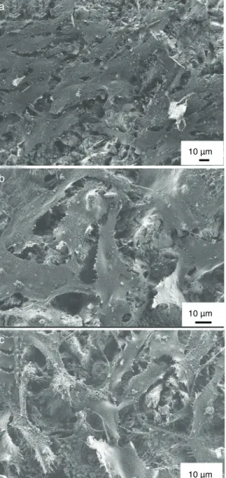
MG-63cellmorphologystudy
Theexpressionofthephenotypeofosteoblast-likecells(MG- 63)wasstudiedbySEMafterincubationonuncoatedtitanium
a
c b
50 µm
50 µm
50 µm
Figure7–Fluorescencemicroscopyimagesofcalcein-AM(greenfluorescent)andnucleus(withDAPI,bluefluorescent)and mergedimagesofMg-63cellsculturedfor2daysinDMEMmediumondifferentsamplessuchastitaniumalloy(a)HAp(b) andmHAp(c)coatings.
a
b
c
10 µm
10 µm
10 µm
Figure8–SEMimagesonMG-63cellsgrownontitanium substrate(a)onHApcoating(b)andonmHApcoatingafter 2daysofcultureinDMEMmedium.
alloy,onpureHApcoatingandonion-modifiedHApcoatings for48h.ItisobviousthatthephenotypeofMG-63osteoblast- likecellswerewell-expressedandcellwerespreadedonthe surfacesofallsamplesandwereinflattenedform.Theshape ofcells mainlypolygonalwithfilopodiaorverythin exten- sions.Thecellscoveredthecoatedsamples’surfacesinathick continuousmonolayerandtheMG-63startedtoformalsoa multilayerinsomeareasofthesample.Ontheotherhand,in thecaseofuncoatedsubstrate,thecoveragewasnotperfect.
Insomeplacesthesurfaceofsubstrateisalsovisiblebeside thecells(seeinFig.8a).Thenumberanddensityofcellsaswell astheextentofspreadingseemedtobealittlehigherinthe caseofcalciumphosphatecoatedsamplesthanforuncoated substrate.Nevertheless,thereisnotmuchvisibledifference
incellmorphologyinthecaseofbothHApandmHApcoated samples. Theseresultsmight confirmthat thecoating can advancecelladherencethuspromotingcellproliferationand provetheresultsfromCalcein/DAPIstaining.
Conclusion
TheSEManalysisrevealedthatthemorphologyofHApand mHAp coatings wasmainly needle-like in nanometresize.
The crosssection analysis (FIB)showed the coatings tobe inhighlyporous,sponge-likestructure,whichresemblesthe structureofnaturalbone.TheEDXelementalanalysiscon- firmedthattheionsdopedHApcoatingcontainedAg,Zn,Sr andMgelementsalsoinunder1At%alongwiththecalcium andphosphorouselements.TheFT-IRspectrashowedsimi- larcharacteristicpeaksofPO43− andOH−anionicgroupsof calciumphosphatephasesandrevealedcarbonateimpurities inbothsamples.TheXRDmeasurementsalsoconfirmedthat thecoatingconsistofmainlynanocrystallinehydroxyapatite phaseandtherewasnovisiblelineshifting,peakbroaden- ingandchanginginpeakintensitywhenmetallicionswere addedtothehydroxyapatitecoating.Accordingtothecorro- sion measurements,the corrosion resistancesofpureHAp andmulti-iondopedHApwerelowerthanthatofuncoated substrateduetothehighlyporouscharacteristicoflayers.
Thebiocompatibletestsshowedthatthecellviabilityval- uesincreasedsignificantlyinthecasesofbothHApandmHAp samplescomparedtobareimplantmaterialsand thehigh- estvaluesweremeasuredinthecaseofmHAp.TheCalcein andDAPIstainingofsamplesrevealeddense,multi-layered, welladheredlivingcellsonallsampleswithnormalmorphol- ogy.TheinvitroresultspresentedheresupportthatHApand multi-iondopedHApcoatingsadvancethegrowthofMG-63 osteoblast-likecells.
Acknowledgements
Theauthorswouldliketoacknowledgethefinancialsupport ofJECSTrustandtheauthorsaregratefulfortheSEM-FIB/EDX measurementsperformedbyLeventeIllés(MTA-EK,Hungary).
references
[1]L.Sun,C.C.Berndt,K.A.Gross,A.Kucuk,Material
fundamentalsandclinicalperformanceofplasma-sprayed hydroxyapatitecoatings:areview,J.Biomed.Mater.Res.B:
Appl.Biomater.58(2001)570–592.
[2]A.C.Tas,Combustionsynthesisofcalciumphosphate bioceramicpowders,J.Eur.Ceram.Soc.12(2000)2389–2394.
[3]E.S.Thian,J.Huang,S.M.Best,Z.H.Barber,W.Bonfield, Surfacemodificationofmagnetron-sputteredhydroxyapatite thinfilmsviasiliconsubstitutionfororthopaedicanddental applications,Surf.Coat.Technol.205(2011)3472–3477.
[4]S.V.Dorozhkin,M.Epple,Biologicalandmedicalsignificance ofcalciumphosphates,Angew.Chem.Int.Ed.41(17)(2002) 3130–3146.
[5]S.Ziani,S.Meski,H.Khireddine,Characterizationof magnesium-dopedhydroxyapatitepreparedbysol–gel process,Int.J.Appl.Ceram.Technol.11(2014)83–91.
[6] N.A.Trujillo,R.A.Oldinski,H.Ma,J.D.Bryers,J.D.Williams, K.C.Popat,Antibacterialeffectsofsilver-doped
hydroxyapatitethinfilmssputterdepositedontitanium, Mater.Sci.Eng.C32(2012)2135–2144.
[7] F.Ren,R.Xin,X.Ge,Y.Leng,Characterizationandstructural analysisofzinc-substitutedhydroxyapatites,ActaBiomater.
5(2009)3141–3149.
[8] K.Ozeki,T.Hoshino,H.Aoki,T.Masuzawa,Phase compositionofsputteredfilmfromamixtureof
hydroxyapatiteandstrontium-apatite,J.Mater.Sci.Technol.
29(2013)1–6.
[9] C.O’Sullivan,P.O’Hare,N.D.O’Leary,A.M.Crean,K.Ryan, A.D.W.Dobson,L.O’Neill,Depositionofsubstitutedapatites withanticolonizingpropertiesontotitaniumsurfacesusing anovelblastingprocess,J.Biomed.Mater.Res.B:Appl.
Biomater.95B(2010)141–149.
[10]J.H.Shepherd,D.V.Shepherd,S.M.Best,Substituted hydroxyapatitesforbonerepair,J.Mater.Sci.Mater.Med.23 (2012)2335–2347.
[11]S.Sprio,A.Tampieri,E.Landi,M.Sandri,S.Martorana,G.
Celotti,G.Logroscino,Physicochemicalpropertiesand solubilitybehaviourofmulti-substitutedhydroxyapatite powderscontainingsilicon,Mater.Sci.Eng.C28(2008) 179–187.
[12]Y.Ando,H.Miyamoto,I.Noda,N.Sakurai,T.Akiyama,Y.
Yonekura,T.Shimazaki,M.Miyazaki,M.Mawatari,T.
Hotokebuchi,Calciumphosphatecoatingcontainingsilver showshighantibacterialactivityandlowcytotoxicityand inhibitsbacterialadhesion,Mater.Sci.Eng.C30(2010) 175–180.
[13]W.K.Jung,H.C.Koo,K.W.Kim,S.Shin,S.H.Kim,Y.H.Park, Antibacterialactivityandmechanismofactionofthesilver ioninStaphylococcusaureusandEscherichiacoli,Appl.Environ.
Microbiol.74(2008)2171–2178.
[14]M.Schierholz,L.J.Lucasj,A.Rump,G.Pulverer,Efficacyof silver-coatedmedicaldevices,J.Hosp.Infect.40(1998) 257–262.
[15]W.C.Chiang,L.R.Hilbert,C.Schroll,T.Tolker-Nielsen,P.
Moller,Bacterialinhibitingsurfacescausedbytheeffectsof silverreleaseand/orelectricalfield,Electrochim.Acta54 (2008)108–115.
[16]S.Seuss,M.Heinloth,A.R.Boccaccini,Developmentof bioactivecompositecoatingsbasedoncombinationofPEEK, bioactiveglassandAgnanoparticleswithantibacterial properties,Surf.Coat.Technol.301(2016)100–105.
[17]K.M.Cheung,W.W.Lu,K.D.Luk,C.T.Wong,D.Chan,etal., Vertebroplastybyuseofastrontium-containingbioactive bonecement,Spine30(2005)S84–S91.
[18]C.T.Wong,W.W.Lu,W.K.Chan,K.M.Cheung,K.D.Luk,D.S.
Lu,A.B.Rabie,L.F.Deng,J.C.Leong,Invivocancellousbone remodelingonastrontium-containinghydroxyapatite (Sr-HA)bioactivecement,J.Biomed.Mater.Res.68A(2004) 513–521.
[19]G.X.Ni,W.W.Lu,K.Y.Chiu,Z.Y.Li,D.Y.Fong,K.D.Luk, Strontium-containinghydroxyapatite(Sr-HA)bioactive cementforprimaryhipreplacement:aninvivostudy,J.
Biomed.Mater.Res.B:Appl.Biomater.77(2006) 409–415.
[20]A.Barbara,P.Delannoy,B.G.Denis,P.J.Marie,Normalmatrix mineralizationinducedbystrontiumranelateinMC3T3-E1 osteogeniccells,Metabolism53(2004)532–537.
[21]J.Abert,C.Bergmann,H.Fischer,Wetchemicalsynthesisof strontium-substitutedhydroxyapatiteanditsinfluenceon themechanicalandbiologicalproperties,Ceram.Int.40 (2014)9195–9203.
[22]R.Baron,Y.Tsouderos,InvitroeffectsofS12911-2on osteoclastfunctionandbonemarrowmacrophage differentiation,Eur.J.Pharmacol.450(2002)11–17.
[23]V.K.Mishra,B.N.Bhattacharjee,O.Parkash,D.Kumar,S.B.
Rai,Mg-dopedhydroxyapatitenanoplatesforbiomedical applications:asurfactantassistedmicrowavesynthesisand spectroscopicinvestigations,J.AlloysCompd.614(2014) 283–288.
[24]D.Gopi,A.Karthika,S.Nithiya,L.Kavitha,Invitrobiological performanceofmineralssubstitutedhydroxyapatitecoating bypulsedelectrodepositionmethod,Mater.Chem.Phys.144 (2014)75–85.
[25]H.X.Wang,S.K.Guan,X.Wang,C.X.Ren,L.G.Wang,Invitro degradationandmechanicalintegrityofMg–Zn–Caalloy coatedwithCa-deficienthydroxyapatitebythepulse electrodepositionprocess,ActaBiomater.6(2010)1743–1748.
[26]F.Marashi-Najafi,J.Khalil-Allafi,M.R.Etminanfar, Biocompatibilityofhydroxyapatitecoatingsdepositedby pulseelectrodepositiontechniqueontheNitinol superelasticalloy,Mater.Sci.Eng.C76(2017)278–286.
[27]R.ChakrabortySrijanSengupta,P.Saha,K.Das,S.Das, Synthesisofcalciumhydrogenphosphateand
hydroxyapatitecoatingonSS316substratethroughpulsed electrodeposition,Mater.Sci.Eng.C69(2016)875–883.
[28]L.Shoujie,L.Hejun,Z.Leilei,Y.Xuemin,G.Yao,Insimulated bodyfluidperformanceofpolymorphicapatitecoatings synthesizedbypulsedelectrodeposition,Mater.Sci.Eng.C 79(2017)100–107.
[29]M.Saremi,S.Mohajernian,S.Hejazi,Controllingthe degradationrateofAZ31Magnesiumalloyandpurityof nano-hydroxyapatitecoatingbypulseelectrodeposition, Mater.Lett.129(2014)111–113.
[30]N.Monasterio,J.L.Ledesma,I.Aranguiz,A.Garcia-Romero,E.
Zuza,Analysisofelectrodepositionprocessestoobtain calciumphosphatelayeronAZ31alloy,Surf.Coat.Technol.
319(2017)12–22.
[31]T.Kokubo,H.Kushitani,S.Sakka,T.Kitsugi,T.Yamamuro, Solutionsabletoreproduceinvivosurface-structure changesinbioactiveglass-ceramicA-W,J.Biomed.Mater.
Res.24(1990)721–734.
[32]L.Berzina-Cimdina,N.Borodajenco,Researchofcalcium phosphatesusingFouriertransforminfraredspectroscopy, in:T.Theophile(Ed.),InfraredSpectroscopy–Materials ScienceEngineeringandTechnology,2012,pp.123–148, ISBN:978-953-51-0537-4.
[33]A.Bigi,G.Falini,E.Foresti,M.Gazzano,A.Ripamonti,N.
Roveri,Rietveldstructurerefinementsofcalcium
hydroxylapatitecontainingmagnesium,ActaCrystallogr.52 (1996)87–92.
[34]F.Z.Ren,R.L.Xin,X.Ge,Y.Leng,Characterizationand structuralanalysisofzinc-substitutedhydroxyapatites,Acta Biomater.5(2009)3141–3149.
[35]Y.Cai,S.Zhang,X.Zeng,Y.Wang,M.Qian,W.Weng, Improvementofbioactivitywithmagnesiumandfluorine ionsincorporatedhydroxyapatitecoatingsviasol–gel depositiononTi6Al4Valloys,ThinSolidFilms517(17)(2009) 5347–5351.
[36]S.Ziani,S.Meski,H.Khireddine,Characterizationof magnesium-dopedhydroxyapatitepreparedbysol–gel process,Int.J.Appl.Ceram.Technol.11(1)(2014)83–91.
[37]Z.Geng,Z.Cui,Z.Li,S.Zhu,etal.,Strontiumincorporation tooptimizetheantibacterialandbiologicalcharacteristicsof silver-substitutedhydroxyapatitecoating,Mater.Sci.Eng.C 58(2016)467–477.
[38]I.C.Lavos-Valereto,I.Costa,S.Wolynec,Theelectrochemical behaviorofTi-6Al-7Nballoywithandwithout
plasma-sprayedhydroxyapatitecoatinginHanks’solution,J.
Biomed.Mater.Res.63(2002)664–670.
[39]Z.Zhang,M.F.Dunn,T.D.Xiao,A.P.Tomsia,E.Saiz, Nanostructuredhydroxyapatitecoatingsforimproved adhesionandcorrosionresistanceformedical
implants,Nanotechnol.Biotechnol.Convers.(2002) 291–296.
[40]C.T.Kwok,P.K.Wong,F.T.Cheng,H.C.Manc,
Characterizationandcorrosionbehaviorofhydroxyapatite coatingsonTi6Al4Vfabricatedbyelectrophoreticdeposition, Appl.Surf.Sci.255(2009)6736–6744.
[41]M.Agostini,F.Romeo,S.Inoue,M.V.Niklison-Chirou,etal., Metabolicreprogrammingduringneuronaldifferentiation, CellDeathDiffer.23(2016)1502–1514.
[42]L.Schneider,S.Giordano,B.R.Zelickson,etal.,
DifferentiationofSH-SY5Ycellstoaneuronalphenotype changescellularbioenergeticsandtheresponsetooxidative stress,FreeRadic.Biol.Med.51(11)(2011)2007–2017.
[43]E.S.Thian,Z.Ahmad,J.Huang,M.J.Edirisinghe,S.N.
Jayasinghe,D.C.Ireland,R.A.Brooks,N.Rushton,W.
Bonfield,S.M.Best,Theroleofsurfacewettabilityand surfacechargeofelectrosprayednanoapatitesonthe behaviourofosteoblasts,ActaBiomater.6(2010)750–755.
[44]K.L.Kilpadi,P.-L.Chang,S.L.Bellis,Hydroxylapatitebinds moreserumproteins,purifiedintegrins,andosteoblast precursorcellsthantitaniumorsteel,J.Biomed.Mater.Res.
57(2001)258–267.
[45]J.Sun,L.Wei,X.Liu,J.Li,B.Li,G.Wang,F.Meng,Influences ofionicdissolutionproductsofdicalciumsilicatecoatingon
osteoblasticproliferation,differentiationandgene expression,ActaBiomater.5(2009)1284–1293.
[46]S.Zhao,Q.Jiang,S.Peel,X.Wang,F.He,Effectsof magnesium-substitutednanohydroxyapatitecoatingon implantosseointegration,Clin.OralImplantsRes.24(2011) 34–41.
[47]F.Yang,W.Dong,F.He,etal.,Osteoblastresponsetoporous titaniumsurfacescoatedwithzincsubstituted
hydroxyapatite,OralSurg.OralMed.OralPathol.OralRadiol.
113(2012)313–318.
[48]V.B.Bueno,R.Bentini,L.H.Catalani,L.R.S.Barbosa,D.F.S.
Petri,Synthesisandcharacterizationof
xanthan–hydroxyapatitenanocompositesforcellular uptake,Mater.Sci.Eng.C37(2014)195–203.
[49]C.Capuccini,P.Torricelli,F.Sima,E.Boanini,C.Ristoscu,B.
Bracci,G.Socol,M.Fini,I.N.Mihailescu,A.Bigi,
Strontium-substitutedhydroxyapatitecoatingssynthesized bypulsed-laserdeposition:invitroosteoblastand
osteoclastresponse,ActaBiomater.4(2008) 1885–1893.
[50]E.S.Thian,Z.Ahmad,J.Huang,M.J.Edirisinghe,etal.,The roleofelectrosprayedapatitenanocrystalsinguiding osteoblastbehaviour,Biomaterials29(2008)
1833–1843.