Contents lists available atScienceDirect
Acta Histochemica
journal homepage:www.elsevier.com/locate/acthis
Non-competitive antagonists of NMDA and AMPA receptors decrease seizure-induced c-fos protein expression in the cerebellum and protect against seizure symptoms in adult rats
Zoltán Tóth, András Mihály
⁎, Adrienne Mátyás, Beáta Krisztin-Péva
Department of Anatomy, Faculty of Medicine, University of Szeged, Szeged, Hungary
A R T I C L E I N F O
Keywords:
4-aminopyridine Cerebellar cortex Mossyfiber Glutamate receptor c-Fos protein
A B S T R A C T
The aim of the present study was to examine the role of ionotropic glutamate receptors in the cerebellum during generalized seizures. Epileptic neuronal activation was evaluated through the immunohistochemical detection of c- fos protein in the cerebellar cortex. Generalized seizures were precipitated by the intraperitoneal injection of 4- aminopyridine. The animals were pretreated with the NMDA receptor antagonists MK-801 (2 mg/kg), amantadine (50 mg/kg), and the AMPA receptor antagonist GYKI 52466 hydrochloride (50 mg/kg). Two hours after 4-ami- nopyridine injection, the number of c-fos immunostained cell nuclei was counted in serial immunohistochemical sections of the cerebellar vermis. The number of c-fos immunostained cell nuclei in the granular layer decreased significantly in animals pretreated with the glutamate receptor antagonists compared to the untreated animals having convulsion. We can conclude that mossyfiber stimulation exerts its seizure-generating action mainly through the ionotropic glutamate receptors of the mossyfiber synapses. Both NMDA and AMPA receptor an- tagonists are effective in reducing glutamate-mediated postsynaptic effects in the cerebellar cortex.
1. Introduction
Epilepsy causes distinct neuropathological alterations in the cere- bellum resulting in the significant loss of Purkinje cells (Honavar and Meldrum, 2002). Nevertheless, there are epileptic seizures which clearly originate from pathological lesions of the cerebellum (Boop et al., 2013). Moreover, stimulation of the cerebellum is used to treat intractable epilepsy (Ge et al., 2013). The double-faced role of the cerebellum in epilepsy can be explained by the double-faced anatomical connections of the cerebellum and the neocortex-diencephalon axis.
The neocortex is projecting to the pontine nuclei, which project to the cerebellum through the pontocerebellar tract (Lee and Mihailoff, 1990).
Axon terminals of the corticopontine tract make excitatory synaptic contacts with the dendrites of the pontine neurons (Brodal and Bjaalie, 1992). The glutamatergic pontocerebellar system terminates as mossy fibers in the granular layer of the cerebellar cortex stimulating mainly granule cells and Golgi cells (Castejon and Castejon, 2000). The cere- bellar efferent informationflows to the deep cerebellar dentate nucleus,
which projects to the thalamus, and the thalamic neurons project to the frontal neocortical areas in order to form excitatory synapses (Geminiani et al., 2017). The dentate nucleus is also connected to the pontine nuclei (Lee and Mihailoff, 1990).
In our previous studies, we proved that generalized tonic–clonic seizures (GTCS) precipitated by the potassium channel blocker 4-ami- nopyridine (4-AP) caused long-lasting, significant increase of c-fos protein expression in the granular layer of the cerebellar cortex (Tóth et al., 2015), very similarly to that seen in the hippocampus and neo- cortex during seizure (Mihály et al., 2001; 2005). We also demonstrated through immunohistochemistry and Western blotting methods that the transection of the middle cerebellar peduncle (MCP) significantly de- creased c-fos expression in the animals with seizure (Tóth et al., 2015).
Thesefindings suggested the primary importance of the mossyfibers in cerebellar seizure initiation (Tóth et al., 2015). Mossyfibers of the MCP use glutamate as a neurotransmitter (Somogyi et al., 1986). Accord- ingly, granule cells in the cerebellar cortex have NMDA and AMPA type ionotropic glutamate receptors (Limatola, 2003;Sanchez-Perez et al.,
https://doi.org/10.1016/j.acthis.2018.02.004
Received 25 September 2017; Received in revised form 12 February 2018; Accepted 14 February 2018
⁎Corresponding author at: Department of Anatomy, Faculty of Medicine, University of Szeged, H-6724 Szeged, Kossuth L. sgt. 40., Hungary.
E-mail address:mihaly.andras@med.u-szeged.hu(A. Mihály).
Abbreviations: AOI, area of interest; AMPA,α-amino-3-hydroxy-5-methyl-4-isoxazolepropionic acid; ANOVA, analysis of variance; 4-AP, 4-aminopyridine; c-fosIR, c-fos im- munoreactive; DAB, 3, 3′-diaminobenzidine tetrahydrochloride; DMSO, dimethyl sulfoxide; EDTA, ethylenediamine tetraacetic acid; GABA,γ-amino butyric acid; GTCS, generalized tonic–clonic seizure; GYKI 52466 or GYKI 52466 hydrochloride, 1-(4-Aminophenyl)-4-methyl-7,8-methylenedioxy-5H-2,3-benzodiazepine hydrochloride; IgG, immunoglobulin G; i.p., intraperitoneal; MCP, middle cerebellar peduncle; MK-801, dizocilpine; NMDA, N-methyl-D-aspartic acid; NR1,2,3, NMDA receptor subunits; PAP, peroxidase-anti-peroxidase; PBS, phosphate buffered saline
0065-1281/ © 2018 Elsevier GmbH. All rights reserved.
T
2005;Koutsouraki et al., 2013). Both NMDA and AMPA receptors are able to induce the expression of transcription factors through strong Ca++influx (Rogawski, 2013; Rothan et al., 2017). This ionotropic glutamate transmission mechanism explains our observations of the significant increase in c-fos protein in the cerebellar cortex in 4-AP seizures (Tóth et al., 2015). The pivotal role of the mossyfibers in this transcription factor expression has been proven through the transection of the MCP, which reduced the ipsilateral c-fos expression by 80% (Tóth et al., 2015). MCP transection has also affected the contralateral cere- bellar hemisphere, and the seizure-generated c-fos expression has de- creased by 30% in the contralateral hemisphere, as detected by Western blotting and immunohistochemistry (Tóth et al., 2015).
Ionotropic NMDA type glutamate receptor antagonist MK-801 (di- zocilpine) is a selective, non-competitive antagonist, which has antic- onvulsant and neuroprotective effects as well (Wong et al., 1986;
Braitman and Sparenborg, 1989;Szakács et al., 2003). The low-affinity NMDA receptor blocker amantadine (1-aminoadamantane) has antic- onvulsant and neuroprotective roles as well (Kornhuber et al., 1994;
Szakács et al., 2003). GYKI 52466 (1-(4-aminophenyl)-4-methyl-7,8- methylenedioxy-5H-2,3-benzodiazepine hydrochloride) is a selective, non-competitive antagonist of the AMPA-type glutamate receptors (Donevan and Rogawski, 1993; Arai, 2001; Weiczner et al., 2008).
Previous experiments performed in our laboratory proved that seizure- induced c-fos protein expression in the neocortex and hippocampus could be significantly inhibited by MK-801, amantadine, and GYKI 52466 pretreatments (Szakács et al., 2003; Weiczner et al., 2008).
Closer investigation of the different receptor blockers revealed different action mechanisms in case of the NMDA (Zádor et al., 2008) and AMPA receptor antagonists (Weiczner et al., 2008). The aim of the present study was to measure the net changes in c-fos expression of cerebellar granule cells in the 4-AP acute seizure model following pretreatment with MK-801, amantadine, and GYKI 52466, and to compare their ef- fectiveness on local neuronal activation (c-fos expression) and on the complex behavioral symptoms of the 4-AP seizures.
2. Material and methods
Adult, male, 220–240 g (12–14-week old) Wistar rats were housed in a light and a temperature-controlled room and had free access to food and water. The animals were kept and the experiments were conducted in accordance with prevailing laws and ethical considera- tions of the European Union (European Community Council Directive;
2010/63/EU). Written permission for the protocols of the experiments was obtained from the Ethics Committee for the Protection of Animals in Research of the University of Szeged. Control, untreated animals (4 rats) received 1 ml physiological saline intraperitoneally. The seizures were induced with a single intraperitoneal (i.p.) injection of 4-amino- pyridine (4-AP; Sigma-Aldrich, St. Louis, MO, USA) dissolved in phy- siological saline (5 mg/kg 4-AP, 0.67 mg/ml 4-AP concentration). In previous investigations, this dose proved to be epileptogenic (Mihály et al., 1990, 2001,2005;Tóth et al., 2015). The rats treated with 4-AP displayed GTCS (ly et al., 1990, 2001;). The NMDA receptor antagonists MK-801 and amantadine (Sigma, St. Louis, MO) were dissolved in physiological saline. The GYKI 52466 hydrochloride (Sigma, St. Louis, MO) was dissolved in 50% DMSO (DMSO: dimethyl sulfoxide; Sigma, St. Louis, MO;de Sarro et al., 1995;Weiczner et al., 2008) in physio- logical saline. The 4-AP-treated animals were divided into six groups with four animals in each group (altogether 24 animals). In thefirst set of three groups, the animals were pretreated with glutamate receptor antagonists MK-801 (2 mg/kg), amantadine (50 mg/kg), and GYKI 52466 hydrochloride (50 mg/kg). After the pretreatment (15 min fol- lowing the administration of the antagonist), the convulsant 4-AP was administered intraperitoneally (dose: 5 mg/kg). In the second set of three groups, the animals received the solvent of the antagonists, and 15 min later, the 4-AP. The experiments werefinished 2 h after the 4-AP injection. The symptoms during seizure were observed, and the latency
of thefirst GTCS was measured. At the end of the observation (2 h following the 4-AP injection), the rats were deeply anesthetized with diethyl-ether (Fluka). The chest was opened, the aorta was cannulated, and the animals were perfused with fixative, which contained 4%
paraformaldehyde in 0.1 M phosphate buffer (pH 7.4). After the transcardiac perfusion, the brain was dissected and postfixed in the samefixative for 1 h at room temperature. The vermis was separated and soaked in 20% sucrose solution (20% sucrose in 0.1 phosphate buffer, pH 7.4.) for 10–12 h. Sagittal plane frozen sections (24μm thickness) were cut on a freezing microtome and used as free-floating sections in immunohistochemistry.
2.1. Behavioral studies
Ten adult male Wistar rats were used in every group (40 animals;
each 230 g, 13 weeks old). The NMDA receptor antagonists MK-801 and amantadine (Sigma, St. Louis, MO) were dissolved in saline, whereas GYKI 52466 hydrochloride (Sigma, St. Louis, MO) was dissolved in 50%
DMSO in saline (de Sarro et al., 1995;Weiczner et al., 2008). The sei- zures were induced with a single i.p. injection of 4-AP, which was dissolved in physiological saline (5 mg/kg 4-AP in 0.67 mg/mL ve- hicle), and this 4-AP dose proved to be epileptogenic (ly et al., 1990, 2001; ;Tóth et al., 2015). The animals were pretreated with glutamate receptor antagonists MK-801 (2 mg/kg), amantadine (50 mg/kg), and GYKI 52466 (50 mg/kg) via i.p. injection. After the pretreatment (15 min later), the convulsant agent 4-AP was administered i.p. (5 mg/
kg). In the control group, the animals received the solvent of the an- tagonists (50% DMSO in 0.9% NaCl solution) and the 4-AP, and the seizure symptoms were registered: the latency of thefirst GTCS was measured from the time of the 4-AP injection. The appearance of the first GTCS and the number of the animals which displayed the GTCS were assessed (Table 1). The latencies of thefirst GTCS were statisti- cally investigated with one-way analysis of variance (ANOVA), whereas the incidence of the generalized tonic–clonic seizure was analyzed by Fisher's exact test using SPSS 9.0 statistical software. During the ana- lysis, the pretreated groups were compared to the control group (4-AP injected only), and the significance level was p < 0.05. These animals survived the experiments: the seizures symptoms decreased gradually, and the animals recovered.
2.2. Immunohistochemistry
Polyclonal c-fos antibody (#sc-52, raised in rabbit; Santa Cruz Biotechnology, Santa Cruz, CA, USA) and the peroxidase–antiperox- idase (PAP) method were used. The specificity of the primary antibody (#sc-52) was tested in our previous experiments with Western blotting (Tóth et al., 2015). The sections were pretreated with 1.5% H2O2and rinsed in 0.1 M phosphate-buffered saline (PBS). Then, they were in- cubated in 20% normal pig serum, next, in primary c-fos antibody (1:1000 in 20% normal pig serum in PBS and 0.2% sodium azide), and then in donkey anti-rabbit IgG (1:40; Jackson Immuno-Research, West Grove, PA, USA). The secondary antibody was detected with the PAP technique (PAP complex diluted to 1:1000; Jackson Immuno-Research, West Grove, PA, USA). The peroxidase reaction was localized with diaminobenzidine tetrahydrochloride (DAB; Sigma-Aldrich), yielding a brown reaction product. The sections were mounted on glass slides and covered with Entellan®(Fluka).
2.3. Morphometric analysis of the immunohistochemical data
From every animal,five sagittal plane sections of the vermis were selected. Areas of interest (AOIs) for counts of c-fos immunoreactive (c- fosIR) neuronal nuclei were from every cerebellar lobule (I–X) from the granule cell layer. The Purkinje-cell layer and the molecular layer were not investigated. These layers will be analyzed in further studies.
Within each AOI, the c-fosIR cell nuclei were counted using a Nikon
Eclipse 600 microscope equipped with a SPOT RT Slider digital camera (1600 × 1200 dpi in 8 bits), with the help of Image Pro Plus 4.5 mor- phometry software (Media Cybernetics, Bethesda, MD, USA). Following background subtraction, the threshold was determined so that all la- beled nuclei could be recognized. The counting was performed blindly of the animals’ treatment. The AOI size was the rectangular image capturingfield of the camera. Cells were counted using a 40 × objec- tive, and the AOI (1 mm2) included a relatively large area of the granular layer. We used the nomenclature of Voogd (2004)for the identification of the cerebellar lobules. The data from the animals in- jected with 4-AP and the solvents of the antagonists were merged into one set. This was possible, because the solvent was 0.9% NaCl. In the case of GYKI 52466 the solvent was 50% DMSO in 0.9% NaCl. This solvent did not interfere with the seizure process, and did not influence the seizure-related c-fos staining (Weiczner et al., 2008). The data were analyzed statistically comparing sets of measurements obtained with the same magnification. The cell counts were normalized to 1 mm2. Differences in the number of c-fosIR cells in the control and in the convulsing animals were analyzed with one-way analysis of variance (ANOVA), followed by the Bonferronipost hoc test. The significance criterion was p < 0.05, and the statistical analysis was performed with the SPSS 9.0 software (SPSS, IBM, Chicago, IL, USA).
3. Results
3.1. Behavioral analysis of the antagonistic effects
The i.p. injection of 4-AP caused characteristic behavioral symptoms (increased exploratory activity, muscle tremor, limb clonuses, and shi- vering) within 15–20 min, similarly to that previously described by Mihály et al. (1990). At the peak of the shivering, the GTCS appeared suddenly and unequivocally, so the latency of the onset of every GTCS could be measured (Table 1). In the group treated with 4-AP, the average latency of the GTCS was 32.2 min. A quiet postictal period followed the GTCS, and approximately 90 min after thefirst GTCS, total recovery was seen. The latency of the GTCS increased significantly only in animals that were pretreated with MK-801 (Table 1). Amantadine and GYKI 52466 did not influence the latency of the GTCS significantly (Table 1). How- ever, the number of the animals displaying GTCS decreased in every pretreated group; in the group pretreated with MK-801, only 80% of the animals displayed GTCS, in the amantadine group, only 70% of the an- imals displayed GTCS, and in the GYKI 52466 group, only 60% of the animals displayed GTCS (Table 1). These variations of the animal num- bers displaying GTCS were not significant as investigated with the Fisher’s test (Table 1). Animals without GTCS displayed only shivering, minor limb clonuses, and increased exploratory activity. In the 4-AP treated group, 100% of the animals displayed GTCS (Table 1).
3.2. Number of c-fos IR cells following pretreatment with glutamate receptor antagonists
The c-fos immunostaining labeled the cell nuclei (Fig. 1). The mo- lecular layer contained few c-fosIR cell nuclei (Fig. 1). Purkinje cells did not contain c-fos staining but their large cell bodies were surrounded by scattered c-fosIR nuclei (Fig. 1). Large numbers of labeled cell nuclei in convulsing animals were detectable in the granular layer in every vermal lobule (Fig. 1A). On the other hand, the granular layer displayed clear-cut alterations following the antagonist treatments (Fig. 1B–D).
Therefore, the granular layer was investigated in the present cell counting experiments. According to our previous investigations (Tóth et al., 2015), the number of c-fosIR cell nuclei raised significantly at 1.5 h and reached the maximum increase between 2–3 h following the administration of 4-AP (Tóth et al., 2015). Therefore, the 2 h survival time was chosen for counting in the present experiments. The sum- mated counts of the entire vermis displayed significant differences:
while the controls injected with 0.9% NaCl solution displayed 559 ± 36 (S.E.M) c-fosIR nuclei in 1 mm2of the granular layer, the 4- AP injected animals displayed 6202 ± 118 (S.E.M.) c-fosIR cell nuclei, which is a highly significant increase (p < 0.05). In comparison to the effects of 4-AP, there was a significant decrease of the number of the c- fosIR cell nuclei in the animals pretreated with the NMDA- and AMPA receptor antagonists. In the GYKI 52466 treated animals the number of c-fosIR nuclei was 4324 ± 87 (S.E.M.), in the MK-801 treated animals the count was 3659 ± 92 (S.E.M.) and in the amantadine pretreated rats the summated counts from the vermis showed 3853 ± 74 (S.E.M.) c-fosIRcell nuclei in 1 mm2of the granular layer (p < 0.05 in every pretreated animal in comparison to the 4-AP treated ones). C-fosIR cell nuclei were also counted separately according to the vermal lobules (Fig. 2). In the control animals (treated with physiological saline), the number of the c-fosIR granule cells was very low (white columns in Fig. 2). Following the 4-AP injections, large numbers of c-fos positive cells were detected in every vermal lobule in the granular layer of the cerebellar cortex (black columns inFig. 2). This c-fosIR cell number is significantly higher than that in non-convulsing control animals in every lobule (p < 0.05;Fig. 2). Pretreatment with GYKI 52466, MK- 801, and amantadine resulted in a significant decrease in the number of c-fosIR positive granule cells compared to the animals treated with the solvents and 4-AP in every vermal lobule (Fig. 2). The antagonist-de- pendent decrease of the c-fosIR granule cell numbers was significant in every vermal lobule (p < 0.01;Fig. 2).
4. Discussion
Previous research performed in our laboratory indicated that the 4- AP model was reliable for the pharmacological investigation of seizure genesis (Mihály et al., 1990;Szakács et al., 2003;Weiczner et al., 2008).
The careful counting of the Fos protein immunostained cell nuclei served as a good indicator of seizure spread in the forebrain (Kovács et al., 2003;Szakács et al., 2003) and cerebellar (Tóth et al., 2015) structures,in vivo. Our previous studies proved that significant seizure- induced increases of the c-fos mRNA and c-fos protein were detectable in the forebrain and in the cerebellum by polymerase chain reaction and Western blotting, respectively (Szakács et al., 2003;Mihály et al., 2005;Tóth et al., 2015).
4.1. c-Fos expression as a marker of neuronal activity
Ourfindings concerning the appearance of c-fos in the convulsing brain are in accord with the data in the literature (Dragunow and Robertson, 1987; Willoughby et al., 1997). The appearance of syn- chronized population spikes was found to correlate well with c-fos mRNA expression, which also correlated with presynaptic glutamate release (Labiner et al., 1993). Therefore, the detection and evaluation of c-fos immunoreactivity appears suitable for the histological mapping of Table 1
Behavioral analysis of the experimental animals treated with 4-AP and receptor antago- nists. The asterisk shows the significant difference between latency times in the control group (treated with 4-AP only) and the pretreated groups. In the control group GTCS oc- cured in 100% of the animals. In case of the second group (pretreated with MK-801), the GTCS latency was significantly longer compared to the control animals (*p < 0.05). In this group GTCS occured in 80% of the animals. In the other groups (pretreated with GYKI- 52466 and amantadine), there was no significant difference in the latency time compared to the control group. The appearance of generalized tonic–clonic seizure (animals dis- playing GTCS) was analyzed by the Fisher's exact test using SPSS 9.0 statistical software. As to the number of the animals with GTCS, no significant differences were found.
Treatment Animal number/
group
GTCS latency (min)
S.E.M. Animals displaying GTCS
4-AP (control) 10 32,21 2,82 10/10
4-AP + MK 801 10 41,37* 4,94 8/10
4-AP + Amantadine 10 27,16 5,32 7/10
4-AP + GYKI 52466 10 39,56 4,57 6/10
epileptic neuronal activity (Labiner et al., 1993;ly et al., 2001, 2005; ; Tóth et al., 2015). Literature data (Herdegen and Leah, 1998;Labiner et al., 1993) suggest that 4-AP induces c-fos expression in part through the increased release of glutamate from the cerebellar synapsesin vivo, and in part through the concomitantly increased Ca2+influx into the postsynaptic cells. Accordingly, decreased seizure-induced c-fos ex- pression following the administration of NMDA- and AMPA antagonists should indicate weakening of the postsynaptic effects of glutamate and the decrease of the concomitant influx of Ca2+ in the postsynaptic neuron (Greenberg and Ziff, 2001).
4.2. Mechanism of seizure protection by non-competitive NMDA and AMPA antagonists in the cerebellum
MK-801 and amantadine decrease the postsynaptic effects of glu- tamate mainly by blocking the NMDA receptor channel. The isotope- labeled derivatives of these compounds are used in human brain ima- ging studies to investigate the glutamate receptor function (Bressan and Pilowsky, 2000). MK-801 is a high-affinity open-channel blocker; it blocks the ion channel of the receptor at the phencyclidine site and inhibits or decreases the ionfluxes, which follow the glutamate binding (Chapman, 1998). Systemic and intracerebroventricular MK-801 ad- ministration has been found to protect against 4-AP seizures in elec- trophysiological experiments (Morales-Villagrán et al., 1996). The present experiments provided further data on the protection against the seizures, and they proved that MK-801 pretreatment inhibited GTCS and reduced c-fos induction in the cerebellar cortex. Im- munohistochemical studies have revealed the distribution of the NMDA
receptor subunits in the cerebellar cortex (Thompson et al., 2000).
Golgi cells express NR1 and NR2B, whereas granule cells are im- munopositive for NR1, NR2A, NR2C and NR2D subunits (Thompson et al., 2000). Purkinje cells display NR1, NR2B, NR2C/D im- munoreactivity (Thompson et al., 2000). MK-801 inhibits effectively the receptors containing NR1/NR2A and NR1/NR2 B subunits (Paoletti and Neyton, 2007), so it should be responsible for the large decrease in the c-fosIR nuclei. On the other hand, MK-801 is the only antagonist that increases GTCS latency significantly; therefore, we think that the granule cell NMDA receptors play an important role in the fast synaptic transmission of the cerebellar mossy fiber synapses during seizures (Delvendahl and Hallermann, 2016).
Amantadine is a low-affinity open-channel antagonist (Parsons et al., 1999). The family of aminoadamantanes displays anti- parkinsonian-like activity as well as a neuroprotective action (Danysz et al., 1997). We know that Ca2+fluxes are able to displace the an- tagonist from the channel resulting in the cessation of its effect (Parsons et al., 1999) therefore, we can conclude that amantadine decreases the Ca2+influx through the NMDA receptor transiently, and this may be reflected in the decrease of the seizure-induced c-fos expression.
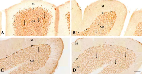
GYKI 52466 is a selective, non-competitive antagonist of the AMPA receptor (Arai, 2001) with good blood–brain barrier permeability (Barna et al., 2000). In our former studies, GYKI 52466 decreased c-fos expression in the rat neocortex and hippocampus in 4-AP seizures (Weiczner et al., 2008). The cerebellar granule cells possess AMPA re- ceptors, the stimulation of which mediated calcium influx into cultured granule cells (Hack and Balázs, 1995). The calcium uptake in these conditions has been successfully blocked by GYKI 52466 (Hack and Fig. 1.Immunohistochemistry of c-fos pro- tein localization in the cerebellar vermis 2 h after 4-AP administration.
(A) The distribution of c-fos immunpositive granule cell nuclei in the anterior lobe, lo- bule III in the rat treated with 4-AP only.
(B) The distribution of c-fos immunpositive granule cell nuclei in the anterior lobe, lo- bule IV in the rat treated with GYKI 52466 and 4-AP.
(C) The distribution of c-fos immunpositive granule cell nuclei in the posterior lobe, lo- bule VII in the rat treated with MK-801 and 4-AP.
(D) The distribution of c-fos immunpositive granule cell nuclei in the posterior lobe, lo- bule VII in the rat treated with amantadine and 4-AP.
Layers of the cerebellar cortex are indicated (M: molecular layer, P: Purkinje cell layer, GR: granular layer). The arrows show the immunopositive cell nuclei in the granular layer. The Purkinje cells are shown by as- terisks. Scale bar: 100μm.
Fig. 2.Quantitative assessment of c-fosIR cell nuclei numbers in vermal lobules I–X, 2 h after 4-AP injection. Ordinate: number of cell nuclei in 1 mm2; abscissa: vermal lobules numbered from I to X. The c- fosIR cells were counted in the granular layer of each cerebellar lo- bule. Control animals treated with physiological saline displayed a small number of c-fos-stained cell nuclei (white columns). Animals treated with 4-AP displayed high numbers of c-fosIR cell nuclei (black columns). Every antagonist pretreatment caused a significant decrease of c-fos expression compared to the 4-AP group. Four animals were investigated in every group. Significant differences compared to the 4- AP treated values are shown by asterisks (*p < 0.05). The standard error of the mean (SEM) is shown on the top of the columns.
Balázs, 1995). The AMPA receptors of the granule cells are relatively resistant to desensitization, and they support high-frequency synaptic transmission in the mossy fiber glomerulus (Delvendahl and Hallermann, 2016). We suppose that granule cell AMPA receptors were successfully blocked in our GYKI 52466 pretreated animals, and the blockade resulted in the significant decrease of c-fos expression in the granular layer. The solvent of GYKI 52466 (dimethyl sulfoxide) did not exert any pharmacological effect and did not influence the c-fos im- munostaining (de Sarro et al., 1995;Weiczner et al., 2008).
4.3. The significance of seizure antagonism in the cerebellum
The pharmacological antagonism of the NMDA and AMPA receptors in the cerebellar cortex decreased the number of the c-fosIR cells sig- nificantly, similarly to that we detected in the contralateral hemisphere after transection of the middle cerebellar peduncle (Tóth et al., 2015).
We conclude that these drugs can decrease the cerebellar activity through postsynaptic mechanisms, acting at the dendrites of the granule cells and decreasing theirfiring upon the mossyfiber activity (Delvendahl and Hallermann, 2016). The pharmacologically inhibited firing of the granule cells is reflected in the decrease of the postsynaptic c-fos protein expression in the granular layer of the cerebellar cortex.
On the other hand, pharmacological action on the level of the pontine nuclei is possible too, as the neurons of the pontocerebellar tract are contacted by the corticopontine axon terminals, which also use gluta- mate (Border and Mihailoff, 1991). The postsynaptic membranes of cerebellar neurons possess not only NMDA, but also AMPA and kainate receptors. These ionotropic receptors respond to extracellular gluta- mate: AMPA receptors trigger depolarization and burst initiation, and then, NMDA receptors become activated, and the opening of the NMDA channel generates large Ca2+ influx (Hwa and Avoli, 1991;Löscher, 1998). The influx of Ca2+has an important role in the triggering of neuronal damage (Chapman, 1998). It is important to note that MK-801 was not only inhibiting neuronal c-fos expression, but it also inhibited the development of the seizure, as we measured significantly longer GTCS latencies in these animals. It may suggest that the blockade of the NMDA channel by this non-competitive antagonist obviously inhibited or delayed the postsynaptic influx of Ca2+, which in turn should have been delayed and inhibited the long lasting neuronal depolarization (Hwa and Avoli, 1991), and it prevented seizure-related neuronal c-fos expression and the behavioral symptoms as well.
It is important to highlight that GTCS, which represents the main symptom of the 4-AP treatment, did not show strict correlation with the inhibition of c-fos expression. Some animals pretreated with NMDA antagonists and exhibiting decreased c-fos expression displayed GTCS;
nonetheless, the latency and the incidence of the symptoms were af- fected significantly. Inhibition of the NMDA receptor channel during this period could inhibit the induction of the c-fos gene, whereas glu- tamate release after this critical period may not induce further c-fos expression, but it may cause the development of the symptoms and maintain them. This could explain the discrepancy between the oc- currence of GTCS and the decrease of c-fos immunoreactivity. Another explanation could be based on the role of non-NMDA receptors in the development and maintenance of the symptoms, as supported by the literature data (Löscher, 1998).
5. Conclusions
1. The cerebellar cortex of the vermis is strongly activated in gen- eralized 4-AP seizures.
2. The activation is primarily due to the stimulation of the pontocer- ebellar mossy fiber pathway, and it can be monitored for 2 h by means of the neuronal c-fos expression.
3. The mossyfibers stimulate their postsynaptic targets through NMDA and AMPA receptors, which mediate the long-lasting postsynaptic c- fos expression.
4. The pharmacological blockade of these receptors attenuates the seizure-related synaptic activation of the granule cell layer.
5. The antagonists of the NMDA and AMPA receptors could be useful in the attenuation of the seizure-related neuronal damage in the cer- ebellum.
Conflict of interest
The authors declare no competing personal orfinancial interests in this study.
Acknowledgements
Mrs. Monica Kara provided valuable technical help during the ex- periments.
The experiments were supported by the Hungarian Government and the European Union (GINOP-2.3.2-15-2016-00034 grant).
References
Arai, A.C., 2001. GYKI 52466 has positive modulatory effects on AMPA receptors. Brain.
Res. 892, 396–400.
Barna, B., Szász, A., Világi, I., Szente, M., 2000. Anticonvulsive effect of AMPA receptor antagonist GYKI 52466 on 4-aminopyridine-induced cortical ictal activity in rat.
Brain Res. Bull. 51, 241–248.
Boop, S.B.A., Wheless, J., Katherine, V.P., McGregor, A., Boop, A.F., 2013. Cerebellar seizures. J. Neurosurg. Pediatrics 12, 288–292.
Border, B.G., Mihailoff, G.A., 1991. Glutamate immunoreactivity in the rat basilar pons:
light and electron microscopy reveals labeled boutons and cells of origin of afferent projections. Neuroscience 45, 47–61.
Braitman, D.J., Sparenborg, S., 1989. MK-801 protects against seizures induced by the cholinesterase inhibitor soman. Brain. Res. Bull. 23, 145–148.
Bressan, R.A., Pilowsky, L.S., 2000. Imaging the glutamatergic system in vivo−relevance to schizophrenia. Eur. J. Nucl. Med. 27, 1723–1731.
Brodal, P., Bjaalie, J.G., 1992. Organization of the pontine nuclei. Neurosci. Res. 13, 83–118.
Castejon, O.J., Castejon, H.V., 2000. Correlative microscopy of cerebellar Golgi cells.
Biocellular 24, 13–30.
Chapman, A.G., 1998. Glutamate receptors in epilepsy. Prog. Brain Res. 116, 371–383.
Danysz, W., Parsons, C.G., Kornhuber, J., Schmidt, W.J., Quack, G., 1997.
Aminoadamantanes as NMDA receptor antagonists and anti-parkinsonian agents.
Neurosci. Biobehav. Rev. 21, 455–468.
de Sarro, G., Gitto, R., de Sarro, A., Chimirri, A., Grasso, S., Giusti, P., Chapman, A.G., 1995. GYKI 52466 and related 2,3-benzodiazepines as anticonvulsant agents in DBA/
2 mice. Eur. J. Pharm. 294, 411–422.
Delvendahl, I., Hallermann, S., 2016. The cerebellar mossyfiber synapse as a model for high-frequency transmission in the mammalian CNS. Trends Neurosci. 39, 722–737.
Donevan, S.D., Rogawski, M.A., 1993. GYKI 52466, a 2,3-benzodiazepine, is a highly selective, noncompetitive antagonist of AMPA/kainate receptor responses. Neuron 10, 51–59.
Dragunow, M., Robertson, H.A., 1987. Kindling stimulation induces c-fos protein(s) in granule cells of the rat dentate gyrus. Nature 329, 441–442.
Ge, Y., Hu, W., Liu, C., Zhang, J.G., Meng, F.G., 2013. Brain stimulation for treatment of refractory epilepsy. Chin. Med. J. 126, 3364–3370.
Geminiani, A., Casellato, C., Antonietti, A., D’Angelo, E., Pedrocchi, A., 2017. A multiple- plasticity spiking neural network embedded in a closed-loop control system to model cerebellar pathologies. Int. J. Neural Syst.http://dx.doi.org/10.1142/
S0129065717500174.
Greenberg, M.E., Ziff, E.B., 2001. Signal transduction in the postsynaptic neuron.
Activity-dependent regulation of gene expression. In: Cowan, W.M., Südhof, T.C., Stevens, C.F. (Eds.), Synapses. The Johns Hopkins University Press, Baltimore, pp.
357–391.
Hack, N., Balázs, R., 1995. Properties of AMPA receptors expressed in rat cerebellar granule cell cultures: Ca2+influx studies. J. Neurochem. 65, 1077–1084.
Herdegen, T., Leah, J.D., 1998. Inducible and constitutive transcription factors in the mammalian nervous system: control of gene expression by Jun, Fos and Krox, and CREB/ATF proteins. Brain Res. Rev. 28, 370–490.
Honavar, M., Meldrum, B.S., 2002. Epilepsy. In: Graham, D.I., Lantos, P. (Eds.), Greenfield’s Neuropathology. Arnold, London, pp. 899–941.
Hwa, G.G.C., Avoli, M., 1991. The involvement of excitatory amino acids in neocortical epileptogenesis: NMDA and non-NMDA receptors. Exp. Brain Res. 186, 248–256.
Kornhuber, J.l., Weller, M., Schoppmeyer, K., Riederer, P., 1994. Amantadine and memantine are NMDA receptor antagonists with neuroprotective properties. J.
Neural. Transm. Suppl. 43, 91–104.
Koutsouraki, E.S., Anastasiades, J.J., Baloyannis, S.J., 2013. An immunohistochemical study of N-methyl-D-aspartate receptors in human cerebellum and hippocampus. Am.
J. Med. Sci. Med. 1, 28–30.
Kovács, A., Mihály, A., Komáromi, Á., Gyengési, E., Szente, M., Weiczner, R., Krisztin- Péva, B., Szabó, G., Telegdy, G., 2003. Seizure, neurotransmitter release and gene expression are closely related in the striatum of 4-aminopyridine-treated rats.
Epilepsy Res. 55, 117–129.
Labiner, D.M., Butler, L.S., Cao, Z., Hosford, D.A., Shin, C., McNamara, J.O., 1993.
Induction of c-fos mRNA by kindled seizures: complex relationship with neuronal burstfiring. J. Neurosci. 13, 744–751.
Lee, H.S., Mihailoff, G.A., 1990. Convergence of cortical and cerebellar projections on single basilar pontine neurons: a light and electron microscopic study in the rat.
Neuroscience 39, 560–577.
Limatola, C., 2003. Neurotrophic effects of AMPA. Cerebellum 2, 2–10.
Löscher, W., 1998. Pharmacology of glutamate receptor antagonists in the kindling model of epilepsy. Prog. Neurobiol. 54, 721–741.
Mihály, A., Bencsik, K., Solymosi, T., 1990. Naltrexone potentiates 4-aminopyridine sei- zures in the rat. J. Neural. Transm. [GenSect] 79, 59–67.
Mihály, A., Szakács, R., Bohota, C., Dobó, E., Krisztin-Péva, B., 2001. Time dependent distribution and neuronal localization of fos protein in the rat hippocampus following 4-aminopyridine seizures. Epilepsy Res. 44, 97–108.
Mihály, A., Borbély, S., Világi, I., Détári, L., Weiczner, R., Zádor, Z., 2005. Neocortical c- fos mRNA transcription in repeated brief, acute seizures: is c-fos a coincidence de- tector? Int. J. Mol. Med. 15, 481–486.
Morales-Villagrán, A., Ureňa-Guerrero, M.E., Tapia, R., 1996. Protection by NMDA re- ceptor antagonists against seizures induced by intracerebral administration of 4- aminopyridine. Eur. J. Pharmacol. 305, 87–93.
Paoletti, P., Neyton, J., 2007. NMDA receptor subunits: function and pharmacology. Curr.
Opin. Pharmacol. 7, 39–47.
Parsons, C.G., Danysz, W., Quack, G., 1999. Memantine is a clinically well tolerated N- methyl-D-aspartate (NMDA) receptor antagonist. Neuropharmacology 38, 735–767.
Rogawski, M.A., 2013. AMPA receptors as a molecular target in epilepsy therapy. Acta Neurol. Scand. Suppl. 197, 9–18.
Rothan, H.A., Amini, E., Faraj, F.L., Golpich, M., Teoh, T.C., Gholami, K., Yusof, R., 2017.
NMDA receptor antagonism with novel indolyl, 2-(1,1-Dimethyl-1,3-dihydro-benzo [e]indol-2-ylidene)-malonaldehyde, reduces seizures duration in a rat model of epi- lepsy. Sci. Rep. 7, 45540.http://dx.doi.org/10.1038/srep45540.
Sanchez-Perez, A., Llansola, M., Cauli, O., Felipo, V., 2005. Modulation of NMDA re- ceptors in the cerbellum. II. Signaling pathways and physiological modulators reg- ulating NMDA receptor function. Cerebellum 4, 162–170.
Somogyi, P., Halasy, K., Somogyi, J., Storm-Mathisen, J., Ottersen, O.P., 1986.
Quantification of immunogold labelling reveals enrichment of glutamate in mossy and paralelfibre terminals in cat cerebellum. Neuroscience 19, 1045–1050.
Szakács, R., Weiczner, R., Mihály, A., Krisztin-Péva, B., Zádor, Z., Zádor, E., 2003. Non- competitive NMDA receptor antagonists moderate seizure-induced c-fos protein ex- pression in the rat cerebral cortex. Brain Res. Bull. 59, 485–493.
Tóth, Z., Molnár, G., Mihály, A., Krisztin-Péva, B., Morvai, M., Kopniczky, Z., 2015.
Immunohistochemistry of cerebellar seizures: mossyfiber afferents play an important role in seizure spread and initiation in the rat. Acta Histochem. 117, 47–55.
Thompson, C.L., Drewery, D.L., Atkins, H.D., Stephenson, F.A., Chazot, P.L., 2000.
Immunohistochemical localization of N-methyl-D-aspartate receptor NR1, NR2A, NR2B and NR2C/D subunits in the adult mammalian cerebellum. Neurosci. Lett. 283, 85–88.
Voogd, J., 2004. Cerebellum. In: Paxinos, G. (Ed.), The Rat Nervous System. Elsevier, pp.
205–242.
Weiczner, R., Krisztin-Péva, B., Mihály, A., 2008. Blockade of AMPA-receptors attenuates 4-aminopyridine seizures, decreases the activation of inhibitory neurons but is in- effective against seizure-related astrocytic swelling. Epilepsy Res. 78, 22–32.
Willoughby, J.O., Mackenzie, L., Medvedev, A., Hiscock, J., 1997. Fos induction following systemic kainic acid: early expression in hippocampus and later widespread expres- sion correlated with seizure. Neuroscience 77, 379–392.
Wong, E.H.F., Kemp, J.A., Priestly, T., Knight, A.R., Woodruff, G.N., Iversen, L.L., 1986.
The anticonvulsant MK-801 is a potent N-methyl-D-aspartate antagonist. Proc. Natl.
Acad. Sci. 83, 7104–7108.
Zádor, Z., Weiczner, R., Mihály, A., 2008. Long-lasting dephosphorylation of connexin 43 inacute seizures is regulated by NMDA receptors in the rat cerebral cortex. Mol. Med.
Rep. 1, 721–727.