1 This is the peer reviewed version of the following article: Árpád Molnár, Zsuzsanna Kolbert, 1
Krisztina Kéri, Gábor Feigl, Attila Ördög, Réka Szőllősi, László Erdei (2018) Selenite- 2
induced nitro-oxidative stress processes in Arabidopsis thaliana and Brassica juncea, 3
Ecotoxicology and Environmental Safety, 148 (2018) 664-674, which has been published in 4
final form at https://doi.org/10.1016/j.ecoenv.2017.11.035. This article may be used for non- 5
commercial purposes in accordance with the terms of the publisher.
6
2 Selenite-induced nitro-oxidative stress processes in Arabidopsis thaliana and Brassica 7
juncea 8
Árpád Molnár+1,2, Zsuzsanna Kolbert*+1, Krisztina Kéri1, Gábor Feigl1, Attila Ördög1, Réka 9
Szőllősi1, László Erdei1 10
1 Department of Plant Biology, University of Szeged, Hungary 11
2 Doctoral School in Biology, Faculty of Science and Informatics, University of Szeged, 12
Szeged, Hungary 13
+ these authors contributed equally to this work 14
* corresponding author, Dr. Zsuzsanna Kolbert, kolzsu@bio.u-szeged.hu 15
16
17
3 Abstract
18
Extremes of selenium (Se) exert toxic effects on plants’ physiological processes;
19
although plant species tolerate Se differently. This study focuses on the effect of Se (0, 20, 50 20
or 100 μM sodium selenite) on secondary nitro-oxidative stress processes mainly using in situ 21
microscopic methods in non-accumulator Arabidopsis thaliana and secondary Se accumulator 22
Brassica juncea. Relative Se tolerance or sensitivity of the species was evaluated based on 23
growth parameters (fresh and dry weight, root growth) and cell viability. Besides, selenite- 24
triggered cell wall modifications (pectin, callose) and stomatal regulations were determined for 25
the first time. In case of Arabidopsis, relative selenite sensitivity was accompanied by decreased 26
stomatal density and induced stomatal opening, callose accumulation, pronounced oxidative 27
stress and moderate nitrosative modifications. In contrast, the selenite-treated, relatively 28
tolerant Brassica juncea showed larger number of more opened stomata, pectin accumulation, 29
moderate oxidative and intense nitrosative stress. These suggest that selenite tolerance or 30
sensitivity is rather associated with oxidative processes than secondary nitrosative 31
modifications in higher plants.
32
Key words: Arabidopsis thaliana; Brassica juncea; cell wall modifications; nitro-oxidative 33
stress; selenite.
34
35
4 1. Introduction
36
Selenium (Se) is a naturally occurring non-metal element, which is in many ways special 37
and exists in many interchangeable oxidized and organic, inorganic forms. Elevated Se 38
concentrations are naturally found in soils derived from Cretaceous shale rock (Kabata-Pendias, 39
1998) and Se may accumulate in the environment as the result of anthropogenic activities 40
(Terry, 2000). Se shows chemical similarities with sulphur (S), therefore plants use their S 41
uptake and metabolism system to assimilate Se. Some species in Brassicaceae family like 42
Brassica juncea are sulphur loving and consequently are capable of accumulating larger amount 43
of Se in their tissues (Pilon-Smits and Quinn, 2010). Additionally, these so-called secondary 44
accumulators show reduced sensitivity to the presence of Se. On the other hand, most plant 45
species, like the model plant Arabidopsis thaliana are non-Se-accumulators since they 46
accumulate less than 25 µg Se/g dry weight and they cannot tolerate elevated Se levels in their 47
environment (El-Ramady et al., 2015). Besides the plant species, also the applied Se form and 48
the plants’ age determine the rate of Se toxicity. The main consequences of Se excess, which 49
are responsible for its toxicity, are the malformation of non-specific selenoproteins, reactive 50
oxygen species (ROS) production and oxidative stress (Van Hoewyk, 2013). For the pro- 51
oxidant properties of Se forms, the depletion of the major antioxidant, glutathione is principally 52
responsible (Van Hoewyk, 2013). Also the disturbance in reactive nitrogen species (RNS) 53
homeostasis and the consequent nitrosative stress are induced as the effect of Se (Lehotai et al., 54
2016a; Kolbert et al., 2016). The term RNS is used to describe the family of nitric oxide (NO.) 55
originated molecules like, inter alia, peroxynitrite (ONOO-), S-nitrosoglutathione (GSNO) or 56
nitrogen dioxide radical (.NO2) (Corpas et al., 2007). The intense production of RNS leads to 57
macromolecule modifications resulting in nitrosative stress (Corpas et al., 2007; Valderrama et 58
al., 2007). Posttranslational modification of proteins caused by tyrosine nitration is becoming a 59
useful marker of nitrosative processes in plant systems (Corpas et al., 2013). Nitration of certain 60
5 tyrosine amino acids occurs in two steps resulting in the formation of 3-nitrotyrosine which 61
induces alterations in protein structure and function and through the prevention of tyrosine 62
phosphorylation it may influence signal transduction as well (reviewed by Kolbert et al., 2017).
63
Plants evolved protection mechanisms against damaging effects of excess Se such as 64
the production and emission of volatile compounds like dimethyl(di)selenide (DMDSe) (El- 65
Ramady et al., 2015). The rate of Se volatilization varies in plant kingdom which is determined 66
also by the forms of Se (de Souza et al., 2000). In addition to protecting against Se toxicity 67
within the plant tissues, Se volatilization may also have important ecological role like deterring 68
herbivores and affecting neighbouring plants (Schiavon and Pilon-Smits, 2017a).
69
Several plant species are capable of modifying the chemical composition of their cell 70
walls in order to prevent heavy metals from entering the cytoplasm. Increased formation and 71
deposition of lignin or callose effectively reduces metal absorption thus facilitates plant survival 72
and at the same time can partly be responsible for growth inhibition in metal-exposed 73
environment (Le Gall et al., 2015). Moreover, heavy metal-triggered alterations in contents and 74
methylesterification, acetylation status of pectins greatly determine heavy metal binding and 75
the porosity of the cell wall thus its capability for growth (Le Gall et al., 2015).
76
Our research was motivated partly by the fact that Se-induced cell wall modifications 77
and their possible correlations with Se tolerance are almost completely unknown. Similarly, our 78
knowledge is incomplete regarding the regulatory effect of Se on stomata; although Se 79
volatilization has been extensively studied in several plant species. Se has shown to induce 80
nitro-oxidative stress in non-accumulator pea (Lehotai et al., 2016a), but the relationship 81
between nitrosative processes and Se tolerance has not been examined so far. Our further aim 82
was to evaluate the possible nitro-oxidative stress-inducing effect of selenite in non- 83
accumulator Arabidopsis thaliana and secondary accumulator Brassica juncea.
84
6 2. Materials and methods
85
2.1.Plant growth conditions 86
Experiments were carried out with Brassica juncea L. Czern (cv. Negro Caballo) and 87
Arabidopsis thaliana L. Heynh (Columbia-0). Seeds of both species were surface sterilised in 88
5% (v/v) sodium hypochlorite, then placed on perlite (in case of Brassica seeds) or on ½ 89
Murashige-Skoog medium (in case of Arabidopsis) in Eppendorf tubes floating on Hoagland 90
solution. In case of Brassica, the seedlings were pre-cultivated for nine days and then treated 91
with 0 (control), 20, 50 or 100 µM sodium selenite (Na2SeO3) for one week meaning that 92
Brassica plants were 16-days-old at the time of the sampling. In order to obtain the appropriate 93
amount of biomass, Arabidopsis plants were grown in Hoagland solution for three weeks before 94
being treated with the same selenite concentrations like mustard for one week. Arabidopsis 95
thaliana plants were 28-days-old at the time of harvesting. Anoxia was prevented with constant 96
aeration of the nutrient solution. Both plant species were grown during controlled conditions 97
(150 µmol m−2/s photon flux density, 12h/12h light/dark cycle, relative humidity 55–60% and 98
temperature 25±2 ºC). All chemicals used during the experiments were purchased from Sigma- 99
Aldrich unless stated otherwise.
100 101
2.2.Se content analysis 102
Leaf and root materials of Arabidopsis and Brassica were harvested separately and 103
washed in distilled water then dried at 70 ºC for 72 hours. Nitric acid (65% w/v, Reanal, 104
Hungary) and hydrogen peroxide (H2O2, 30%, w/v, VWR Chemicals, Hungary) were added to 105
dried plant material. The samples were destructed at 200 ºC and 1600 W for 15 min. After 106
appropriate dilutions, Se concentrations were determined by inductively coupled plasma mass 107
spectrometry (ICP-MS) (Agilent 7700 Series, Santa Clara, USA). Se concentrations are given 108
in µg/g dry weight (DW).
109
7 110
2.3.Evaluation of growth parameters, root system morphology and root cell viability 111
Fresh and dry weights of plant materials were measured using a balance and the values 112
are given in mg. Primary root length was measured manually and expressed as centimetre. Also 113
the number of visible lateral roots were counted manually and expressed as pieces/root.
114
In order to evaluate Se tolerance of the species, cell viability in root apical meristem 115
was determined by using fluorescein diacetate (FDA) fluorophore according to Lehotai et al.
116
(2011). Root tips were incubated in 10 µM FDA solution (prepared in 10/50 mM MES/KCl 117
buffer, pH 6.15) for 30 min in darkness and were washed four times in buffer.
118 119
2.4.Microscopic visualization of cell wall modifications in the root system 120
Callose was detected with aniline blue fluorescent dye according to Cao et al. (2011) 121
with slight modifications. The stain was used in 0.1% (w/v) solution containing 1 M of glycine.
122
Root tips were incubated in dye solution for 5 minutes at room temperature, then washed once 123
with distilled water.
124
Cell wall pectin content was visualized using 0.05% (w/v) ruthenium red (RR) solution 125
prepared with distilled water. Root samples were incubated in RR solution for 15 minutes and 126
were washed once with distilled water according to Durand et al. (2009).
127 128
2.5.Examination of stomatal parameters 129
Plant leaves were submerged in MES/KCl buffer (10/50 mM, pH 6.15) and the 130
epidermal layers were carefully removed using forceps. In every case, strips were prepared from 131
the same part of the leaf blade, avoiding leaf veins. The epidermal cell layers were put on slides 132
using the previous buffer. Pictures were taken with a microscope (Zeiss Axiovert 200M) using 133
10x and 40x object lenses. Image analysis was carried out using Axiovision Rel. 4.8 software.
134
8 Stomatal density (pieces/mm2) was analysed by counting all stomata in a 200 µm diameter 135
circle. For the stomatal opening analysis, the widths of the stomatal pores were measured and 136
the data are given as µm.
137 138
2.6.In situ detection of ROS, glutathione, cell-wall peroxidase activities, lipid 139
peroxidation and RNS in the root tips 140
141
Dihydroethidium (DHE) at 10 µM concentration was applied for the detection of 142
superoxide anion levels. Root segments were incubated in darkness at 37 ºC for 30 min, and 143
washed two times with Tris-HCl buffer (10 mM, pH 7.4) (Kolbert et al., 2012).
144
Hydrogen peroxide levels were examined using 50 µM Amplex Red (10-acetyl-3,7 145
dihydroxyphenoxazine) dye solution in sodium phosphate buffer (50 mM, pH 7.5), then washed 146
once with the same buffer according to Lehotai et al. (2012).
147
Cellular glutathione levels were detected with the help of monobromobimane (MBB) 148
fluorophore. Root tips were stained in 100 µM dye solution (prepared in distilled water) for 60 149
min, and then washed once (Lehotai et al., 2016a).
150
Cell wall peroxidase (POD) activity was examined using 0.2% (w/v) pyrogallol solution 151
containing 0.03% (v/v) hydrogen peroxide prepared in 10 mM phosphate buffer (pH 7.0).
152
Samples were incubated for 15 minutes in room temperature and washed two times with 153
distilled water (Eleftheriou et al., 2015).
154
Reactive aldehydes produced during lipid peroxidation were visualized using 155
Schiff’s reagent according to Arasimowicz-Jelonek et al. (2009). Root tips were incubated in 156
dye solution for 20 minutes and then the reagent was replaced by 0.5% (w/v) K2S2O5 (prepared 157
in 0.05M HCl) for a further 20 minutes.
158
9 Nitric oxide level of the root tips was monitored with the help of 4-amino-5- 159
methylamino- 2′,7′-difluorofluorescein diacetate (DAF-FM DA) according to Kolbert et al.
160
(2012). Root segments were incubated in 10 µM dye solution for 30 min (darkness, 25±2 oC), 161
and washed twice with Tris-HCl (10 mM, pH 7.4).
162
Peroxynitrite was visualised with 10 µM dihydrorhodamine 123 (DHR) prepared in 163
Tris-HCl buffer. After 30 min of incubation, root tips were washed with buffer two times 164
(Sarkar et al., 2014).
165
Microscopic analysis of epidermal strips and stained root tips was accomplished under 166
Zeiss Axiovert 200 M inverted microscope (Carl Zeiss, Jena, Germany) equipped with a high- 167
resolution digital camera (AxiocamHR, HQ CCD, Carl Zeiss, Jena, Germany). Filter set 10 168
(exc.: 450–490, em.: 515–565 nm) was used for FDA, DAF-FM and DHR, filter set 9 169
(exc.:450–490 nm, em.:515–∞ nm) for DHE and Amplex Red and filter set 49 (exc.: 365 nm, 170
em.: 445/50 nm) was applied for MBB and aniline blue. Circles of 100 µm radii were applied 171
for measuring of the pixel intensity on digital photographs using Axiovision Rel. 4.8 software 172
(Carl Zeiss, Jena, Germany).
173 174
2.7.Detection of nitrated proteins using SDS-PAGE and western blotting 175
Fresh leaf and root tissues of Arabidopsis and Brassica were grounded with double 176
volume of extraction buffer (50 mM Tris–HCl buffer pH 7.6–7.8) containing 0.1 mM EDTA, 177
0.1% Triton X-100 and 10% glycerol and centrifuged at 12,000 rpm for 20 min at 4°C. The 178
protein extract was treated with 1% protease inhibitor cocktail and stored at -20 °C. Protein 179
concentration was determined using the Bradford (1976) assay with bovine serum albumin as 180
a standard.
181
25 µg of denaturated root and shoot protein were subjected to sodium dodecyl sulphate- 182
polyacrylamide gel electrophoresis (SDS-PAGE) on 12 % acrylamide gels. The proteins were 183
10 transferred to PVDF membranes using the wet blotting procedure (25 mA, 16h) for 184
immunoblotting. After transfer, membranes were used for cross-reactivity assays with rabbit 185
polyclonal antibody against 3-nitrotyrosine diluted 1:2000. Immunodetection was performed 186
by using affinity isolated goat anti-rabbit IgG-alkaline phosphatase secondary antibody in 187
dilution of 1:10000, and bands were visualized by using NBT/BCIP reaction. Nitrated bovine 188
serum albumin served as positive control.
189
2.8.Statistical analysis 190
All results are shown as mean±SE. Data were statistically evaluated by Duncan’s 191
multiple range test (One-way ANOVA, P≤0.05) using SigmaPlot 12 or by Student’s T-test 192
applying Microsoft Excel 2010. All experiments were carried out at least two times with at least 193
3-10 samples each.
194 195
196
197
198
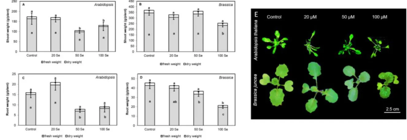
11 3. Results and Discussion
199
3.1. Se accumulation and translocation in selenite-treated Arabidopsis and Brassica 200
In the root and shoot tissues of both 28-days-old Arabidopsis and young Brassica juncea 201
plants, Se concentration increased as the effect of exogenous selenite treatments (Fig 1AB).
202
Total Se concentrations showed 28-, 125- and 300-fold accumulation in shoot tissues of 20, 50 203
and 100 µM selenite-treated Arabidopsis, respectively (Fig 1A). In Brassica shoots (Fig 1B), 204
the degree of accumulation proved to be smaller (19-, 26- and 68-fold in case of 20, 50 and 100 205
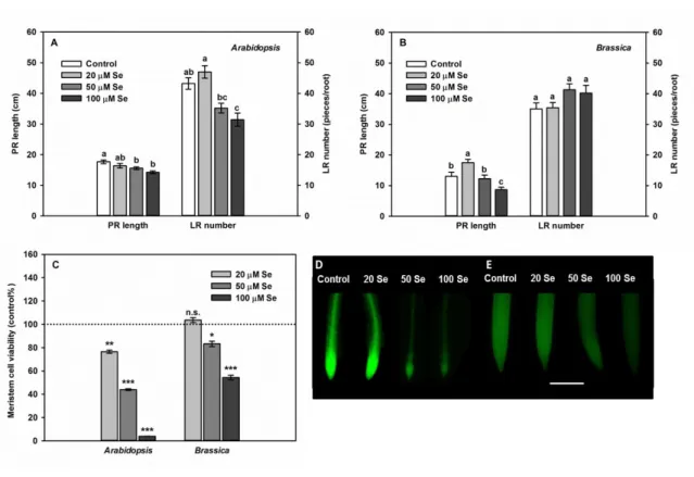
µM selenite treatment, respectively) compared to Arabidopsis. In roots, Se accumulations were 206
70-, 128 and 220-fold in case of Arabidopsis, and 57-, 133- and 264-fold in case of 20, 50 and 207
100 µM selenite treated Brassica. This means that the Se accumulation capacity of young 208
Brassica juncea roots a bit surpasses that of the older Arabidopsis. At the same time, it is quite 209
surprising that the accumulation capacity of young, secondary Se accumulator Brassica plant 210
is comparable with that of the older, non-accumulator Arabidopsis. This means that Brassica 211
plants at the early growth stage do not show such rate of accumulation capacity that is typical 212
for secondary accumulators. The roots of both species showed much higher Se contents 213
compared to shoots and also the degree of accumulation proved to be higher in the root system 214
than in the aerial plant parts, which is the result of the poor translocation capacity of selenite as 215
described earlier (de Souza et al., 1998; Zayed et al., 1998). Indeed, selenite is retained in the 216
root system where it is rapidly converted to organic forms, particularly to selenomethionine 217
(Zayed et al., 1998).
218
3.2.Selenite affects organ development and viability of Arabidopsis and Brassica 219
The lowest applied selenite concentration did not influence shoot and root growth, while 220
higher selenite doses significantly inhibited growth in both organs of mature (28-days-old) 221
Arabidopsis thaliana. The shoot fresh weight decreased to one fifth and the root fresh weight 222
12 showed 50% reduction as the effect of 100 µM selenite (Fig 2AC), while in case of young 223
Brassica juncea plants, 20 and 50 µM selenite did not decrease shoot fresh and dry weights and 224
100 µM Se caused diminution only in fresh weight but not in dry weight (Fig 2B). Both the 50 225
and the 100 µM Se resulted in serious (~50%) losses of root weights in Arabidopsis (Fig 2C), 226
while in Brassica, 50 µM selenite reduced only fresh weight and 100 µM selenite resulted in 227
significant fresh and dry weight reductions (root fresh weight was halved compared to control, 228
Fig 2D). Therefore, critical Se concentration, which induces 50% growth reduction was ~120 229
µg/g dry weight in Arabidopsis and was ~250 µg/g dry weight in case of Brassica indicating 230
the pronounced Se sensitivity of Arabidopsis comparted to Brassica. Moreover, in case of 50 231
and 100 µM selenite, leaf area, leaf number and petiole length decreased (Fig 2E, data not 232
shown), but visible symptoms of selenite toxicity such as chlorosis or necrosis could not be 233
observed in leaves of the species (Fig 2E). Indeed, asymptomatic Se accumulation in a wide 234
concentration range is typical for many plant species (Gupta and Gupta, 2017).
235
As to the root system, primary root (PR) elongation, lateral root (LR) number and root 236
meristem cell viability was determined in younger Brassica juncea and older Arabidopsis 237
thaliana plants (Fig 3).
238
In Arabidopsis, 20 µM selenite did not cause PR shortening, but slightly induced LR 239
formation (Fig 3A) resulting in larger fresh weight (Fig 2C). In Brassica root system, the 240
positive effect of low Se dose was manifested in induced elongation of the primary root (Fig 241
3B). Moreover, 50 and 100 µM selenite resulted in a slight, non-significant induction of LR 242
formation (Fig 3B). More severe selenite exposure (both 50 and 100 µM) led to the inhibition 243
of PR elongation and to the reduction of LR number (Fig 3A) which together resulted in root 244
weight loss (Fig 2C) of Arabidopsis, but only 100 µM Se was able to inhibit PR elongation and 245
Se had no influence at all on LR emergence of Brassica (Fig 3B). The observed alterations in 246
root developmental processes faithfully reflect Se tolerance (Tamaoki et al., 2008; Freeman et 247
13 al., 2010; Zhang et al., 2007) of Brassica and relative sensitivity of Arabidopsis. Selenite- 248
induced inhibition of primary root elongation is partly due to the decrease in root apical 249
meristem (RAM) cell division as it was observed in Arabidopsis model plant (Lehotai et al., 250
2016b).
251
Cell viability of the root meristem as an indicator of stress tolerance was determined by 252
microscopy. All applied concentrations decreased viability in Arabidopsis root tips, while in 253
case of Brassica juncea, 50 and 100 µM selenite caused only moderate viability loss (Fig 3CD).
254
Besides inhibited cell division, the observed viability loss in RAM cells may also contribute to 255
the growth inhibition at the organ level. In addition, the viability of RAM cells shows a good 256
correlation with the Se tolerance or sensitivity of the plant species. Based on the effects of 257
selenite on growth parameters, on PR elongation and on meristem cell viability, 28-days-old 258
Arabidopsis plants showed sensitivity relative to younger Brassica plants, which is in 259
accordance with earlier literature data (reviewed by Schiavon and Pilon-Smits, 2017).
260
3.3. Processes to reduce the amount of absorbed Se: stomatal regulation 261
The number and the opening of stomata determine the rate of Se volatilization, which is 262
an effective way to reduce tissue Se content. Therefore, the density and the opening of stomata 263
were examined in selenite-exposed Arabidopsis thaliana and Brassica juncea (Fig 4).
264
All applied selenite concentrations decreased the amount of stomata in Arabidopsis leaf 265
epidermis (Fig 4A). In case of higher selenite doses, 1.5-times less stomata could be observed 266
than in control plants (Fig 4 AEF); however, the sizes of stomatal apertures proved to be 267
significantly larger as the effect of all applied selenite concentrations (Fig 4 CEF). In contrast, 268
the lowest exogenous selenite concentration significantly increased both the sizes of stomatal 269
apertures (Fig 4D) and the number of stomata per unit area (Fig 4B) in young Brassica juncea 270
resulting in larger number of more opened stomata compared to control. Similarly, 100 µM 271
14 selenite caused enhancement of stomatal number and opening (Fig 4 BD). Microscopic images 272
representing the data are shown in Fig 4 GH. Microscopic analysis of stomatal parameters is 273
much less examined compared to stomatal conductance, which is determined by stomatal 274
density and the stomatal size. Se treatments caused increased stomatal conductance in tobacco 275
or in alfalfa (Jiang et al., 2015; Hajiboland et al., 2015). In other plants, like cucumber or maize, 276
treatments with Se forms resulted in decreased stomatal conductance (Haghighi et al., 2016;
277
Jiang et al., 2017) suggesting that the effect of Se forms on stomatal parameters depends on the 278
examined species. In our experiments, the more tolerant Brassica juncea showed 20 and 100 279
μM selenite-induced increment in stomatal density and size supposedly creating decreased 280
stomatal conductance and possibly contributing to enhanced Se volatilization. In 281
hydroponically grown, 40 μM selenite-treated Brassica juncea, the rate of Se volatilization was 282
60 μg/g fresh weight/day (Van Huysen et al., 2003) on the basis of which it can be considered 283
as well-volatilizing species (Terry et al., 1992). Moreover, the rate of volatilization was shown 284
to correlate with endogenous Se content of the leaf (Terry et al., 1992) suggesting that Se or its 285
volatile forms serve as direct signals for stomatal opening rather than an indirect regulation 286
through the disturbance of water status. Although, regarding the complex signal mechanism of 287
Se-regulated stomatal movements, we have no solid literature evidence yet. Occasionally purple 288
discolorations appeared on the leaves of 100 µM selenite-treated Brassica juncea, which could 289
be the results of anthocyanin accumulation (Fig 4 I) as it was reported in maize (Hawrylak- 290
Nowak, 2008) or in purple lettuce (Liu et al., 2017). In the latter species, expressions of genes 291
involved in anthocyanin biosynthesis like UDP glycose flavonoid glycosyl transferase (UFGT) 292
and flavanone 3-hydroxylase (F3H) were upregulated by selenite (Liu et al., 2017). Because of 293
their competitive uptake system selenite exposure may trigger phosphorous deficiency (Li et 294
al., 2008) which in turn may result in purple pigmentation of the leaves. The accumulated 295
anthocyanins can act as antioxidants, UV protectors or metal chelators (Winkel-Shirley, 2002).
296
15 Anthocyanin formation was visible in epidermis of Brassica leaves (Fig 4 IJ) where they can 297
interact with the metals (Landi, 2015) or in our case with the accumulated Se. Namely high 298
concentrations of Se were observed in large epidermal storage cells (Freeman et al., 2006;
299
2010), where anthocyanin accumulation seems also to be localized (Fig 4 IJ).
300
3.4. Processes to reduce the amount of absorbable selenite in the root system: cell wall 301
modifications 302
Modifications in cell wall composition and structure may contribute to tolerance in the 303
element-exposed environment and is concomitantly able to inhibit root growth. Pectin, callose 304
and lignin were microscopically visualized in lateral root tips of both plant species (Fig 5).
305
In the roots of four-weeks-old Arabidopsis, 20 and 100 µM selenite did not influence 306
pectin staining pattern of control root tips, while 50 µM selenite slightly increased pink 307
coloration indicating pectin presence in the meristem and root cap region (Fig 5A, indicated by 308
white arrowhead). In contrast, in 20 µM selenite-treated young Brassica roots, pectin staining 309
became remarkably intense in the whole lateral root (Fig 5B). In case of 50 and 100 µM selenite, 310
the intensified presence of pectin staining could be observed only in the elongation zone of the 311
LRs (Fig 5B). For the increase in pectin content, Se-triggered down-regulation of 312
pectinesterases (At5g04960; At2g45220; At3g10710) may be partly responsible as was found 313
in a transcriptome analysis of Arabidopsis thaliana (Van Hoewyk et al., 2008). Pectin may act 314
as a metal (e.g. Pb, Cd, Al) binding element of the cell wall contributing to the protection of 315
cytoplasm against high metal dosages (Polec-Powlak et al., 2007; Douchiche et al., 2010, 316
Hossain et al. 2006). Interestingly, in case of 20 μM selenite-treated Brassica, intensified 317
formation of pectin in lateral roots was accompanied by enhanced elongation. In general, 318
selenite-induced pectin formation was more intense in Brassica compared to Arabidopsis, 319
16 which supports that increased pectin content of root cell walls is a protection mechanism of 320
tolerant plant species (El-Moneim et al., 2014).
321
The presence of callose in cell walls was evaluated by aniline blue staining (Fig 5C). In 322
Arabidopsis, concentration-dependent callose accumulation (~3.5-fold) was detected in the root 323
system while in cell walls of young Brassica roots was evident only in case of 100 µM selenite 324
exposure and proved to be slighter (~1.8-fold) than in Arabidopsis roots (Fig 5C). Callose 325
deposition causes the appearance of extra carbohydrate layers leading to thickening of the walls 326
and consequently preventing the metal from entering the cytoplasm (Kartusch, 2003). Callose 327
is synthetized by callose synthase the genes of which (Os01g48200.1; Os06g02260.2) were 328
identified as putative Se responsive genes in Lolium perenne (Byrne et al., 2010). At the same 329
time callose deposition at the site of plasmodesmata may inhibit molecule transport which in 330
turn may contribute to growth inhibition (Zavaliev et al., 2011; Fig 3A). According to the 331
literature, callose accumulation is typical for sensitive species (Llugany et al., 1994; Pirselová 332
et al., 2012) like Arabidopsis thaliana in our experimental system (Fig 5C). Additionally, lignin 333
deposition may be a protection mechanism in element-exposed environment (Le Gall et al., 334
2015). During one-week selenite treatment, lignification could not be microscopically observed 335
either in Arabidopsis or in Brassica root system, but longer treatment period resulted in slight 336
lignin deposition in tolerant Brassica (data not shown).
337
3.5. Selenite influences ROS levels, peroxidases and induces lipid peroxidation in the 338
root system of Arabidopsis and Brassica 339
In the roots of Arabidopsis, the lowest applied selenite concentration resulted in 340
significantly higher superoxide anion levels compared to control just like the more severe 341
selenite doses (Fig 6A). In young Brassica roots, 20 and 50 μM selenite did not significantly 342
influence superoxide levels, but 100 µM selenite exposure resulted in 2.5-fold formation of 343
17 superoxide anion compared to control (Fig 6B). Moreover, the levels of H2O2 increased as the 344
effect of 50 µM selenite and showed a notable, 3-fold accumulation in 100 µM selenite-treated 345
Arabidopsis roots (Fig 6C), but it showed a modest (1.5-fold) elevation only in 100 µM selenite- 346
treated Brassica roots (Fig 6D). These results support that Se-compounds may evolve pro- 347
oxidant effects disturbing the redox status of plant cells (Van Hoewyk, 2013). Intense 348
formations of different ROS like superoxide radical and hydrogen peroxide were reported in 349
both organs of several plant species (Tamaoki et al., 2008; Freeman et al., 2010; Lehotai et al., 350
2012; 2016ab; Chen et al., 2014; Dimkovikj and Van Hoewyk, 2014). Glutathione levels 351
(examined in MBB-stained root tips) significantly decreased as the effect of the most severe 352
applied Se treatment in Arabidopsis (Fig 6E), but both 50 and 100 µM selenite effectively 353
decreased GSH content in Brassica roots (Fig 6F). Beyond the present experimental system, 354
Se-triggered depletion of endogenous glutathione pool was observed in plant species like 355
Arabidopsis thaliana, Stanleya pinnata, Brassica napus, Pisum sativum (Tamaoki et al., 2008;
356
Freeman et al., 2010; Dimkovikj and Van Hoewyk, 2014; Lehotai et al., 2016). The reason for 357
the selenite-triggered decrease in GSH content may be the interaction between reduced GSH 358
and selenite yielding selenodiglutathione and superoxide radical (Terry et al., 2000; Schiavon 359
and Pilon-Smits, 2017). This is supported by the increased formation of superoxide as the effect 360
of 100 μM selenite treatment especially in Brassica juncea (Fig 6B). Glutathione is a major 361
plant antioxidant (Noctor et al., 2012), therefore the decrease in its endogenous content can be 362
a significant cause of Se-induced oxidative stress (Van Hoewyk, 2013). Additionally, since 363
glutathione is associated with auxin transport and is involved in the maintenance of root growth 364
(Koprivova et al., 2010), its depletion within the root tip tissues may contribute to the inhibition 365
of root elongation in both plant species (Fig 3AB).
366
367
18 Brown pyrogallol staining indicates activity of cell-wall peroxidases (PODs), which 368
were induced by all Se concentrations in Brassica (Fig 6H) and by 50 and 100 µM selenite in 369
Arabidopsis (Fig 6G). Cell-wall peroxidases are involved in polymerization of lignin monomers 370
using H2O2 (Siegel, 1953); however, their selenite-induced activation did not result in 371
microscopically detectable lignification neither in Arabidopsis thaliana nor in Brassica juncea 372
roots (data not shown). Cell wall-associated PODs also influence oxidative coupling reactions 373
involving phenolics that are esterified to cell wall polysaccharides and formation of 374
isodityrosine bridges that are thought to crosslink structural proteins (Fry, 1986). Based on their 375
actions, PODs are considered to be involved in wall rigidification (Cosgrove, 1997) and thus 376
their activation may result in growth inhibition (Fig 3AB). Pink staining showing the intensity 377
of lipid peroxidation proved to be pronounced in 50 and 100 µM selenite-treated Arabidopsis 378
root tips (Fig 6I). In Brassica, 50 µM selenite treatment caused only slight pink coloration in 379
the root tip, while the highest applied selenite dose triggered more intense lipid peroxidation 380
(Fig 6J). Enhanced formation of reactive oxygen species causes peroxidation of 381
polyunsaturated fatty acids, yielding aldehydes such as 4-hydroxynonenal (4-HNE) and 382
malondialdehyde (MDA) (Hartley et al. 1999). These aldehydic products of lipid peroxidation 383
are widely accepted markers of oxidative stress and can be microscopically detected by Schiff’s 384
staining method (Pompella et al., 1987).
385
Results show that selenite proved to be pro-oxidant in both species. However, if we 386
compare the susceptibility of the species with the degree of lipid peroxidation in the examined 387
species, we can conclude that the sensitive Arabidopsis showed more intense selenite-induced 388
lipid peroxidation compared to Brassica juncea. In case of both Arabidopsis and Brassica, 389
peroxidation of lipids seems to be associated with the simultaneous formation of selenite- 390
triggered superoxide and hydrogen peroxide in the root tips (Fig 6).
391
392
19 3.6. Selenite alters reactive nitrogen species levels in Arabidopsis and Brassica root tips 393
The formation of ROS is closely related to the production and signalling of reactive 394
nitrogen species, therefore the level of RNS forms and their nitration effect on proteome was 395
examined (Fig 7).
396
Milder selenite doses caused significant diminution of nitric oxide levels, while 100 µM 397
selenite did not influence NO production in Arabidopsis root tips (Fig 7A). In young Brassica 398
roots, none of the Se concentrations influenced NO levels (Fig 7B). The effects of Se forms on 399
NO. levels are diverse since selenite-induced NO. production was reported in pea and Brassica 400
rapa roots (Lehotai et al., 2016a; Chen et al., 2014), while similarly to the data of the present 401
study, NO. levels significantly decreased as the effect of Se exposure in other species like agar- 402
grown Arabidopsis seedlings (Lehotai et al., 2016b). In the latter experimental system, data 403
suggested that NO. diminution was independent from nitrate reductase activity and selenite- 404
induced cytokinin accumulation negatively influences NO. levels (Lehotai et al., 2016b) 405
possibly through a direct chemical reaction (Liu et al., 2013). In contrast, peroxynitrite- 406
dependent fluorescence showed notable increment as the effect of all selenite concentrations in 407
Arabidopsis (Fig 7C) and as the effect of higher Se doses in Brassica (Fig 7D). This means that 408
selenite was able to induce its significant peroxynitrite production in the root system of both 409
species; however, the rate of its accumulation seems to be related to Se sensitivity.
410
Protein tyrosine nitration as the consequence of peroxynitrite accumulation was 411
examined in whole protein extracts of the shoot and root using western blot analysis (Fig 7E).
412
In both organs of untreated Arabidopsis and Brassica, several protein bands showed 413
immunoreactivity against 3-nitrotyrosine (Fig 7E) which indicates that in case of both species, 414
both organs contain physiological tyrosine nitroproteome meaning that a certain protein pool is 415
in nitrated state even under stress-free conditions. Similarly, basal tyrosine nitration in case of 416
20 control sunflower, pea and pepper plants have earlier been reported (Chaki et al., 2009; Begara- 417
Morales et al., 2013; Chaki et al., 2015). Based on the mostly negative impact of nitration on 418
protein activity (reviewed by Kolbert et al., 2017), physiological tyrosine nitroproteome of 419
healthy plants can be considered as an inactive part of the endogenous protein pool. Moreover, 420
protein pool of the control root system seems to be more affected by tyrosine nitration compared 421
to the shoot (Lehotai et al., 2016a) especially in Arabidopsis plants. In Arabidopsis shoot, signal 422
intensification of the immunoreactivity towards 3-nitrotyrosine antibody could be observed 423
only in 50 µM selenite-treated shoot sample in case of two bands (indicated by grey arrows).
424
In contrast, in Brassica shoots both 20 and 50 µM selenite caused intensified protein tyrosine 425
nitration in seven bands. In the root proteome, selenite-induced intensification of protein 426
nitration was observed in similar number of protein bands were observed in both species 427
(indicated by arrows), but the rate of Se-triggered nitration compared to controls proved to be 428
more intense in case of Brassica (Fig 7E). These indicate the pronitrant effect of exogenous 429
selenite treatment in both species; although it caused the appearance of newly nitrated protein 430
band neither in Arabidopsis thaliana nor in Brassica juncea. Surprisingly, selenite exerted more 431
severe effects on protein tyrosine nitration in both organs of young, more selenite tolerant 432
Brassica plants compared to older, relatively sensitive Arabidopsis. Protein tyrosine nitration 433
was formerly considered to be a biomarker for secondary nitrosative stress (Corpas et al., 2007;
434
2013) and as such may be related to the degree of impairment during stress. The more intense 435
Se-triggered protein tyrosine nitration as well as the notably high nitric oxide and peroxynitrite 436
levels (Fig 7BD) compared to Arabidopsis can be attributed to the young age of Brassica juncea 437
which may make it susceptible to nitrosative stress.
438
439
440
21 4. Conclusions
441
In the present experimental system, the effect of selenite exposure was examined in 16- 442
days-old Brassica juncea and in older (4-weeks-old) Arabidopsis thaliana plants. Comparisons 443
between the species therefore can only be made taking into account the age difference. Both 444
examined plant lines took up selenite from the external medium and translocated it into the 445
shoot system. Based on the slighter growth inhibition (fresh/dry weight, critical Se 446
concentration, root length, lateral root number) and moderate viability loss, young Brassica 447
juncea plants proved to be more tolerant to selenite exposure than older Arabidopsis thaliana.
448
In case of Arabidopsis, relative selenite sensitivity was accompanied by decreased stomatal 449
density and induced stomatal opening, callose accumulation, pronounced oxidative stress and 450
moderate nitrosative modifications. In contrast, selenite-treated, relative tolerant Brassica 451
juncea showed larger number of more opened stomata, pectin accumulation, moderate 452
oxidative and intense nitrosative stress. These suggest that selenite tolerance or sensitivity is 453
rather related to oxidative processes than secondary nitrosative modifications in higher plants.
454
455
Funding: This work was supported by the János Bolyai Research Scholarship of the Hungarian 456
Academy of Sciences (Grant no. BO/00751/16/8) by the National Research, Development and 457
Innovation Fund (Grant no. NKFI-6, K120383) and by the EU-funded Hungarian grant EFOP- 458
3.6.1-16-2016-00008. Zs. K. was supported by UNKP-17-4 New National Excellence Program 459
of the Ministry of Human Capacities.
460 461
22 5. References
462
Arasimowicz-Jelonek, M., Floryszak-Wieczorek, J., Kubiś, J., 2009. Involvement of nitric 463
oxide in water stress-induced responses of cucumber roots. Plant. Sci. 177, 682–690.
464
Begara-Morales, J.C., Chaki, M., Sánchez-Calvo, B., Mata-Pérez, C., Leterrier, M., Palma, 465
J.M., Barroso, JB., Corpas, F.J., 2013. Protein tyrosine nitration in pea roots during 466
development and senescence. J. Exp. Bot. 64, 1121-1134. doi: 10.1093/jxb/ert006 467
Bradford, M.M., 1976. A rapid and sensitive method for the quantification of microgram 468
quantities of protein utilizing the principle of protein-dye-binding. Anal. Biochem. 72, 469
248–255. doi: 10.1016/0003-2697(76)90527-3 470
Byrne, S.L., Durandeau, K., Nagy, I. et al., 2010. Identification of ABC transporters from 471
Lolium perenne L. that are regulated by toxic levels of Se. Planta 231, 901-911.
472
doi:10.1007/s00425-009-1096-y 473
Cao, Y., Lou, Y., Han, Y. et al., 2011. Al toxicity leads to enhanced cell division and changed 474
photosynthesis in Oryza rufipogon L. Mol. Biol. Rep. 38, 4839. doi:10.1007/s11033- 475
010-0618-9 476
Chaki, M., de Morales, P.Á., Ruiz, C., Begara-Morales, J.C., Barroso, J.B., Corpas, F.J., Palma, 477
J.M., 2015. Ripening of pepper (Capsicum annuum) fruit is characterized by an 478
enhancement of protein tyrosine nitration. Ann. Bot. 116, 637-47.
479
doi:10.1093/aob/mcv016 480
Chaki, M., Valderrama, R., Fernández-Ocana, A.M., et al., 2009. Protein targets of tyrosine 481
nitration in sunflower (Helianthus annuus L.) hypocotyls. J. Exp. Bot. 60, 4221–4234.
482
doi: 10.1093/jxb/erp263 483
Chen, Y., Mo, H.-Z., Zheng, M.-Y., Xian, M., Qi, Z.-Q., Li, Y.-Q., et al., 2014. Se Inhibits Root 484
Elongation by Repressing the Generation of Endogenous Hydrogen Sulfide in Brassica 485
rapa. PLoS ONE 9, e110904. doi:10.1371/journal.pone.0110904 486
23 Corpas, F.J., Carreras, A., Valderrama, R., Chaki, M., Palma, J.M., del Río, L.A., Barroso, J.B., 487
2007. Reactive nitrogen species and nitrosative stress in plants. Plant Stress 1, 37-41.
488
Corpas, F.J., Palma, J.M., del Río, L.A., Barroso,J.B., 2013. Protein tyrosine nitration in higher 489
plants grown under natural and stress conditions. Front. Plant. Sci. 4, 29. doi:
490
10.3389/fpls.2013.00029 491
Cosgrove, D.J., 1997. Assembly and enlargement of the primary cell wall in plants. Annu. Rev.
492
Cell. Dev. Biol. 13, 171–201. doi: 10.1146/annurev.cellbio.13.1.171 493
de Souza, M.P, Pilon-Smits, E.A.H., Lytle, C.M., Hwang, S., Tai, J., Honma, T.S.U., Yeh, L., 494
Terry, N., 1998. Rate-limiting steps in Se assimilation and volatilization by Indian 495
mustard. Plant. Phys. 117, 1487–1494. doi: 10.1104/pp.117.4.1487 496
de Souza, M.P., Pilon-Smits, E.A.H., Terry, N., 2000. The Physiology and Biochemistry of Se 497
Volatilization by Plants. In: Ensley, B.D., Raskin, I. (Eds.), Phytoremediation of toxic 498
metals: Using plants to clean up the environment. Wiley & Sons, New York, pp 171- 499
190.
500
Dimkovikj, A., Van Hoewyk, D., 2014. Selenite activates the alternative oxidase pathway and 501
alters primary metabolism in Brassica napus roots: evidence of a mitochondrial stress 502
response. BMC Plant Biol. 14, 259. doi:10.1186/s12870-014-0259-6 503
Douchiche, O., Driouich, A., Morvan, C., 2010. Spatial regulation of cell wall structure in 504
response to heavy metal stress: Cadmium-induced alteration of the methyl-esterification 505
pattern of homogalacturonans. Ann. Bot. 105, 481–491. doi: 10.1093/aob/mcp306 506
Durand, C., Vicré-Gibouin, M., Follet-Gueye, M.L., Duponchel, L., Moreau, M., Lerouge, P., 507
Driouich, A., 2009. The organization pattern of root border-like cells of Arabidopsis is 508
dependent on cell wall homogalacturonan. Plant Physiol. 150, 1411-21.
509
doi:10.1104/pp.109.136382 510
24 Eleftheriou, E.P., Adamakis, I.-D. S., Panteris, E., Fatsiou. M., 2015. Chromium-induced 511
ultrastructural changes and oxidative stress in roots of Arabidopsis thaliana. Int. J. Mol.
512
Sci. 16, 15852–15871. doi: 10.3390/ijms160715852 513
El-Moneim, D.A., Contreras, R., Silva-Navas, J., Gallego, J.F., Figueiras, A.M., Benito, C., 514
2014. Pectin methylesterase gene and aluminum tolerance in Secale cereal. Environ.
515
Exp. Bot. 107, 125-133.
516
El-Ramady, H., Abdalla, N., Alshaal, T., et al., 2015. Se and its role in higher plants. In:
517
Lichtfouse, E., et al., (Eds.), Pollutants in Buildings, Water and Living Organisms, 518
Environmental Chemistry for a Sustainable World 7, Springer, pp 238-285. doi:
519
10.1007/978-319-19276-5_6 520
Freeman, J.L., Zhang, J.H., Marcus, M.A., Fakra, S., McGrath, S.P., Pilon-Smits, E.A.H., 2006.
521
Spatial imaging, speciation, and quantification of Se in the hyperaccumulator plants 522
Astragalus bisulcatus and Stanleya pinnata. Plant. Phys. 142, 124-134 doi:
523
10.1104/pp.106.081158 524
Freeman, J.L., Tamaoki, M., Stushnoff, C., Quinn, C.F., Cappa, J.F., Devonshire, J., Fakra, 525
S.C., Marcus, M.A., McGrath, S.P., Van Hoewyk, D., Pilon-Smits, E.A.H., 2010.
526
Molecular mechanisms of Se tolerance and hyperaccumulation in Stanleya pinnata.
527
Plant. Phys. 153, 1630-1652 doi: 10.1104/pp.110.156570 528
Fry, S.C., 1986. Cross-linking of matrix polymers in the growing cells of angiosperms. Annu.
529
Rev. Plant. Physiol. 37, 165–186. doi: 10.1146/annurev.pp.37.060186.001121 530
Gupta, M., Gupta, S., 2017. An overview of se uptake, metabolism, and toxicity in plants. Front.
531
Plant. Sci. 7, 2074 doi: 10.3389/fpls.2016.02074 532
Haghighi, M., Sheibanirad, A., Pessarakli, M., 2016. Effects of Se as a beneficial element on 533
growth and photosynthetic attributes of greenhouse cucumber. J. Plant. Nutr. 39, 1493- 534
1498.
535
25 Hajiboland, R., Rahmat, S., Aliasgharzad, N., Hartikainen, H., 2015. Se-induced enhancement 536
in carbohydrate metabolism in nodulated alfalfa (Medicago sativa L.) as related to the 537
glutathione redox state. Soil Sci. Plant. Nutr. 61, 676-687. doi:
538
10.1080/00380768.2015.1032181 539
Hartley, D.P., Kolaja, K.L., Reichard, J., Petersen, D.R., 1999. 4-Hydroxynonenal and 540
malondialdehyde hepatic protein adducts in rats treated with carbon tetrachloride:
541
immunochemical detection and lobular localization. Toxicol. Appl. Pharmacol. 161, 23- 542
33. doi: 10.1006/taap.1999.8788 543
Hawrylak-Nowak, B., 2008. Changes in anthocyanin content as indicator of maize sensitivity 544
to Se. J. Plant. Nutr. 31, 1232-1242.
545
Hossain, A.K.M.Z., Koyama, H., Hara, T., 2006. Growth and cell wall properties of two wheat 546
cultivars differing in their sensitivity to aluminium stress. J. Plant. Physiol. 163, 39–47.
547
Jiang, C., Zu, C., Shen, J., Shao, F., Li, T., 2015. Effects of Se on the growth and photosynthetic 548
characteristics of flue-cured tobacco (Nicotiana tabacum L.). Acta. Soc. Bot. Pol. 84, 549
71–77 DOI: 10.5586/asbp.2015.006 550
Jiang, J., Tang, X., Xue, Y., Lin, G., Xiong, Y.L., 2017. Dietary linseed oil supplemented with 551
organic Se improved the fatty acid nutritional profile, muscular Se deposition, water 552
retention, and tenderness of fresh pork. Meat. Sci. 131, 99-106.
553
Kabata-Pendias, A., 1998. Geochemistry of Se. J. Environ. Pathol. Toxicol. Oncol. 17, 173- 554
177.
555
Kartusch, R., 2003. On the mechanism of callose synthesis induction by metal ions in onion 556
epidermal cells. Protoplasma. 220, 219-225.
557
Kolbert, Zs., Pető, A., Lehotai, N., Feigl, G., Ördög, A., Erdei, L., 2012. In vivo and in vitro 558
studies on fluorophore-specificity. Acta. Biol. Szeged. 56, 37–41.
559
26 Kolbert, Zs., Lehotai, N., Molnár, Á., Feigl, G., 2016. "The roots" of Se toxicity: a new concept.
560
Plant. Signal. Behav. 11, e1241935. doi: 10.1080/15592324.2016.1241935 561
Kolbert, Zs., Feigl, G., Bordé, Á., Molnár, Á., Erdei, L., 2017. Protein tyrosine nitration in 562
plants: Present knowledge, computational prediction and future perspectives. Plant.
563
Physiol. Biochem. 113, 56–63.
564
Landi, M., 2015. Can anthocyanins be part of the metal homeostasis network in plant? Am. J.
565
Agr. Biol. Sci. 10, 170-177.
566
Le Gall, H., Philippe, F., Domon, J.-M., Gillet, F., Pelloux, J., Rayon, C., 2015. Cell wall 567
metabolism in response to abiotic stress. Plants. 4, 112-166.
568
Lehotai, N., Pető, A., Erdei, L., Kolbert, Zs., 2011. The effect of Se (Se) on development and 569
nitric oxide levels in Arabidopsis thaliana seedlings. Acta. Biol. Szeged. 55, 105–107.
570
Lehotai, N., Kolbert, Zs., Pető, A., Feigl, G., Ördög, A., Kumar, D., Tari, I., Erdei, L., 2012.
571
Selenite-induced hormonal and signaling mechanisms during root growth of 572
Arabidopsis thaliana L. J. Exp. Bot. 63, 5677–5687. doi: 10.1093/jxb/ers222 573
Lehotai, N., Lyubenova, L., Schröder, P., Feigl, G., Ördög, A., Szilágyi,K., Erdei, L., Kolbert, 574
Zs., 2016a. Nitro-oxidative stress contributes to selenite toxicity in pea (Pisum sativum 575
L.). Plant. Soil. 400, 107-122. doi: 10.1007/s11104-015-2716-x 576
Lehotai, N., Feigl, G., Koós, Á., Molnár, Á., Ördög, A., Pető, A., Erdei, L., Kolbert, Zs., 2016b.
577
Nitric oxide-cytokinin interplay influences selenite sensitivity in Arabidopsis. Plant.
578
Cell. Rep. 35, 2181-2195. Doi:10.1007/s00299-016-2028-5 579
Li, H.F., McGrath, S.P., Zhao, F.J., 2008. Se uptake, translocation and speciation in wheat 580
supplied with selenate or selenite. New. Phytol. 178, 92–102. doi:10.1111/j.1469- 581
8137.2007.02343.x 582
27 Liu, D., Li, H., Wang, Y., Ying, Z., Bian, Z., Zhu, W., Liu, W., Yang, L., Jiang, D., 2017. How 583
exogenous Se affects anthocyanin accumulation and biosynthesis-related gene 584
expression in purple lettuce?. Pol. J. Environ. Stud. 26, 717-722.
585
Liu, W.-Z., Kong, D.-D., Gu, X.-X., et al., 2013. Cytokinins can act as suppressors of nitric 586
oxide in Arabidopsis. PNAS. 110, 1548-1553. doi:10.1073/pnas.1213235110 587
Llugany, M., Massot, N., Wissemeier, A.H., Poschenrieder, C., Horst, W.J., Barceló, J., 1994.
588
Aluminium Tolerance of Maize Cultivars as Assessed by Callose Production and Root 589
Elongation. J. Plant. Nutr. Soil. Sci. 157, 447–451.
590
Noctor, G., Mhamdi, A., Chaouch, S., Han, Y., Neukermans, J., Marquez-Garcia, B., Queval, 591
G., Foyer, C.H., 2012. Glutathione in plants: an integrated overview. Plant. Cell.
592
Environ. 35, 454-84.
593
Pilon-Smits, E.A.H., Quinn, F.C., 2010. Se metabolism in plants. In: Hell, R., Mendel, R.-R., 594
(Eds.), Cell Biology of Metals and Nutrients. Plant Cell Monographs 17, Springer- 595
Verlag Berlin Heidelberg, pp 225-241.
596
Piršelová, B., Veronika, Mistríková., Libantová, J., Moravčíková, J., Matušíková, I., 2012.
597
Study on metal-triggered callose deposition in roots of maize and soybean. Biologia 67, 598
698—705.
599
Polec-Powlak, K., Ruzik, R., Lipiec, E., Ciurzynska, M., Gawronska, H., 2007. Investigation 600
of Pb(II) binding to pectin in Arabidopsis thaliana. J. Anal. Atom. Spectrom. 22, 968–
601
972.
602
Pompella, A., Maellaro, E., Casini, A.F., Comporti, M., 1987. Histochemical detection of lipid 603
peroxidation in the liver of bromobenzene-poisoned mice. Am. J. Pathol. 129, 295–301.
604
Ribeiro, D.M., Silva Júnior, D.D., Cardoso, F.B., Martins, A.O., Silva, W.A., Nascimento, 605
V.L., Araújo, W.L., 2016. Growth inhibition by Se is associated with changes in primary 606
28 metabolism and nutrient levels in Arabidopsis thaliana. Plant. Cell. Environ. 39, 2235–
607
2246.
608
Sarkar, T.S., Biswas, P., Ghosh, K.S., Ghosh, S., 2014. Nitric oxide production by necrotrophic 609
pathogen Macrophomina phaseolina and the host plant in charcoal rot disease of jute:
610
complexity of the interplay between necrotroph-host plant interactions. PLOS One, 9, 611
e107348. doi:10.1371/journal.pone.0107348 612
Schiavon, M., Pilon-Smits, E.A.H., 2017. The fascinating facets of plant Se accumulation – 613
biochemistry, physiology, evolution and ecology. New. Phytol. 213, 1582–1596.
614
Siegel, S.M., 1953. On the biosynthesis of lignins. Physiol. Plant. 6, 134–139.
615
Tamaoki, M., Freeman, J.L., Pilon-Smits, E.A.H., 2008. Cooperative ethylene and jasmonic 616
acid signaling regulates selenate resistance in Arabidopsis. Plant. Phys. 146, 1219–1230.
617
Terry, N., Carlson, C., Raab, T.K., Zayed, A.M., 1992. Rates of Se volatilization amongst crop 618
species. J. Environ. Quality. 21, 341-344.
619
Terry, N., Zayed, A.M., de Souza, M.P., Tarun, A.S., 2000. Se in higher plants. Annu. Rev.
620
Plant. Physiol. Plant. Mol. Biol. 51, 401–432.
621
Valderrama, R., Corpas, F.J., Carreras, A., Fernández-Ocaña, A., Chaki, M., Luque, F., Gómez- 622
Rodríguez, MV., Colmenero-Varea, P., del Río, L.A., Barroso, J.B., 2007. Nitrosative 623
stress in plants. FEBS Lett. 581, 453-461.
624
Van Hoewyk, D., 2013. A tale of two toxicities: malformed selenoproteins and oxidative stress 625
both contribute to Se stress in plants. Ann. Bot. 112, 965–972.
626
Van Huysen, T., Abdel-Ghany, S., Hale, K.L., et al., 2003. Overexpression of cystathionine-γ- 627
synthase enhances Se volatilization in Brassica juncea. Planta 218, 71-78 628
Winkel-Shirley, B., 2002. Biosynthesis of flavonoids and effects of stress. Curr. Opin. Plant.
629
Biol. 5, 218-23.
630
29 Zavaliev, R., Ueki, S., Epel, B.L., et al., 2011. Biology of callose (β-1,3-glucan) turnover at 631
plasmodesmata. Protoplasma 248, 117-130.
632
Zayed, A., Lytle, C.M., Terry, N., 1998. Accumulation and volatilization of different chemical 633
species of Se by plants. Planta 206, 284–292.
634
Zhang, L., Ackley, A.R., Pilon-Smits, E.A.H., 2007. Variation in Se tolerance and accumulation 635
among 19 Arabidopsis thaliana accessions. J. Plant. Physiol. 64, 327-36.
636
637 638
30 Figures and captures
639
640
Fig 1 Concentrations of total Se in 28-days-old Arabidopsis thaliana (A) and 16-days-old 641
Brassica juncea (B) treated with 0 (control), 20, 50 or 100 µM sodium selenite for 7 days.
642
Different letters indicate significant differences according to Duncan’s test (n=6, P≤0.05).
643
644
Fig 2 Shoot (A, B) and root (C, D) fresh and dry weights of control and selenite-treated 28- 645
days-old Arabidopsis (A, C) and 16 days-old Brassica (B, D). Different letters indicate 646
significant differences according to Duncan’s test (n=20, P≤0.05). Representative images of 647
control, 20, 50 or 100 µM selenite-treated Arabidopsis thaliana and Brassica juncea shoots (E).
648
Bar=2.5 cm.
649
31 650
Fig 3 Primary root length and lateral root number of 4-weeks-old Arabidopsis thaliana (A) and 651
16-days-old Brassica juncea (B) grown in hydroponics and treated with 0 (control), 20, 50 or 652
100 µM selenite for 7 days. Different letters indicate significant differences according to 653
Duncan’s test (n=20, P≤0.05). (C) Cell viability of meristem cells in selenite-treated 654
Arabidopsis and Brassica roots. The lack of significance (n.s.) or significant differences 655
according to Student’s t-test (n = 10, *P≤0.05, **P≤0.01, ***P ≤ 0.001) are indicated.
656
Representative fluorescent microscopic images of Arabidopsis (D) and Brassica (E) root tips 657
stained with fluorescein diacetate. Bar=200 µm.
658