Ecotoxicology and Environmental Safety
ZnO nanoparticles induce cell wall remodeling and modify ROS/ RNS signalling in roots of Brassica seedlings
--Manuscript Draft--
Manuscript Number: EES-20-2078R1
Article Type: Research paper
Section/Category: Ecotoxicology
Keywords: Brassica juncea; Brassica napus; cell wall remodeling; nitro-oxidative signalling; zinc oxide nanoparticles
Corresponding Author: Zsuzsanna Kolbert, Ph.D.
Szegedi Tudomanyegyetem Termeszettudomanyi es Informatikai Kar Szeged, HUNGARY
First Author: Árpád MOLNÁR
Order of Authors: Árpád MOLNÁR
Andrea RÓNAVÁRI Péter BÉLTEKY Réka SZŐLLŐSI Emil VALYON Dóra OLÁH Zsolt RÁZGA Attila ÖRDÖG Zoltán KÓNYA
Zsuzsanna Kolbert, Ph.D.
Abstract: Cell wall-associated defence against zinc oxide nanoparticles (ZnO NPs) as well as nitro-oxidative signalling and its consequences in plants are poorly examined.
Therefore, this study compares the effect of chemically synthetized ZnO NPs (~45 nm, 25 or 100 mg/L) on Brassica napus and Brassica juncea seedlings. The effects on root biomass and viability suggest that B. napus is more tolerant to ZnO NP exposure relative to B. juncea . This may be due to the lack of Zn ion accumulation in the roots, which is related to the increase in the amount of lignin, suberin, pectin and in
peroxidase activity in the roots of B. napus . TEM results indicate that root cell walls of 25 mg/L ZnO NP-treated B. napus may bind Zn ions. Additionally, callose
accumulation possibly contribute to root shortening in both Brassica species as the effect of 100 mg/L ZnO NPs. Further results suggest that in the roots of the relatively sensitive B. juncea the levels of superoxide radical, hydrogen peroxide, hydrogen sulfide, nitric oxide, peroxinitrite and S-nitrosoglutathione increased as the effect of high ZnO NP concentration meaning that ZnO NP intensifies nitro-oxidative signalling.
In B. napus; however, reactive oxygen species signalling was intensified, but reactive nitrogen species signalling wasn’t activated by ZnO NPs.
DEPARTMENT OF PLANT BIOLOGY FACULTY OF SCIENCE
UNIVERSITY OF SZEGED H-6726 SZEGED
HUNGARY
Phone/Fax: +36-62-544-307 E-mail: kolzsu@bio.u-szeged.hu Dr. Zsuzsanna Ördögné Kolbert
________________________________________________________________________________________________
Dear Editorial Board of Ecotoxicology and Environmental Safety,
Hereby, please find the revised version of our manuscript entitled „ZnO nanoparticles induce cell wall modifications and modify ROS/ RNS signalling in roots of Brassica seedlings”
written by Árpád Molnár et al. for consideration to publish in EES.
We investigated ZnO nanoparticle induced cell wall modification in detail for the first time and provide new evidence for nitro-oxidative stress-inducing effect of ZnO nanoparticles in plants.
Therefore, we believe that our study falls into the scope of the journal (ecotoxicology).
We revised the manuscript to our best knowledge and we are confident about its positive evaluation.
Szeged, 6th of August, 2020
Dr. Zsuzsanna Kolbert Associate professor
Corresponding author
Cover Letter
Responses to Reviewer’s Comments Reviewer #1: Comments to Authors:
The present paper investigates the effect of ZnO nanopartcles on cell wall remodeling and ROS/RNS signalling in roots of Brassica seedlings. Overall the paper is very interesting and complex with a lot of different laboratory methods that give a good insight in the topic. The novelty of the paper is the role of ZnO nanoparticles in modification of the cell wall (lignification, pectin accumulation, lignin-suberin deposition, callose accumulation) that is concentration and species- dependent. The paper is well written, the methods are described in detail and the results are clearly presented. The only minor objection is the Discussion part where some lack of explanations and possible mechanisms are present.
The impact of higher dose of ZnO NP (100 mg/L) is well described and supported by previous research through the whole Discussion part. On the other hand, the impact of the lower ZnO NP dose is only mentioned in the text. The authors showed beneficial effect of low ZnO NP dose on root elongation in both species and fresh weight and root width in B. napus (Ln 268). In the same time (Ln320) the intra cellular Zn2+ level in the B. napus did not increase while POD activity increased and H2O2 level decreased. In the same time RNS system is not affected by this treatment (Fig 5C and Fig 6) . There are no possible explanations and mechanisms for this results nowhere in the Discussion part. When authors improve this part of the Discussion, in my opinion, the paper can be accepted for publishing in Ecotoxicology and Environmental Safety.
We highly appreciate the positive evaluation of our manuscript and we agree with the suggestion.
We improved the discussion and conclusion parts by evaluating the results of the low NP dose in more detail.
Here we summarize our results obtained in case of 25 mg/L ZnO NP concentration as follows: In our opinion our results support that in the presence of 25 mg/L ZnO NP, B. napus alters its cell wall composition in order to be able to bind most of the Zn2+ inthe apoplast. Due to the binding, intracellular Zn2+ levels only slightly elevates (creating beneficial concentrations) leaving the ROS and RNS homeostasis undisturbed. Thus the beneficial effect on growth and biomass production may prevail. In case of B. juncea, much slighter cell wall modifications are induced by the low ZnO NP dosage resulting in the notable elevation of intracellular Zn2+ level, in the accumulation of NO which may contribute to viability loss and to the mitigation of the beneficial effect of low ZnO NP concentration.
We changed the text as follows:
L 297-299: “The above results indicate that 25 mg/L applied NP dose positively affected root biomass in both species; however, it was more significant in B. napus compared to B. juncea where root fresh weight and thickness remained at control-level and root viability decreased.”
Response to Reviewers Click here to access/download;Response to
Reviewers;Responses to Reviewer_EES 2020.docx
L 376-378: “Collectively, the low ZnO NP concentration (25 mg/L) resulted in lignification, pectin and suberin accumulation in the roots of B. napus, which may contribute to the observed Zn2+- binding in the cell wall and to the beneficial effect on root biomass production.”
L 419-424: “The fact that the beneficial concentration of ZnO NPs didn’t influence ROS and RNS levels in B. napus indicates that this species is able to maintain a healthy ROS/RNS homeostasis.
In case of B. juncea, ROS levels are unaffected by the low ZnO NP dosage and GSNO decomposition may lead to the observed NO formation which doesn’t induce protein nitration but may contribute to the mitigation of the beneficial effect of 25 mg/L ZnO NP in this species.”
L 444-449: “Interestingly, both ZnO NP doses caused reduction in nitration level in the relative tolerant B. napus (indicated by decreased immunopositive signals) as well as in B. juncea exposed to low ZnO NP dose regardless of the state of ROS/RNS metabolism indicating that a process may regulate nitration level independently from ROS/RNS. Such mechanism can be the intensified proteasomal degradation of nitrated proteins reversing the damage (Tanou et al., 2012; Castillo et al., 2015).”
L 463-466: “Due to these alterations in cell wall composition, Zn2+ may be bounded by the cell walls. These may result in beneficially elevated Zn2+ levels in the cytoplasm of root cells which cause undisturbed ROS and RNS metabolism allowing the positive effects on biomass production.”
We highlighted all relevant changes in the manuscript by yellow color.
Graphical Abstract
Highlights
ZnO NPs induce cell wall modifications in the relatively tolerant Brassica napus
Cell wall remodeling may contribute to Zn-binding and ZnO NP tolerance
ZnO NPs disturb ROS/RNS metabolism in sensitive B. juncea
Nitro-oxidative signalling is associated with ZnO NP tolerance of Brassica species
Highlights (for review)
Title: ZnO nanoparticles induce cell wall remodeling and modify ROS/ RNS signalling 1
in roots of Brassica seedlings 2
3
Árpád MOLNÁR1, Andrea RÓNAVÁRI2, Péter BÉLTEKY2, Réka SZŐLLŐSI1,Emil 4
VALYON1, Dóra OLÁH1, Zsolt RÁZGA3, Attila ÖRDÖG1, Zoltán KÓNYA2, Zsuzsanna 5
KOLBERT1* 6
7
1 Department of Plant Biology, Faculty of Science and Informatics, University of Szeged, H- 8
6726 Szeged, Közép fasor 52., Hungary 9
2 Department of Applied and Environmental Chemistry, Faculty of Science and Informatics, 10
University of Szeged, H-6720 Szeged, Rerrich Bela ter 1., Hungary 11
3 Department of Pathology, Faculty of Medicine, University of Szeged, H-6725 Szeged, 12
Állomás u. 2., Hungary 13
*correspondence: Zsuzsanna Kolbert kolzsu@bio.u-szeged.hu 14
15
Árpád Molnár molnara@bio.u-szeged.hu 16
Andrea Rónavári ronavari@chem.u-szeged.hu 17
Péter Bélteky beltekyp@chem.u-szeged.hu 18
Réka Szőllősi szoszo@bio.u-szeged.hu 19
Emil Valyon emil6555@citromail.hu 20
Dóra Oláh olahdora.csorvas@gmail.com 21
Zsolt Rázga razgazst44@hotmail.com 22
Attila Ördög aordog@bio.u-szeged.hu 23
Zoltán Kónya konya@chem.u-szeged.hu 24
25
Manuscript Click here to
access/download;Manuscript;renamed_87d65.docx Click here to view linked References
1 2 3 4 5 6 7 8 9 10 11 12 13 14 15 16 17 18 19 20 21 22 23 24 25 26 27 28 29 30 31 32 33 34 35 36 37 38 39 40 41 42 43 44 45 46 47 48 49 50 51 52 53 54 55 56 57 58 59
Abstract 26
Cell wall-associated defence against zinc oxide nanoparticles (ZnO NPs) as well as 27
nitro-oxidative signalling and its consequences in plants are poorly examined. Therefore, this 28
study compares the effect of chemically synthetized ZnO NPs (~45 nm, 25 or 100 mg/L) on 29
Brassica napus and Brassica juncea seedlings. The effects on root biomass and viability suggest 30
that B. napus is more tolerant to ZnO NP exposure relative to B. juncea. This may be due to the 31
lack of Zn ion accumulation in the roots, which is related to the increase in the amount of lignin, 32
suberin, pectin and in peroxidase activity in the roots of B. napus. TEM results indicate that 33
root cell walls of 25 mg/L ZnO NP-treated B. napus may bind Zn ions. Additionally, callose 34
accumulation possibly contribute to root shortening in both Brassica species as the effect of 35
100 mg/L ZnO NPs. Further results suggest that in the roots of the relatively sensitive B. juncea 36
the levels of superoxide radical, hydrogen peroxide, hydrogen sulfide, nitric oxide, peroxinitrite 37
and S-nitrosoglutathione increased as the effect of high ZnO NP concentration meaning that 38
ZnO NP intensifies nitro-oxidative signalling. In B. napus; however, reactive oxygen species 39
signalling was intensified, but reactive nitrogen species signalling wasn’t activated by ZnO 40
NPs. Collectively, these results indicate that ZnO NPs induce cell wall remodeling which may 41
be associated with ZnO NP tolerance. Furthermore, plant tolerance against ZnO NPs is 42
associated rather with nitrosative signalling than oxidative modifications.
43
44
Keywords: Brassica juncea, Brassica napus, cell wall modifications, nitro-oxidative 45
signalling, zinc oxide nanoparticles 46
47 1 2 3 4 5 6 7 8 9 10 11 12 13 14 15 16 17 18 19 20 21 22 23 24 25 26 27 28 29 30 31 32 33 34 35 36 37 38 39 40 41 42 43 44 45 46 47 48 49 50 51 52 53 54 55 56 57 58 59 60
1. Introduction 48
Zinc oxide nanoparticles (ZnO NPs) are released into the environment where sessile 49
plants are particularly affected by their toxic effects. Plants can come in contact with ZnO NPs 50
through foliage or mostly through their root system. In the presence of plant roots, ZnO NPs 51
release Zn ions (Zn2+) (López-Moreno et al., 2010) which are absorbed by the roots with 52
specific transporters (Milner et al. 2013). The internalization of ZnO NPs smaller than cell wall 53
pores (5-30 nm) may also happen (Fleischer et al., 1999; Nair et al., 2010) as well as the 54
decomposition of larger NPs or aggregates into smaller ones. Additional mechanisms of NP 55
uptake such as endocytosis, pore formation and carrier proteins-mediated internalization have 56
been proposed (Pérez-de-Luque, 2017; Lv et al., 2019). Within the root tissue, ZnO NPs move 57
via symplastic pathway involving plasmodesmata, although their root-to shoot translocation 58
within the vascular tissues has not been supported by experimental data (Wang et al., 2013; Lv 59
et al., 2015; Singh et al., 2018). ZnO NPs can positively or negatively affect root and shoot 60
development depending on the concentration, particle size, surface area, stability, 61
physicochemical properties, plant species and developmental stage (Singh et al., 2018;
62
Sturikova et al., 2018).
63
Plants have the ability to actively protect their cells against stressors by modifying the 64
composition and structure of their cell walls (Tenhaken, 2014; Le Gall et al., 2015; Houston et 65
al., 2016). In general, cell wall remodeling form an effective barrier against heavy metal (HM) 66
accumulation in the root tissues due to binding of HMs (pectin/lignin accumulation, Loix et al., 67
2017) or preventing symplastic (callose deposition, Vatén et al., 2011) or apolastic transport of 68
HM ions (exodermal lignification and suberinization, Cheng et al., 2014). Cell wall-associated 69
class III peroxidases (cwPOD) contribute to lignin and suberin formation due to oxidizing small 70
phenolic compounds coniferyl, sinapyl, and p-coumaryl alcohols as precursors of lignin and 71
ferulic acid, caffeic acid and p-coumaric acid as precursors of suberin (Shigeto and Tsusumi, 72
1 2 3 4 5 6 7 8 9 10 11 12 13 14 15 16 17 18 19 20 21 22 23 24 25 26 27 28 29 30 31 32 33 34 35 36 37 38 39 40 41 42 43 44 45 46 47 48 49 50 51 52 53 54 55 56 57 58 59
2016). Additionally, PODs oxidize flavonol substrates (Takahama and Oniki, 2000). Flavonols 73
proved to be multitasking secondary metabolites protecting against UV-B radiation, regulating 74
hormone levels and signalling, participating in metal binding depending on the chemical 75
structure (Brown et al., 1998; Aherne and O’Brien, 2000; Soczynska-Kordala et al., 2001;
76
Michalak, 2006; Korkina, 2007) and modulating reactive oxygen species (ROS) homeostasis 77
together with PODs (Brunetti et al., 2019).
78
Overproduction of ROS (like hydrogen peroxide, H2O2 or superoxide radial, O2.-) due 79
to the downregulation of the antioxidant machinery leads to oxidative modification of proteins, 80
nucleic acids and lipids. Lipid peroxidation can be considered as a marker of the intensification 81
of ROS-mediated oxidative signalling (Foyer et al., 2017). Besides ROS, reactive nitrogen 82
species (RNS) such as nitric oxide (NO), peroxynitrite (ONOO-) and S-nitrosoglutathione 83
(GSNO) as well as hydrogen sulfide (H2S) as a representative of reactive sulphur species (RSS) 84
are important modulators in the redox signalling matrix where ROS, RNS and RSS signalling 85
is tightly connected (Hancock and Whiteman, 2016). Indeed, ONOO- is formed in the reaction 86
between NO and O2.- (Vandelle and Delledonne, 2011) while the reaction of NO with 87
glutathione yields GSNO (Khajuria et al., 2019). The bioactivity of RNS is transferred by 88
posttranslational protein modifications such as S-nitrosation during which thiol groups in 89
specific cysteines are reversibly converted into S-nitrosothiols by NO (Feng et al., 2019).
90
During the irreversible protein nitration, mainly tyrosine amino acids are affected indirectly by 91
ONOO- leading to the formation of 3-nitrotyrosine and as a result protein structure is modified 92
and protein activity is lost in most known cases (Kolbert et al., 2017). The intensity of protein 93
tyrosine nitration was found to be correlated with stress severity (Lehotai et al., 2016; Molnár 94
et al., 2018a; b) and with the sensitivity of plant species (e.g. Kolbert el. al., 2018; 2020) thus 95
protein nitration can be considered as a biomarker of nitrosative (or nitro-oxidative) signalling 96
(Valderrama et al., 2007).
97 1 2 3 4 5 6 7 8 9 10 11 12 13 14 15 16 17 18 19 20 21 22 23 24 25 26 27 28 29 30 31 32 33 34 35 36 37 38 39 40 41 42 43 44 45 46 47 48 49 50 51 52 53 54 55 56 57 58 59 60
Despite the considerable amount of research data being accumulated in recent years on 98
the effects of NPs on plants, the molecular mechanisms e.g. cell wall modifications or changes 99
in nitro-oxidative signalling are poorly known. Thus, this study aims to investigate the effect of 100
chemically synthesized ~45 nm ZnO NPs on cell wall composition, on cell wall-associated 101
oxidative and on nitrosative signalling in a model system comparing Brassica species using in 102
situ labelling techniques.
103
104
105
106
107
108
109 1 2 3 4 5 6 7 8 9 10 11 12 13 14 15 16 17 18 19 20 21 22 23 24 25 26 27 28 29 30 31 32 33 34 35 36 37 38 39 40 41 42 43 44 45 46 47 48 49 50 51 52 53 54 55 56 57 58 59
2. Materials and Methods 110
2.1.Chemical synthesis of ZnO NPs 111
ZnO nanoparticles were prepared based on the work of Srivastava et al. (2013). In a 112
typical synthesis, 100 mL of 0.2 M zinc chloride was prepared in a beaker. Once the salt was 113
completely dissolved, 25 % ammonia solution was dropwise added to the mixture under 114
vigorous stirring until no further precipitation was observed (typically, the volume of the added 115
ammonia solution was around 1.25 mL). This mixture was further stirred for 15 minutes, 116
afterwards, the precipitate was washed once with ion exchanged water using 117
ultracentrifugation. The precipitate was put in a drying oven on 60 °C overnight to remove any 118
remaining solvent, then was ground with a hand mortar and calcinated at 450 °C for 2 h using 119
a tube furnace in air. Ultimately, the product was once again ground into a fine powder and kept 120
in room temperature until further use.
121
122
2.2.Characterization of ZnO NPs 123
To investigate the particle size, chemical composition and crystallinity of the 124
synthesized nanoparticles, transmission electron microscopic (TEM) images and electron 125
diffraction (ED) patterns were captured by a FEI Tecnai G2 20 X-Twin instrument (FEI 126
Corporate Headquarters, Hillsboro, OR, USA) using 200 kV accelerating voltage. The X-ray 127
diffraction (XRD) of the prepared particles was measured to verify their composition by cross 128
referencing their crystallinity with the literature using Cu Kα radiation in a Rigaku MiniFlex II 129
powder diffractometer (Rigaku Corporation, Tokyo, Japan). Characteristic light absorbance 130
properties were investigated with UV-Visible spectrophotometry by an Ocean Optics 355 DH- 131
2000-BAL spectrophotometer (Halma PLC, Largo, FL, USA).
132
133 1 2 3 4 5 6 7 8 9 10 11 12 13 14 15 16 17 18 19 20 21 22 23 24 25 26 27 28 29 30 31 32 33 34 35 36 37 38 39 40 41 42 43 44 45 46 47 48 49 50 51 52 53 54 55 56 57 58 59 60
2.3.Preparation of ZnO NP treatment suspensions 134
In distilled water, adequate amount of ZnO NPs was dissolved, resulting in a 135
heterogeneous suspension containing large ZnO NP aggregates, which was dispersed using an 136
ultrasound sonicator. The pH of the treatment suspension was set to 5.7-5.8 and its volume was 137
adjusted to a final concentration of 25 mg/L or 100 mg/L ZnO NP.
138
139
2.4.Plant material and growth conditions 140
Two Brassica species, Indian mustard (Brassica juncea L. Czern. cv. Negro Caballo) 141
and oilseed rape (Brassica napus L. cv. GK Gabriella) were used as plant objects. Seeds were 142
surface sterilized (70 v/v % ethanol of 1 min followed by 5% sodium hypochlorite for 15 min) 143
and placed in Petri dishes (9 cm diameter) filled with filter paper. Filter paper was moistened 144
with 5 ml distilled water (control) or with equal volume of aqueous solutions of ZnO NPs. Petri 145
dishes were placed in control conditions (150 µmol m−2 s-1 photon flux density, 12h/12h 146
light/dark cycle, relative humidity 55–60% and temperature 25±2 ºC) for 5 days. All analyses 147
were performed using 5-day-old seedlings.
148
149
2.5.Determining root growth parameters and viability 150
Primary root length of Brassica seedlings was measured manually and expressed as 151
centimetre (cm). Root fresh weight (FW) was measured using an analytical balance and 152
expressed as milligram (mg). Root width (µm) was determined under microscope by measuring 153
the diameter of root cross sections derived from the differentiation zone of the primary root.
154
Viability of root meristem cells was estimated by labelling root tips with 10 µM fluorescein 155
diacetate (FDA) solution for 30 min in the dark. Samples were washed four times in 20 min 156
1 2 3 4 5 6 7 8 9 10 11 12 13 14 15 16 17 18 19 20 21 22 23 24 25 26 27 28 29 30 31 32 33 34 35 36 37 38 39 40 41 42 43 44 45 46 47 48 49 50 51 52 53 54 55 56 57 58 59
with MES/KCl buffer (10 mM/50 mM, pH 6.15) and prepared on microscopic slides (Lehotai 157
et al. 2011).
158
159
2.6.Visualization of cell wall components (callose, lignin, suberin, pectin), peroxidase 160
activity and quercetin level in roots 161
For callose detection, root tips (~2 cm-long) were incubated in aniline blue solution 162
(0.1% aniline blue (w/v) and 1 M glycine, dissolved in distilled water) for 5 min at room 163
temperature in the dark (Cao et al., 2011) and washed once in distilled water. The level of lignin 164
was visualized in the roots using phloroglucinol-HCl solution. Roots were incubated in 1 % 165
(w/v) phloroglucinol solution prepared in 6 N HCl for 5 min, washed with distilled water and 166
placed on slides (Rogers et al., 2005). Cross sections of roots were prepared similarly to Barroso 167
et al. (2006). 5 mm pieces of mature roots were subjected to 4 % (w/v) paraformaldehyde 168
fixative and then washed with distilled water. Samples were embedded in 5 % (w/v) bacterial 169
agar according to the slightly modified method of Zelko et al. (2012). Embedded samples were 170
cut with vibratome (VT 1000S, Leica) to acquire 100 µm thick root cross sections. Auramine- 171
O staining was applied for the in situ visualization of lignin plus suberin in root cross sections 172
prepared with vibratome. Cross sections were stained in dye solution (0.01% (w/v) prepared in 173
10 mM Tris-HCl buffer, pH 7.4) for 10 minutes in dark (Rahoui et al., 2017). Pectin content of 174
root tips was visualised using Ruthenium Red (RR) according to Durand et al. (2009). Roots 175
were incubated in 0.05 % (w/v) RR solution for 15 minutes, washed with distilled water and 176
placed on slides. The activity of cell wall peroxidases (cwPODs) was detected with pyrogallol 177
(Eleftheriou et al. 2015). Root tips were incubated for 15 min in staining solution (0.2 % w/v 178
pyrogallol, 0.03 % (v/v) hydrogen peroxide, dissolved in 10 mM phosphate buffer, pH 7.0).
179
Samples were washed two times and placed on slides with distilled water. Quercetin was 180
visualized by incubating Brassica root tips in diphenylboric acid 2-amino-ethylester (DPBA) 181
1 2 3 4 5 6 7 8 9 10 11 12 13 14 15 16 17 18 19 20 21 22 23 24 25 26 27 28 29 30 31 32 33 34 35 36 37 38 39 40 41 42 43 44 45 46 47 48 49 50 51 52 53 54 55 56 57 58 59 60
solution (0.25 %(w/v) DPBA with 0.005 % (v/v) Triton X-100 prepared in distilled water) for 182
7 similarly to Sanz et al. (2014). Samples were washed with distilled water for 7 minutes once 183
and placed on microscopic slides. Gold fluorescence corresponds to quercetin.
184
185
2.7.Estimation of free intracellular Zn2+ content in the roots and TEM analysis of ZnO 186
NPs in Brassica root and hypocotyl cells 187
Zinc-specific fluorophore, Zinquin was used to detect free, intracellular Zn2+ in Brassica 188
root tips. Specimens were stained with 25 µM Zinquin (prepared in 1 x PBS, pH 7.4) for 60 189
min at room temperature in the dark and washed once with buffer before placing on slides 190
(Sarret et al., 2006). Segments from the mature zone of the root and from the hypocotyl were 191
prepared and fixed with 3% glutardialdehyde (in PBS, pH 7.4). Following embedding in 192
Embed812 (EMS, Hatfield, PA, USA), 70 nm thin sections were prepared with an Ultracut S 193
ultra-microtome (Leica, Vienna, Austria). Specimens were stained with uranyl acetate and lead 194
citrate, and the sections were observed with a Jeol 1400 plus transmission electron microscope 195
(Jeol, Tokyo, Japan).
196
2.8.Determination ROS, hydrogen sulfide and RNS levels in Brassica roots 197
Dihidroethidium (DHE) was used for the detection of superoxide levels according to 198
Kolbert et al. (2012). Samples were stained with 10 µM DHE solution (in 10 mM Tris-HCl 199
buffer) and washed twice with buffer before microscopic analysis. Hydrogen peroxide levels 200
were estimated with 50 µM 10-acetyl-3,7 dihydroxyphenoxazine (ADHP or Amplex Red) 201
fluorescent probe solution prepared in 50 mM sodium phosphate buffer (pH 7.5). After staining 202
the root tips were washed in buffer and placed on slides (Lehotai et al., 2012). Hydrogen sulfide 203
was visualised using WSP-1 (Washington State Probe-1). Root tips were stained for 40 minutes 204
in WSP-1 solution (15 µM, in 20 mM Hepes-NaOH buffer, pH 7.5), washed with distilled water 205
1 2 3 4 5 6 7 8 9 10 11 12 13 14 15 16 17 18 19 20 21 22 23 24 25 26 27 28 29 30 31 32 33 34 35 36 37 38 39 40 41 42 43 44 45 46 47 48 49 50 51 52 53 54 55 56 57 58 59
three times and examined under microscope (Li et al., 2014). The nitric oxide content of root 206
tips was analysed with 4-amino-5-methylamino-2′,7′-difluorofluorescein diacetate (DAF-FM 207
DA). Samples were stained for 30 minutes in fluorophore solution (10 µM, prepared in 10 mM 208
Tris-HCl buffer, pH 7.4), washed two times with buffer and placed on slides (Kolbert et al., 209
2012). For the detection of peroxynitrite, aminophenyl fluorescein (APF) was used according 210
to Chaki et al. (2009). 10 µM APF solution was prepared in 10 mM Tris-HCl and the root tips 211
were incubated in it for 60 minutes. After incubation samples were washed two times with 212
buffer and analysed under the microscope. The detection of S-nitrosogluthatone was performed 213
on root cross sections prepared with vibratome as described above. Samples were incubated in 214
1:2500 rat anti-GSNO (VWR Chemicals, Poole, England) antibody prepared in TBSA-BSAT 215
(5 mM Tris, 9% (w/v) NaCl, 0.05% (w/v) sodium azide, 0.1% (w/v) bovine serum albumin 216
(BSA) and 0.1% (v/v) Triton X-100, pH 7.2) overnight at room temperature. After washing 217
with buffer three times, 1:1000 anti-rat IgG antibody conjugated with fluorescein 218
isothiocyanate (Agrisera, Vännäs, Sweden) was used as secondary antibody (Corpas et al., 219
2008). Cross sections were placed on slides with PBS:glycerine (1:1), and analysed under 220
microscope. 250 µM GSNO treated sections were used as positive control and treated for 1 hour 221
before immunohistochemistry.
222
223
2.9.Detection of 3-nitro-tyrosine and lipid peroxidation 224
Sections for 3-nitrotyrosine localisation were prepared as described above. As primary 225
antibody, anti-3-nitrotyrosine (polyclonal, produced in rabbit, Sigma-Aldrich, St. Louis, USA) 226
was used. Samples were incubated in 1:300 antibody solution (prepared in TBSA-BSAT) for 3 227
days at 4 oC. Cross sections were washed three times and labelled with 1:1000 FITC conjugated 228
goat anti-rabbit IgG antibody (Agrisera, Vännäs, Sweden). Samples were examined on 229
microscopic slides in PBS:glycerin 1:1 solution. As positive and negative controls of antibody 230
1 2 3 4 5 6 7 8 9 10 11 12 13 14 15 16 17 18 19 20 21 22 23 24 25 26 27 28 29 30 31 32 33 34 35 36 37 38 39 40 41 42 43 44 45 46 47 48 49 50 51 52 53 54 55 56 57 58 59 60
specificity, 1 mM 3-morpholino-sydnonimine (SIN) and 2 mM urate treatment was applied 231
before the primary antibody staining (Kolbert et al., 2018).
232
Commercial Schiff reagent was used to detect lipid peroxidation in root tips. Samples 233
were incubated in staining solution for 20 minutes, which was changed to K2S2O5 solution (0.5 234
% (w/v) in 0.05 M HCl) for 20 minutes. After the staining, root tips were examined under 235
microscope (Arasimowicz-Jelonek et al., 2009).
236
237
2.10. Microscopy 238
All analyses were performed using Axiovert 200M invert fluorescent microscope (Carl 239
Zeiss, Jena, Germany). Filter set 10 (exc.: 450–490, em.: 515–565 nm) was used for FDA, 240
DAF-FM, APF, Auramine-O and for FITC, filter set 9 (exc.:450–490 nm, em.:515–∞ nm) for 241
DHE, DPBA, filter set 20HE (exc.: 546/12 nm, em.: 607/80 nm) for ADHP and filter set 36 242
(exc. em.) for Zinquin and aniline blue. Pixel intensity was measured in area of circles. The 243
radii of circles were set in order to cover the largest sample area. Axiovision Rel. 4.8 software 244
(Carl Zeiss, Jena, Germany) was applied for measuring of the pixel intensity on digital 245
photographs.
246
247
2.11. Statistical analysis 248
All results are shown as mean values of raw data (±SE). For statistical analysis, Duncan's 249
multiple range test (OneWay ANOVA, P<0.05) was used in SigmaPlot 12. For the assumptions 250
of ANOVA, we used Hartley's Fmax test for homogeneity and Shapiro-Wilk normality test.
251 1 2 3 4 5 6 7 8 9 10 11 12 13 14 15 16 17 18 19 20 21 22 23 24 25 26 27 28 29 30 31 32 33 34 35 36 37 38 39 40 41 42 43 44 45 46 47 48 49 50 51 52 53 54 55 56 57 58 59
3. Results and Discussion 252
3.1.NP synthesis and characterization 253
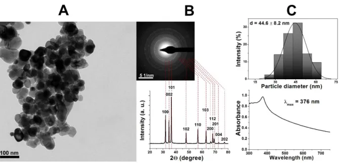
The TEM image of ZnO NPs (Supplementary Fig 1A) demonstrated that quasi-spherical 254
nanoparticles could be formed using this simple synthesis method, with an average diameter of 255
~45 nm illustrated by their size distribution histogram (Supplementary Fig 1C). The ED and 256
XRD results (Supplementary Fig 1B) that were collected to verify the crystallinity and chemical 257
composition of the particles were analogous with one another and proved to be in good 258
agreement with the literature (Talam et al., 2012; Kumar et al., 2013; Zhang et al., 2014; Ersan 259
et al., 2015), thus confirming the synthesis product as nanosized zinc oxide. The observed UV- 260
Vis spectra further proved the chemical composition of the sample, as an absorption maximum 261
around 370 nm is a characteristic value according to the literature (Talam et al., 2012; Kumar 262
et al., 2013).
263
It is worth mentioning, that all our results showed strong resemblance to one of our 264
recent contributions (Molnár et al., 2020), where smaller (~8 nm) ZnO NPs were synthesized.
265
While zinc oxide nanoparticles from both projects demonstrated similar XRD and UV-Vis 266
characteristics, some discrepancies were observed that can highlight how particle size may 267
affect certain properties. The full width at half maximum (FWHM) of XRD reflexion peaks 268
decrease with increasing particle size, due to the higher amount of similar crystal facets, and 269
the exact wavelength of the light absorption maximum of the characteristic UV-Vis spectra is 270
also NP size dependent. Due to their larger size, the ZnO NPs discussed in this research possess 271
sharper, more defined XRD reflexions and a moderately red shifted UV-Vis maximum 272
compared to smaller (and perhaps more commonly investigated) particles. Based on these 273
observations, the assumption could be made, that the as-prepared particles of this research may 274
have other distinct characteristics, which could affect biological activity.
275
276 1 2 3 4 5 6 7 8 9 10 11 12 13 14 15 16 17 18 19 20 21 22 23 24 25 26 27 28 29 30 31 32 33 34 35 36 37 38 39 40 41 42 43 44 45 46 47 48 49 50 51 52 53 54 55 56 57 58 59 60
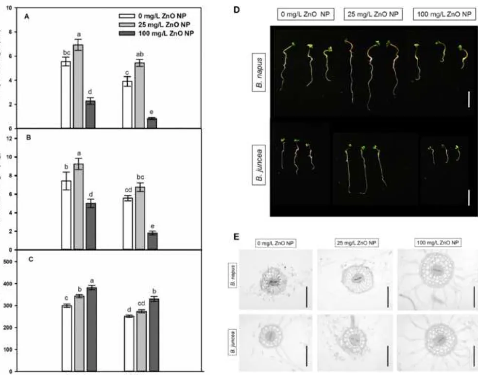
3.2.The effects of ZnO NPs doses on biomass production is similar in Brassica species 277
During stress-free circumstances, the primary root length of B. juncea is significantly 278
smaller than that of B. napus (Fig 1A and D). The beneficial effect of low ZnO NP dose (25 279
mg/L) proved to be similar in both Brassica species, since it induced primary root (PR) 280
elongation by 35-40%. High ZnO NP concentration (100 mg/L) caused PR shortening by 85%
281
in B. juncea, while in B. napus, the negative effect proved to be significantly slighter (53%).
282
The tendencies in ZnO NP-triggered changes of root fresh weight were similar to changes in 283
PR length. The ZnO NPs at 25 mg/L concentration induced 20-25% increase in root fresh 284
weight of both species; however, this was significant only in B. napus (Fig 1B). As the effect 285
of 100 mg/L ZnO NP root fresh weight decreased by 74% in B. juncea, and by 46% in B. napus.
286
Interestingly, ZnO NP treatments exerted no significant effects on shoot fresh weight compared 287
to controls (data not shown). Root shortening was accompanied by root thickening in both 288
species as indicated by the cross sections prepared from the differentiation zone of the primary 289
root (Fig 1E). In case of B. napus, root width significantly increased as the effect of both ZnO 290
NP concentrations (Fig 1C). Brassica juncea seedlings grown in the presence of 25 mg/L ZnO 291
NP did not show significantly thickened roots, however, 100 mg/L ZnO NP exposure notably 292
increased root width.
293
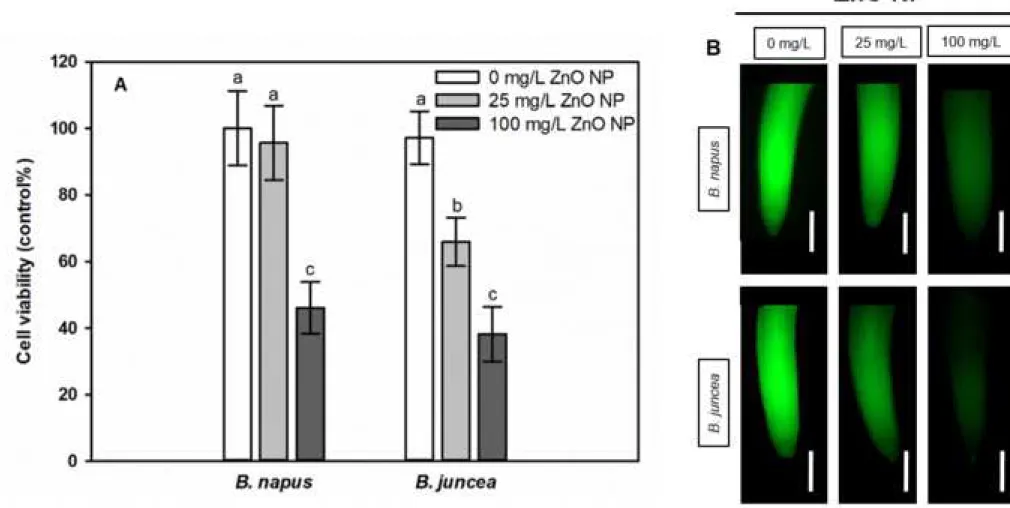
Viability of root meristem tissues significantly decreased (by 50-60%) due to 100 mg/L 294
ZnO NP exposure in both species, but the low ZnO NP dose caused 35% reduction in B. juncea 295
and practically no decrease in B. napus compared to control (Fig 2).
296
The above results indicate that 25 mg/L applied NP dose positively affected root 297
biomass in both species; however, it was more significant in B. napus compared to B. juncea 298
where root fresh weight and thickness remained at control-level and root viability decreased.
299
The four-times higher ZnO NP concentration has detrimental effects on root biomass which are 300
more pronounced in B. juncea compared to B. napus. Beneficial and toxic concentrations of 301
1 2 3 4 5 6 7 8 9 10 11 12 13 14 15 16 17 18 19 20 21 22 23 24 25 26 27 28 29 30 31 32 33 34 35 36 37 38 39 40 41 42 43 44 45 46 47 48 49 50 51 52 53 54 55 56 57 58 59
ZnO NPs slightly differ among experimental conditions, but in general, low doses (1-50 mg/L) 302
ZnO NPs induce germination, seedling growth, shoot and root biomass of Brassica species 303
while higher doses (>50 mg/L) reduce those parameters (Lin and Xing, 2007; Kouhi et al., 304
2014; Zafar et al., 2016; Rahmani et al., 2016; Singh et al., 2017) similarly to our results.
305
Thickening of the primary root indicates that secondary cell wall modifications may have 306
occurred (Somssich et al., 2016) in the presence of ZnO NPs which may influence Zn2+ uptake.
307
308
3.3.ZnO NPs-induced Zn2+ uptake and cell wall modifications in roots of Brassica 309
seedlings 310
Due to biotransformation in the presence of the root, ZnO NPs release Zn2+ which is 311
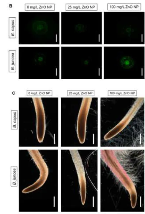
taken up by the root (Ma et al., 2013; Kouhi et al., 2015). Free, intracellular Zn2+ levels were 312
visualized using Zinquin fluorophore (Fig 3A and B). Despite the increasing external ZnO NP 313
concentrations, the intracellular Zn2+ level in B. napus roots did not increase, but in B. juncea, 314
both 25 and 100 mg/L ZnO NP caused two-fold elevation of Zn2+ levels compared to control.
315
Elevation in tissue Zn2+ level due to Zn2+ release from ZnO NPs was observed, inter alia, in 316
maize (Lv et al., 2015; Wang et al., 2016), alfalfa (Bandyopaghyay et al., 2015) and wheat 317
(Dimkpa et al., 2012).
318
Next, we examined whether the presence of ZnO NPs in root and stem cells (especially 319
in cells walls) can be detected. Nanoparticles with the size of ~45 nm possibly cannot enter the 320
cell wall pores having 5-30 nm width; however, their degradation to smaller NPs due to 321
biotransformation is conceivable (Fleischer et al., 1999; Nair et al., 2010). The TEM images 322
revealed that cell walls in case of 25 mg/L ZnO NP-treated B. napus became electron dense 323
indicating the possible binding of NPs (indicated by white arrows in Fig 3C). This was not 324
observed in root samples of 25 mg/L ZnO NP-exposed B. juncea. In root cells of untreated 325
plants and in hypocotyl cells, no signs of NP internalization were found in TEM images (Fig 326
1 2 3 4 5 6 7 8 9 10 11 12 13 14 15 16 17 18 19 20 21 22 23 24 25 26 27 28 29 30 31 32 33 34 35 36 37 38 39 40 41 42 43 44 45 46 47 48 49 50 51 52 53 54 55 56 57 58 59 60
3C and D) supporting the immobility of ZnO NPs between root and shoot (Wang et al., 2013;
327
Lv et al., 2015; Singh et al., 2018). Presumably, cell wall remodeling may contribute to Zn ion 328
binding and to the prevention of ZnO NP internalization.
329
Plant cell wall is a flexible macromolecule complex and its composition is finely 330
regulated but can be adapted to the environmental cues (Zhao et al., 2019). Cell wall 331
modifications permit the bounding and exclusion of heavy metals from the sensitive cytoplasm.
332
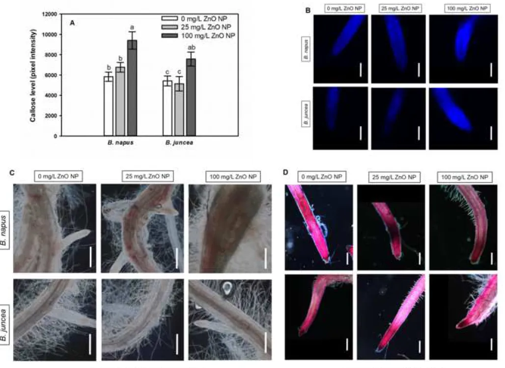
In both species treated with 100 mg/L ZnO NP, callose accumulation was detected in 333
walls of root tip cells as indicated by the elevation of callose-associated fluorescence (Fig 4A 334
and B). Additionally, 25 mg/L ZnO NP treatment caused no effect in callose level of both 335
species. Callose regulates the permeability of plasmodesmata and consequently the intercellular 336
transport (Vatén et al., 2011). In the study of Yanik and Vardar (2015) callose accumulation 337
was correlated with growth inhibition in the root of Al2O3 nanoparticles. Therefore, we suspect 338
that in Brassica species, 100 mg/L ZnO NP-induced callose accumulation may contribute to 339
the serious root shortening due to the reduction of symplastic molecule movement via 340
plasmodesmata. There was no correlation between Zn2+ levels and callose accumulation in ZnO 341
NP-treated Brassica suggesting that callose accumulation is potentially not involved in Zn2+
342
binding; however, callose may inhibit their movement via plasmodesmata. Lignification might 343
form an effective barrier against heavy metals preventing their entry into the cytoplasm, and 344
lignin also binds heavy metals (Parrotta et al., 2015; Loix et al. 2017). Lignin accumulation in 345
cells walls was microscopically detectable in root differentiation zone of B. napus exposed to 346
25 mg/L or 100 mg/L ZnO NP (Fig 4C). In contrast, B. juncea roots showed no detectable ZnO 347
NP-induced lignification. Lignin enrichment in the cell walls of the elongation zone may be 348
partly responsible for root growth diminution in the presence of heavy metals (Schützendübel 349
et al., 2001). In B. napus, lignin deposition was detected in the root parts close to the shoot, thus 350
ZnO NP-induced lignification may not have a role in root growth inhibition in this experimental 351
1 2 3 4 5 6 7 8 9 10 11 12 13 14 15 16 17 18 19 20 21 22 23 24 25 26 27 28 29 30 31 32 33 34 35 36 37 38 39 40 41 42 43 44 45 46 47 48 49 50 51 52 53 54 55 56 57 58 59
system. Among nanomaterials, CuO NPs has been found to trigger lignin and callose 352
accumulation in S. lycopersicum, B. oleracea (Singh et al., 2017) and A. thaliana (Nair and 353
Chung, 2014).
354
Pectin is a component of the cell wall matrix with a complex structure having high water 355
and Ca2+ binding properties (Voragen et al., 2009). Similar to lignification, pectin content was 356
significantly increased by 25 mg/L ZnO NP treatment in B. napus roots (Fig 4D). Moreover, 357
slight induction of pectin formation was detectable by Ruthenium Red staining also in B. juncea 358
in case of both ZnO NP doses. Accumulation of pectic substances in the cell walls may result 359
in more efficient Zn2+ binding due to the replacement of bounded Ca2+ (Dronett et al., 1996;
360
Krezlowska, 2011; Loix et al., 2017). Based on this, we can assume that ZnO NP-induced 361
formation of pectin in B. napus roots may result in Zn2+ binding in the cell wall and 362
consequently itsexclusion from the cytoplasm.Auramine-O staining can be applied for in situ 363
visualization of suberin and lignin in cell walls. In cross sections from the differentiation root 364
zones of both species, lignin and suberin-associated fluorescence increased as the effect of 100 365
mg/L ZnO NP exposure; however, the induction was more pronounced in B. napus (Fig 4E and 366
F). As for the lower ZnO NP dose, it induced lignin and suberin deposition in the root cell walls 367
of B. napus, but it decreased it in B. juncea (Fig 4E and F). Generally, lignin/suberin deposition 368
within the exodermis significantly contributes to the formation of an apoplastic transport barrier 369
(Hose et al., 2001). In roots of 25 and 100 mg/L ZnO NP-exposed B. napus the appearance of 370
exodermal lignin/suberin was detectable suggesting the role of this cell wall modification in 371
delaying Zn2+ entry into the roots as suggested by Cheng et al. (2014). Our results strengthen 372
the hypothesis that lignification/suberinization correlates with metal tolerance (Cheng et al., 373
2014) since the more tolerant B. napus showed more pronounced lignin/suberin deposition 374
compared to the relative sensitive B. juncea.
375 1 2 3 4 5 6 7 8 9 10 11 12 13 14 15 16 17 18 19 20 21 22 23 24 25 26 27 28 29 30 31 32 33 34 35 36 37 38 39 40 41 42 43 44 45 46 47 48 49 50 51 52 53 54 55 56 57 58 59 60
Collectively, the low ZnO NP concentration (25 mg/L) resulted in lignification, pectin 376
and suberin accumulation in the roots of B. napus, which may contribute to the observed Zn2+- 377
binding in the cell wall and to the beneficial effect on root biomass production. Callose 378
deposition was triggered only by the high ZnO NP dose in both Brassica species. Based on 379
these, B. napus shows more intense cell wall modification due to ZnO NP treatment.
380
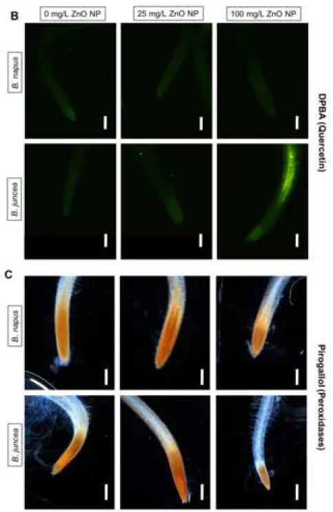
Additional cell-wall related defence processes such as flavonol contents and peroxidase 381
activities were examined. Flavonols play multifunctional roles during heavy metal stress since 382
they act as antioxidants in co-operation with cell wall peroxidases (cwPOD) and as chelators 383
they bind metals (Brown et al., 1998; Aherne and O'Brien, 2000; Soczynska-Kordala et al., 384
2001; Michalak, 2006; Korkina, 2007; Cherrak et al., 2016). Additionally, flavonols regulate 385
auxin transport consequently influencing growth (Gayomba et al., 2017). Elevation in quercetin 386
levels was detectable only in B. juncea roots exposed to 100 mg/L ZnO NP (Fig 5A and B), 387
while B. napus roots did not accumulate this flavonol molecule even in the presence of ZnO 388
NPs. Beyond their participation in lignification and suberinization, cell wall-associated class III 389
peroxidases (cwPODs) can oxidize flavonol substrates and convert H2O2 into H2O (Liox et al., 390
2017). In 100 mg/L ZnO NP-treated B. juncea, quercetin accumulation was accompanied by 391
decreased activity of cwPOD compared to control (Fig 5C) suggesting that cwPOD activity and 392
quercetin levels are associated and inactivation of cwPOD may contribute to quercetin 393
induction under ZnO NP stress (Fig 5A and B). Since cwPODs exert additional antioxidant 394
functions, it is not surprising that the 25 mg/L ZnO NP-induced slight cwPOD activation (Fig 395
5C) was accompanied by unmodified quercetin levels (Fig 5A and B). Similar to ZnO NP, Ag 396
NP treatment also caused activation of cwPODs in the leaves of Halophila stipulacea as 397
indicated by the intensification of pyrogallol staining (Mylona et al., 2020).
398
399
3.4.ZnO NPs induce species specific alterations in ROS and RNS signalling 400
1 2 3 4 5 6 7 8 9 10 11 12 13 14 15 16 17 18 19 20 21 22 23 24 25 26 27 28 29 30 31 32 33 34 35 36 37 38 39 40 41 42 43 44 45 46 47 48 49 50 51 52 53 54 55 56 57 58 59