Nanomolar inhibition of human OGA by 2-acetamido-2-deoxy- D - glucono-1,5-lactone semicarbazone derivatives
Mariann Kiss
a, Erna Szab o
b, Bogl arka Bocska
a, Luu Thanh Sinh
b,
Conceicao Piedade Fernandes
c, Istv an Tim ari
a, Joseph M. Hayes
c,**, L aszl o Soms ak
a,*, Ter ez Barna
b,***aDepartment of Organic Chemistry, University of Debrecen, H-4002, POB 400, Debrecen, Hungary
bDepartment of Genetics and Applied Microbiology, University of Debrecen, H-4002, POB 400, Debrecen, Hungary
cSchool of Pharmacy&Biomedical Sciences, University of Central Lancashire, Preston, PR1 2HE, United Kingdom
a r t i c l e i n f o
Article history:
Received 12 March 2021 Received in revised form 11 June 2021
Accepted 12 June 2021 Available online 15 June 2021
Keywords:
hOGA Expression Inhibitor
Glyconolactone semicarbazone QM/MM-PBSA
Halogen
HalogenHydrogen bond donor
a b s t r a c t
O-GlcNAcylation is a dynamic post-translational modification mediated byO-linkedb-N-acetylglucos- amine transferase (OGT) andO-GlcNAc hydrolase (OGA), that adds or removes a singleb-N-acetylglu- cosamine (GlcNAc) moiety to or from serine/threonine residues of nucleocytosolic and mitochondrial proteins, respectively. The perturbed homeostasis ofO-GlcNAc cycling results in several pathological conditions. Human OGA is a promising therapeutic target in diseases where aberrantly low levels ofO- GlcNAc are experienced, such as tauopathy in Alzheimer's disease. A new class of potent OGA inhibitors, 2-acetamido-2-deoxy-D-glucono-1,5-lactone (thio)semicarbazones, have been identified. Eight inhibitors were designed and synthesized infive steps starting fromD-glucosamine and with 15e55% overall yields.
A heterologous OGA expression protocol with strain selection and isolation has been optimized that resulted in stable, active and full length human OGA (hOGA) isomorph. Thermal denaturation kinetics of hOGA revealed environmental factors affecting hOGA stability. From kinetics experiments, the synthe- sized compounds proved to be efficient competitive inhibitors of hOGA withKi-s in the range of ~30 e250 nM and moderate selectivity with respect to lysosomalb-hexosaminidases.In silicostudies con- sisting of Prime protein-ligand refinements, QM/MM optimizations and QM/MM-PBSA binding free energy calculations revealed the factors governing the observed potencies, and led to design of the most potent analogue 2-acetamido-2-deoxy-D-glucono-1,5-lactone 4-(2-naphthyl)-semicarbazone 6g (Ki¼36 nM). The protocol employed has applications in future structure based inhibitor design targeting OGA.
©2021 The Authors. Published by Elsevier Masson SAS. This is an open access article under the CC BY- NC-ND license (http://creativecommons.org/licenses/by-nc-nd/4.0/).
1. Introduction
O-GlcNAcylation has emerged as a major posttranslational modification (PTM) that indicates the energy, nutritional and stress status of the cell, and involves transfer of a singleN-acetylglucos- amine moiety from UDP-GlcNAc to a serine or threonine side chain of proteins [1]. The two most striking features ofO-GlcNAcylation, which distinguish this PTM fromN- andO-glycosylations are its
localisation and its dynamic nature. It is localized to cytosol, nu- cleus and mitochondria rather than the endoplasmic reticulum or Golgi apparatus [2]. Similar to phosphorylation, two opposing enzyme actions determine the level ofO-GlcNAcylation of a pro- tein: OGT (O-GlcNAc transferase), the transferase, which transfers theN-acetylglucosamine moiety and creates theb-glycosidic link- age between the protein and GlcNAc, and the hydrolase OGA (O- GlcNAcase), which cleaves the bond [3]. UDP-GlcNAc, produced by the hexosamine pathway, provides the GlcNAc moiety for OGT to transfer to the aliphatic hydroxyl groups of the proteins to be modified. This UDP-GlcNAc is the nutrient sensor of the cell andO- GlcNAcylation, in response to the hexosamine flux, modulates metabolism and affects numerous cellular processes such as tran- scription regulatory pathways, cell cycle progression, embryonic
*Corresponding author.
**Corresponding author.
***Corresponding author.
E-mail addresses: jhayes@uclan.ac.uk (J.M. Hayes), somsak.laszlo@science.
unideb.hu(L. Somsak),barna.terez@science.unideb.hu(T. Barna).
Contents lists available atScienceDirect
European Journal of Medicinal Chemistry
j o u r n a l h o m e p a g e :h t t p : / / w w w . e l s e v i e r .c om/l ocate /ej mec h
https://doi.org/10.1016/j.ejmech.2021.113649
0223-5234/©2021 The Authors. Published by Elsevier Masson SAS. This is an open access article under the CC BY-NC-ND license (http://creativecommons.org/licenses/by-nc- nd/4.0/).
development, the regulation of the epigenome, respiration and proteosomal degradation [4e8]. O-GlcNAcylation does not only switch the biological function of key regulatory proteins on and off, but finely tunes their activities and interaction partners in the intracellular network. Imbalance in the GlcNAcylation cycle has been shown to manifest itself in diseases, where the hyperactivity of either OGA or OGT is observed. In nearly all cancers, an elevated level ofO-GlcNAcylation was reported [9]. The trend is opposite in the brain, in multiple neurodegenerative diseases where decreased levels ofO-GlcNAc modifications on numerous proteins have been reported, such as the proteins of the cytoskeleton [10]. The shift of homeostasis of O-GlcNAcylation to the increased hydrolysis of GlcNAc from the modified proteins is one of the main mechanisms contributing to neurodegeneration [11]. As a consequence of the complementary and competitive nature of phosphorylation andO- GlcNAcylation at many serine/threonine sites, low levels of O- GlcNAcylation leads to hyperphosphorylation of cytoskeleton pro- teins in the brain, including the microtubule protein tau. The hyperphosphorylated form of tau has been found in neurofibrillary tangles, characterized as tauopathy. An increase ofO-GlcNAcylation levels by inhibiting the activity of OGA has significantly decreased the aggregation propensity of tau [12,13]. The considerable thera- peutic relevance of dissecting the mechanism ofO-GlcNAcylation, motivates biological studies, and design and synthesis of potent OGA inhibitors.
Human OGA (hOGA) is a neutral hexosaminidase and belongs to family 84 of glycoside hydrolases (GH84) [14]. The enzyme follows a two-step substrate-assisted mechanism, through a dissociative oxocarbenium ion-like transition state, resulting in the net reten- tion of the anomeric configuration [15]. This mechanism is shared by the acidicN-acetylhexosaminidases, a member of GH20 [16]. In the GH84 family two neighbouring aspartates and in the GH20 enzymes, one aspartate and one glutamate, function as the key catalytic nucleophile and the acid/base residues [16e18]. There are specific humanN-acetylhexosaminidases that reside in lysosomes and are expressed in several tissues, mostly as a mixture of two isoenzymes: N-acetylhexosaminidase A (HexA) andN-acetylhex- osaminidase B (HexB) [19]. GH20N-acetylhexosaminidases catal- yse the hydrolysis of terminal, non-reducing N-acetyl-b-D- glucosamine and N-acetyl-b-D-galactosamine residues from pro- teoglycans [16,20]. Both isoforms appear in plasma and in synovial fluid and become elevated in inflammatory processes. In the sy- novial fluid of patients with osteoarthritis, HexA and HexB are released by chondrocytes into the extracellular compartment, mainly responsible for the degradation of glycosaminoglycans (GAG) [21,22]. Development of potent inhibitors against HexA and HexB are also highly relevant in studying the physiological role of these enzymes and their potential therapeutic applications.
PUGNAc (Phenyl Urethane of GlcNAc,Chart 1) was one of the first inhibitors of OGA [23] with aKivalue of around 50 nM, how- ever, it is not selective, since it inhibits GH20 N-acetylhex- osaminidases in a similar manner. TheN-phenylcarbamate moiety
can form hydrogen bonds between the oxime nitrogen atom and catalytic site residues, and the phenyl moiety, which protrudes from the active site pocket, takes advantage of hydrophobic in- teractions with aromatic side chains [24]. 2-Acetamido-2-deoxy-D- gluconhydroximo-1,5-lactone (LOGNAc), which lacks the phenyl carbamate group, is an around 30 times less potent inhibitor [23], showing that this moiety makes an important contribution to binding. Several other inhibitors are known in the literature, mostly transition state analogue compounds (NAG-thiazoline, NButGT, Thiamet-G), glucoimidazoles (GlcNacstatins) and non-carbohy- drate-based inhibitors have also been reported [25]. Surprisingly, there are only a few examples of PUGNAc analogues where the aromatic moiety is changed [26]. In addition, to the best of our knowledge, the effects of modifications of the urethane linker on OGA inhibition have not yet been studied.
Though PUGNAc is a well established OGA inhibitor, its synthesis has some drawbacks. The first eight-step synthetic route [27]
(overall yield: 14%) included two critical steps: a) the incomplete and low yielding transformation of the starting compound in the oxidation step towards the hydroximo-1,5-lactone (50% with 35%
unreacted starting material); b) a debenzylidenation with Na/NH3. The scale-up of both transformations to multigram synthesis led to lower yields and involved inconvenient workup procedures [28]. In an improved six-step synthesis [28] (overall yield: 15%) the hydroximo-1,5-lactone was obtained in an 59% yield only, and for this step keeping the 40 C temperature was crucial. At lower temperature the conversion was incomplete, while at higher tem- perature the 1,4-lactone oxime was formed.
With these preliminaries in mind we envisaged the synthesis of aryl substituted (thio)semicarbazones of 2-acetamido-2-deoxy-D- glucono-1,5-lactone (target compounds inChart 1) whose prepa- ration was expected to lack critical steps such as those for PUGNAc.
Kinetics experiments were performed to determine the potencies for hOGA inhibition, as well as selectivity studies considering also N-acetylhexosaminidases. High level in silico calculations using quantum mechanics/molecular mechanics (QM/MM) methods were performed to understand the hOGA binding properties and relative inhibitory efficiencies of the compounds.
2. Results and discussion
2.1. Synthesis
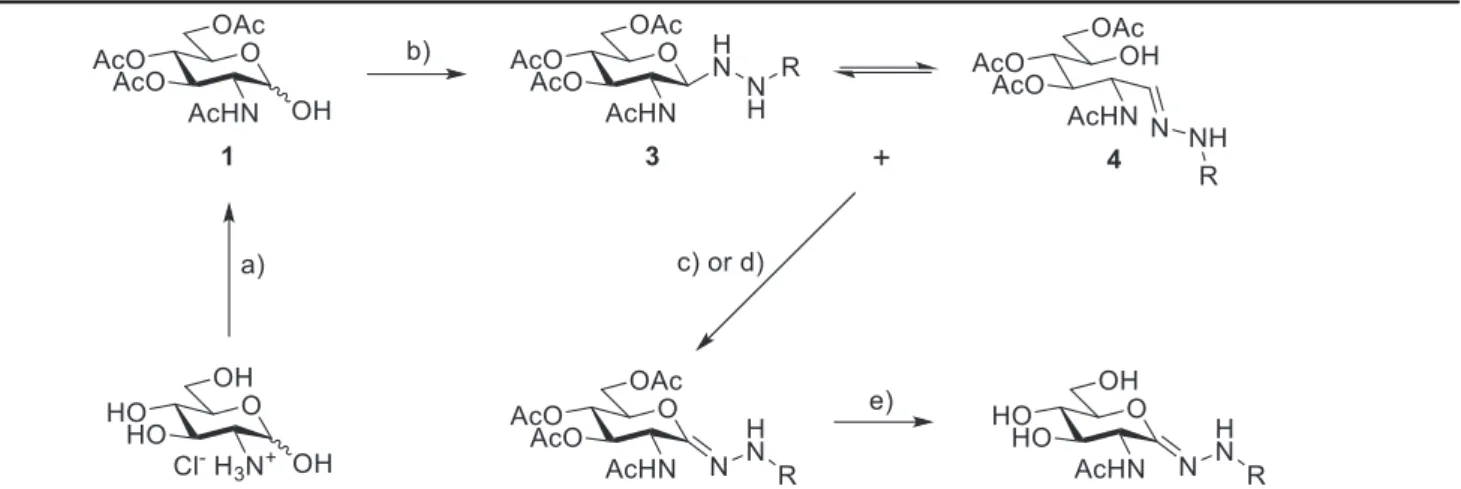
A single representative of our target compounds (Chart 1, Ar¼ Ph, X¼O) was located in the literature, therefore, the synthesis was based on the adaptation of this published procedure [29] (Table 1).
The starting material 2-acetamido-3,4,6-tri-O-acetyl-2-deoxy-D- glucopyranose (1) was obtained by a known two-step procedure [30,31] fromD-glucosamine hydrochloride. Compound1was then transformed by a 4-arylsemicarbazide (2, either commercial chemicals or obtained from the corresponding aromatic amines as described in the supporting material) in the presence of 10 mol% of
Chart 1.The structure of some OGA inhibitors and the target compounds of this study.
o, B. Bocska et al. European Journal of Medicinal Chemistry 223 (2021) 113649
p-toluenesulfonic acid as catalyst to get the N1-glycosyl semi- carbazides3. In place of the originally proposed 1,2-dichloroethane the use of dichloromethane, chloroform and toluene was tested for this transformation. In toluene the solubility of the starting mate- rial was insufficient, while the reaction in boiling CH2Cl2resulted in lower yields than in CHCl3 at reflux temperature, therefore, the latter solvent was used in the reactions. With the semicarbazide derivatives2a-gthe equilibrating open-chain tautomers4a-gwere also observed in the1H NMR spectra, while the reaction with thi- osemicarbazide 2h was devoid of this product. No attempt was made to separate compounds3and4. The NMR spectra of these mixtures contained signals which were analogous to those ob- tained for pure3aand4ain the literature [29]. Theb(D) configu- ration of compounds3followed from the large3JH-1eH-2couplings of 9e10 Hz (c.f.Experimental section). Oxidation of the mixtures containing3and4with CrO3-pyridine complex gave good yields of the expected lactone semicarbazones 5a-g. Regrettably, these oxidation conditions yielded complex reaction mixtures in case of the thio derivative 3h. Several oxidizing agents were tested (I2/ NEt3, PIDA, ICl/Cs2CO3) andfinally MnO2proved to be efficient to furnish the required compound5h. The lactone semicarbazones5 wereO-deprotected by a solution of NH3in MeOH to result in the test compounds6. The overall yields of these five-step synthetic sequences to give target compounds6fromD-glucosamine fell in the 15e55% range.
To verify the configuration of the C]N bond in compounds6, a Nuclear Overhauser Effect (NOE) based NMR approach was applied to6c(Fig. 1). TheE-Zisomeric forms of compounds6are expected to be distinguished on the basis of NOE cross-peaks between the N(2)H and distinctive protons of the sugar part, since the maximum spatial distance which can provide an NOE signal is about 5 Å. First,
a 2D1He13C HMBC spectrum [32] (Fig. S1) was recorded for the unambiguous assignment of N(2)H (9.8 ppm). Then a 2D1He1H EASY ROESY experiment [33] was performed, which is a powerful alternative of NOE measurements for small molecules. In the
1He1H ROESY spectrum (Fig. S2), the N(2)H resonance provided NOE cross-peaks with the H(50) and O(6’)H resonances (Fig. 1), which indicated the vicinity in space of these protons, and so, the presence of theZisomeric form of compound6c. Since both the1H and13C NMR spectra for the other compounds6closely resemble those of6c, one can safely conclude the presence of theZisomer also in those compounds.
2.2. Heterologous expression of hOGA
A heterologous expression system for hOGA in E. coli was Table 1
Synthesis of the test compounds6.
Reagents and conditions: a) 1. Ac2O, abs. Py, r.t.; 2. 4 Å molecular sieves, abs. MeOH, r.t. (83% for two steps) [30,31]; b) NHwitha-g: CrO3-Py, CH2Cl2, 0C/r.t.; d) with h: MnO2, abs. CH2Cl2, reflux; e) NH32/MeOH, r.t.NHR (2), 0.1 equiv. pTsOH$H2O, CHCl3, reflux; c)
R Products and yields (%)
3þ4 Ratioaof3:4 5 6 Overall yield of6fromD-glucosamine
a CONHPh 90 5:1 83 79 49
b CONH(p-MePh) 83 7:1 92 86 55
c CONH(p-CF3Ph) 81 5:1 26 87 15
d CONH(p-OMePh) 87 7:3 86 83 52
e CONH(p-ClPh) 77 4:1 78 74 37
f CONH(1-naphthyl) 94 b 64 83 41
g CONH(2-naphthyl) 80 b 93 75 46
h CSNHPh 87c e 78 83 47
aDetermined from the1H NMR spectra of the worked-up reaction mixtures.
bFormation of the open-chain products4fand4gwas detected by TLC but due to signal overlaps and small amounts of these compounds the3:4ratio was not established.
c Compound3formed as a single product.
Fig. 1.Nuclear Overhauser Effects (NOEs) observed in compound6c.
o, B. Bocska et al. European Journal of Medicinal Chemistry 223 (2021) 113649
established in order to gain soluble, active, full-length wild-type enzyme with considerable thermostability for the kinetic evalua- tion of the inhibitory potency of new inhibitors6a-hagainst hOGA.
Bacteriophage T7 polymerase promoter based expression system such as BL21(DE3)/pET system was chosen for this purpose. The pET-28 based vector construct carrying the N-terminally His- tagged hOGA encoded gene was transformed into five E. coli BL21(DE3) strains and the cultivation conditions were optimized, as well as the environmental parameters of the downstream pro- cesses. The small scale expression trials of all the transformed BL21(DE3) strains carried out at 23 C unravelled two main ob- jectives to be solved in this expression system. Namely, the high tendency of the recombinant hOGA to form inclusion body aggre- gates during the protein production phase and the significant proteolytic degradation of the recombinant enzyme sufferedin vivo and in vitro (Fig. 2). We did experience the latter phenomenon, despite all the strains used were deficient in two proteases, an outer membrane protease (ompT) and an ATP dependent protease (Lon), both of which have been reported as responsible for degradation of misfolded proteins and interference with production of some full- length recombinant proteins [34e36]. In this respect, the worst performing strain was BL21(DE3), where full length recombinant enzyme was hardly produced (Fig. 2). Beside the non-specific proteolysis, significant production of a 70 kDa long truncated hOGA fragment was observed with BL21 Star and pLysS strains referring to an existence of a sequence specific proteolytic cleavage site. This fragment bound to the affinity column and exhibitedb-N- acetylglucosaminidase activity (see supporting material,Fig. S3).
The size and activity of this comparatively stable hOGA fragment suggests the presence of caspase-3-like protease activity ofE. coli cells that might be activated upon the overproduction of hOGA, causing a proteotoxic stress for the cell [37e40]. Two strains, RIPL and Rosetta, where the translation was optimized for genes that differ in codon usage from that ofE. coliby carrying rare tRNA genes on plasmids, showed increased production of recombinant enzyme.
However, only the Rosetta strain could increase the synthesis of the enzyme in the active, soluble form (Fig. 2). The productivity of the Star and pLysS strains was very similar with a relatively high yield of active hOGA compared to the aggregate form. This highlights the importance of the increased life-time of mRNA in Star and the very tight control of expression in pLysS (Fig. 2). However, Star and pLysS exhibited the highest site-specific proteolytic activity producing
the 70 kDa long hOGA fragment.Table S1summarizes the b-N- acetylglucosaminidase activity of the cell lysates of the selected E. colistrain. It can be misleading to rely only on these results and rank the productivity of the strains according to these activity values. For instance, the BL21 (DE3) strain, which yielded the sec- ond highest b-N-acetylglucosaminidase activity, produced the lowest level of soluble, full-length recombinant enzyme. From all these observations it can be concluded that even though the strains have the same origin, they displayed different tolerance to the overproduction of misfolded protein, due to the activation of an extensive network of proteolytic housekeeping and regulating en- zymes [41,42].
Since the Rosetta strain proved to be the best performing expression host, systematic cultivation parameters were optimized for this strain. The effect of temperature on the recombinant pro- tein production was investigated below 30C, at three tempera- tures (16, 23 and 28C). The low protein concentrations in theory should decrease the partial folding and misfolding of proteins, thus, preventing the aggregation to inclusion body [43]. Decreasing the cultivation temperature only favoured full-length and active enzyme production at 23 C. At lower temperature (16 C), the misfolding of the human enzyme was accelerated and the chap- erone systems of E. coliwere not able to prevent the aggregate formation (see supplementary material,Fig. S4,Table S2).
The induction phase of the culture growth also influenced the yield of hOGA production. When the inducer was employed at later phase than the mid-exponential one (atOD6000.9 instead of 0.6), we could achieve 50% higher yield (expressed in totalb-N-acetyl- glucosaminidase activity: 35 units for the mid-exponential phase induction and 53 units for the later phase induction in 250 ml cell culture) without compromising greater proteolytic degradation or formation of inclusion-body aggregates (Fig. S5).
All the optimized conditions were built in thefinal heterologous expression protocol for hOGA, in which aggregate formation of the overproduced enzyme was minimized and an efficient full-length, active hOGA production was ensured.
2.3. Stability studies on wild-type hOGA
The most important issue during the downstream processes was undoubtedly minimization of the action of E. coli proteases.
Nevertheless, the identification of stabilizing and destabilizing factors involved in maintaining hOGA in its native state was also essential. Therefore, we carried out stability studies on hOGA by varying buffer compositions and we followed the heat-induced denaturation kinetics of the enzyme at 50C. The time-course of the enzyme residual activities was determined and the thermal denaturation kinetic constants (kD) or the half-lifes (t1/2) were calculated. By increasing the ionic strength, the thermal denatur- ation rate of the enzyme increased too, as was detected in phos- phate buffer upon increasing KCl concentration (Fig. S6a and Table S3). There was no significant difference in the rate of thermal denaturation of hOGA in the presence of either potassium or so- dium cation (Fig. S6bandTable S3). Treating hOGA solution with reducing agents such as mercaptoethanol and DTT proved to be an important protective factor increasing the protein half-life by up to 25% (Table S3andFig. S7b). An even greater stabilizing effect was observed when phosphate buffer was supplemented with 15%
glycerol to increase the enzyme half-life by 80% (Table S3). On the contrary, detergent Triton X-100 greatly destabilized hOGA native state and accelerated its thermal denaturation rate fromkD¼0.05 tokD¼0.17 min1, whereas the presence of Tween 20 proved to be an innocent factor in the structural disintegration of the enzyme (Fig. S7aandTable S3). The effect of the majority of environmental factors on thermal denaturation kinetics of hOGA could be Fig. 2.Gel electrophoretic (8% SDS-PAGE) analysis of the soluble and insoluble frac-
tions of the cell lysates produced by differentE. coliBL21(DE3) strains grown at 23C, induced at mid-exponential phase with 0.4 mM IPTG and harvested at early stationary phase.
Lanes: 1. protein marker; 2.E. coliBL21(DE3) insoluble fraction; 3.E. coliBL21(DE3) soluble fraction; 4.E. coliBL21(DE3) RIPL insoluble fraction; 5.E. coliBL21(DE3) RIPL soluble fraction; 6.E. coliBL21(DE3) Rosetta insoluble fraction; 7.E. coliBL21(DE3) Rosetta soluble fraction; 8.E. coliBL21 Star (DE3) insoluble fraction; 9.E. coliBL21 Star (DE3) soluble fraction; 10. E. coliBL21(DE3) pLysS insoluble fraction; 11. E. coli BL21(DE3) pLysS soluble fraction.
o, B. Bocska et al. European Journal of Medicinal Chemistry 223 (2021) 113649
modelled by a single-transient process between the native and unfolded states that obeys afirst-order kinetics. When phosphate buffer alone was present, the heat denaturation of hOGA followed sigmoidal kinetics, which applies for an autocatalytic process (indicating the autocatalytic nature of the unfolding event). We did not investigate the mechanistic details further, however suggested explanation for the existence of cooperativity in domain confor- mational changes is discussed in the supplementary material.
We applied the stabilizing supplementations including the presence of reducing agent, Tween 20 and the preferable envi- ronmental factors such as low ionic strength, during the cell disruption process. It is also worth emphasizing the strict use of broad spectrum protease inhibitors during the cell lysis. During the affinity chromatography step, high salt concentration was neces- sary to avoid unspecific protein binding. The collected fractions of high b-N-acetylglucosaminidase activity were immediately ultra- filtrated to change the elution buffer into the‘long storage buffer’ containing all the evaluated stabilizing supplementations including glycerol and kept at 20 C. The concentration of the enzyme samples was not larger than 2 mg/ml. The single chromatographic purification step resulted in higher than 90% homogenous, full- length and active hOGA sample (Fig. S8). Following the quality of the enzyme samples during the long time storage, their enzyme activity and homogeneity were maintained even after 12 months (Fig. S8).
2.4. Inhibitory potency of 2-acetamido-2-deoxy-D-glucono-1,5- lactone semicarbazone derivatives on hOGA and HexA/HexB activities
Michaelis-Menten kinetic parameters forp-nitrophenyl-GlcNAc (pNP-GlcNAc) as substrate in the presence of hOGA established in potassium-phosphate buffer at pH 7.5 resulted in 20mM forKMand 1.6 s1forkcatvalues (Table 2). The determinedkcatvalue correlated well with the previously published data [44]. However, the enzyme in this study displayed much higher binding affinity towardpNP- GlcNAc than was previously reported for different conditions (Table 2). Kinetic analysis of hOGA and HexA/HexB inhibition by 2- acetamido-2-deoxy-D-glucono-1,5-lactone semicarbazone de- rivatives was performed by using Dixon method [45] and an improved method presented by Cornish-Bowden [46] (Fig. 3).
Plotting the reciprocal initial rate against inhibitor concentration, the intersection point of the obtained curves at three different substrate concentrations was located in the fourth quadrant and gave the inhibitor constants (Ki) for all the investigated compounds (Table 3). The location of the intersection point also refers to the mode of inhibition, either competitive or mixed. Since Dixon plot cannot distinguish unambiguously between these inhibitory modes, we also plotted the kinetic data using the Cornish-Bowden method (Fig. 3). The resulting lines are parallel in the plot of [S]/v against inhibitor concentration indicating competitive inhibition of hOGA and HexA/HexB enzymes by each of the 2-acetamido-2- deoxy-D-glucono-1,5-lactone semicarbazone derivatives6.
Inhibitor constants (Ki) for most of the compounds 6 against hOGA are in the submicromolar, in some cases in the nanomolar range, and slightly higherKivalues have been obtained for HexA/
HexB (Table 3). The most potent inhibitors of hOGA are semi- carbazones6eand6g, while thiosemicarbazone6hproved to be the weakest one.
For the determination of the inhibitor constants of the most potent inhibitors (6eand6g), the Dixon method could not be used in the presence of thepNP-GlcNAc substrate because the inhibitor concentration was comparable to the enzyme concentration.
Therefore, the fluorogenic 4-methylumbelliferyl-GlcNAc (4-MU- GlcNAc) substrate was used to enable the application of the more sensitivefluorometric detection method. This allowed a significant decrease in the applied enzyme concentration for the inhibition studies. In a separate experiment with6fwe demonstrated that substituting the chromogenic substrate (pNP-GlcNAc) with afluo- rogenic one (4-MU-GlcNAc) did not influence the binding affinity.
The obtainedKivalues for6fin the presence ofpNP-GlcNAc and 4- MU-GlcNAc were 0.506 ± 0.020 mM and 0.509 ± 0.036 mM, respectively (Fig. S9).
The selectivity of inhibition of the two enzymes was moderate, and this feature resembles PUGNAc. It is worth noting the consid- erable difference in the inhibition by naphthyl derivatives6fand 6g, of which the 2-naphthyl6gis more than one order of magni- tude more potent than the 1-naphthyl6fcounterpart. On the other hand,6fis more selective towards HexA/HexB.
2.5. In silico predictions analysis
In order to gain an understanding of the factors governing the observed potencies for hOGA inhibition, computations were per- formed in the form of Prime protein-ligand refinements [48], fol- lowed by higher level QM/MM optimizations and QM/MM-PBSA binding free energy calculations (DGbind, Eq.(1)).
DGbind¼DEQM/MMþDGsolveTDSMM [1]
Eq. (1) includes contributions from gas-phase protein-ligand interaction energies (DEQM/MM), changes in solvation free energy on binding (DGsolv), as well as the ligand entropy changes (TDSMM). In these calculations, side chains Tyr219, Phe223, Val254, Val255 and Tyr286 in the vicinity of6a-gligand phenyl or naphthyl rings were included as part of the QM region, selected due to the importance of these protein-ligand interactions to the observed potency differ- ences, and to better describep-pinteractions with Tyr286. Because of the potential for a better description of binding site interactions using QM methods, QM/MM-PB(GB)SA approaches have recently been applied in a number of studies [49], including where protein- ligandp-pstacking interactions play a key role [50,51]. Here, the QM region was modelled using DFT at the M06-2X/6-31þG**level of theory, with the M06-2X functional recognised for its description of non-covalent interactions [52e54]. We should note that native ligand docking calculations with Glide [48] failed to reproduce the PUGNAc ligand crystallographic conformation (PDB code: 5UHO);
more specifically the critical orientation of the phenylurethane (-O- CO-NH-Ph) moiety, so that the Prime refinement, QM/MM opti- mization and QM/MM-PBSA approach as fully outlined in the experimental details was applied.
The results of the QM-MM/PBSA calculations for PUGNAc and
Table 2
Michaelis-Menten kinetic parameters of hOGA forpNP-GlcNAc.
Buffer 50 mM
K2HPO4- KH2PO4buffer, pH 7.5
PBS (10 mM phosphate buffer, 137 mM NaCl, 2.7 mM KCl) pH 7.4 [44]
0.5 M citrate/phosphate buffer, pH 6.5 [47]
KM(mM) 20±2 170±10 1100±100
kcat(s1) 1.6±0.1 1.8±0.1 3.4±0.3
o, B. Bocska et al. European Journal of Medicinal Chemistry 223 (2021) 113649
the eight inhibitors6a-hstudied in this work are shown inTable 4.
Thefinal predictedDGbindvalues for each inhibitor are broken down into the different contributions fromDEQM/MM,DGsolv, andTDSMM. In
terms of reproducing the relative potencies, good agreement with experiment was obtained (the absolute values of predictedDGbind
are larger than experiment, an expected consequence of the Fig. 3.Plotting hOGA inhibition data in the presence of6aat three differentpNP-GlcNAc concentrations (-75mM;40mM;:20mM) by using Dixon method (A) and Cornish- Bowden method (B).
Table 3
The binding affinities of 2-acetamido-2-deoxy-D-glucono-1,5-lactone semicarbazone derivatives toward hOGA and HexA/HexB enzymes (Ki[mM]).
Compound hOGA HexA/HexB
6a 0.190±0.008 0.205±0.014 (lit.: 0.130±0.020) [29]
6b 0.155±0.003 0.332±0.017
6c 0.167±0.006 0.125±0.004
6d 0.270±0.010 0.413±0.014
6e 0.083±0.004a 0.170±0.004
6f 0.506±0.020 0.154±0.006
6g 0.036±0.001a 0.047±0.002
6h 27±3 30±2
a4-MU-GlcNAc was used as substrate.
o, B. Bocska et al. European Journal of Medicinal Chemistry 223 (2021) 113649
method approximations [50,51,55]). PUGNAc was correctly pre- dicted as more potent than the phenylsemicarbazone analogues 6a-e. PUGNAc and6adiffer structurally only in terms of their -O- C(O)-NH- and -NH-C(O)eNH- moieties, respectively. Compared to PUGNAc,6a(and the other semicarbazone analogues,c.f.6ecom- plex inFig. 4 (A)) is able to exploit a hydrogen bond interaction from N2H (c.f.numbering scheme inTable 4) with the Tyr219 side chain hydroxyl O atom. While protein-ligand interactions based on DEQM/MM are predicted as stronger for 6a (140.7 kcal/mol) compared to PUGNAc (136.2 kcal/mol), the desolvation costs for 6a are greater, as well as entropy costs accounted for as loss of ligand vibration, rotation and translation on binding. Thio- semicarbazone6h(a C]O to C]S replacement in the linker) was correctly predicted as the least potent ligand. Although its protein-
ligand (DEQM/MM) interactions are predicted as strong (145.9 kcal/
mol), the desolvation cost of binding (119.0 kcal/mol) was the highest of all inhibitors inTable 4.
Analyzing theDGbindvalues for all the phenyl analogues6a-e, compounds6dand6eare correctly predicted as the least and most potent inhibitors within this series, respectively; the remaining inhibitors in this series (6a-c) displayed similar experimental po- tencies (0.155e0.190mM) and had predicted potency ranks in be- tween. Considering the electron donating or withdrawing properties of6a-epara-substituents, there is no obvious correlation between these properties and the observed potencies. Indeed, considering the contributions toDGbind, the relative potencies of 6a-eare predicted to be a sensitive balance between protein-ligand interactions (DEQM/MM) and desolvation effects; entropy to a lesser Table 4
Quantum Mechanics⁄Molecular MechanicsePoissoneBoltzmann Surface Area (QM⁄MM-PBSA) results for the estimation of hOGAeligand binding free energies using Eq.(1).
The QM region was modelled at the M06-2X/6-31þG**level of theory; MM region using the OPLS-AA (2005) forcefield. All energy values are in kcal/mol.
Ligand Predicted Experimental
DEQM/MM DGsolv TDSMMa DGbind DGbind
PUGNAc 136.2 96.2 21.1 18.9 10.4
6a 140.7 101.9 21.8 17.0 9.5
6b 143.1 103.3 23.0 16.8 9.7
6c 152.7 111.8 23.3 17.6 9.6
6d 147.7 109.2 22.9 15.6 9.3
6e 148.4 107.8 22.2 18.4 10.0
6f 152.2 105.8 23.0 23.4 8.9
6g 153.8 106.8 22.7 24.3 10.6
6h 145.9 119.0 19.8 7.1 6.5
aValues calculated at 310 K to be consistent with the performed kinetic experiments. ExperimentalDGbind¼RT ln Ki.
o, B. Bocska et al. European Journal of Medicinal Chemistry 223 (2021) 113649
degree. Given the good agreement between relative DGbind pre- dicted and experimental values for the phenyl substituted com- pound series (Spearman rank correlation Rs¼0.893 (p<0.01);
Pearson correlation Rp¼0.993 (p<0.01)efor PUGNAc,6a-eand 6h), the predicted binding interactions could be analysed with confidence.
The most potent phenyl analogue6ehas the strongest protein-
ligand interactions (DEQM/MM¼ 148.4 kcal/mol), and its predicted protein-ligand complex is shown inFig. 4 (A). The crystallographic N-acetyl-glucosamine moiety interactions in hOGAePUGNAc (PDB code: 5UHO) are, as expected, conserved in the predicted com- plexes for the newly designed ligands 6a-h. These include key hydrogen bond interactions of inhibitor O40and O60hydroxyls (c.f.
Table 4) with Asp285 side chain carboxylate; O40-hydroxyl O with Fig. 4.Predicted model complexes from Prime, QM/MM and QM/MM-PBSA calculations:(A)hOGA-6eand(B)hOGA-6g.
o, B. Bocska et al. European Journal of Medicinal Chemistry 223 (2021) 113649
the Asn313 side chain amide; and O30-hydroxyl with both Gly67 backbone O and Lys98 side chain. TheN-acetyl group is involved in hydrogen bonds with Asp174 side chain carboxylate and Asn280 side chain amide. Considering the binding interactions of the phenyl substituents of 6a-6e (and6h), there are favourable in- teractions with the hydrophobic side chains of Tyr219, Phe223, Val254, Val255 and Tyr286. Of particular significance are edge-to- face T-shapedp-pstacking with Tyr286. Although the rings are not ideally aligned, thep-pstacking ring centroidering centroid distances for6ae6ewere in the range 5.3e5.5 Å (5.4 Å for6ein Fig. 4 (A)), close to theoretical optimum intermonomer distances (~4.8e5.0 Å) in simple model systems [56] and within the observed geometric preferences (5.0e5.6 Å) of protein e ligand (Phe e phenyl) T-shapedp-pinteractions from Protein Data Bank analysis [57]. The protein-ligand ring planes had intersection angles 83.5e86.8, that are also consistent with the T-stacking Protein Data Bank analysis [57]. Accounting for phenyl substituent effects for T-shaped configurations can be somewhat more complex than for sandwich arrangements [58]; in our situation (6a-e), the ligand p ep interactions with Tyr286, are accompanied by additional direct interactions between the ligand phenyl substituents and the phenol ring of Tyr286.
Of further significance, we observed the potential for binding site loop Val255 (backbone) NHepinteractions with the designed compounds. For the 6e complex (Fig. 4 (A)), the Val255 NH to centroid of6ephenyl ring predicted distance was relatively long at 3.8 Å. This analysis, in part, was the basis of our decision to explore naphthyl substituents that could better exploit such interactions (vide infra). Compound6e, however, had the additional potential to form halogen Clehydrogen bond donor (HBD) interactions with Val255 NH. A quite recent QM study and survey of crystal structure data demonstrated that halogen (X)eHBD interactions can make significant contributions to ligandprotein binding [59]. Geometric parameters considered by the authors included the angle CeXeHBD and the distance HBDeX, with the HBD defined by its oxygen, nitrogen or sulfur atom. The survey revealed CeXeHBD angle maxima in the plots in the range ~80e110. While the pre- dicted CeXeHBD angle (66) and HBDeX distance (4.9 Å) for the hOGA e 6epredicted model complex are also not optimum for halogen eHBD interactions, these interactions can occur over a broad range of angles and we must also consider that Val255 is part of a loop. Additionally, from the crystal structure data analysis, 60e70angles for nitrogen as HBD were associated with mainly longer 4.0e4.5 Å distances. We can therefore postulate that the Val255 NHeCl interaction may contribute to the improved potency of6ecompared to the other analogues6a-6d.
As mentioned, it was decided to explore whether substitution of the phenyls of6a-6ewith naphthyl moieties could better exploit potential NHepcontacts with Val255, but also the hydrophobic nature of other surrounding residues. Indeed, predictions for both 1- (6f) and 2-naphthyl (6g) suggested improved potency, with predictedDGbindvalues of23.4 and24.3 kcal/mol, respectively.
And while the prediction was incorrect for6f, potentially due to conformational entropy effects (over-prediction of 1-naphthalene compound analogues extends to other methods and systems [60]),6gwas experimentally the most potent of all ligands tested.
The predicted binding of 6g with a 2-naphthyl moiety did as anticipated lead to better Val255 NHepcontacts (3.4 Å to centroid of ring extension), as well as preserving the edge-to-face T-shaped contacts with Tyr286 (Fig. 4 (B)). TheDEQM/MMvalue of153.9 kcal/
mol was considerably more favourable (>5 kcal/mol) to those ob- tained for the substituted phenyls. An alternative parallel confor- mationp-pbinding mode with Tyr286 was also predicted for6g, but was less favourable (DGbind¼ 11.4 kcal/mol) compared to the T-shaped conformation (DGbind¼ 25.0 kcal/mol); in agreement, T-
shaped binding conformations are reported as more predominant for solved protein-ligand complexes [57].
3. Conclusion
A series of 2-acetamido-2-deoxy-D-glucono-1,5-lactone 4- aryl(thio)semicarbazone derivatives was obtained by a five-step synthetic sequence from D-glucosamine with overall yields of 15e55%. These compounds represent thefirst analogues of PUGNAc with modified linkers between the sugar and the aromatic moiety.
An efficient heterologous expression protocol for full length and active recombinant hOGA was developed byE. colistrain selection and optimization of cultivation conditions. Thermal denaturation studies revealed environmental factors that most affected hOGA stability, so that hOGA samples displayed long-term stability and ensured reproducibility in the evaluation of inhibitory potencies.
Kinetic studies with hOGA and hexosaminidases A and B showed competitive inhibition for all synthesized semicarbazone de- rivatives (hOGA: range ofKi-s ~30e250 nM; HexA/HexB: range of Ki-s ~50e400 nM) with a modest selectivity towards hOGA. These lactone semicarbazones are therefore alternatives of the OGA in- hibitor PUGNAc in terms of both synthetic efficiency and inhibitory efficacy. A computational approach consisting of Prime protein- ligand refinements, post-processed by QM/MM optimizations and calculation of binding free energies using QM/MM-PBSA proved effective, for the most part, to describe the interactions governing the observed ligand potencies. The most potent inhibitor 6g (Ki¼36 nM) had a 2-naphthyl substituent that better exploited interactions (including NHepand T-shapedpepstacking), with surrounding hydrophobic residues. Further computational and synthetic work to enhance hOGA selectivity is in progress and will be disclosed in due course.
4. Experimental
4.1. Syntheses 4.1.1. General methods
Melting points were measured on a Kofler hot-stage and are uncorrected. Optical rotations were determined with a PerkineElmer 241 or Jasco P-2000 (Easton, MD, USA) polarimeters at room temperature. NMR spectra were recorded by Bruker AM Avance 360 (360/90 MHz for1H/13C) spectrometer. Chemical shifts are referenced to TMS as the internal reference (1H), or to the re- sidual solvent signals (13C). 2D 1He13C HMBC experiment [32]
optimized to long-range heteronuclear coupling of 8 Hz, and 2D
1He1H EASY ROESY experiment [33] with 300 ms mixing time were performed on a Bruker Avance II 500 MHz spectrometer equipped with a 5 mm z-gradient TXI probe. Mass spectra were recorded with MicroTOF-Q type Qq-TOF MS and maXis II UHR ESI-QTOF MS (Bruker Daltonik, Bremen, Germany) instruments in positive ion mode with electrospray ionization or atmospheric pressure chemical ionization technique. TLC plates were visualized under UV light, and by spray reagent with gentle heating (the plate was sprayed with the following solution: abs. EtOH (95 ml), cc H2SO4
(5 ml), anisaldehyde (1 ml)). For column chromatography Kieselgel 60 (Merck, particle size (0.063e0.200 mm) was applied. Dichloro- methane was distilled from P2O5and stored over 4 Å molecular sieves. Pyridine was distilled from KOH and stored over KOH. CrO3
was stored over P2O5. 4-Phenylsemicarbazide (2a) and 4- phenylthiosemicarbazide (2h) were purchased from Sigma- Aldrich Ltd. (Budapest, Hungary). 2-acetamido-3,4,6-tri-O-acetyl- 2-deoxy-a,b-D-glucopyranose (1) [30,31], substituted phenyl- semicarbazides (2b-e) and 1- or 2- naphthylsemicarbazides (2f-g) [61] were synthesized according to literature procedures. Unless
o, B. Bocska et al. European Journal of Medicinal Chemistry 223 (2021) 113649
otherwise indicated, all chemicals used in the biochemical experi- ments were analytical-grade and purchased from Sigma-Aldrich Ltd. (Budapest, Hungary).
4.1.2. General procedure I. for the synthesis of 1-(2-acetamido- 3,4,6-tri-O-acetyl-2-deoxy-b-D-glucopyranosyl)-(thio)
semicarbazides (3a-h)
A solution of 200 mg (0.58 mmol) 2-acetamido-3,4,6-tri-O- acetyl-2-deoxy-a,b-D-glucopyranose (1), 0.87 mmol (thio)semi- carbazide and 0.058 mmolp-toluenesulfonic acid in 4 ml chloro- form were boiled under reflux until the reactions were complete (monitored by TLC hexane: acetone¼3 : 2). The mixtures were cooled in ice-bath, the white precipitates werefiltered. If the re- agents contaminated the products, column chromatography was used (hexane: acetone 1 : 1).
4.1.3. General procedure II. for the synthesis of 2-acetamido-3,4,6- tri-O-acetyl-2-deoxy-D-glucono-1,5-lactone semicarbazones (5a-h) Method A:A solution of 4 equivalents of anh. pyridine in 2 ml of anh. CH2Cl2was stirred at 25C as 2 equivalents of dry CrO3were added in one portion. The reaction was slightly exothermic, and the solution turned burgundy. After 20 min at 25C, the solution was cooled to 0C and a suspension of 100 mg compound in 3 ml of anh.
CH2Cl2was added in one portion. The reaction mixture was stirred, while it slowly warmed up to room temperature. After the reaction was complete (monitored by TLC hexane: acetone 1 : 1) it was diluted with 4 ml of Et2O. The mixture wasfiltered under vacuum and thefiltrate concentrated. The pyridine was removed under high vacuum, and the residue was purified by column chromatography (hexane: acetone 2:1).
Method B:Activated MnO2was added to a solution of 100 mg compound in anh. CH2Cl2. The mixture was boiled until the reaction was complete (hexane: acetone 1:1), the MnO2wasfiltered under vacuum and thefiltrate concentrated, the residue was purified by column chromatography (hexane: acetone 2:1).
4.1.4. General procedure III. for the synthesis of 2-acetamido-2- deoxy-D-glucono-1,5-lactone semicarbazones (6a-h)
A solution of 100 mg compound in 1 ml of MeOH was stirred at 25C with 1 ml of a sat. NH3/MeOH solution. After the reaction was complete (monitored by TLC, CHCl3: MeOH 7:3) the solvent was removed and the residue was purified by column chromatography or was recrystallized (MeOH).
4.1.5. Synthesis and characterization of the new compounds Detailed synthetic procedures and compound characterization data can befound in the electronic supplementary information.
4.2. Biochemical materials and methods 4.2.1. Microorganisms and culture conditions
Five differentEscherichia colistrains were used for the expres- sion of the long isomorph of hOGA: E. coli BL21(DE3), E. coli BL21(DE3) pLysS (from Novagen), E. coliBL21- CodonPlus (DE3)- RIPL (from Stratagene) andE. coliBL21 STAR(DE3) (from Invitrogen) andE. coliBL21(DE3) Rosetta. hOGA encoding gene was inserted between NdeI and XhoI restriction site of pET28b plasmid carrying kanamycin resistance. The resulting vector construction was kindly provided by prof. D. J. Vocadlo from Simon Fraser University (Bur- naby, Canada) [62]. The vector construct carrying hOGA encoding gene was transformed into thefiveE. colistrains by the heat-shock methods [63]. Resulting transformant cells of the different strains, were subjected to the protein expression procedure.
4.2.2. Heterologous expression of human OGA in E. coli
4.2.2.1. Small scale protein expression. Small scale expressions were performed in 250 ml Luria broth (LB) growth medium supple- mented with 50 mg/ml kanamycin antibiotics in 1L Erlenmeyer flasks and inoculated with 1 ml starter cultures of the different E. colistrains. They were grown overnight from a single colony, at 37C. The growing temperature of the small-scale expression was varied between 16 and 37C, specifically 16C, 23C and 28C.
OGA enzyme was expressed by induction with 0.4 mM IPTG at about mid-log phase, OD600~0.6 or 0.9. Bacterial cells were har- vested at stationary phase (OD600 ~ 1.4) by centrifugation at 6000 rpm for 20 min at 4C. The isolated pellet (~1.2e1.6 g) was kept at20C for the later isolation procedures. The frozen pellet was re-suspended in 30 ml of lysis buffer (50 mM potassium- phosphate buffer, pH 7.5, containing 1 mM DTT, 0.5% Tween 20, Protease inhibitor cocktail (P8849, Sigma Aldrich, Hungary) and 0.1 mg/ml lysozyme) and kept on ice for 30 min. In addition to lysozyme, lysis was also performed by using an Ultrasonic Ho- mogenizer UW 2070 (from Bandelin Electronic), at 4 C. The resulting lysates were incubated with DNase in the presence of 1 mM MgCl2, for 120 min, at 10C. Soluble fraction of the lysate was separated by centrifugation at 15,000 rpm, at 4C and was kept at 4C for the chromatographic separation which followed. During the expression and lysis, samples were collected for further char- acterization by gel electrophoresis, hexosaminidase activity testing and protein content determination, by the Bradford method, using BSA for the calibration curve [64].
4.2.2.2. Large-scale protein expression. Starter culture of E. coli Rosetta (DE3) strain was prepared (72 ml) to inoculate 500 ml kanamycine (50mg/ml) supplemented LB medium in Erlenmeyer flasks (7500 ml). The cells were grown at 23C, until OD600
reached mid-log phase (~0.9), whereupon by adding 0.4 mM IPTG the induction was carried out. The incubation of the growth was continued until the stationary phase, when cells were harvested by centrifugation at 5000 rpm for 20 min at 4C. The collected pellet was weighed (~27 g) and kept frozen at20C. Lysis was carried out using the frozen sample by following the same procedure mentioned above (“small scale protein expression”section) except less lysis buffer was used to suspend the pellet in relation to“pellet mass/lysis volume”ratio (27 g pellet was dissolved in 400 ml lysis buffer).
4.2.3. Chromatography of hOGA
The long isomorph of hOGA protein was expressed as a fusion protein with anN-terminal His-tag comprising of hexapeptide of histidine. HiPrep IMAC Fast Flow (diameter 16 mm, length 10 mm;
GE-Healthcare) column was charged up with Co2þ and pre- equilibrated with 20 mM potassium-phosphate buffer, pH¼7.7, containing 0.5 M KCl, 50 mM imidazole and 0.5% Tween 20 as binding buffer. The bound hOGA protein was eluted with a 50e500 mM linear imidazole gradient in 20 mM potassium- phosphate buffer, pH¼7.7, containing 0.5 M KCl over 20 column volumes. The N-acetylglucosaminidase activity and the protein content of the fractions were determined. Those with the highest N-acetylglucosaminidase activity were pooled together and ultra- filtrated (Amicon Ultrae 100 kDa, Merck Millipore) for a buffer exchange, into 50 mM potassium-phosphate buffer, pH¼7.5 con- taining 1 mM PMSF, 1 mM DTT, 1 mM EDTA and 30% glycerol. The enzyme sample was stored at20C in small aliquots.
4.2.4. N-acetylglucosaminidase activity assay
The N-acetylglucosaminidase activity of hOGA enzyme was determined usingpNP-GlcNAc substrate. Upon enzyme action,p- nitrophenol is liberated from the substrate in the enzyme-substrate
o, B. Bocska et al. European Journal of Medicinal Chemistry 223 (2021) 113649
reaction mixture. Quenching the enzyme effect by adding strong alkaline solution to the reaction mixture, the liberated p-nitro- phenol product was detected by measuring the absorbance at 400 nm. For quantitative product estimation, the p-nitrophenol calibration curve was determined in a solution that contains one volume of the assay buffer and two volumes of the 0.2 M Na2HPO4/ NaOH solution, pH ¼ 12.0. N-acetylglucosaminidase activity determination was performed in 200 ml of 50 mM potassium phosphate buffer, pH¼7.5, containing 0.2 mMpNP-GlcNAc as a substrate, which was pre-incubated in 37C. The enzyme reaction was started by adding a 50ml hOGA sample to the above solution, incubating the reaction mixture at 37C, for 5 min. This reaction was then stopped by adding 500ml of 0.2 M Na2HPO4/NaOH solu- tion, pH¼12.0. The absorbance at 400 nm of the quenched reaction was followed and the enzyme activity was calculated in units of mmol/min.
4.2.5. Stability studies on hOGA
Thermal stability of hOGA was measured by incubating the enzyme at 50C in different solutions of varying the present cation and its concentration, the type of reducing agents, the type of de- tergents and their concentrations. In all cases, the stability studies were carried out by incubating 0.33 mg/ml hOGA at 50C, and, at regular intervals (5 min for thefirst half hour, then every 10 min) samples (10ml) were taken and were placed on ice. For the incu- bation of hOGA samples, Thermalmixer MKR 13 (by HLC BioTech) was used by applying 200 rpm shaking. To the incubated samples, placed on ice, 40ml distilled water was added then theN-acetyl- glucosaminidase activity was measured by following the protocol described above. The sodium and potassium ion influence on the enzyme thermal stability was tested in 50 mM phosphate buffer, pH 7.5, containing 0.3 M KCl and 0.3 M NaCl. Thermal stability of hOGA in the presence of different reducing agents such as DTT (1 mM) andb-mercaptoethanol (1 mM) was also determined in 50 mM phosphate buffer, pH 7.5 as well as the effect of different detergents including 0.5% Tween 20, 0.5% Triton X-100 and glycerol in the same buffer. In all measurements the ratios of the N-ace- tylglucosaminidase activity determined at a given time interval (At) and at zero time (A0) were calculated as residual activities (At/A0).
Residual activity was then plotted against time. The data points were fitted to a single step transition, from the native enzyme conformation to the denatured enzyme form (EN/ED), that fol- lowedfirst order kinetics:d[EN]/t¼-kD[EN], wherekDis the thermal denaturation rate (min1). The active enzyme concentration ([EN]) is expressed as the residual enzyme activity (At/A0). Fitting residual activity vs incubation time according to the single exponential decay curve,ln At/A0will be constant and giveskD. For datafitting and graphic representation Origin-Pro-8 (OriginLab Corp. North- umpton, MA, USA) was used.
Data points for residual activity versustime, giving sigmoidal curve, were modelled by using logistic equation (Eq.(2).):
y¼A1þ A2A1
1þ10ðlogx0xÞp [2]
whereA1is the bottom asymptote,A2top asymptote,logxocenter, p: slope factor that decides the steepness of the curve [65]. The logistic function is used for describing the rate of autocatalytic processes [66]. In this caseyis residual activity,xis time,logxothe time needed for the denaturation of the half protein population (half-life).
Residual activityversustime, which resulted in a biphasic sig- moid curve, can befitted by biphasic logistic equation (Eq.(3).):
y¼A1þ ðA2A1Þ
p
1þ10ðlogx01xÞh1þ 1p 1þ10ðlogx02xÞh2
[3]
whereA1is the bottom asymptote,A2top asymptote,logxo1centre of thefirst phase,logxo2centre of the second phase,h1slope of the first phase,h2slope of the second phase,pproportion between the phases. In this case, two-state transition was observed during the denaturation of the protein, the inflexion points (logxo1,logxo2) of the biphasic sigmoid curve gives the time (t1/2) needed to reach half-maximal of each phase.
4.2.6. Kinetic studies
For determining the Michaelis-Menten constants of OGA for pNP-GlcNAc and 4-MU-GlcNAc substrates, the enzymatic reactions were performed in 50 mM potassium phosphate buffer (pH 7.5) in a final volume of 1 ml, at 37C, containing variedfinal substrate concentrations. These ranged from 2 mM to 5mM (for both sub- strates) in the presence of two hOGA concentrations, namely 33 nM and 82 nM in case ofpNP-GlcNAc and 3 nM in case of 4-Mu-GlcNAc.
UsingpNP-GlcNAc substrate the reaction was conducted in 1 ml cuvettes held in the thermostated cell holder of AnalytikJena Spe- cord 250 plus double beam spectrophotometer. On addition of the enzyme, the reactions were monitored continuously at 400 nm.
Usingfluorogenic 4-MU-GlcNAc substrate, the reactions were set up in afluorescence cuvette, held in thermostated cell holder of Jasco FP-8200fluorescence spectrophotometer equipped with a Xe lamp light source at 37C. The reactions were initiated by the addition of 4-MU-GlcNAc and the liberatedfluorescent product, 4- methylumbelliferone, was followed with excitation at 360 nM, and emission was detected at 450 nM every 10 s for 10 min. For determining the Michaelis-Menten constants of HexA/HexB for pNP-GlcNAc substrate, the enzymatic reactions were performed in 50 mM potassium phosphate buffer (pH 5.8) in afinal volume of 1 ml, at 37C. The substrate concentrations used ranged from 5 mM to 60mM in the presence of 5.0 mU of HexA/HexB (from bovine kidney, Sigma Aldrich). Initial velocities were calculated within the linear region of the reaction progress curves obtained from using both the chromogenic and fluorogenic substrates, then MichaeliseMenten kinetic parameters were calculated through nonlinear regression of Michaelian saturation curves using Origin- Pro-8 (OriginLab Corp. Northumpton, MA, USA). Initial velocities were expressed inmM of liberated product in 1 s as it was deter- mined from the calibration curves of the liberated pNP. All the measurements were performed in triplicates.
4.2.7. Inhibition studies
All target compounds were evaluated for their inhibitory ac- tivities against hOGA and HexA/HexB. Inhibition studies were performed at three substrate concentrations (20mM, 40mM and 75mM forpNP-GlcNAc and 7.5mM, 12.5mM and 25mM for 4-MU- GlcNAc) in a reaction mixture containing 50 mM potassium phos- phate buffer (pH 7.5),final concentration of synthesized inhibitors (6a-g) ranging from 0 to 3mM or from 0 to 200mM for6h, in the presence of 33 nM hOGA. In experiments usingfluorogenic sub- strate, 3 nM hOGA was applied in the presence of6eand6gin- hibitors, their concentrations ranged from 0 nM to 100 nM. In case of HexA/HexB the pNP-GlcNAc concentrations were 0.5 mM, 1.0 mM and 1.5 mM, except for6eand6h, where concentrations were 0.8 mM, 1.0 mM and 1.5 mM. The concentration of the in- hibitors was in the 0e1mM range, except for6hwhere the range was from 0 to 50mM.
In all cases the enzyme and the inhibitors were pre-incubated at 37C. The reactions were initiated by the addition of the substrate solution and the kinetic curves were recorded as described above.
o, B. Bocska et al. European Journal of Medicinal Chemistry 223 (2021) 113649
Initial velocities were estimated and the reciprocal of initial ve- locities plotted against inhibitor concentrations. All measurements were performed in triplicates. Inhibition constants (Ki) were determined by linear regression of data from Dixon plots [45]. The modality of the inhibition were assessed for the investigated compounds according to the Cornish-Bowden method [46].
4.3. Computational details 4.3.1. Protein preparation
hOGA was prepared for the initial Prime refinement calculations using Schr€odinger's Protein Preparation Wizard [48] and the solved 3.21 Å resolution co-crystallized complex of the protein (chains A- D) with PUGNAc (PDB code 5UHO). The waters within 5 Å of PUGNAc were initially retained (deleted for subsequent calcula- tions), bond orders assigned and hydrogens added, with proton- ation states for basic and acidic residues based on calculated pKa's at pH 7 from PROPKA [67]. Subsequent optimization of hydroxyls, histidine protonation states and C/N atomflips, as well as side chain O/N atomflips of Gln and Asn residues was based on optimizing hydrogen bonding patterns. The system was then minimized using OPLS-AA (2005) forcefield [68] with the RMSD (heavy atoms) maintained to within 0.3 Å of the crystallographic positions.
4.3.2. Prime protein-ligand complex refinements
Initial models of protein-ligand complexes for each of the in- hibitors6a-hwere prepared by mutation of the relevant atoms in the PUGNAc ligand in chain A of the prepared crystallographic hOGA-PUGNAc complex (PDB code: 5UHO; section4.1.1). For the naphthyl analogues, rings were extended by fusing at both ortho- meta positions for 1-napthalene and both meta-para positions for 2-napthalene. Prime v5.4 protein-ligand refinements [48] were then performed in local optimization mode for each new complex.
Default settings were employed that included the OPLSe forcefield [69] and VSGB model of solvation [70]. Residues within 5 Å of PUGNAc in 5UHO were selected to be free in all refinements (same 360 atoms), with the rest of the protein atoms constrained. The method was also applied to the native PUGNAc ligand; comparing the Prime refined protein-ligand model with the crystal structure complex (following backbone superimposition), small RMSDs (heavy atoms) of 0.821 Å and 0.430 Å were obtained for ligand and flexible binding site residues, respectively.
4.3.3. QM/MM and QM/MM-PBSA calculations
The Prime refined protein-ligand complexes for each ligand were used as input for QM/MM gas phase optimizations. In these calculations, the same protein residues were free and constrained as per the Prime refinements described above. The QM region consisted of the respective ligand and side chains of key binding site residues (Try219, Phe223, Val254, Val255 and Tyr286 of chain A) surrounding the phenyl/naphthyl ligand substituents, and was modelled using DFT with the M06-2X functional [53] and the 6- 31þG** basis set [71,72]; the rest of the systems were modelled using MM with the OPLS-AA(2005) forcefield [68]. For the QM with MM interface, hydrogen caps on the QM region residue side chains were used. No cut-off was used for non-bonded interactions. The MM region effectively polarizes the QM region, with interactions between QM and MM regions including electrostatic effects be- tween the MM point charges and the QM wavefunction, as well as van der Waals terms between QM and MM atoms [73]. The opti- mized complexes were then used as input for single-point QM/
MM-PBSA calculations [55], where the binding free energies (DGbind) were calculated as the difference in energies between the bound and the unbound states of the protein-ligand complexes using Eq.Eq. (1).
In Eq.(1),EQM/MMis the gas phase QM/MM energy; andGsolv, the solvation free energy. For the latter, bulk solvation (water) effects were included using Poisson-Boltzmann Surface Area (PBSA) approach [74], with the default solute (internal) dielectric constant of 1.0 used. Otherwise, the QM and MM regions were modelled the same as for the preceding QM/MM optimizations. Thefinal term in Eq.(1)includes a MM estimate (OPLS-AA(2005) forcefield [68]) for the loss of ligand entropy (DSMM) on binding calculated using the Rigid Rotor Harmonic Oscillator (RRHO) approximation, which considers the vibrational, rotational and translational (VRT) en- tropy of the ligands; RRHO calculations were run using Macro- Model v12.2 [48]. All QM/MM and QM/MM-PBSA calculations were performed using QSite (Jaguar v10.2; Impact v81012) [48]. Cor- rections for basis set superimposition error (BSSE) as a result of incomplete basis sets are not included with the Schrodinger qsi- te_binding_energies.py script used for QM/MM-PBSA calculations.
However, it was important to probe the potential effects of neglect of BSSE on the predicted relative potencies (reportedDGbindvalues).
Accordingly, Jaguar v10.2 [48] gas phase QM calculations (M06-2X/
6-31þG**) on simplified model complexes of the inhibitors were performed using an approach previously described [50]. In these calculations the complexes consisted only of hydrogen capped side chains of residues from the QM region (Try219, Phe223, Val254, Val255 and Tyr286) and the ligands. Estimated BSSE effects calcu- lated using the Boys-Bernardi counterpoise (CP) method [75]
revealed values ranging 2.5e3.0 kcal/mol andDEQM/MMvalues were corrected accordingly using the following equation [50]:
D
EQM=MM¼D
EuncorrQM=MMþBSSE: [4]Declaration of competing interest
The authors declare that they have no known competing financial interests or personal relationships that could have appeared to influence the work reported in this paper.
Acknowledgement
This work wasfinancially supported by the National Research, Development and Innovation Office of Hungary (Grants: 109450, FK-125067 and PD 135034) and by the EU and co-financed by the European Regional Development Fund under the projects GINOP- 2.3.2-15-2016-00008 and GINOP-2.3.3-15-2016-00004. I.T. ac- knowledges the support of the Janos Bolyai Research Scholarship of the Hungarian Academy of Sciences (BO/00372/20/7) and the ÚNKP-20-5-DE-262 New National Excellence Program of the Min- istry for Innovation and Technology from the source of the National Research, Development and Innovation Fund.
Appendix A. Supplementary data
Supplementary data to this article can be found online at https://doi.org/10.1016/j.ejmech.2021.113649.
References
[1] X. Yang, K. Qian, ProteinO-GlcNAcylation: emerging mechanisms and func- tions, Nat. Rev. Mol. Cell Biol. 18 (2017) 452e465.
[2] J. Ma, C. Wu, G.W. Hart, Analytical and biochemical perspectives of proteinO- GlcNAcylation, Chem. Rev. 121 (2021) 1513e1581.
[3] M.S. Macauley, D.J. Vocadlo, Enzymatic characterization and inhibition of the nuclear variant of human O-GlcNAcase, Carbohydr. Res. 344 (2009) 1079e1084.
[4] G.W. Hart, Nutrient regulation of signaling and transcription, J. Biol. Chem.
294 (2019) 2211e2231.
o, B. Bocska et al. European Journal of Medicinal Chemistry 223 (2021) 113649