Characterization of the interactions of DNA-targeted molecules
PhD Thesis
Ádám Péter Orosz
Semmelweis University
Doctoral School of Pharmaceutical Sciences
Supervisor: Gabriella Csík, CSc.
Official reviewers: Budai Lívia, Ph.D.
Katalin Uray, Ph.D.
Head of the Final Examination Committee: Romána Zelkó, DSc Members of the Final Examination Committee: László Grama, Ph.D.
Zoltán Kukor, Ph.D.
Budapest
2018
1. Introduction
Interaction with the DNA is an important part of the mechanism of action of many drugs used in the treatment of cancer. They can have direct or indirect effect on the DNA, the latter may involve inhibition of enzymes important in the maintaining the stability of the polynucleotide or ensuring the synthesis of its components, but they can also achieve antitumor activity through producing reactive free radicals. In most cases binding occurs between DNA and the drug molecule, which can be covalent or non- covalent. The most important forms of non-covalent interaction are intercalation between the base pairs and minor groove-binding. Decades of experience have been collected with molecules used in antitumor chemotherapy, however their efficacy is still in need of further development. They possess many and sometimes severe adverse effects, and due to their genotoxicity, could induce secondary tumors. The cause of many these side effects can also be found in their DNA-damaging properties, and in their non-selective accumulation in cancerous cells. The development and modification of drugs focuses on achieving greater selectivity for the targeted cells. After designing and synthesizing new molecules, it is important to understand how the new drug interacts with the DNA, peptides and other biological structures to predict their physiological and therapeutic effects. As a first step it is imperative to study in detail their in vitro interactions with isolated DNA and DNA in the presence of proteins, nucleoprotein complexes, to aid further drug design. The present doctoral thesis examines two groups of molecules known for their binding to DNA: cationic porphyrins and anthracyclines aiming to analyze the interactions of modified compounds.
2 1.1. Cationic porphyrins and their conjugates
Scientific attention was drawn to cationic porphyrins already several years ago when their DNA-binding ability was first described. They are amphiphilic molecules with good water-solubility, and can form various types of binding complexes with the polynucleotide. Porphyrins are also photoreactive and can produce reactive oxygen species which can damage biological membranes, proteins and nucleic acids, inhibiting their biological functions. Their photodynamic effect can be utilized to damage neoplastic cells through photochemical reactions on the cell membrane, in lysosomes and mitochondria which can lead to necrosis or apoptosis. They clearly have potential in microbiological uses, cationic porphyrins can eliminate Gram- positive and Gram-negative bacteria through photoreactions, as well as inactivating viruses.
Recently some bioactive moieties were introduced onto the periphery of cationic porphyrins in order to either utilize porphyrins as DNA targeting agents for example in gene therapy or increase their cellular uptake and selectivity.
Conjugating cationic porphyrins with branched chain polypeptides with polylysine backbone is a promising opportunity for improving the efficiency of photodynamic therapy and other applications because they were proven to be efficient carrier peptides, enhancing cellular uptake.
However DNA binding of peptide conjugates of this type was not described in detail before, although these modifications can potentially alter or hinder the ability to bind to the polynucleotides by altering charge distribution and size of the molecules.
1.2. Daunorubicin and its conjugates
Anthracyclines are among the most researched DNA-intercalating molecules. Their therapeutic use is in treatment of breast cancer, some types of leukemia and Kaposi sarcoma. They damage the bone marrow, skin and intestinal cells and cardiocytes. Anthracycline induced cardiomyopathy can be severe. Their mode of action is through DNA-intercalation and inhibition of the topoisomerase II.
Our group is studying several targeting peptides to enhance tumor- specificity and decrease adverse effects of these molecules. During the synthesis of conjugates we also introduce a spacer peptide sequence, which can be cleaved by proteases in the lysosomes. Only one amino acid remains on the drug molecule. However this amino acid modifies a part of daunorubicin which plays an essential role in DNA binding, the decision for the type of this amino acid must be supported by binding studies.
4
2. Objectives
The aim of our work is to characterize the interactions of cationic porphyrins and their peptide conjugates and daunorubicin amino acid conjugates. We had the following questions:
2.1. Cationic porphyrins and their peptide conjugates
1. Can the new compounds bind to isolated DNA and nucleoprotein complex?
2. What are the modes of binding?
3. How the charge distribution and the size of the conjugates influence the binding?
4. Does the binding alter the structure of DNA and nucleoprotein complex?
5. Are there any interactions with proteins?
6. What are the characteristics of cellular uptake? How charge and molecule size influence the uptake?
7. What is the pattern of intracellular localization?
2.2. Daunorubicin amino acid conjugates
1. Can the new compounds bind to isolated DNA and nucleoprotein complex?
2. What influence does the structure of the conjugate have on the binding?
3. Does the binding alter the structure of DNA and nucleoprotein complex?
4. Are there any interactions with proteins?
5. Which amino acid is the best choice as the last element of the spacer sequence?
3. Methods
3.1. Cationic porphyrins and their peptid conjugates
We used five compounds: a meso-tri(4-N-methylpyridyl)-mono-(4- carboxyphenyl)porphyrin (TMPCP), its tetrapeptide conjugate (TMPCP-4P) and polypeptide conjugate (TMPCP-AK); a meso-5,10-bis(4-N- methylpyridyl)-15,20-di-(4-carboxyphenyl)porphyrin (BMPCP) and its tetrapeptide conjugate (BMPCP-4P2). The TMPCP has three, BMPCP two positive charges on the methyl-pyridyl groups. The tetrapeptide consists of three alanine and one lysine. The conjugation is through the carboxyl group, as a result TMPCP-4P has one, BMPCP-4P2 has two tetrapeptides.
A poly[Lys-(DL-Ala)m], later „AK”, branched chain polypeptide with polylysine backbone was bound to TMPCP molecules, the average degree of polymerization was 250.
3.2. Daunorubicin amino acid conjugates
We synthesized three amino acid conjugates of daunorubicin.
Amino acids were bound through amide bonds to the daunosamine part of the molecule. The three conjugates: Daunorubicin-Arginine (Dau-Arg), Daunorubicin-Glycine (Dau-Gly) and Daunorubicin-Leucine (Dau-Leu).
3.3. Spectroscopy
Absorption spectra of the compounds were recorded Tris-HCl buffer (pH=7,4) in room temperature with Cary 4E (Varian, Mulgrave, Australia) spectrophotometer at constant drug and changing base pair concentration. The solutions were characterized with base pair/porphyrin
6
molar ratio, “r”. During measurement with daunorubicin we used:
bp/Dau=([base pair])/([daunorubicin]).
Absorption spectra were decomposed to component curves with the Microcal origin software. Our goal was to use the least amount of components, so we only analyzed the band between λ=370-490 nm. We found that the only parameter changing with the amount of base pairs in the sample is the area under the curves.
Emission spectra were recorded with Fluorolog 4 (Jobin Yvon, France) spectrometer. The samples were excited at the absorption maximum of each molecule. In the case of daunorubicin binding constants were calculated using the neighbour exclusion model of McGhee - von Hippel.
Fluorescence energy transfer of porphyrins were recorded with excitation at λ=260 nm, the absorption maximum of the DNA.
Circular dichroism (CD) measurements were made with Jasco-810 dichrograph. Induced CD signals were recorded in the λ=380-500 nm range.
To look for DNA and bacteriophage structural changes we recorded CD spectra in the λ=200-500 nm range. Concentration of the samples was given in porphyrin/base pair (1/r) and daunorubicin/base pair (Dau/bp) ratios.
3.4. Flow cytometry
In vitro cellular uptake measurements were conducted with flow cytometer (BD LSR II, BD Biosciences, USA) on HL-60 cells. Cells were treated with 2,5-20 µM of the drugs, incubation time was between 0,5-5 hours.
3.5. Confocal microscopy
Intracellular localization of the porphyrins was investigated with confocal microscope (Zeiss LSM-710, Carl Zeiss Microscopy, Jena,
Germany) on HT-29 cells. Before the measurement cells were treated for 3 hours with c=20 µM of the drugs in RPMI medium. Nuclei were labeled with SYBR Green I, lysosomes with Lyso Tracker Green DND-26 and mitochondria with MitoTracker Deep Red FM.
8
4. Results
4.1. Binding of cationic porphyrins to isolated DNA and nucleoprotein complex
Soret-band of the absorption spectra of cationic porphyrins exhibit bathochromic and hypochromic shifts in the presence of nucleic acids.
These changes indicate binding to DNA. The process stops at a certain base pair/porphyrin ration, saturation can be reached.
Figure 1. Absorption spectra of BMPCP-4P2 at r=0-10 base pair/porphyrin ratios.
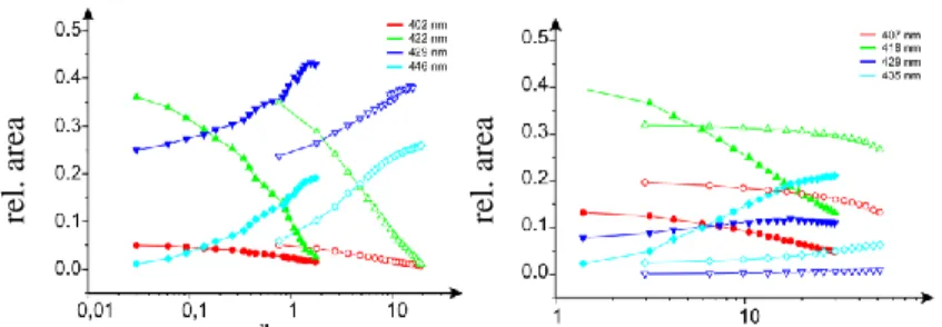
More details about the binding process can be obtained by decomposition of the Soret-bands. The band of free TMPCP, BMPCP and their tetrapeptide conjugates was fitted with 3 Gaussians while the band of TMPCP-AK could be best fitted with 4 Gaussians. In the presence of nucleic acids the number of components was extended with two component curves, the only exception being TMPCP-AK where only one new Gaussian appeared. We plotted the area under the component curves against the base pair/porphyrin ration to achieve an analytical overlook of the changes. We can observe the
absorbance
wavelength (nm)
plots in the case of isolated DNA and TMPCP-4P, TMPCP, BMPCP-4P2, BMPCP on Figure 2.
Figure 2. Relative area under the component curves as a function of base pair/porphyrin ratio (isolated DNA).
In the case of T7 phage decomposition of the spectra also provides evidence of binding, however only one clearly increasing component can be separated. This component presumably represents the dominant binding form. Only with decomposition we cannot rule out other forms of binding in the case of TMPCP and TMPCP-4P. No spectral shift can be detected with BMPCP and TMPCP-AK in the presence of bacteriophage.
To further investigate the binding forms we also recorded fluorescence energy transfer signals. If a porphyrin molecule comes in close contact with the base pairs of the DNA their fluorescence quantum yield increases upon excitation at 260 nm, the absorption maximum of DNA. This phenomenon is only possible if the energy is transferred from the base pairs to the porphyrin. This is only possible in close contact - such as intercalation - so this can be considered proof of this binding mode. The integrated fluorescence intensity of the compounds was plotted against the base pair/porphyrin ratio.
rel. area rel. area
10
Figure 3. Change in fluorescence intensity with excitation at 260 nm in the case of isolated DNA (A) and T7 phage (B) in the function of base
pair/porphyrin ratio
BMPCP and TMPCP and their tetrapeptide conjugates are not chiral molecules, however binding to the chiral double helix of the DNA induced CD signals can be detected in the Soret-band. A positive induced CD band can be considered as a sign of minor-groove binding, while the presence of a negative CD band indicates intercalative binding. The relative amplitudes of these bands can represent the ratio of the two binding modes relative to each other. Figure 4. shows the CD spectra of three porphyrins in presence of isolated DNA and bacteriophage.
4. Figure Induced CD spectra in the Soret-band in the presence of isolated DNA (A) and T7 phage (B).
A B
A B
wavelength (nm) wavelength (nm)
rel. intensity rel. intensity
Absorption and CD melting curves were recorded to investigate possible interactions with capsid proteins. Absorption melting could not prove such binding, however the more sensitive and protein specific CD melting curves changed significantly in the presence of the porphyrins.
4.2. Cellular uptake and intracellular localization of cationic
Flow cytometry data suggest that the cellular uptake of the new derivatives is time and concentration dependent in HL-60 cells. BMPCP and BMPCP-4P2 exhibited the greatest amount of uptake. Confocal microscopy images showed that after the uptake the derivatives accumulate in the lysosomes and the mitochondria. TMPCP-AK accumulates in mitochondria and other cytoplasmic vesicles but not in the lysosomes.
Figure 5. Intracellular localization of BMPCP-4P2
BMPCP-4P2 nuclei BMPCP-4P2 + nuclei
12
4.3. Binding of daunorubicin amino acid conjugates to isolated DNA and nucleoprotein complex
Upon increasing base pair/daunorubicin ratios the λ=400-600 nm daunorubicin absorption band exhibits bathochromic and hypochromic shifts. This can be considered characteristic of DNA binding of these molecules. All three amino acid conjugates showed similar spectral tendencies, with small differences in the case of each derivative. In the presence of bacteriophage the amplitude of the spectral changes are more moderate.
The emission intensities of the three amino acid conjugates were significantly quenched when bound to isolated DNA, while interaction with T7 phage caused only modest spectral shift.
Figure 6. Dau-Arg fluorescence emission spectra in the presence of isolated DNA (A) and T7 phage (B).
Using the data of emission intensity changes the calculation of binding constants is possible with the model suggested by McGhee - von Hippel. Table 1. shows the calculated binding constants for three conjugates and unmodified daunorubicin.
B A
wavelength (nm) wavelength (nm)
rel. intensity rel. intensity
Table 1. Binding constants (Ka) of unmodified daunorubicin and Dau-Arg, Dau-Leu, Dau-Gly in the case of isolated DNA and T7 phage.
Absorption melting curves were also registered for T7 phage- daunorubicin interaction. In the presence of daunorubicin conjugates strand separation temperature of the T7 DNA increases, proving that DNA-binding is also possible in the vicinity of capsid proteins. Thermal denaturation temperature of the capsid proteins is also shifted to higher temperatures, indicating protein-daunorubicin interaction.
14
5. Conclusions
This study focuses on two groups of molecules. The new scientific conclusions regarding cationic porphyrin peptide conjugates a daunorubicin amino acid conjugates can be summarized in the following points:
5.1. Cationic porphyrins and peptide conjugates
1. We synthesized tetrapeptide conjugates of bi-cationic (BMPCP) and tri- cationic (TMPCP) porphyrins, furthermore we conjugated TMPCP to branched chain polypeptide with polylysine backbone. The new derivatives bind to isolated DNA. In the case of T7 phage only the binding of TMPCP, TMPCP-4P and BMPCP-4P2 molecules could be determined.
2. TMPCP, TMPCP-4P and BMPCP-4P2 interact through intercalation and groove-binding with isolated DNA, while TMPCP-AK exhibits only groove-binding. BMPCP has weak affinity to DNA and binds through intercalation. BMPCP-4P2 binds to encapsidated with groove-binding, intercalation is only possible after denaturation of capsid proteins. TMPCP, TMPCP-4P exhibit both binding modes also in the presence of nukleoprotein complex, at higher concentrations intercalation becomes dominant.
3. Tri-cationic derivatives have higher binding affinity to isolated DNA than their bi-cationic counterparts; conjugation with tetrapeptide improves the binding through elimination of negative charges. Conjugation with the more bulky polypeptide however decreases the affinity, TMPCP-AK shows weaker binding than TMPCP, TMPCP-4P or BMPCP-4P2. Binding of
BMPCP is the weakest. The effect of capsid proteins was also investigated.
TMPCP, TMPCP-4P, BMPCP-4P2 was not blocked by the proteins but binding affinity decreased. More positive charges are preferential for the binding, however the presence of tetrapeptides becomes hindering. BMPCP and TMPCP-AK do not interact with bacteriophage.
4. Conformation of the DNA is altered by intercalative binding with the loss of stacking interaction and loss of helicity. In T7 phage the binding induces structural changes of the capsid and conformation changes of DNA.
5. Interaction between capsid proteins and porphyrins is presumed.
6. BMPCP-4P2 has the most favorable uptake profile. Bi-cationic derivatives exhibit higher association constants to model membranes with neutral phospholipids and experience increased uptake. The bulkier TMPCP-AK containing several TMPCP molecules, therefore more positive charges shows only moderate uptake.
7. The investigated derivatives cannot be detected accumulating in the nuclei, their intracellular localization is focused on lysosomes and mitochondria, with TMPCP-AK being a slight exception, with its accumulation in mitochondria and cytoplasmic vesicles but not in lysosomes.
16 5.2. Daunorubicin amino acid conjugates
1. We synthesized three conjugates of daunorubicin binding amino acids through amide bond to the drug. The new derivatives bind to isolated and encapsidated DNA with intercalative binding mode.
2. Binding constants calculated for the interaction with encapsidated DNA are an order of magnitude smaller than those calculated for isolated DNA.
Similar difference can be observed between unmodified and conjugated daunorubicin with both types of nucleic acids.
3. Binding of conjugates changes the helical structure of the DNA while the two stranded structure is stabilized. Significant change in the structure of T7 phage could not be detected upon interaction with the derivatives.
4. The new conjugates bind to capsid proteins.
5. Daunorubicin-arginine conjugate has the highest affinity for the polynucleotide, therefore we suggest arginine as the amino acid used in the spacer peptide during further development of targeting moieties.
6. Publications
I. Publications related to the thesis:
1. Orosz Á, Mező G, Herényi L, Habdas J, Majer ZS, Mysliwa- Kurdziel B, Tóth K, Csík G. Binding of new cationic porphyrin–
tetrapeptide conjugates to nucleoprotein complexes.
BIOPHYSICAL CHEMISTRY 177-178: pp. 14-23. (2013), IF:
2,319
2. Orosz Á, Bősze Sz, Mező G, Szabó I, Herényi L, Csík G. Oligo- and polypeptide conjugates of cationic porphyrins: binding, cellular uptake, and cellular localization. AMINO ACIDS 49:(7) pp. 1263-1276. (2017), IF: 2,906
3. Orosz Á, Csik G. Peptide/protein conjugates of photosensitizers.
In: Maxim Ryadnov, Ferenc Hudecz (szerk.) Amino Acids, Peptides and Proteins. Cambridge: Royal Society of Chemistry Publishing, 2016. pp. 100-145. (40.) (ISBN:978-1-78262-059-4)
II. Publications not related to the thesis:
1. Szabó R, Sebestyén M, Kóczán G, Orosz Á, Mező G, Hudecz F.
Cellular Uptake Mechanism of Cationic Branched Polypeptides with Poly[l-Lys] Backbone. ACS COMBINATORIAL SCIENCE 19:(4) pp. 246-254. (2017), IF: 3,5