Review
Microparticles, Microspheres, and Microcapsules for Advanced Drug Delivery
Miléna Lengyel1, Nikolett Kállai-Szabó1, Vince Antal1, András József Laki2,3 and István Antal1,*
1 Department of Pharmaceutics, Semmelweis University, H˝ogyes E. str 7, 1092 Budapest, Hungary
2 Pázmány Péter Catholic University, Faculty of Information Technology and Bionics, Práter str 50/A, 1083 Budapest, Hungary
3 Department of Biophysics and Radiation Biology, Semmelweis University, T ˝uzoltóstr. 37-47, 1094 Budapest, Hungary
* Correspondence: antal.istvan@pharma.semmelweis-univ.hu; Tel.:+36-12-170-914
Received: 11 July 2019; Accepted: 1 August 2019; Published: 9 August 2019 Abstract: Microparticles, microspheres, and microcapsules are widely used constituents of multiparticulate drug delivery systems, offering both therapeutic and technological advantages.
Microparticles are generally in the 1–1000µm size range, serve as multiunit drug delivery systems with well-defined physiological and pharmacokinetic benefits in order to improve the effectiveness, tolerability, and patient compliance. This paper reviews their evolution, significance, and formulation factors (excipients and procedures), as well as their most important practical applications (inhaled insulin, liposomal preparations). The article presents the most important structures of microparticles (microspheres, microcapsules, coated pellets, etc.), interpreted with microscopic images too. The most significant production processes (spray drying, extrusion, coacervation, freeze-drying, microfluidics), the drug release mechanisms, and the commonly used excipients, the characterization, and the novel drug delivery systems (microbubbles, microsponges), as well as the preparations used in therapy are discussed in detail.
Keywords: multiparticulate formulations; polymer excipients; processes for microparticles in therapy;
structure and drug release; microfluidics; microbubbles; microsponge
1. Introduction
Microparticles, microspheres, and microcapsules are common constituents of multiparticulate drug delivery systems offering numerous advantages based on their structural and functional abilities [1], and their application is suitable for convenient and tolerable drug administration via several routes.
Depending on the formulation, they can be incorporated into different pharmaceutical dosage forms such as solids (capsules, tablets, sachets), semisolids (gels, creams, pastes), or liquids (solutions, suspensions, and even parenterals).
An advantage of microcarriers over nanoparticles is that they do not traverse into the interstitium over the size of 100 nm transported by the lymph, and thus act locally [2]. Possibly toxic substances can be carried encapsulated and liquids can be handled as solids in the form of dried microparticles.
In the case of multiparticulates, the dose is distributed in many small separate particles, which carry and liberate a part of the dose, hence the malfunction of an individual subunit does not cause the failure of the whole dosage.
Multiparticulate drug delivery systems offer outstanding advantages to experts and patients, such as:
- choice of dosage form for the desired drug delivery route (peroral tablets, parenteral injections);
- modified and targeted (even site-specific) drug release and delivery;
Sci. Pharm.2019,87, 20; doi:10.3390/scipharm87030020 www.mdpi.com/journal/scipharm
- more expectable pharmacokinetics with reduced intra- or inter-subject variability;
- more homogenous distribution in the physiological environment;
- stable fixed-dose combinations of drugs;
- dose titration and less dose-dumping;
- patient centricity through better compliance (e.g., patients with dysphagia) and adherence;
- individual therapy (e.g., for pediatric or geriatric population);
- improving stability of the medicinal preparations;
- isolating the constituents to ensure better compatibility;
- innovative products with a prolonged life cycle through patent protection.
From the viewpoint of technology, microencapsulation provides several advantages: microparticles are formulated in order to protect the core from the environment; masking an unpleasant taste;
preserving volatiles or the viability of the cells; separating incompatible substances; protecting the body from the side effects; and optimizing, prolonging, or targeting the effect of a drug.
The polymer excipient protects the active pharmaceutical ingredient (API) from the environment (oxidation, temperature, pH) or the body from the irritative, or mucosa-damaging effect of the drug substance. The lesion (e.g., bisectioning) of the multiparticulate solid dosage form (i.e., micropellets in spansule or compressed) affects only a small number of units, thus does not result in a significant change of the blood level.
However, there are some limitations, such as higher product costs due to the more expensive excipients in the formulations or to the more sophisticated equipment and processes, as well as stricter quality control. In addition, some constituents may not meet the requirements for biocompatibility and biodegradation.
2. Construction and Structure
Microparticles’ sizes range from 1 to 1000µm and the well-known matrix or reservoir structure they exist in have various different structures (Figure1). Beyond the excipients used, the structure and the shape determines the function as well. Multiparticulate drug delivery systems (micropellets, microgranules, microspheres, microcapsules, microsponges, liposomal preparations) attract attention because of their wide range of favorable technological properties.
Sci. Pharm. 2019, 87, x FOR PEER REVIEW 2 of 31
‐ choice of dosage form for the desired drug delivery route (peroral tablets, parenteral injections);
‐ modified and targeted (even site-specific) drug release and delivery;
‐ more expectable pharmacokinetics with reduced intra- or inter-subject variability;
‐ more homogenous distribution in the physiological environment;
‐ stable fixed-dose combinations of drugs;
‐ dose titration and less dose-dumping;
‐ patient centricity through better compliance (e.g., patients with dysphagia) and adherence;
‐ individual therapy (e.g., for pediatric or geriatric population);
‐ improving stability of the medicinal preparations;
‐ isolating the constituents to ensure better compatibility;
‐ innovative products with a prolonged life cycle through patent protection.
From the viewpoint of technology, microencapsulation provides several advantages:
microparticles are formulated in order to protect the core from the environment; masking an unpleasant taste; preserving volatiles or the viability of the cells; separating incompatible substances;
protecting the body from the side effects; and optimizing, prolonging, or targeting the effect of a drug.
The polymer excipient protects the active pharmaceutical ingredient (API) from the environment (oxidation, temperature, pH) or the body from the irritative, or mucosa-damaging effect of the drug substance. The lesion (e.g., bisectioning) of the multiparticulate solid dosage form (i.e., micropellets in spansule or compressed) affects only a small number of units, thus does not result in a significant change of the blood level.
However, there are some limitations, such as higher product costs due to the more expensive excipients in the formulations or to the more sophisticated equipment and processes, as well as stricter quality control. In addition, some constituents may not meet the requirements for biocompatibility and biodegradation.
2. Construction and Structure
Microparticles’ sizes range from 1 to 1000 µm and the well-known matrix or reservoir structure they exist in have various different structures (Figure 1). Beyond the excipients used, the structure and the shape determines the function as well. Multiparticulate drug delivery systems (micropellets, microgranules, microspheres, microcapsules, microsponges, liposomal preparations) attract attention because of their wide range of favorable technological properties.
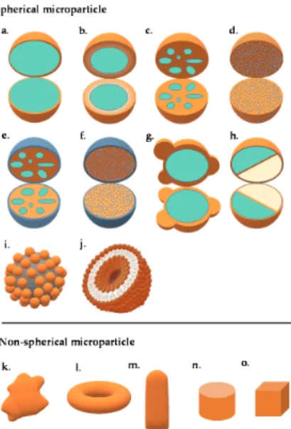
Figure 1. Schematic illustration of the different microparticle structures: (a) mononuclear/single core/core-shell, (b) multi-wall, (c) polynuclear/multiple core, (d) matrix, (e) coated polynuclear core, (f) coated matrix particle, (g) patchy microparticle, (h) dual-compartment microcapsule, (i) colloidosome, (j) giant liposome, (k) irregular-shaped microparticle, (l) torus-shaped microparticle, (m) bullet-shaped microparticle, (n) microtablet, and (o) cubic-shaped microparticle.
Sci. Pharm.2019,87, 20 3 of 31
Microparticles may be characterized as either a homogenous or heterogenous structure depending on the formulation and processing. Usually the spheroid shape is preferred since it makes the further processing (e.g., coating) easier.
2.1. Microspheres and Microcapsules
Microspheres can be characterized as matrix systems in which the drug is homogeneously dispersed, either dissolved or homogenously suspended [3]. Microcapsules are heterogenous particles where a membrane shell is surrounding the core forming a reservoir (Figure2) [4].
Figure 1. Schematic illustration of the different microparticle structures: (a) mononuclear/single core/core-shell, (b) multi-wall, (c) polynuclear/multiple core, (d) matrix, (e) coated polynuclear core, (f) coated matrix particle, (g) patchy microparticle, (h) dual-compartment microcapsule, (i) colloidosome, (j) giant liposome, (k) irregular-shaped microparticle, (l) torus-shaped microparticle, (m) bullet-shaped microparticle, (n) microtablet, and (o) cubic-shaped microparticle.
Microparticles may be characterized as either a homogenous or heterogenous structure depending on the formulation and processing. Usually the spheroid shape is preferred since it makes the further processing (e.g., coating) easier.
2.1. Microspheres and Microcapsules
Microspheres can be characterized as matrix systems in which the drug is homogeneously dispersed, either dissolved or homogenously suspended [3]. Microcapsules are heterogenous particles where a membrane shell is surrounding the core forming a reservoir (Figure 2) [4].
.
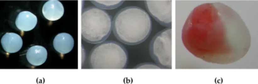
Figure 2. Structures of calcium alginate microparticles: (a) microspheres, (b) microcapsules W/O/W emulsion in core surrounded by calcium alginate shell, and (c) “Janus particle” (the particles were prepared by a Büchi B-390 Microencapsulator).
A classical microsphere structure contains solid or liquid API dispersed or dissolved in a matrix.
Microcapsules are reservoirs of microscopic size surrounded by a wall that is able to control the release from the reservoir.
The size plays a role in the gastrointestinal performance: microparticles under 800 µm get through the pylorus without the influence of gastric emptying, thus eliminating the interpersonal and intrapersonal (nutrition-based) differences.
Particles larger than 100 nm stay at the site of administration until the phagosomal clearance.
Lymphatic uptake and node accumulation is most significant between 10–80 nm [2]. The foreign body response is decreased by the application of spheres with a diameter of 1.5–2.5 mm compared to that of smaller diameter spheres [5].
The surface charge has a key role in the aggregation of the particles. Aggregation hinders optimal administration and drug delivery (e.g., compromises content uniformity of doses, occludes normal blood flow).
Porosity also has a significance in the in vivo performance in cell transplantation. Porous materials facilitate vascularization relative to non-porous biomaterials. A porosity of 30–40 µm leads to the polarization of macrophages, causing elevated tissue repair [5].
Several unique anisotropic colloidal microstructures have lately been created with special properties.
2.1.1. Janus Particles
These microparticles with a certain shape and phase anisotropy, created by alloying the distinct properties (hydrophilic, hydrophobic) of the separated excipients, are used as multifunctional imaging probes and sensors [6,7]. The anisotropic response to external signals has also been exploited in the preparation of color-changing traffic signals.
2.1.2. Patchy Particles
Figure 2.Structures of calcium alginate microparticles: (a) microspheres, (b) microcapsules W/O/W emulsion in core surrounded by calcium alginate shell, and (c) “Janus particle” (the particles were prepared by a Büchi B-390 Microencapsulator).
A classical microsphere structure contains solid or liquid API dispersed or dissolved in a matrix.
Microcapsules are reservoirs of microscopic size surrounded by a wall that is able to control the release from the reservoir.
The size plays a role in the gastrointestinal performance: microparticles under 800 µm get through the pylorus without the influence of gastric emptying, thus eliminating the interpersonal and intrapersonal (nutrition-based) differences.
Particles larger than 100 nm stay at the site of administration until the phagosomal clearance.
Lymphatic uptake and node accumulation is most significant between 10–80 nm [2]. The foreign body response is decreased by the application of spheres with a diameter of 1.5–2.5 mm compared to that of smaller diameter spheres [5].
The surface charge has a key role in the aggregation of the particles. Aggregation hinders optimal administration and drug delivery (e.g., compromises content uniformity of doses, occludes normal blood flow).
Porosity also has a significance in the in vivo performance in cell transplantation. Porous materials facilitate vascularization relative to non-porous biomaterials. A porosity of 30–40µm leads to the polarization of macrophages, causing elevated tissue repair [5].
Several unique anisotropic colloidal microstructures have lately been created with special properties.
2.1.1. Janus Particles
These microparticles with a certain shape and phase anisotropy, created by alloying the distinct properties (hydrophilic, hydrophobic) of the separated excipients, are used as multifunctional imaging probes and sensors [6,7]. The anisotropic response to external signals has also been exploited in the preparation of color-changing traffic signals.
2.1.2. Patchy Particles
Spherical geometries with concave, convex, or flat patches on the polystyrene sphere’s surface have also been created via colloidal fusion and concomitant shrinking (Figure1). The patchy particles can form clusters, and by patch-to-patch bonding between oil patches, can build supracolloidal architectures (tetrahedral, hexagonal) that can withstand drying forces and show the ability to reversibly expand
several times their original volume in a swelling agent [8]. Microfluidic techniques give rise to the creation of anisotropic geometries.
Magnetic nanoparticles embedded into nonplanar, bullet-shaped microneedles give access to mild invasive therapy [9].
2.2. Liposomes
Liposomes are lipid vesicles with one or more phospholipid bilayers and their structure comprises small unilamellar vesicles (SUV: 20–100 nm), large unilamellar vesicles (LUV:>100 nm), multilamellar vesicles (MLV:>500 nm), oligolamellar (OLV: 0.1–1µm), giant unilamellar liposomes (GUV:>1µm), and multivesicular vesicles (MVV:>1µm).
For ophthalmic delivery, they represent ideal drug delivery systems for both hydrophilic and hydrophobic APIs by performing a cell-membrane like structure. The negative or positive charge can contribute to the bioavailability: cationic liposomes (e.g., fabricated with didodecyldiethylammonium chloride, stearylamine) exhibited better efficacy at the surface of cornea with a negative charge.
Liposomes can interact with the cells via several mechanisms: interaction with the cell surface components, fusion with the membrane, endocytosis via phagocytic cells, or swap by bilayer components. The methods of preparation are also versatile: solvent evaporation, double emulsion-evaporation, or reverse-phase evaporation [10].
2.3. Colloidosomes
Colloidosomes are microcapsules that contain a hollow or hydrogel core and their wall is composed of self-assembled colloidal particles. Their sizes range from 10–20 nm to micrometers. It was found that colloidosomes have selective permeability where drugs can diffuse through their shells via size exclusion [11]. They can be stabilized by solid excipients (e.g., Fe2O3, CaCO3, colloidal anhydrous silica) forming a Pickering emulsion [12]. Besides the emulsification, depending on the excipient thermal annealing and chemical cross-linking take part in the immobilization of the active ingredient.
3. Types and Mechanism of Drug Release
The process of drug release of microparticulates, produced by special manufacturing technologies and/or possibly containing special excipient(s), is the result of various phenomena and mechanisms (dissolution/diffusion, osmotically driven release, erosion) (Figure3). Generally, these mechanisms take place side by side and one or the other mechanism provides a greater role during drug release [13].
In the microparticulate, when the active pharmaceutical ingredient is embedded in a polymer matrix (Figure3), the behavior of the polymer system is crucial during dissolution, but depends on many factors (drug properties, formulation, release medium, etc.) [14].
In the case of a polymer matrix, the diffusion of the active ingredient can be through the intact polymer network or through the pores filled with water. Water-soluble drugs may also dissolve in the aqueous pore networks. Water uptake causes polymer chains to swell, indicating the formation of new pores and/or osmotic pressure. During swelling, the volume increases, the effective diffusion coefficient of the drug is increased, and more pharmacon molecules enter the aqueous part. Erosion of the polymer matrix (bulk/surface) is also possible.
In the case of polymer coated microparticles, the film-forming polymer may dissolve in the medium or act as a water-insoluble, permeable or semipermeable membrane. In the former case, the diffusion is predominantly due to the release of the active ingredient. In the case of a semipermeable coating, the osmotic phenomenon should be taken into account. It is also possible to use water-soluble pore formers, which, by creating pores, accelerate the dissolution profile [15,16].
Figure 3. Release mechanisms in microencapsulated products.
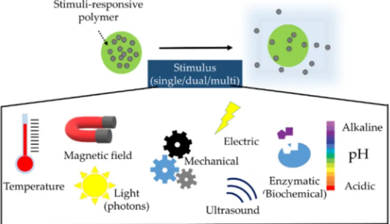
In the case of smart drug delivery microparticle systems, the release of the drug occurs via a stimulus. It is possible that one, two, or more (multiple) stimuli are required for dissolution (Figure 4).
The stimulus for drug release may be internal or external and be classified as physical, chemical, or even microbiological. Opening and closing signals of these systems are also possible, creating feedback [17].
Figure 4. Smart release stimuli via microencapsulation.
Magnetic Microcapsules
Magnetic microspheres are supramolecular particles that circulate through capillaries without causing occlusion in the form of emboly (<4 µm) but show a ferromagnetic character such that they can be dragged into the target tissue with a magnetic field of 0.5–0.8 T [18]. Magnetic microparticles for medical applications have been developed, such as magnetic resonance imaging (MRI) and drug delivery, and they are a promising choice in tumor therapy accompanied by hyperthermia. The
Figure 3.Release mechanisms in microencapsulated products.
In the case of smart drug delivery microparticle systems, the release of the drug occurs via a stimulus. It is possible that one, two, or more (multiple) stimuli are required for dissolution (Figure4).
The stimulus for drug release may be internal or external and be classified as physical, chemical, or even microbiological. Opening and closing signals of these systems are also possible, creating feedback [17].
Figure 3. Release mechanisms in microencapsulated products.
In the case of smart drug delivery microparticle systems, the release of the drug occurs via a stimulus. It is possible that one, two, or more (multiple) stimuli are required for dissolution (Figure 4).
The stimulus for drug release may be internal or external and be classified as physical, chemical, or even microbiological. Opening and closing signals of these systems are also possible, creating feedback [17].
Figure 4. Smart release stimuli via microencapsulation.
Magnetic Microcapsules
Magnetic microspheres are supramolecular particles that circulate through capillaries without causing occlusion in the form of emboly (<4 µm) but show a ferromagnetic character such that they can be dragged into the target tissue with a magnetic field of 0.5–0.8 T [18]. Magnetic microparticles for medical applications have been developed, such as magnetic resonance imaging (MRI) and drug delivery, and they are a promising choice in tumor therapy accompanied by hyperthermia. The
Figure 4.Smart release stimuli via microencapsulation.
Magnetic Microcapsules
Magnetic microspheres are supramolecular particles that circulate through capillaries without causing occlusion in the form of emboly (<4µm) but show a ferromagnetic character such that they can be dragged into the target tissue with a magnetic field of 0.5–0.8 T [18]. Magnetic microparticles
for medical applications have been developed, such as magnetic resonance imaging (MRI) and drug delivery, and they are a promising choice in tumor therapy accompanied by hyperthermia. The release can be fine-tuned by the strength of the applied magnetic heating: with a gentle magnetic effect, the particles react with shrinkage and slow drug release, and with intense heating, the structure disruption induces the shrinking, which leads to a burst release [19].
Their great advantage lies in the effective method of targeting the drug molecule to the desired site to be treated (i.e., the tumor) with higher therapeutic efficacy and lower toxicity. The reduction of dosing frequency enhances the patient’s compliance.
The production involves emulsion methods (multiple and phase separation polymerization, solvent extraction, hot melt microencapsulation, dispersion copolymerization).
4. Formulation and Manufacturing
4.1. Composition and Excipients
The use of plant, animal, or microbial-origin biopolymers are propitious; semi-synthetic cellulose derivatives and biodegradable or non-biodegradable synthetic polymers are used as well.
The formulation is usually based on polysaccharides or proteins, but waxes and lipids also play a role in the construction. Nonpolymer excipients play a role in crosslinking the polymer chains (CaCl2, glutaraldehyde, poly-l-lysine, etc.), thus forming and hardening the polymer network of the drug delivery systems. The most commonly used polymers and their important microencapsulation-related properties are summarized in Tables1–5.
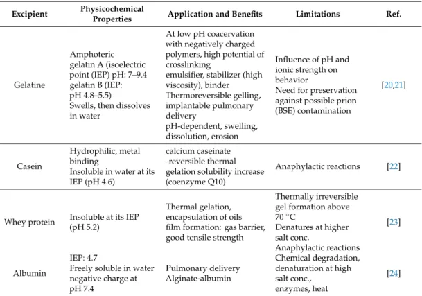
Table 1.Examples of proteins and waxes of plant or animal origin used in microencapsulation.
Excipient Physicochemical
Properties Application and Benefits Limitations Ref.
Gelatine
Amphoteric gelatin A (isoelectric point (IEP) pH: 7–9.4 gelatin B (IEP:
pH 4.8–5.5)
Swells, then dissolves in water
At low pH coacervation with negatively charged polymers, high potential of crosslinking
emulsifier, stabilizer (high viscosity), binder Thermoreversible gelling, implantable pulmonary delivery
pH-dependent, swelling, dissolution, erosion
Influence of pH and ionic strength on behavior
Need for preservation against possible prion (BSE) contamination
[20,21]
Casein
Hydrophilic, metal binding
Insoluble in water at its IEP (pH 4.6)
calcium caseinate –reversible thermal
gelation solubility increase (coenzyme Q10)
Anaphylactic reactions [22]
Whey protein Insoluble at its IEP (pH 5.2)
Thermal gelation, encapsulation of oils film formation: gas barrier, good tensile strength
Thermally irreversible gel formation above 70◦C
Denatures at higher salt conc.
Anaphylactic reactions
[23]
Albumin
IEP: 4.7
Freely soluble in water negative charge at pH 7.4
Pulmonary delivery Alginate-albumin
Chemical degradation, denaturation at high salt conc.,
enzymes, heat
[24]
Table 1.Cont.
Excipient Physicochemical
Properties Application and Benefits Limitations Ref.
Zein
α,β,γ,δ, zein with different Mw
amphiphilic character IEP: 6.8
hydrophobic
Oral controlled release matrix and wall
Brittle, rigid wall, complex with a gelling component to plasticize
[25,26]
Soy protein
Partly soluble in water, depending on the extraction process;
IEP 4.5
Oral controlled release matrix and wall
Emulsifier, foaming agent
Sensitivity [27]
Gluten Water-insoluble;
IEP:7.5
Wall material, good elastic, good thermoplastic properties
Gluten sensitivity [28]
Bees-wax (Apis mellifera)
mp (melting point):
62–64◦C HLBrequired=12
Edible, easy use, smooth surface,
hot melt extrusion prolonged release for hydrophilic substances, protection from chemical degradation
Oxidation [29]
Carnauba wax (Copernica
cerifera)
mp: 78–85◦C HLBrequired=12
Good compatibility hot melt extrusion, embedding water soluble components, taste masking
Oxidation [30,31]
Paraffin (hard)
(mineral) mp: 50–61◦C
Embedding water soluble components
liquid paraffin in the emulsification process
Sensitivity [32]
Table 2.Examples of polysaccharides of various origin used in microencapsulation.
Excipient Physicochemical
Properties Applications and Benefits Limitations Ref.
Chitosan
(deacylated chitin)
Soluble in weak acids Mucoadhesive reacts with negatively charged surfaces
Antifungal, antibacterial, reduces LDL (low-density lipoprotein), tissue regenerative, pulmonary delivery
Ionotropic gelation, coacervation with anions, modified emulsification
pH dependence (insoluble above pH 6.5)
Addition of electrolytes precipitates chitosan in solution,
hygroscopic
[33–35]
Sodium hyaluronate
Anionic character, soluble in water, high viscosity at low concentration
In microspheres nasal, vaginal, ophthalmic delivery systems
Very hygroscopic, when heated, emits Na2O
[36,37]
Starch (wheat, corn, potato, rice, tapioca)
Starch from different origins
Differ in particle size and shape
Soluble in hot water after a time of gelatinization
Spray drying, extrusion, molecular inclusion, coacervation with proteins, hydrocolloid-forming, release via swelling, diffusion, erosion
Hygroscopic [38]
Guar gum
Water soluble, nonionic, galactomannan forms a thixotropic solution, stable at pH 4–10.5
Controlled-release, colon-targeted release, appetite suppressant, thermoreversible
Needs preservation, borate hinders swelling
[39]
Table 2.Cont.
Excipient Physicochemical
Properties Applications and Benefits Limitations Ref.
Locust bean gum (LBG)/carob, ceratonia/
Nonionic, galactomannan, dispersible in hot water, soluble at higher temperature
Controlled release in combination pseudoplastic,
gelling with the addition of borate, solubility not affected by pH or ionic concentration
Low water solubility,
hypersensitivity [40]
Konjac gum Water soluble
Elevation of temperature increases gelation, antioxidant properties
Indigestable [41,42]
K,ι,λ−, Carrageenan
Anionic polymer, ι−Carrageenan: shear thinning thixotropic gel forming
λ−Carrageenan: highest anionic charge, high solubility, but no gelling, κ,ι−Carrageenan: elastic gel is formed with K+, Ca2+, thermoreversible gel forming
Release by erosion (physical
contact-Scentcaps®), effective against HPV (human papilloma virus)
Acid-catalyzed hydrolyses, especially at higher temperature and pH<3
Administration of
>2 g/kg orally Induces intestinal ulcers, not biodegradable
[43,44]
Agarose Soluble in hot water
swelling,
Thermoreversible gelation (at≈37◦C) with hysteresis
Poor biodegradability [45]
Sodium alginate
Water soluble, anionic coacervation with ions (Ca2+, Sr2+, Ba2+), polycations (chitosan) or poly-l-lysine disintegrant, binder, viscosity increasing
swelling ability (200–300× of its own weight from water)
pH-dependent swelling nontoxic, nonirritant low density (capable of floating in gastric juice) Diffusion, erosion, in situ forming hydrogels
pH dependence (insoluble in acidic medium)
heat-sensitivity (hydrolysis)
microbial spoilage on storage
(depolymerization)
[46]
Tragacanth
Water soluble arabinogalactan and swellable bassorin components,
swells quickly in water
Acid-resistance emulsifier,
encapsulation of oils, stabilizer in emulsions
Di/trivalent cations cause drop in viscosity because of
precipitation
[47]
Gum arabic/ Acacia gummi (Ph. Eur.)
Water soluble, clear solution of pH 4.5, protective colloid
Emulsifier, spray-dry encapsulation of flavor and of essential oils, 30% solution has a relatively low viscosity, Newtonian flow permeable coating dietary fiber
Variation dependent on source
pH, ionic strength influences viscosity (max at≈pH 6–7)
[48–50]
Pectin (low or high methoxylated) (apple, citrus peel, beet)
Negatively charged molecule
Gelation depends on the degree of esterification, cation (Ca,Zn)
concentration in solution, temperature and pH In situ gelling, sustained delivery, dietary fiber, drug delivery in colorectal carcinoma (5-FU), antiviral activity
Gelling occurs at low pH (<3.5),
presence of sugars necessary for gelation of HM pectin
[51,52]
Table 3.Examples of polysaccharides of microbial fermentation used in microencapsulation.
Excipient Physicochemical
Properties Applications and Benefits Limitations Ref.
Xanthan gum
Soluble in warm and cold water, not affected by pH
anionic polyelectrolyte
No gelation at room temp, cryogelation possible, stable viscosity over wide pH range, surface activity, emulsion stabilizer, controlled-release, colon-targeted swelling, diffusion, matrix erosion
Hydrolysis
Acceptable daily intake (WHO) 10 mg/kg BW Contains cellulase
[53]
Gellan gum Anionic polyelectrolyte soluble in water
Thermoreversible gelation with cations
sol-gel transition under physiological conditions bioadhesive
nasal administration
Poor biodegragability [54]
Dextran
Neutral polymer, solubility depends on degree of
polymerization
Colon-targeted delivery, formation of porous particle
pulmonary delivery
At parenteral adiminitration:
possible platelet adhesiveness
[55,56]
Pullulan
Neutral polymer underivarized pullulan has high water solubility
Emulsifier sustained-release preparations
Relatively high price [57,58]
Table 4.Examples of cellulose derivatives applied in microencapsulation.
Excipient Physicochemical
Properties Applications and Benefits Limitations Ref.
Methylcellulose (MC)
Soluble in cold water Tsol-gel: 80◦C amphiphilic
Emulsifier, pseudoplastic solution, pH-independent gel formation above 50◦C, high variety in Mw
Complexation with surface-active components, laxative
[59]
Carboxymethyl- cellulose sodium
(CMC-Na)
Anionic cellulose ether, dispersible in water (forms a colloidal solution)
Injectable thermoreversible gel-forming, mucoadhesive
Microbial instability
hygroscopicity [60,61]
Hydroxypropyl- cellulose (HPC)
Soluble in cold water, compatible with waxes, oils, Tsol-gel: 55◦C
High surface activity film-forming ability
Incompatible with alkaline substances, substituted phenol derivatives, anionic polymers increase viscosity
[62]
Hydroxypropyl- methylcellulose
(HPMC)
Water soluble, nonionic
Reversible thermal gelation surface activity, emulsion stabilizer, film-forming ability
No complex with metallic salts or ionic organics
[63]
Ethylcellulose Water-insoluble, hydrophobic coating
Film forming ability, membrane-controlled diffusion, modified release, floating, gastroretentive systems
Organic solvent
residuals [64]
Cellulose acetate butyrate
Insoluble in water, soluble in aceton-water blends
Semipermeable coating, extended release formulations, diffusion, matrix erosion
Organic solvent
residuals [65]
Table 5.Synthetic polymers applied in microencapsulations.
Excipient Physicochemical
Properties Applications and Benefits Limitations Ref.
Poly (lactic acid) (PLA)
Insoluble in water degrades to CO2and H2O over
12–24 months
Biodegradability, prolonged-release in im or sc injections, implants, oral solid dispersions
Digestive tract influences degradation, parenteral
administration is favorable, initial burst release may occur
[64,65]
Polylactic acid-glycolic acid copolymer (PLGA)
Insoluble in water lactic acid-glycolic acid ratio influences degradation ability to thermoplastic gel forming
Injectable or implantable systems (microparticles, gels) for human and veterinary use, pH- responsive/non-pH -responsive polymer
degradation,
bone tissue engineering
Degrades into by-products that can induce inflammation
[66–69]
Polyacrylic acid (Carbopol)
Neutralization with alkaline chemicals for gelling
Bioadhesive, targeted delivery,
intranasal administration with microencapsulation
Neutralization at preparation, preservative needed
[70]
Polymethacrylates
Soluble in organic solvents, most of them are miscible with water
pH-dependent solubility, permeability
gastric or enteric targeted delivery possible
Water insolubility [71]
Poly-(N-
isopropylacrylamide) LCST: 35–40◦C Thermoresponsive Not biodegradable [72,73]
Polyethylene glycols
Liquid or solid grades depending on the Mw, PEG (polyethylene glycol) 1500 freezing point at 37–41◦C
Plasticizer in wall of compressible microcapsules, oral insulin delivery cell delivery in combination
Not biodegradable [74]
Fumaryl diketopiperazine
(FDKP)
High solubility at pH≥6,
no biological activity
Self-assembled highly porous microparticles, Technosphere®carrier
Not biodegradable,
excreted via urine [75]
LCST: lower critical solution temperature.
The combination of polymers of different properties is a common technique to improve the particle characteristics and performance. Because of their opposite charges, alginate and chitosan at low pH form a polyelectrolyte complex [76], thus decreasing the porosity of the polymer network and delay the release of the API (Table6).
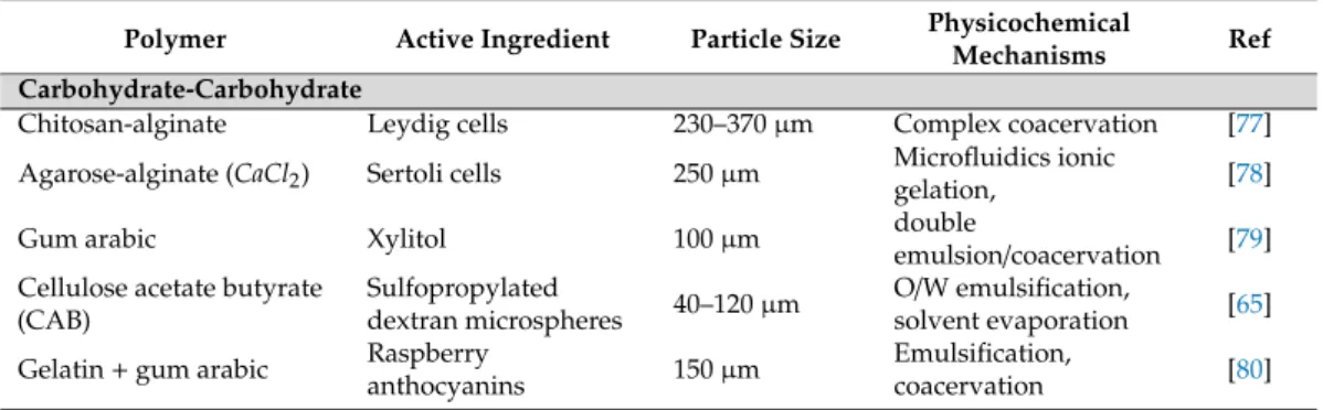
Table 6.Synergistic polymer combinations.
Polymer Active Ingredient Particle Size Physicochemical
Mechanisms Ref
Carbohydrate-Carbohydrate
Chitosan-alginate Leydig cells 230–370µm Complex coacervation [77]
Agarose-alginate (CaCl2) Sertoli cells 250µm Microfluidics ionic
gelation, [78]
Gum arabic Xylitol 100µm double
emulsion/coacervation [79]
Cellulose acetate butyrate (CAB)
Sulfopropylated
dextran microspheres 40–120µm O/W emulsification,
solvent evaporation [65]
Gelatin+gum arabic Raspberry
anthocyanins 150µm Emulsification,
coacervation [80]
Table 6.Cont.
Polymer Active Ingredient Particle Size Physicochemical
Mechanisms Ref
Carbohydrate-Carbohydrate
Maltodextrin+gum arabic Lavender oil 10–20µm S/O/W emulsification,
spray-drying [50]
CMC-Na+xanthan gum Diclofenac sodium 1000–1500µm
Emulsification, ionic gelation,
Interpenetrating network
[81]
LBG+PVA Buflomedil
hydrochloride (BH) 350–750µm
emulsification, Interpenetrating network
[40]
Chitosan+pectin Insulin 0.24–2µm electrostatic
self-assembly [82]
Carbohydrate-Protein
Gelatin+gum arabic Aspartame 100µm
Double
emulsion/complex coacervation
[83]
Gelatin+chitosan Citronella oil 100µm Coacervation [33]
whey Protein+
maltodextrin Flaxseed oil ≈10µm emulsification, spray
drying [84]
Whey protein+alginate Flaxseed oil <10µm
Double layer emulsification spray drying
[85]
Alginate+gelatin
Whey peptides (antihypertensive activity
1000µm Dripping, coacervation [86]
Alginate+zein Bifidobacterium
bifidum 1200–1700µm Extrusion coacervation
shell-core [87]
Chitosan-zein Oral gene delivery 10µm
Zein-sodium tripolyphosphate
W/O emulsion [88]
Poly-l-lysine
Poly (methyl vinyl ether-alt-maleic anhydride) (PMM0)
400µm prilling, coacervation [89]
Poly (l-ornithine)+ alginate+PLA, PLGA
Superoxide dismutase,
ketoprofen 500µm W/O/W, O/W,
solvent evaporation [90]
Poly (l-ornithine)+ ursodeoxycholic acid, Polystyrene sulfonate, polyallilamine
Pancreatic ß-cells 700µm Complex coacervation, vibrational jet [91]
Poly (ethylene glycol) (PEG)-anthracene alginate
Coomassie blue no data Coacervation [92]
Vinyl-sulfone terminated PEG+alginate
Human foreskin
fibroblast 550µm Simple coacervation [93]
Alginate+
Poly-ε-caprolactone Theophylline 800µm Complex coacervation [94]
Polypropylene+PMMA+
ethylcellulose Verapamil 150–200µm O/W solvent
evaporation [95]
PLGA-alginate Rifampicin 15–50µm Microfluidics [96]
Funami et al. [97] proved that galactomannans, including guar gum, tara gum, LBG, or konjac gum (glucomannan), influence both the short- and long-term retrogradation process of starch by controlling the gelation, hindering the crystallization, and improving the water-holding capacity of starch.
Farris et al. [88] developed a peroral gene delivery system, where DNA was encapsulated in chitosan nanoparticles and the particles were protected from the gastric environment by embedding the nanoparticles into zein matrix microparticles with a W/O emulsification method. Zein forms a relatively brittle film; however, alginate was successfully administered to decrease the rigidity of the wall [87].
Several studies show that copolymerization between synthetic and natural polymers helps to increase biocompatibility and cell viability (agarose-Carbopol®, hyaluronic acid-polyethyleneglycol) [98]. Hydrophobic excipients (poly-ε-caprolactone) can modify the release of low-solubility drugs from alginate by forming hydrophobic interactions with the drug molecule, thus prolonged release can be reached [94].
Controlled-release, porous, floating microparticles were formulated by combining polypropylene-Eudragit®RS, ethylcellulose polymers [95].
Mahou et al. successfully studied the addition of vinyl-sulfone terminated polyethyleneglycol to alginate and thus eliminated the use of polycations for human foreskin fibroblast cell encapsulation from a cell culture medium [93].
Wells et al. reached prolonged drug release and light-sensitive drug delivery with the chemical modification of PEG and chemical crosslinking with alginate.
Dalpiaz et al. studied the nasal absorption of zidovudine by deoxycholic-acid-conjugated chitosan microparticles and found that the particles could get through the blood–brain barrier, in contrast with active efflux transporters in rats [99].
Alginate microcapsules show shrinking and lower stability in an acidic medium.κ-carrageenan locust bean gum gel beads show lower sensitivity to acidic conditions than alginate beads. A limitation of their use is that the formation of κ-carrageenan locust bean gum beads requires a higher amount of potassium or calcium ions, which in terms of a healthy diet, are not acceptable in high amounts. In cell transplantation, alginate is applied because of its excellent biocompatibility and biodegradability. Alginate, however, has some limitations: uneven porosity, poor mechanical strength, weak wall-formation, and easy rupture on reaction to osmotic change have been reported.
To overcome these drawbacks, excipients such as the polyelectrolytes polystyrene sulfonate (PSS) and polyallylamine (PAA) were examined in cell microencapsulation, which improved the physical structure of polyelectrolyte gels and complexes by reducing pore size in the microcapsule membrane.
Osmotic or mechanical stress caused lower cell leakage of these microcapsules [91]. The incorporation of PSS and PAA were shown to attract inflammatory cells and to trigger an immune response. The same study showed that ursodeoxycholic acid increased mechanical stability and did not affect cell viability.
Poly-l-ornithine in combination with alginate has been shown to prevent post-transplant inflammatory response and has protected the microcapsules in vivo over the first week after grafting [90].
As artificial cell therapy, an alginate structure strengthened by poly-l-lysine was used for encapsulation [100]. The alkylamino groups of the polyamide chain bonds via electrostatic interactions with alginate, thus the matrix structure has reduced porosity and good immunoprotection.
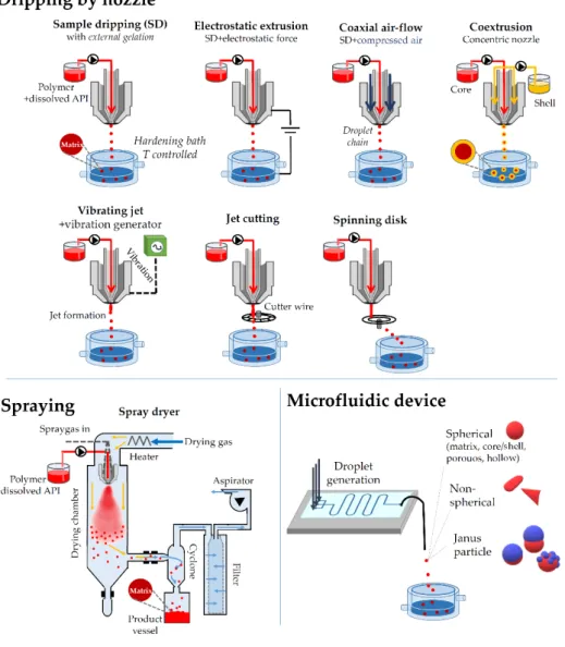
4.2. Processes for the Particle Formation
From the point of view of the industrial performance, the most important methods for microencapsulation are the air suspension method, spray-drying, and coacervation. Figure 5 summarizes the most important techniques.
Figure 5. Preparation processes through droplet formation at an orifice.
4.2.1. Coacervation
Coacervation related to the formation of calcium alginate can be considered a classical method for the preparation of microspheres and microcapsules. Calcium-ions form crosslinks between the α-L-guluronic acid and β-D-mannuronic acid units of alginate, thus organizing the polymer chains into an “eggbox-structure.” This chemical bonding gives the possibility of forming stable microspheres or core-shell structured microcapsules (Figures 6 and 7)
Figure 5.Preparation processes through droplet formation at an orifice.
4.2.1. Coacervation
Coacervation related to the formation of calcium alginate can be considered a classical method for the preparation of microspheres and microcapsules. Calcium-ions form crosslinks between the α-l-guluronic acid andβ-d-mannuronic acid units of alginate, thus organizing the polymer chains into an “eggbox-structure.” This chemical bonding gives the possibility of forming stable microspheres or core-shell structured microcapsules (Figures6and7).
Sci. Pharm.2019,87, 20 14 of 31 Figure 5. Preparation processes through droplet formation at an orifice.
4.2.1. Coacervation
Coacervation related to the formation of calcium alginate can be considered a classical method for the preparation of microspheres and microcapsules. Calcium-ions form crosslinks between the α-L-guluronic acid and β-D-mannuronic acid units of alginate, thus organizing the polymer chains into an “eggbox-structure.” This chemical bonding gives the possibility of forming stable microspheres or core-shell structured microcapsules (Figures 6 and 7)
Figure 6.(a) Scheme for the interaction between calcium and alginate forming an eggbox-like structure.
(b) Internal structure of the microsphere using scanning electron microscopy (SEM, magnification:
130×, scale bar indicates 500µm).
Sci. Pharm. 2019, 87, x FOR PEER REVIEW 15 of 31
Figure 6. (a) Scheme for the interaction between calcium and alginate forming an eggbox-like structure. (b) Internal structure of the microsphere using scanning electron microscopy (SEM, magnification: 130×, scale bar indicates 500 µm).
Figure 7. Images of a liquid core calcium alginate microcapsule prepared by vibration nozzle technique (prepared by Büchi B-390 Microencapsulator) followed by coacervation: (a) oil-filled hollow particles, (b) calcium alginate shell after removal of oil core, and (c) calcium alginate shell cut in half (average wall thickness: 8 µm) (Images: Nikon SMZ 1000 Microscope).
Coacervation is defined as the separation of liquid phases in colloidal solutions [101]. During coacervation, the active ingredient can be dispersed in the coating polymer solution and at a specific environmental influence (ionic, pH, thermal change), phase separation occurs, while the core material is encapsulated by the wall-forming polymer. Simple coacervation is based on incompatibilities between the polymers. In most cases, it is caused by salting-out (di- or trivalent cation, as it happens with alginate and Ca2+ or Ba2+, pectin and Ca2+). The particle size mainly depends on the excipients’
properties (viscosity, surface tension) and stirrer setup [102,103].
During complex coacervation, polyelectrolyte polymers with opposite charges form an insoluble complex and meanwhile encapsulate the active ingredient. pH is important in complex coacervation, as the isoelectric point of the polymers has to be taken into consideration.
Hydrophobic components are often encapsulated with coacervation. In the case of hydrophilic components, a double emulsion process followed by a coacervation proves to be successful way to form a core–shell structure microcapsule [79]. As a result of coacervation, the product contains a high amount of solvent, which needs to be evaporated from the product.
For drying, besides the common processes, lyophilization (freeze-drying) offers a choice. This is an expensive method, but effective for heat-sensitive actives. Shrinking of the particles can be partly avoided; the end product, however, has a high porosity, which is an advantage in fast release preparations to promote the water uptake, but is an obvious drawback in the formulation of sustained-release preparations.
4.2.2. Air Suspension Method/Fluid-Bed Coating
(Wurster (1959)) [104] has successfully administered fluidization in macroencapsulation for the coating of solid particles, but it was also administered to encapsulate small particles in the size range of 74–250 µm by Hinkes et al. [105]. The great advantage is that during the process, the particles are suspended in an air (or inert gas) stream, in constant mixing, relative independently from each other and the wall material solution or dispersion is sprayed on their surface, then dried inside the coating chamber. The particle size can be tuned by the well-controllable process parameters: properties of the core (density, hygroscopicity, surface area, particle size and shape, melting point, wettability, solubility, volatility, compressibility, crystallinity, hardness, cohesiveness, adsorption, friability and flowability of the core material). Concentration and quantity of wall material, inlet, outlet air temperature, and spray settings are also critical for the process.
4.2.3. Extrusion through a Nozzle
Figure 7.Images of a liquid core calcium alginate microcapsule prepared by vibration nozzle technique (prepared by Büchi B-390 Microencapsulator) followed by coacervation: (a) oil-filled hollow particles, (b) calcium alginate shell after removal of oil core, and (c) calcium alginate shell cut in half (average wall thickness: 8µm) (Images: Nikon SMZ 1000 Microscope).
Coacervation is defined as the separation of liquid phases in colloidal solutions [101]. During coacervation, the active ingredient can be dispersed in the coating polymer solution and at a specific environmental influence (ionic, pH, thermal change), phase separation occurs, while the core material is encapsulated by the wall-forming polymer. Simple coacervation is based on incompatibilities between the polymers. In most cases, it is caused by salting-out (di- or trivalent cation, as it happens with alginate and Ca2+ or Ba2+, pectin and Ca2+). The particle size mainly depends on the excipients’
properties (viscosity, surface tension) and stirrer setup [102,103].
During complex coacervation, polyelectrolyte polymers with opposite charges form an insoluble complex and meanwhile encapsulate the active ingredient. pH is important in complex coacervation, as the isoelectric point of the polymers has to be taken into consideration.
Hydrophobic components are often encapsulated with coacervation. In the case of hydrophilic components, a double emulsion process followed by a coacervation proves to be successful way to form a core–shell structure microcapsule [79]. As a result of coacervation, the product contains a high amount of solvent, which needs to be evaporated from the product.
For drying, besides the common processes, lyophilization (freeze-drying) offers a choice. This is an expensive method, but effective for heat-sensitive actives. Shrinking of the particles can be partly avoided; the end product, however, has a high porosity, which is an advantage in fast release preparations to promote the water uptake, but is an obvious drawback in the formulation of sustained-release preparations.
4.2.2. Air Suspension Method/Fluid-Bed Coating
(Wurster (1959)) [104] has successfully administered fluidization in macroencapsulation for the coating of solid particles, but it was also administered to encapsulate small particles in the size range of 74–250µm by Hinkes et al. [105]. The great advantage is that during the process, the particles are suspended in an air (or inert gas) stream, in constant mixing, relative independently from each other and the wall material solution or dispersion is sprayed on their surface, then dried inside the coating chamber. The particle size can be tuned by the well-controllable process parameters: properties of the core (density, hygroscopicity, surface area, particle size and shape, melting point, wettability, solubility, volatility, compressibility, crystallinity, hardness, cohesiveness, adsorption, friability and flowability of the core material). Concentration and quantity of wall material, inlet, outlet air temperature, and spray settings are also critical for the process.
4.2.3. Extrusion through a Nozzle
Extrusion through a nozzle is a common method in the formation of gel particles. Many factors influence the formation of microgels: the concentration, feeding rate, and surface tension of the polymer solution, solvent, temperature, and nozzle diameter [3,106,107]. The solidification of the formed microgel particles is performed in an additional step. For solidification of the gel, coacervation (either simple or complex) is an optimal choice. The formed particles are collected in a solidification liquid (ionic or polymer solution). The size and shape control of particles is dependent on various factors. The distance, concentration, surface tension of the solidification liquid, and the time of the process have a significance in the particle size and the physical properties of the beads (gel strength, porosity, etc.). The most important limitations of the extrusion are the viscosity of the polymer because of potential blockage of the nozzle; as such, the settings for optimal, narrow particle size distribution, and shape are required. For the scale-up of the process, multiple-nozzle solutions have been presented.
4.2.4. Vibrational Jet/Electrostatic Extrusion
The particle formation via extrusion is completed by various processes: vibrational jet technique means that a changing frequency vibration separates the laminar jet into beads). At the electrostatic extrusion [108] the jet is subjected to an electric field, where having reached a certain voltage (5–6 kV), particles are created, the repulsive forces additionally hinder their aggregation. The applied elevated electric potential does not cause cell death [109].
4.2.5. Spinning Disk and Cutting Wire
These devices mechanically aid the particle formation and disaggregation [110].
4.2.6. Spray Drying
Spray drying is widely used in the industry for microencapsulation of volatiles, probiotics, and viable cells. Besides the obvious drawback (high loss, low yield), the numerous advantages make this technology very popular (uniform particle size, all steps carried out in one apparatus, use of organic solvents, the capability of encapsulating heat-labile materials). The emulsion is sprayed into a chamber, where warm air dries the particles, and as a result, regular shaped, micron-sized, uniform particles are created.
The extruded wax particles can be solidified using congealing, which offers a solution for embedding hydrophilic component to perform sustained release via the slow erosion of the wall in the biological medium.
4.2.7. Supercritical Fluid Precipitation
This technique is capable of constructing very uniform particles, but its use is more significant in nanoencapsulation [111].