III./3.1.2. Parkinsonian syndrome (parkinsonism, atypical parkinsonian disorders) in neurodegenerative diseases
III./3.1.2.1. Multiple System Atrophy (MSA)
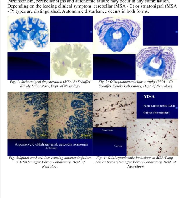
MSA is a sporadic, adult onset degenerative neurological disease belonging to the group of alpha-synucleinopathies. MSA has been formerly called striatonigral degeneration (with dominant parkinsonian symptoms), or olivopontocerebellar degeneration (with dominant cerebellar symptoms) or Shy-Drager syndrome (with dominant autonomic symptoms). These conditions were considered as one entity and referred to as multiple system atrophy since 1968 (Oppenheimer). The unified concept of MSA was confirmed by the histopathological examinations of Papp and coworkers, who demonstrated oligodendroglial inclusions in all forms of the disease. These oligodendroglial inclusions (Papp–Lantos bodies) do not occur in any other neurological diseases.
Parkinsonism, cerebellar signs and autonomic failure may occur in any combination.
Depending on the leading clinical symptom, cerebellar (MSA - C) or striatonigral (MSA - P) types are distinguished. Autonomic disturbance occurs in both forms.
Fig. 1: Striatonigral degeneration (MSA-P) Schaffer Károly Laboratory, Dept. of Neurology
Fig. 2: Olivopontocerebellar atrophy (MSA – C) Schaffer Károly Laboratory, Dept. of Neurology
Fig. 3:Spinal cord cell loss causing autonomic failure in MSA Schaffer Károly Laboratory, Dept. of
Neurology
Fig. 4: Glial cytoplasmic inclusions in MSA(Papp–
Lantos bodies) Schaffer Károly Laboratory, Dept. of Neurology
Video 1: MSA-C type. Leading clinical symptoms are gait ataxia and symmetric dysmetry on all extremities.
Red flags for MSA:
Falls from the beginning of the disease Rapid progression
Orofacial dystonia Camptocormia Pisa–sign Dystonia
Inspiratory stridor Myoclonus
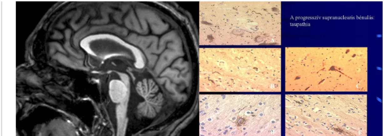
III./3.1.2.2. Progressive Supranuclear Palsy (PSP)
PSP belongs to the group of taupathies (misfolded protein diseases). PSP usually appears in the seventh decade, never before the age of 40. Clinical symptoms include early postural instability with falls, vertical supranuclear gaze palsy, pseudobulbar palsy, frontal subcortical dementia, and levodopa – unresponsive parkinsonism with
bradykinesia and pronounced axial rigidity.
The “doll’s head” phenomenon is positive (the eyes move down with the passive movement of the head). A common symptom is “eye-lid apraxia”: patients cannot open their eyes in the absence of weakness of the levator palpebrae muscle.
Video 2: Progressive supranuclear palsy: parkinsonism and vertical gaze palsy
felirat:]]
Fig. 5: MRI in PSP: mesencephalon atrophy– colibri or penguin’s sign
Fig. 6: Histopathological changes in PSP
III./3.1.2.3. Corticobasal Degeneration (CBD)
CBD is another clinical form of the taupathies. Symptoms of CBD include basal ganglia and cortical, mainly dominant parietal lobe symptoms. It is typically characterized by unilateral levodopa–unresponsive parkinsonism, limb apraxia, dystonia, myoclonus, sensory or visual neglect, cortical sensory loss, and alien limb syndrome (complex, involuntary movements of the limb). Speech disorder is also common, but it is not associated with dysphagia. Severe cognitive decline occurs in most patients (e.g. frontal lobe behavioral disorder, aphasia, attention deficit, frontal dementia).
Fig. 7: Dystonic hand posture in CBD
Fig. 8: Asymmetric cortical atrophy in CBD on MRI Fig. 9. Asymmetric cortical atrophy in CBD on PET
III./3.1.2.4 Parkinsonism in dementias of degenerative origin
Parkinsonian symptoms may occur in Alzheimer’s disease, or Alzheimer’s disease and Parkinson’s disease may be comorbid. In Alzheimer’s disease, cortical symptoms
(apraxia, aphasia, dyscalculia, dyslexia, etc.) are seen in addition to the movement disorder.
Diffuse Lewy body dementia (DLBD)
DLBD is characterized by cognitive decline, episodic delirium, visual hallucinations from the very beginning of the disease, with or without parkinsonism. If visual hallucinations occur within one year after the onset of parkinsonian motor symptoms, DLBD should be considered (“one year rule”). First symptoms may be memory and attention deficit, and visuospatial disorientation. Recall, execution and problem solving are mainly affected. An akinetic type parkinsonism may develop. Neuroleptic treatment generally worsens parkinsonian symptoms, which is called “neuroleptic sensitivity”.
Lewy bodies are found mainly in the prefrontal cortex. Lewy body disease spectrum:
brain stem localization of Lewy bodies is characteristic for PD (movement disorder), cortical localization of Lewy bodies is characteristic for frontal dementia.
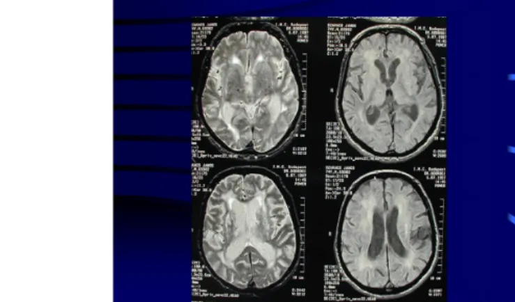
Fig. 10: Lacunar infarcts in both hemispheres and in the basal ganglia on CT
Frontotemporal dementia (FTD)
FTD is characterized by personality changes and the alteration of social behavior (disinhibition, attention deficit, lack of insight). Patients show perseveration and stereotype behavior, and they are irritable. Rarely akinetic – rigid parkinsonism may occur.
Recently, PSP and CBD have also been classified as part of the frontotemporal dementia spectrum.
Final diagnosis is provided by histopathology.
III./3.1.2.5. Parkinsonism of known origin III./3.1.2.5.1. Neuroleptic parkinsonism
In 30-50% of cases, parkinsonim is caused by neuroleptic agents that cause dopamine depletion in the striatum (drugs with dopamine antagonistic effects).
Clinically, this syndrome is difficult to distinguish from PD, but some signs may help.
History is very important. The risk of parkinsonism is substantially lower with the modern, ʽatypical neuroleptics’, as opposed to the old, ʽtypical neuroleptics’. Symptoms are generally symmetric and remit after the withdrawal of the drug. This improvement may take several months. If parkinsonian symptoms are still present 6 months after the withdrawal of the drug, PD should be considered. In such dubious cases, functional imaging methods (PET, SPECT) may be of help, using presynaptic neuron tracers:
activity is decreased in PD, while in drug induced parkinsonism, it is normal.
Neuroleptic parkinsonism generally responds well to anticholinergic drugs.
Drugs that may cause parkinsonism:
neuroleptics
metoclopramide, cinnarizine valproate formulations certain antidepressants
Neuroleptics may cause akathisia and tardive dyskinesia as well, in addition to akinetic–rigid symptoms.
III./3.1.2.5.2. Toxic parkinsonism
Parkinsonism caused by carbon monoxide or manganese intoxication occurs
exceptionally. Parkinsonism caused by MTPT (methyl–phenyl–tetrahydropyridine) is of greater importance. MPTP itself is not a toxic substance, but it passes through the blood-brain barrier and is converted into the toxic MPP+ ion in two steps in the CNS, with the help of MAO-B enzyme. MPP+ selectively damages substantia nigra cells, causing an oxidative stress. Parkinsonism caused by MPTP is an accepted animal model of the disease. The inhibition of MAO-B activity may affect the production of toxic hydroxyl radicals (razagiline, selegiline).
The toxic effect of MPTP was first described in young people consuming illicit drugs.
III./3.1.2.5.3. Vascular parkinsonism
Vascular parkinsonism constitutes about 6–12% of all cases with parkinsonism. Its incidence increases with age, and affects more males than females.
Clinical symptoms are dominated by a gait disorder (“lower body parkinsonism”). Gait disorder, postural instability, frequent falls, dementia, pyramidal signs, urinary
incontinence are the most characteristic features.
Patients generally do not respond well to levodopa treatment, but if response is seen, levodopa treatment is given on a long-term.
Imaging methods may help in the diagnosis: diffuse, paraventricular white matter lesions (leukoaraiosis) and bilateral lacunar basal ganglia infarcts are seen, as a consequence of small vessel disease. Thus, the motor circuit is disrupted in a different way.
The substantia nigra is normal in vascular parkinsonism, thus it is referred to as ʽsupranigral parkinsonism’.
III./3.1.2.5.4. Postencephalitic parkinsonism
The classic cases were described following Economo’s encephalitis lethargica (Spanish influenza) in Europe in 1914. In about 50 % of survivors, symptoms of PD developed.
Slow progression, dystonia, pyramidal signs, fluctuating delirium were characteristic of the disease.
Nowadays, postencephalitic parkinsonism is no longer important.
III./3.1.2.5.5. Parkinsonism in prion diseases
The most common form of prion diseases is sporadic Creutzfeldt–Jakob’s disease (CJD).
The clinical picture is diverse: rapid cognitive decline, behavioral changes, cortical symptoms, parkinsonism, myoclonus, cortical blindness, cerebellar ataxia, pyramidal signs. EEG may help in the diagnosis, if periodic triphasic waves typical for CJD are
seen. Antibodies and pathological proteins can be detected in the cerebrospinal fluid.
The “new variant” observed in the UK has a younger age of onset and psychiatric symptoms dominate. Familiar cases are also known.
III./3.1.2.5.6. Parkinsonism and hydrocephalus
In normal pressure hydrocephalus (NPH), the clinical triad of Hakim is characteristic:
rapid frontal type dementia, gait disorder (frontal apraxia), and urinary incontinence.
Symptoms improve after the implantation of a ventriculo–peritoneal shunt.
Red flags to differentiate Parkinson’s disease from parkinsonian syndromes (parkinsonism)
Falls from the very beginning of the disease: atypical Parkinson plus syndrome Early dementia: diffuse Lewy body dementia (DLBD), PSP, CBD
Early autonomic disturbance (sexual dysfunction, orthostatic hypotension):
MSA
Acute onset: cerebrovascular disorder Absence of resting tremor
Symmetrical symptoms Axial PS symptoms: PSP Poor levodopa response Rapid progression
References
Litvan I., Bathia KP., Burn DJ. Et al: SIC Task Force Appraisal of Clinical Diagnostic Criteria for Parkinsonian Disorders. Movement Disorders Society Scientific Issues Committee Report. Mov Disord 2003, 18: 467-486.
W Poewe, GK Wenning: Atypical Parkinsonian Disorders. Mov Disorders Vol 20/Suppl 12 12, 2005
J. Jankovic, E. Tolosa: Parkinson's Disease & Movement Disorders, Lippincott Williams & Wilkins, Philadelphia, USA, 2007
Köllensberger M., Geser F., S. Klaus et al: Red Flags for Multiple System Atrophy.
Mov Disord Vol 23 No 8 2008, pp 1093 -1099.