Full Terms & Conditions of access and use can be found at
https://www.tandfonline.com/action/journalInformation?journalCode=ienz20
Journal of Enzyme Inhibition and Medicinal Chemistry
ISSN: (Print) (Online) Journal homepage: https://www.tandfonline.com/loi/ienz20
Synthesis and evaluation of AKR1C inhibitory properties of A-ring halogenated oestrone derivatives
Maša Sinreih, Rebeka Jójárt, Zoltán Kele, Tomaž Büdefeld, Gábor Paragi, Erzsébet Mernyák & Tea Lanišnik Rižner
To cite this article: Maša Sinreih, Rebeka Jójárt, Zoltán Kele, Tomaž Büdefeld, Gábor Paragi, Erzsébet Mernyák & Tea Lanišnik Rižner (2021) Synthesis and evaluation of AKR1C inhibitory properties of A-ring halogenated oestrone derivatives, Journal of Enzyme Inhibition and Medicinal Chemistry, 36:1, 1500-1508, DOI: 10.1080/14756366.2021.1937142
To link to this article: https://doi.org/10.1080/14756366.2021.1937142
© 2021 The Author(s). Published by Informa UK Limited, trading as Taylor & Francis Group.
View supplementary material
Published online: 06 Jul 2021. Submit your article to this journal
Article views: 951 View related articles
View Crossmark data
RESEARCH PAPER
Synthesis and evaluation of AKR1C inhibitory properties of A-ring halogenated oestrone derivatives
Masa Sinreiha, Rebeka Jojartb, Zoltan Kelec, Tomaz B€udefelda, Gabor Paragid,e, Erzsebet Mernyakband Tea Lanisnik Riznera
aFaculty of Medicine, Institute of Biochemistry and Molecular Genetics, University of Ljubljana, Ljubljana, Slovenia;bDepartment of Organic Chemistry, University of Szeged, Szeged, Hungary;cDepartment of Medicinal Chemistry, University of Szeged, Szeged, Hungary;dMTA-SZTE Biomimetic Systems Research Group, University of Szeged, Szeged, Hungary;eInstitute of Physics, University of Pecs, Pecs, Hungary
ABSTRACT
Enzymes AKR1C regulate the action of oestrogens, androgens, and progesterone at the pre-receptor level and are also associated with chemo-resistance. The activities of these oestrone halides were investigated on recombinant AKR1C enzymes. The oestrone halides with halogen atoms at both C-2 and C-4 positions (13b-, 13a-methyl-17-keto halogen derivatives) were the most potent inhibitors of AKR1C1. The lowest IC50 values were for the 13a-epimers 2_2I,4Br and 2_2I,4Cl (IC50, 0.7lM, 0.8lM, respectively), both of which selectively inhibited the AKR1C1 isoform. The 13a-methyl-17-keto halogen derivatives 2_2Br and 2_4Cl were the most potent inhibitors of AKR1C2 (IC50, 1.5lM, 1.8lM, respectively), with high selectivity for the AKR1C2 isoform. Compound 1_2Cl,4Cl showed the best AKR1C3 inhibition, and it also inhibited AKR1C1 (Ki: AKR1C1, 0.69lM; AKR1C3, 1.43lM). These data show that halogenated derivatives of oestrone represent a new class of potent and selective AKR1C inhibitors as lead compounds for further optimisations.
ARTICLE HISTORY Received 18 February 2021 Revised 18 May 2021 Accepted 26 May 2021 KEYWORDS Aldo-keto reductase;
halogenated oestrone derivatives; inhibition;
structure–activity relationships
Introduction
The enzymes of the aldo-keto reductase (AKR) superfamily catalyse NADPH-dependent reductions in carbonyl-group-containing sub- strates, to provide their alcohols1. There are four human members of the AKR1C subfamily; AKR1C1–AKR1C3 are widely expressed, and AKR1C4 is liver specific. The AKR1C enzymes act as 3-keto, 17-keto, and 20-keto steroid reductases, through which they regu- late the actions of androgens, oestrogens, and progestagens at the pre-receptor level2. Differential expression of the genes that encode the AKR1C isoforms has been reported for a wide variety of cancers, including breast, prostate, and endometrial cancers, and also in benign pathologies, including endometriosis2–4.
The AKR1C enzymes are associated with chemoresistance to platin-based drugs (e.g. cisplatin, carboplatin)5–8, and they are also involved in resistance to the anthracycline chemotherapeutics daunorubicin, doxorubicin, idarubicin, and epirubicin9,10. Studies
in model cell lines and in explant mouse models have shown that AKR1C inhibitors can reverse the chemoresistance in cervical, colon, bladder, and oral cancers, and that AKR1C3 inhibitors can restore cytotoxicity of daunorubicin and idarubicin in lung, liver, breast, and colon cancers, and in leukaemia cell lines9,11–17. These actions of the AKR1C enzymes can be explained by their involve- ment in inactivation of cellular stressors, especially lipid peroxides, through reduction of 4-hydroxy-2-nonenal and inactivation of chemotherapeutics18.
The AKR genes are up-regulated by stress responses via the Nrf2–Keap1 pathway, which explains the overexpression of AKR1C1–AKR1C3 in chemoresistant cell lines and tumour sam- ples18. Chemoresistance is the hallmark of cancers19,20, and thus specific or pan-AKR1C inhibitors are needed to alleviate the ser- ious and very frequent problems of resistance to platinum-based chemotherapeutics and individual anthracyclines.
CONTACTTea Lanisnik Rizner tea.lanisnik-rizner@mf.uni-lj.si Faculty of Medicine, Institute of Biochemistry and Molecular Genetics, University of Ljubljana, Vrazov trg 2, 1000 Ljubljana, Slovenia; Erzsebet Mernyak bobe@chem.u-szeged.hu Department of Organic Chemistry, University of Szeged, Dom ter 8., 6720 Szeged, Hungary; Gabor Paragi paragi@sol.cc.u-szeged.hu MTA-SZTE Biomimetic Systems Research Group, University of Szeged, Dom ter 8, Szeged H-6720, Hungary;
Institute of Physics, University of Pecs, Ifjusagutja 6, Pecs H-7624, Hungary Supplemental data for this article can be accessedhere.
ß2021 The Author(s). Published by Informa UK Limited, trading as Taylor & Francis Group.
This is an Open Access article distributed under the terms of the Creative Commons Attribution-NonCommercial License (http://creativecommons.org/licenses/by-nc/4.0/), which permits unrestricted non-commercial use, distribution, and reproduction in any medium, provided the original work is properly cited.
https://doi.org/10.1080/14756366.2021.1937142
Inhibitors against enzymes involved in the regulation of the actions of oestrogens at the pre-receptor level can be designed based on their oestrone substrates. However, one of the major
risks of oestrone-based inhibitors is their oestrogenic side-effects.
This might be avoided by the use of core-modified synthetic oes- trone derivatives that lack hormonal activity. Owing to its modi- fied conformation, 13a-oestrone meets these requirements, in terms of low affinity for nuclear oestrogen receptors21,22. We reported recently on the synthesis and biochemical assessment of oestrone A-ring halogenated derivatives (Figures 1 and 2)23–25. The 2- and 4-halogenated, and the 2,4-bis-halogenated derivatives were subjected to biochemical investigations into their effects on the enzymes involved in oestrogen biosynthesis. Important struc- ture–activity relationships were defined, and certain potent inhibi- tors of 17b-hydroxysteroid dehydrogenase 1 and steroid sulphatase were identified25.
Previous transformation of substrates1and2(Figures 1and2) with Selectfluor as reagent produced the 10b-fluoro-oestra-1,4- dien-3-ones (Figure 3)26. The 10-fluoro and 10-chloro 13-epimeric 1,4-dien-3-ones have also been investigated for inhibition of the HO
A Cl
RO A
H
H H
O
RO A
H
H H
O
HO A Br
HO A I
HO A Cl
HO Br A
HO A
I
HO A
Cl HO
Br A
HO A
I
Cl Br I
RO A
H
H H
R = H: 1
1_2Cl
2_2Cl 1_2Br
2_2Br 1_2I
2_2I
1_4Cl
2_4Cl 1_4Br
2_4Br 1_4I
2_4I
1_2Cl,4Cl 2_2Cl,4Cl 3_2Cl,4Cl
1_2Br,4Br 2_2Br,4Br 3_2Br,4Br
1_2I,4I 2_2I,4I 3_2I,4I
R = Me:1Me R = H: 2
R = Me:2Me
R = H: 3 R = Me:3Me
Figure 1. Structures of the oestrones and their halogenated derivatives considered in the present study.
H3CO A
H
H H
O
H3CO A Br
H3CO A I 2Me
H3CO A Cl
2Me_2Cl 2Me_2Br 2Me_2I
H3CO A
H3CO H3CO A
A
2Me_4Cl 2Me_4Br 2Me_4I
Cl Br I
Figure 2. Structure of the halogenated 13a-methyl-oestrone 3-methyl ethers con- sidered in the present study.
O
H
H H
O
4 O
H
H H
O
5
F F
10
13
Figure 3. Structures of the epimeric 10-fluoro-1,4-dien-3-ones4and5.
JOURNAL OF ENZYME INHIBITION AND MEDICINAL CHEMISTRY 1501
human aromatase enzyme, which is responsible for aromatisation of androgens to oestrogens26. For this aromatase, the 13b-methyl group of these compounds appeared to be crucial, as only the 13b-methyl compounds were potent inhibitors, with submicromo- lar or micromolar IC50values.
Several halogenated 13a-methyl-oestrones have been eval- uated for inhibition of organic anion transporting polypeptide 2B1 (OATP2B1), which is involved in cellular transport of oestrone sul- phate24. The OATP2B1 inhibitory potential greatly depended on the structure of the tested derivative. The most potent derivative showed outstanding OATP2B1 inhibition with submicromolar IC50. Considering all of the available data for these A-ring halogenated derivatives, and especially those of 13a-oestrone, this compound group appears to be particularly promising for the design of anti- tumoral agents with multiple inhibitory actions. Furthermore, based on our recent data, structurally different enzymes might be inhibited by the same synthetic compounds.
With these considerations in mind, we aimed here to investi- gate the inhibitory properties against the enzymes AKR1C1–3 of the recently and newly synthesised halogenated derivatives of oestrone. The test compound set included the 13b-methyl-oes- trone (1) and 13a-methyl-oestrone (2) halogenated derivatives and the 17-deoxy-13a-methyl (3) counterparts, with the halogens at C-2 and/or C-4. Additional to the investigations of these base compounds, a selection of their 3-methoxy derivatives (1Me,2Me, 3Me) and the 13-epimeric 10-fluoro 1,4-dien-3-ones (4, 5) were halogenated and included. Finally, for selected ligands, computa- tional investigations were used to gain insight into the molecular backgrounds of the enzyme selectivities using docking and molecular dynamics (MD). On the assumption that greater bio- logical activity can be achieved through stronger ligand–protein interactions, the ligands with well-defined selectivities were con- sidered for each enzyme. Concerning specific interactions, the selected ligands and the enzymes were analysed initially by dock- ing calculations and then by MD.
Materials and methods Chemistry
Melting points were determined with a Kofler hot-stage apparatus, and are uncorrected. Elemental analysis was performed with an organic elemental analyser (2400 CHN; Perkin-Elmer, Waltham, MA). Thin-layer chromatography was run on silica gel 60 F254 plates (layer thickness, 0.2 mm; Merck, Darmstadt, Germany) with 30% ethyl acetate/70% hexane as eluent. Detection was with I2or UV light (365 nm) after spraying with 5% phosphomolybdic acid in 50% aqueous phosphoric acid, and heating to 100–120C for 10 min. Flash chromatography was run on silica gel 60 plates (40–63lm; Merck, Darmstadt, Germany).1H nuclear magnetic res- onance (NMR) spectra were recorded in dimethylsulphoxide (DMSO)-d6 or CDCl3 solution (DRX-500; Bruker, Billerica, MA) at 500 MHz, with Me4Si as internal standard. 13C NMR spectra were recorded with the same instrument at 125 MHz, and under the same conditions. Mass spectrometry provided the full scan mass spectra of the compounds using a triple quadrupole mass spec- trometer (TSQ-7000; Finnigan-MAT, San Jose, CA), and were acquired in the range of 50 m/z to 1000 m/z, using an electro- spray ionisation source (Finnigan, San Jose, CA). The analyses were performed in negative ion mode using flow injection mass spec- trometry, with a mobile phase of 50% aqueous acetonitrile. The flow rate was 0.3 mL/min. Five microlitre aliquots of the samples were loaded into the flow. The electrospray ionisation capillary
was adjusted to 4.5 kV, and N2was used as the nebuliser gas. The oestrone derivatives were synthesised as described elsewhere, except for those bearing different halogens at positions C-2 and C-423,27.
Synthesis of 2-bromo-4-chloro-3-hydroxy-13a-oestra-1,3,5(10)- trien-17-one (2_2Br,4Cl) and 4-bromo-2-chloro-3-hydroxy-13a-oes- tra-1,3,5(10)-trien-17-one (2_2Cl,4Br)
4-Chloro-13a-methyl-oestrone 2_4Cl (152 mg, 0.50 mmol) or 2- chloro-13a-methyl-oestrone 2_2Cl (152 mg, 0.50 mmol) was dis- solved in dichloromethane (5 mL), and N-bromosuccinimide (0.50 mmol) was added. The mixture was stirred at room tempera- ture for 2 h, the solvent was evaporated off, and the crude prod- uct2_2Br,4Cl or2_2Cl,4Br was subjected to flash chromatography, with 10% ethyl acetate/90% hexane as eluent.
Product 2_2Br,4Cl (175 mg, 91%) was obtained as an oil.
Rf¼0.46. Anal Calcd. for C18H20BrClO2: C, 56.34; H, 5.75. Found C, 56.47, H, 5.82%.1H NMR (DMSO-d6)dppm: 0.96 (s, 3H, H-18), 2.55 and 2.81 (2xm, 2x1H, H-6), 7.38 (s, 1H, H-1), 9.64 (s, 1H, 3-OH).13C NMR (DMSO-d6) d ppm: 20.4 (CH2), 24.4 (C-18), 27.1 (CH2), 27.9 (CH2), 28.1 (CH2), 31.4 (CH2), 32.8 (CH2), 40.6 (CH), 48.1 (2C, 2x CH), 49.2 (C-13), 108.8 (C), 121.9 (C), 128.3 (C-1), 134.0 (C), 135.0 (C), 147.4 (C-3), 220.5 (C-17). MS: [M–H]– 381 (35Cl/79Br) and 383 (35Cl/81Br).
Product 2_2Cl,4Br (171 mg, 89%) was obtained as an oil.
Rf¼0.46. Anal Calcd. for C18H20BrClO2: C, 56.34; H, 5.75. Found C, 56.49, H, 5.84%.1H NMR (DMSO-d6)dppm: 0.96 (s, 3H, H-18), 2.56 and 2.82 (2xm, 2x1H, H-6), 7.38 (s, 1H, H-1), 9.65 (s, 1H, 3-OH).13C NMR (DMSO-d6) d ppm: 20.4 (CH2), 24.4 (C-18), 27.1 (CH2), 27.9 (CH2), 28.1 (CH2), 31.4 (CH2), 32.8 (CH2), 40.6 (CH), 48.1 (2C, 2x CH), 49.2 (C-13), 108.8 (C), 121.9 (C), 128.3 (C-1), 134.1 (C), 135.0 (C), 147.5 (C-3), 220.5 (C-17). MS: [M–H]– 381 (35Cl/79Br) and 383 (35Cl/81Br).
Synthesis of 4-chloro-3-hydroxy-2-iodo-13a-oestra-1,3,5(10)-trien- 17-one (2_2I,4Cl) and 2-chloro-3-hydroxy-4-iodo-13a-oestra- 1,3,5(10)-trien-17-one (2_2Cl,4I)
4-Chloro-13a-methyl-oestrone 2_4Cl (152 mg, 0.50 mmol) or 2- chloro-13a-methyl-oestrone 2_2Cl (152 mg, 0.50 mmol) was dis- solved in trifluoroacetic acid (5 mL), and N-iodosuccinimide (0.50 mmol) was added. The mixture was stirred at room tempera- ture for 2 h, and then poured into 100 mL water, and extracted with dichloromethane. The organic phase was separated, neutral- ised with ammonia solution, and washed with a saturated solution of sodium thiosulphate in water. The organic phase was dried over anhydrous sodium sulphate, filtered, and evaporated. The crude product was subjected to flash chromatography with 10%
ethyl acetate/90% hexane as eluent.
Product 2_2I,4Cl (198 mg, 92%) was obtained as an oil.
Rf¼0.50. Anal Calcd. for C18H20ClIO2: C, 50.19; H, 4.68. Found C, 50.26, H, 4.78%.1H NMR (DMSO-d6)dppm: 0.96 (s, 3H, H-18), 2.56 and 2.82 (2xm, 2x1H, H-6), 7.55 (s, 1H, H-1), 9.64 (s, 1H, 3-OH).13C NMR (DMSO-d6) d ppm: 20.4 (CH2), 24.4 (C-18), 27.2 (CH2), 28.0 (CH2), 28.2 (CH2), 31.4 (CH2), 32.8 (CH2), 37.1 (CH2), 40.5 (CH), 48.1 (2C, 2x CH), 49.2 (C-13), 84.1 (C-4), 120.3 (C-2), 134.2 (C-1), 134.9 (C), 135.7 (C), 149.8 (C-3), 220.5 (C-17). MS m/z (%): 429 (100, [M–H]–).
Product 2_2Cl,4I (194 mg, 90%) was obtained as an oil.
Rf¼0.52. Anal Calcd. for C18H20ClIO2: C, 50.19; H, 4.68. Found C, 50.27, H, 4.76%.1H NMR (DMSO-d6)dppm: 0.96 (s, 3H, H-18), 2.56 and 2.75 (2xm, 2x1H, H-6), 7.30 (s, 1H, H-1), 9.75 (s, 1H, 3-OH).
13C NMR (DMSO-d6)dppm: 20.4 (CH2), 24.3 (C-18), 28.1 (CH2), 28.3 (CH2), 31.5 (CH2), 32.8 (CH2), 37.1 (CH2), 40.9 (CH), 48.1 (2C, 2x CH), 49.3 (C-13), 96.2 (C), 117.2 (C), 126.8 (C-1), 134.2 (C), 139.2 (C), 149.6 (C-3), 220.5 (C-17). MSm/z(%): 429 (100, [M–H]–).
Synthesis of 4-bromo-3-hydroxy-2-iodo-13a-oestra-1,3,5(10)-trien- 17-one (2_2I,4Br) and 2-bromo-3-hydroxy-4-iodo-13a-oestra- 1,3,5(10)-trien-17-one (2_2Br,4I)
4-Bromo-13a-methyl-oestrone 2_4Br (174 mg, 0.50 mmol) or 2- bromo-13a-methyl-oestrone 2_2Br (174 mg, 0.50 mmol) was dis- solved in trifluoroacetic acid (5 mL), and N-iodosuccinimide (0.50 mmol) was added. The mixture was stirred at room tempera- ture for 2 h, and then poured into 100 mL water and extracted with dichloromethane. The organic phase was separated, neutral- ised with ammonia solution, and washed with a saturated solution of sodium thiosulphate in water. The organic phase was dried over anhydrous sodium sulphate, filtered, and evaporated. The crude product was subjected to flash chromatography with 10%
ethyl acetate/90% hexane as eluent.
Product 2_2I,4Br (216 mg, 91%) was obtained as an oil.
Rf¼0.50. Anal Calcd. for C18H20BrIO2: C, 45.50; H, 4.24. Found C, 45.62, H, 4.33%.1H NMR (DMSO-d6)dppm: 0.96 (s, 3H, H-18), 2.56 and 2.76 (2xm, 2x1H, H-6), 7.59 (s, 1H, H-1), 9.41 (s, 1H, 3-OH).13C NMR (DMSO-d6) d ppm: 20.4 (CH2), 24.4 (C-18), 27.5 (CH2), 28.1 (CH2), 31.5 (CH2), 32.8 (CH2), 39.5 (CH), 40.5 (CH), 48.1 (CH), 49.2 (C-13), 84.4 (C-2), 113.6 (C-4), 135.0 (C-1), 135.6 (C-10), 137.4 (C-5), 150.6 (C-3), 220.5 (C-17). MS: [M–H]–472 (79Br) and 474 (81Br).
Product 2_2Br,4I (221 mg, 93%) was obtained as an oil.
Rf¼0.52. Anal Calcd. for C18H20BrIO2: C, 45.50; H, 4.24. Found C, 45.60, H, 4.30%.1H NMR (DMSO-d6)dppm: 0.96 (s, 3H, H-18), 2.54 and 2.73 (2xm, 2x1H, H-6), 7.43 (s, 1H, H-1), 9.52 (s, 1H, 3-OH).13C NMR (DMSO-d6) d ppm: 20.4 (CH2), 24.3 (C-18), 28.2 (CH2), 28.3 (CH2), 31.5 (CH2), 32.8 (CH2), 37.2 (CH2), 40.8 (CH), 48.1 (2C, 2x CH), 49.3 (C-13), 96.7 (C-2), 107.3 (C-4), 129.9 (C-1), 134.9 (C-10), 139.9 (C-5), 150.5 (C-3), 220.5 (C-17). MS: [M–H]– 472 (79Br) and 474 (81Br).
Inhibition assays for the AKR1C enzymes
The AKR1C1-3 recombinant enzymes were prepared as described previously28. The in vitro catalytic activities of AKR1C1–3 were determined spectrophotometrically by measuring increased NADH absorbance (ek340¼6220 M1 cm1) in the presence of the chiral artificial substrate 1-acenaphthenol. The enzymatic reactions (300 mL) were performed in 0.09 M potassium phosphate buffer (pH 9.0), 0.005% (v/v) Triton X-114, 0.05% (v/v) DMSO, 2.3 mM NADþ, and 1-acenaphthenol (all Sigma). The final substrate concentra- tions (i.e. 1-acenaphthenol) were 90mM, 180mM, and 250mM, for AKR1C1, AKR1C2, and AKR1C3, respectively. Five microlitres of each tested compound in DMSO was added to the reaction mix- ture, with the reactions started by addition of the enzymes, at 0.1 mM, 0.3 mM, and 1.5 mM for AKR1C1, AKR1C2, and AKR1C3, respectively. The measurements were performed in duplicate and were repeated as two independent experiments, using a micro- plate reader (PowerWave XS; Biotek, Winooski, VT). The initial velocities were calculated and the IC50 values were determined from the plots of residual activity (RA) versus log10(inhibitor con- centration), using GraphPad Prism, version 7.00 (GraphPad Software, Inc., San Diego, CA). Type of inhibition, KM, KI, and a were determined using either GraphPad Prism, version 7.00 (GraphPad Software, Inc., San Diego, CA) or SigmaPlot, version 14.0 (Systat Software, Inc., San Jose, CA).
Computer simulation
Docking calculations were performed with the Glide pro- gramme29–31 from the Schrodinger suite32, using the XP protocol.
Docking grid generation was based on the X-ray crystal geometry from the protein database (http://www.rcsb.org), and the graphical user interface Protein Preparation Wizard tool in Maestro33 was applied to determine the positions of the missing hydrogens, side chains, and loops. The Protein Preparation Wizard step was aug- mented with 5-ns-long MD running with the Desmond module34 from the Schrodinger suite32. The last frame of the MD calculation was considered as the target protein in the docking investigations.
The XP docking protocol was applied in each case with the enhanced sampling method, and the energy window for the ring sampling was also increased to 100 kcal/mol, and the number of final outputs per ligand was increased to 10. Following the dock- ing calculations, the best docking pose of each ligand was consid- ered as the starting structure for a 500-ns-long MD simulation. To calculate the binding free energy (DG) of the ligand–protein com- plex, the molecular mechanics generalised Born surface area (MMGBSA) method was applied for each MD trajectory, for 2500 dynamic trajectory points. The OPLS3e force field and simple point charge water model were applied in all of the MD calculations, and the MMGBSA values were determined by the thermal_- mmgbsapython script from the Schrodinger suite. Standard error of means and meanDG values of a complex were determined by bootstrap calculations using an in-house script, where 100,000 bootstrap iterations were performed with eight randomly selected datapoints from the DG values. All of the figures were prepared with the Maestro programme33, which is the GUI part of the Schrodinger programme package.
Results and discussion
Synthesis of the halogenated derivatives of oestrone
Electrophilic substitutions with N-halosuccinimides were carried out in our recent studies, starting from the 13b-methyl-oestrone (1), its 13a-methyl epimer (2) and the 17-deoxy-13a-methyl-oes- trone (3; Figure 2)26,27. Halogenations occurred at the orthoposi- tions relative to the phenolic OH group. Mono-substituted and bis-substituted derivatives were formed. Starting from the 3- methyl ethers (1Me, 2Me, 3Me), mono-halogenated derivatives were obtained exclusively. Some of these halogenated oestrone derivatives showed potent inhibition of aromatase, oestrone sul- phatase, and 17b-hydroxysteroid dehydrogenase, and/or OATP2B1 actions26,27. Important structure–activity relationships were identi- fied. These studies indicated that the 13a-methyl epimer of the natural oestrone might be superior to its 13b-methyl counterpart, as it is readily available and hormonally inactive and has other promising biological properties. The group of 2,4-bis halogenated compounds provided the most promising inhibitors from the bio- logical point of view.
Encouraged by these data, we continued our interest in the synthesis of A-ring halogenated derivatives of 13a-methyl-oes- trone here. 2,4-Disubstituted compounds that included different halogens were synthesised (Scheme 1). The order of the halogen introduction had to be defined, whereby the smaller halogen had to be introduced first. Chlorination of 13a-methyl-oestrone (2) resulted in the 2-chloro and 4-chloro derivatives, which were sub- jected to bromination. The 4-bromo-2-chloro (2_2Cl,4Br) and 2- bromo-4-chloro (2_2Br,4Cl) compounds were obtained in high yields. Iodination of the chloro derivatives led to the 2-chloro-4- iodo (2_2Cl,4I) and 4-chloro-2-iodo (2_2I,4Cl) derivatives. Bis JOURNAL OF ENZYME INHIBITION AND MEDICINAL CHEMISTRY 1503
compounds that included bromo and iodo substituents were syn- thesised starting from the 2-bromo and 4-bromo substrates.
Iodination with N-iodosuccinimide provided the desired 2-bromo- 4-iodo (2_2Br,4I) and 4-bromo-2-iodo (2_2I,4Br) products in excel- lent yields.
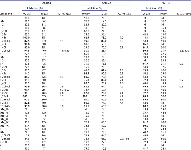
Halogenated derivatives of oestrone inhibit the AKR1C enzymes Along with the three starting oestrones (1, 2, 3) and their 3- methyl ether derivatives (1Me,2Me,3Me), we evaluated 35 halo- genated derivatives of oestrone as inhibitors of the recombinant enzymes AKR1C1, AKR1C2, and AKR1C3. There were 29 13a- methyl-oestrones, which included 3-hydroxy,17-keto-oestrones (16 compounds), 3-methoxy, 17-keto-oestrones (seven compounds) and 17-deoxy-oestrones (five compounds). There were also 12 13b-methyl-17-keto-oestrones. The screening data for these oes- trones and their derivatives for inhibition of recombinant enzymes AKR1C1-AKR1C3 are presented inTable 1.
Initial screening at 100 mM oestrones revealed that a number of these acted as potent inhibitors: 13 compounds showed80%
inhibition of AKR1C1 (six compounds at 10 mM), 11 compounds showed 80% inhibition of AKR1C2 (six compounds at 10 mM), and 10 compounds showed 80% inhibition of AKR1C3 (none at 10mM). When the compounds were screened at 10lM, additional potent inhibitors of AKR1C2 were revealed that had shown lower
inhibitory activities at the higher concentration (1_4Br, 1_4Cl, 2_2Br), which was probably caused by solubility issues.
Among the 13b-methyl-17-keto-oestrones, five showed potent inhibition of AKR1C1, as80% at 100lM, with IC50 values for the two most promising compounds, 1_2Br,4Br and 1_2Cl,4Cl, of 5.4lM and 1.6lM, respectively. Neither of these two were select- ive inhibitors of AKR1C1, as 1_2Br,4Br also showed strong inhib- ition of AKR1C2 (IC50¼5.6lM), and 1_2Cl,4Cl, of AKR1C3 (IC50¼6.3lM). Two other 13b-methyl-17-keto oestrones,1_4Br and 1_4Cl, inhibited AKR1C2 in the low micromolar range (IC50¼7.0, 5.3lM, respectively), while no inhibition was seen for these at 10lM with AKR1C1, and low inhibition was seen for AKR1C3 (25%
and 50%, respectively).
The 13a-methyl-17-keto-oestrones in the 3-methyl ether series generally showed weaker inhibition of the AKR1C enzymes com- pared to their 3-hydroxy counterparts. Indeed, only two of these showed inhibition 80% at 100lM, for AKR1C3 (2Me_2Cl, 2Me_4Cl), with much weaker inhibition of AKR1C1 and AKR1C2.
The more potent inhibition by the 13a-methyl-17-keto-oestrones in the 3-hydroxy series included six compounds with80% inhib- ition of AKR1C1 at 10lM, where the best two had IC50 of 0.7lM and 0.8lM (2_2I,4Br, 2_2I,4Cl, respectively). Five of these com- pounds inhibited AKR1C2 by 80% at 10lM, where the best inhibitor had an IC50 of 1.5lM (2_2Br). This group of oestrone derivatives did not include any potent AKR1C3 inhibitors (i.e. none 80% inhibition at 10lM).
HO A
Cl H
H H
O
HO A Br Cl
HO A Cl
H
H H
O
HO A
Br H
H H
O
HO A Br
H
H H
O
HO A
I Cl
HO A Cl Br
HO A Cl I
HO A Br I HO
A I Br
2_2Cl
2_4Cl
2_2Br
2_4Br
2_2Cl,4Br 2_2Cl,4I
2_2Br,4Cl 2_2I,4Cl
2_2Br,4I
2_2I,4Br NBS
NIS
NBS
NIS
NIS
NIS CH2Cl2
rt, 2 h
CH2Cl2 rt, 2 h
TFArt, 2 h TFA
rt, 2 h
TFArt, 2 h
TFArt, 2 h
Scheme 1. Synthesis of the 2,4-disubstituted 13a-methyl-oestrone derivatives with the different halogens.
Finally, we screened five 13a-methyl-17-deoxy derivatives. For the initial testing at 100lM, some promising results were shown, but only for AKR1C2 (three compounds showed around 75%
inhibition). At 10lM, 3_2Br,4Br and 3_2Cl,4Cl showed 68.0% and 66.2% inhibition, respectively, with the best compound,3_2Br,4Br, with an IC50of 0.9lM.
To further evaluate these oestrone derivatives as inhibitors of the AKR1C enzymes, we conducted detailed kinetic studies on three different compounds, which were chosen as they were the most potent inhibitors of AKR1C1, AKR1C2, and AKR1C3. The first was compound 1_2Cl,4Cl, or bis-chloro-13b-methyl-17-keto-oes- trone, which inhibited both AKR1C1 and AKR1C3 potently, but showed low inhibition of AKR1C2. These inhibition studies revealed a mixed type of inhibition of 1_2Cl,4Cl for AKR1C1 (Ki ¼ 0.69lM), which was instead competitive for inhibition of AKR1C3 (Ki¼1.43lM) (Table S1). The second compound chosen here was 2_2I,4Br, a 3-hydroxy disubstituted 13a-methyl-17-keto-oestrone, which at 10lM showed 90.7% inhibition of AKR1C1, 59.5% inhib- ition of AKR1C2 and 40.0% of AKR1C3. Its IC50 for AKR1C1 was 0.7lM, with Ki of 0.57lM. Here,2_2I,4Br showed a mixed type of inhibition of AKR1C1. Finally, the third compound chosen was 3_2Br,4Br, a 13a-methyl-17-deoxy bis-bromo oestrone, which
showed selectivity for AKR1C2, with IC50 of 0.9lM.3_2Br,4Br also showed mixed type of inhibition, with Ki of 1.98lM.
Structure–activity relationships
On the basis of these inhibition data, we were able to postulate the initial structure–activity relationship for all three AKR1C iso- forms. The most potent of these AK1C1 inhibitors were a 13b- methyl-17-keto-oestrone (1_2Cl,4Cl) and a 13a-methyl-17-keto-oes- trone (2_2I,4Br), with the halogen atoms at both C-2 and C-4 (Figure 4). For the 13b-methyl-17-keto-oestrone, the activity was highest with bromine and chlorine, which indicated that iodine would probably be too large, and would result in lower enzyme inhibition. Here, the most potent inhibitor had chlorine at both positions, for its IC50 of 1.6lM. For the equivalent 13a-methyl epi- mers, the lowest IC50were achieved with iodine on C-2 and brom- ine or chlorine on C-4 (IC50: 2_2I,4Br, 0.7lM; 2_2I,4Cl, 0.8lM).
These oestrone derivatives were selective for AKR1C1. The loss of the 17-keto group (3, and its derivatives) or methylation of the 3- hydroxy group (2Me, 3Me, and their derivatives) had negative impact on the AKR1C inhibition.
Table 1. Inhibition of the AKR1C enzymes by the halogenated oestrone derivatives.
AKR1C1 AKR1C2 AKR1C3
Inhibition (%)
IC50/Ki (lM)
Inhibition (%)
IC50/Ki (lM)
Inhibition (%)
IC50/Ki (lM)
Compound 100lM 10lM 100lM 10lM 100lM 10lM
1 10.0 NI 50.0 NI NI NI
1Me 22.5 8.2 70.0 4.8 NI 16.7
1_2I 22.5 31.3 52.5 20.2 NI NI
1_4I 10.0 8.3 NI NI NI 36.4
1_2I,4I 25.0 42.2 62.2 21.3 NI 13.6
1_2Br 65.0 51.2 25.0 36.4 40.2 13.0
1_4Br 100.0 NI 62.5 73.0 7.0 8.3 25.0
1_2Br,4Br 82.5 67.0 5.4 99.8 83.0 5.6 58.3 50.0
1_2Cl 91.6 50.0 88.0 53.0 86.8 NI
1_4Cl 80.0 NI 25.0 70.0 5.3 91.7 50.0
1_2Cl,4Cl 94.8 46.9 1.6/0.69 50.0 23.4 90.4 21.9 6.3, 1.43
2 22.5 7.5 65.0 3.5 29.8 22.5
2Me 20.0 1.3 35.0 NI 59.9 22.9
2_2I 42.5 37.8 50.0 22.4 NI 33.8
2_4I 22.5 2.3 75.0 16.5 85.7 35.1 12.3
2_2I,4I 77.5 NI 45.0 NI 50.0 2.6
2_2Br 22.5 47.2 65.0 81.4 1.5 23.8 62.6
2_4Br 14.2 NI 98.3 80.0 2.2 58.2 25.0
2_2Br,4Br 88.7 82.5 5.3 86.0 70.3 7.3 50.8 27.9
2_2Cl 45.1 50.0 75.4 83.0 2.1 72.3 60.0 8.7
2_4Cl 22.5 18.5 80.0 73.9 1.8 43.6 5.3
2_2Cl,4Cl 95.9 84.8 2.8 81.5 84.1 4.6 89.8 69.9 12.8
2_2I,4Br 93.4 90.7 0.7/0.57 74.7 59.5 43.6 40.0
2_2I,4Cl 99 90.0 0.8 87.5 73.0 7.1 72.2 50.0
2_2Br,4I 100 67.0 7.2 100 75.0 4.4 88.9 50.0
2_2Br,4Cl 92.3 80.0 1.7 99.0 80.0 3.1 89.4 50.0
2_2Cl,4I 82.6 50.0 89.2 73.0 4.6 58.0 NI
2_2Cl,4Br 93.9 80.0 1.9 91.9 65.0 88.0 25.0
2Me_2Br NI 7.1 NI NI 18.3 13.6
2Me_4Br 12.5 12.9 15.0 NI 39.7 44.6
2Me_2I NI 1.0 5.0 NI 50.8 NI
2Me_4I NI 5.1 NI NI 19.8 NI
2Me_2Cl 35.7 17.0 76.3 60.0 91.0 60.0
2Me_4Cl 21.4 NI 78.8 50.0 90.0 50.0
3 12.5 13.8 NI NI NI 23.4
3Me NI NI 75.0 NI 49.2 21.1
3_2Cl,4Cl 2.8 39.4 76.8 66.2 44.6 NI
3_2Br,4Br 64.3 33.0 74.4 68.0 0.9/1.98 26.7 50.0
3_2I,4I 9.8 17.5 11.7 22.9 27.3 11.4
4 25.0 NI 30.0 NI NI NI
5 50.0 7.1 75.0 10.5 41.7 24.1
NI: no inhibition.
Data in bold, compounds showing80% inhibition for the respective AKR1C.
JOURNAL OF ENZYME INHIBITION AND MEDICINAL CHEMISTRY 1505
We established here that with the 13b-methyl epimers, no sub- stitution or bromine substitution at C-2 was effective for inhibition of AKR1C2, while bromine or chlorine at C-4 was beneficial, but not iodine (Figure 4). The larger halogens at C-4 thus appeared to be advantageous. The loss of the keto group at position 17 halved the IC50. The best two AKR1C2 inhibitors among the 13a-methyl- 17-keto-oestrone epimers had only a bromine at the C-2 position and only a chlorine at the C-4 position (IC50:2_2Br, 1.5lM;2_4Cl, 1.8lM). These two compounds also showed high selectivity for the AKR1C2 isoform.
These structure–activity relationship studies also revealed that chlorine substitution of 13b-methyl-17-keto oestrones at C-2 and/
or C-4 was required for AKR1C3 inhibition, with improvement for both at the same time (IC50: 1_2Cl,4Cl, 6.3 mM; Figure 4). With bromine substitution showing low inhibition of AKR1C3, all inhib- ition was lost with either no substitution or iodine substitution.
For 13a-methyl-3-methoxy-17-keto-oestrones, bromine or chlorine substitution at C-2 and the larger halogens at C-4 (i.e. chlorine, iodine) were required for AKR1C3 inhibition, while this inhibition of AKR1C3 was almost completely lost when the 17-keto group was removed. Methylation of 3-hydroxy group greatly improved the potency of the AKR1C3 inhibition.
Computational simulations
For an atomic level investigation of the structural background of the biological activities, computational simulations were carried out for the ligands that showed well-defined enzyme selectivities.
First, docking calculations were performed using the Glide pro- gramme29–31, where receptor models were based on experimental X-ray crystal structures from the PDB database (http://www.rcsb.
org). The following structures were selected for AKR1C1, AKR1C2, and AKR1C3: 1MRQ (AKR1C1 in complex with NADPH and 20a- hydroxyprogesterone); 4L1W (AKR1C2 in complex with NADPþand progesterone); and 1XF0 (AKR1C3 in complex with NADPþand androstenedione). The structures were prepared for docking calcu- lations according to the details in the “Materials and methods”
section. It is worth noting that various structures can be retrieved for each enzyme in the PDB database, but the crystal structures were always selected here, with co-crystallisation with a ster- oid ligand.
The precision of the docking protocol was verified by redock- ing the original crystal ligand into the binding pocket, where the XP method with the applied settings accurately reproduced the binding poses of the original co-crystallised ligands. Then, all the selected ligands (2_2I,4Br, 2_4Cl, 2_4I) were docked into each relaxed target, where the scoring function of the docking program (glide-score) correlates with the binding free energy (DG) and therefore with the enzyme activity. Unfortunately, even the ligand with the highest biological activity for the relevant receptor could not be properly predicted by the docking calculations (see Glide score values inTable 2).
Thus, a more advanced DG calculation scheme was applied, namely the MMGBSA method, which used 500-ns-long MD trajec- tories for all the nine ligand–target complexes. The starting struc- tures of the MD simulations were always the best pose geometries from the docking calculations, and the MMGBSA bind- ing free energies were computed as 2500 snapshots for each tra- jectory. Statistical descriptors were calculated for all the nine ligand–target complexes, including means and standard error of means of DG, with these data shown in Table 2. It can be seen that the MMGBSA calculations always defined the most active lig- and with the strongest binding affinity for each enzyme. So, these 500-ns-long MD simulations can correctly represent the ligand–tar- get interactions. It can also be mentioned that the tertiary struc- tures of the three enzymes were very similar; each of them contained a central b-barrel structure that was surrounded by eighta-helices and a nicotine-adenosine-diphosphate co-factor on one of the tops of the barrel. The steroid binding site was located on the top side of the b-barrel, adjacent to the co-factor. The binding pocket was formed by flexible loops in all three cases, which emphasises the dynamic nature of the ligand binding. On the basis of these impressions of the binding geometry of a ster- oid in an AKR1C enzyme, we have presented here the starting Figure 4. Structure–activity relationship for inhibition of the AKR1C1-3 enzymes.
poses of the MD calculations for all of the three ligands in their
“preferred”enzyme in theSupplementary Information(Figure S1).
As the MMGBSA calculations successfully identified the most active ligands as the strongest binding ones, we examined all of the nine 500-ns-long trajectories using the Simulation Interaction Diagram tool from the Schrodinger suite. Here, among the other analyses, the protein–ligand interaction diagram was obtained in each case, where the occurrence of all of the important interac- tions were determined along the trajectories concerned (hydrogen bonds, hydrophobic interactions, water bridges, etc.). Moreover, the positions of the significant interactions on the ligands were also demonstrated in simplified 2D diagrams (Supplementary Figures S2–S4, boxed insets), as shown together with the inter- action diagrams in the Supporting Information (Figures S2–S4).
Comparisons were also made between the interaction dia- grams of the selective compounds and the diagrams of the less active compounds at each enzyme. However, neither a new sec- ondary bond formation nor a unique interaction that might be solely responsible for the selectivity were found for any of the enzymes. It seemed that the patterns of the different interactions can help to explain the possible sources of the selectivity here.
For example, for AKR1C1, the p–p staking of the A-ring of com- pound 2_2I,4Br and the significant hydrophobic interactions, as well as the largest number of water-bridge connections, might together have resulted in2_2I,4Br having the strongest interaction with the AKR1C1 enzyme (Supplementary Figure S2). For AKR1C2, with 2_4Cl, the greater hydrophobic interactions with residue Trp86 and the larger number of water bridges might together override the similar interaction patterns of 2_4I and 2_2I,4Br, although these last two also show extra p–p stacking (Supplementary Figure S3). Finally, for AKR1C3, the stabile inter- action of the keto-oxygen in the D-ring with residue Ser118, and the less stable, but probably stronger, interaction with the nega- tively charged residue Asp224 might have been responsible for the strong binding.
In summary here, we can say that there was no formation of new hydrogen or halogen bonds with the flexible loops that might have been responsible for the selectivity, although hydro- phobic interactions might have particularly important roles in the selectivities of these compounds. Hence, the variations of atoms at positions 2 and 4 in the sterane skeletons might fine-tune these effects.
Conclusions
The AKR1C enzymes are promising drug targets, as they are involved in the development of different cancers and several benign pathologies, and they are also associated with chemore- sistance to platin and anthracycline-based drugs. Here, we syn- thesised 35 halogenated oestrone derivatives with low affinities for the nuclear oestrogen receptors, and evaluated their inhibitory actions against AKRC1, AKR1C2, and AKR1C3. Some potent
inhibitors were identified, with occasional dual or triple inhibitory properties. Selective compounds were found against each of these three enzymes. Atomic level computational simulations showed that neither a well-defined unique chemical connection nor a spe- cific interaction with a single amino acid could be identified in search of a source of selectivity regarding potent ligands. Overall, these data indicate that these halogenated oestrones represent a new class of potent and selective AKR1C inhibitors, and thus have the potential for development of new antitumour agents.
Acknowledgements
The authors thank Ajda Godec and Anastazija Zajec for help with enzyme inhibition assays, andSpela Kos for technical support.
Disclosure statement
The authors report no conflict of interest.
Funding
The work of Erzsebet Mernyak on this project was supported by the Janos Bolyai Research Scholarship of the Hungarian Academy of Sciences. This work was supported by the National Research, Development and Innovation Office-NKFIH through project OTKA SNN 124329 and project N1-0066 from the Slovenian Research Agency.
References
1. Penning TM. The aldo-keto reductases (AKRs): overview.
Chem Biol Interact 2015;234:236–46.
2. Rizner TL, Penning TM. Role of aldo-keto reductase family 1 (AKR1) enzymes in human steroid metabolism. Steroids 2014;79:49–63.
3. Hevir N, Vouk K, Sinkovec J, et al. Aldo-keto reductases AKR1C1, AKR1C2 and AKR1C3 may enhance progesterone metabolism in ovarian endometriosis. Chem Biol Interact 2011;191:217–26.
4. Rizner TL, Penning TM. Aldo-keto reductase 1C3-assessment as a new target for the treatment of endometriosis.
Pharmacol Res 2020;152:104446.
5. Shiiba M, Yamagami H, Yamamoto A, et al. Mefenamic acid enhances anticancer drug sensitivity via inhibition of aldo- keto reductase 1C enzyme activity. Oncol Rep 2017;37:
2025–32.
6. Matsumoto R, Tsuda M, Yoshida K, et al. Aldo-keto reductase 1C1 induced by interleukin-1b mediates the invasive Table 2. Scoring values of docking calculations, mean and standard error of means (SEM) of binding energies (in kcal/mol) determined by MMGBSA calculations, as well as biological inhibition (at 100lM) for the tested compounds are presented.
Glide score DGMMGBSA Biological activity
AKR1C1 AKR1C2 AKR1C3
Compound AKR1C1 AKR1C2 AKR1C3 Mean SEM Mean SEM Mean SEM AKR1C1 AKR1C2 AKR1C3
2_2I,4Br –8.3 –7.8 –8.4 –60.3 3.5 –57.7 1.7 –55.8 4.0 93.4 74.7 43.6
2_4Cl –8.7 –8.5 –7.9 –55.5 1.6 –59.9 1.6 –56.1 2.5 22.5 80.0 43.6
2_4I –8.4 –8.2 –7.3 –53.8 1.9 –52.7 1.83 –72.1 2.0 22.5 75.0 85.7
The enzyme structures in docking studies were based on X-ray crystal structures from the pdb database (http://www.rcsb.org; AKR1C1, 1MRQ; AKR1C2, 4L1W;
AKR1C3, 1XF0).
JOURNAL OF ENZYME INHIBITION AND MEDICINAL CHEMISTRY 1507
potential and drug resistance of metastatic bladder cancer cells. Sci Rep 2016;6:34625.
7. Matsunaga T, Hojo A, Yamane Y, et al. Pathophysiological roles of aldo-keto reductases (AKR1C1 and AKR1C3) in devel- opment of cisplatin resistance in human colon cancers.
Chem Biol Interact 2013;202:234–42.
8. Chen CC, Chu CB, Liu KJ, et al. Gene expression profiling for analysis acquired oxaliplatin resistant factors in human gas- tric carcinoma TSGH-S3 cells: the role of IL-6 signaling and Nrf2/AKR1C axis identification. Biochem Pharmacol 2013;86:
872–87.
9. Hofman J, Malcekova B, Skarka A, et al. Anthracycline resist- ance mediated by reductive metabolism in cancer cells: the role of aldo-keto reductase 1C3. Toxicol Appl Pharmacol 2014;278:238–48.
10. Plebuch M, Soldan M, Hungerer C, et al. Increased resistance of tumor cells to daunorubicin after transfection of cDNAs coding for anthracycline inactivating enzymes. Cancer Lett 2007;255:49–56.
11. Hintzpeter J, Seliger JM, Hofman J, et al. Inhibition of human anthracycline reductases by emodin–a possible remedy for anthracycline resistance. Toxicol Appl Pharmacol 2016;293:
21–9.
12. Veitch ZW, Guo B, Hembruff SL, et al. Induction of 1C aldo- ketoreductases and other drug dose-dependent genes upon acquisition of anthracycline resistance. Pharmacogenet Genomics 2009;19:477–88.
13. Heibein AD, Guo B, Sprowl JA, et al. Role of aldo-keto reduc- tases and other doxorubicin pharmacokinetic genes in doxo- rubicin resistance, DNA binding, and subcellular localization.
BMC Cancer 2012;12:381–10.
14. Novotna E, Bukum N, Hofman J, et al. Roscovitine and pur- valanol A effectively reverse anthracycline resistance medi- ated by the activity of aldo-keto reductase 1C3 (AKR1C3): a promising therapeutic target for cancer treatment. Biochem Pharmacol 2018;156:22–31.
15. Matsunaga T, Yamaguchi A, Morikawa Y, et al. Induction of aldo-keto reductases (AKR1C1 and AKR1C3) abolishes the efficacy of daunorubicin chemotherapy for leukemic U937 cells. Anticancer Drugs 2014;25:868–77.
16. Verma K, Zang T, Gupta N, et al. Selective AKR1C3 inhibitors potentiate chemotherapeutic activity in multiple acute mye- loid leukemia (AML) cell lines. ACS Med Chem Lett 2016;7:
774–9.
17. Verma K, Zang T, Penning TM, Trippier PC. Potent and highly selective aldo-keto reductase 1C3 (AKR1C3) inhibitors act as chemotherapeutic potentiators in acute myeloid leu- kemia and T-cell acute lymphoblastic leukemia. J Med Chem 2019;62:3590–616.
18. Penning TM. Aldo-keto reductase regulation by the Nrf2 sys- tem: implications for stress response, chemotherapy drug resistance, and carcinogenesis. Chem Res Toxicol 2017;30:
162–76.
19. Brasseur K, Gevry N, Asselin E. Chemoresistance and tar- geted therapies in ovarian and endometrial cancers.
Oncotarget 2017;8:4008–42.
20. Ji X, Lu Y, Tian H, et al. Chemoresistance mechanisms of breast cancer and their countermeasures. Biomed Pharmacother 2019;114:108800.
21. Schonecker B, Lange C, Kotteritzsch M, et al. Conformational design for 13alpha-steroids. J Org Chem 2000;65:5487–97.
22. Ayan D, Roy J, Maltais R, Poirier D. Impact of estradiol struc- tural modifications (18-methyl and/or 17-hydroxy inversion of configuration) on the in vitro and in vivo estrogenic activ- ity. J Steroid Biochem Mol Biol 2011;127:324–30.
23. Bacsa I, Jojart R, Schneider G, et al. Synthesis of A-ring halo- genated 13a-estrone derivatives as potential 17b-HSD1 inhibitors. Steroids 2015;104:230–6.
24. Laczko-Rigo R, Jojart R, Mernyak E, et al. Structural dissec- tion of 13-epiestrones based on the interaction with human organic anion-transporting polypeptide, OATP2B1. J Steroid Biochem Mol Biol 2020;200:105652.
25. Bacsa I, Herman BE, Jojart R, et al. Synthesis and structure- activity relationships of 2- and/or 4-halogenated 13b- and 13a-estrone derivatives as enzyme inhibitors of estrogen biosynthesis. J Enzyme Inhib Med Chem 2018;33:1271–82.
26. Jojart R, Traj P, Kovacs E, et al. Synthesis, biological evalu- ation and docking studies of 13-epimeric 10-fluoro- and 10- chloroestra-1,4-dien-3-ones as potential aromatase inhibitors.
Molecules 2019;24:1783.
27. Hong Y, Chen S. Aromatase, estrone sulfatase, and 17beta- hydroxysteroid dehydrogenase: structure-function studies and inhibitor development. Mol Cell Endocrinol 2011;340:
120–6.
28. Brozic P, Smuc T, Gobec S, Rizner TL. Phytoestrogens as inhibitors of the human progesterone metabolizing enzyme AKR1C1. Mol Cell Endocrinol 2006;259:30–42.
29. Friesner RA, Banks JL, Murphy RB, et al. Glide: a new approach for rapid, accurate docking and scoring. 1. Method and assessment of docking accuracy. J Med Chem 2004;47:
1739–49.
30. Friesner RA, Murphy RB, Repasky MP, et al. Extra precision glide: docking and scoring incorporating a model of hydro- phobic enclosure for protein-ligand complexes. J Med Chem 2006;49:6177–96.
31. Halgren TA, Murphy RB, Friesner RA, et al. Glide: a new approach for rapid, accurate docking and scoring. 2.
Enrichment factors in database screening. J Med Chem 2004;47:1750–9.
32. Schr€odinger. Schr€odinger release 2020–4. New York (NY):
Schr€odinger LLC; 2020.
33. Maestro S. Schr€odinger release 2020–3. New York (NY):
Schr€odinger LLC; 2020.
34. DESR. Desmond molecular dynamics system, Schr€odinger release 2020–3. Maestro-Desmond interoperability tools.
New York (NY): Schr€odinger; 2020.