Synthesis and investigation of FITC-labeled and crizotinib-conjugated GnRH analogues and their
relevance in targeted cancer therapy Ph.D. thesis
József Murányi Semmelweis University
Doctoral School of Pharmaceutical Sciences
Supervisor: Dr. Tibor Vántus, Ph.D.
Dr. György Kéri
†, D.Sc.
Official reviewers: Dr. György Mészáros, Ph.D.
Dr. Ildikó Szabó, Ph.D.
Theoretical exam committee:
President: Dr. Tamás Török, D.Sc.
Members: Dr. Imre Klebovics, D.Sc.
Dr. Zoltán Bánóczi, Ph.D.
Budapest
2018
1. Introduction
Signal transduction therapy is an intensively developing field of cancer treatment, approved kinase inhibitors has several advantages compared to classical chemotherapy. However the limited selectivity, consequently the serious side effects and the fast developing resistance still represent a general problem.
Crizotinib is a first-generation tyrosine kinase inhibitor that was initially designed as an inhibitor of c-Met. However, it is approved for treatment of ALK or ROS-1 positive, advanced non-small cell lung carcinoma as a multi-target inhibitor. The serious side effects of crizotinib justify their targeted delivery into cancer cells.
Moreover, the increase in the selectivity of crizotinib could enhance their appliance in combination therapy in the future.
Among anti-cancer drug delivery systems GnRH analogues have great potential, because they are deliver and target the drug in the same time. The overexpression of GnRH-R creates an opportunity for targeted delivery which is frequently occur at cancer cells. In the case of GnRH-R expressing cancers the direct anti-tumour effect of synthetic GnRH analogues provides further therapeutic advantages.
Surprisingly, experiments focusing on the amount of the drugs which could be delivered into the various cancer cells are very rare in the literature. Fluorescent labelling of GnRH analogues offers great possibilities to investigate the GnRH- R-targeted drug delivery, and predict its efficiency in vitro. Despite this, besides the strongly cytotoxic anthracycline-derivate containing GnRH conjugates, I did not find further literary sources about fluorescently detectable GnRH analogues.
There are numerous open questions about GnRH-R-targeted drug delivery which meant a great challenge for my PhD research to design and investigate the crizotinib-GnRH conjugates. Despite the promising preclinical results of anti- cancer GnRH conjugates, none of them achieved clinical approval, which calls the attention for the numerous possible pitfalls. The limited background and the several uncertainties about GnRH-based drug delivery systems gave strategic significance for my PhD research.
2. Aims
In my doctoral thesis I investigated the therapeutic possibilities of GnRH-R targeted cancer therapy. Synthesis and evaluation of fluorescently labelled GnRH analogues were aimed to the prediction of GnRH-R targeted therapeutic efficiency on cancer cell models in vitro.
Purposes of my PhD research:
1. Labelling D-Lys6-GnRH-I, D-Lys6-GnRH-II and GnRH-III analogues with fluorescein isothiocyanate.
2. Characterisation of the physicochemical properties of the synthesized, novel FITC-GnRH conjugates.
3. To investigate the localisation and the amount of FITC-GnRH conjugates on cancer cell lines in vitro.
In my further research I have dealt with the synthesis and the in vitro evaluation of novel crizotinib-GnRH conjugates in order to combine the mechanism of action of crizotinib and the therapeutic advantages of GnRH-targeted drug delivery
Additional purposes of my PhD research:
4. Synthesis of crizotinib analogues which could be conjugated to GnRH.
5. Characterisation of the physicochemical properties of crizotinib analogues and investigate them on in vitro cancer cells.
6. Conjugation of the most promising crizotinib analogues with GnRH, applying different spacers and chemical linkages.
7. To characterize the physicochemical properties of the synthesized crizotinib-GnRH conjugates, investigate them on cancer model in vitro and evaluate their effectiveness in connection with the results of FITC- GnRH conjugates.
3. Methods
Analytical methods
In order to follow chemical synthesis, to purify and measure the purity of chemical products, I applied HPLC. To measure the solubility, stability and permeability HPLC was used also, while I developed three separation methods.
Separation by column chromatography and certain reactions were followed by thin-layer chromatography.
In order to chemical structure proof mass spectrometry and 1H NMR spectrometry were used in collaboration with ELTE Department of Analytical Chemistry, SE Department of Pharmaceutical Chemistry and Vichem Chemie Researcher Ltd.
Syntheses
Crizotinib and crizotinib analogues were synthesized by classical organic synthesis. Certain steps of crizotinib total synthesis were reproduced by literature data, further steps were modified in favour of the GnRH conjugation. The modified total synthesis allowed to synthesize novel, selectively Boc-protected crizotinib analogues as well. Further elaboration on the synthesis methods is included in the PhD dissertation.
The synthesis of D-Lys6-GnRH-I, D-Lys6-GnRH-II and GnRH-III was performed by Fmoc solid-phase peptide synthesis. Kaiser-test was applied to monitor completeness of amino acid coupling.
The FITC labelling of GnRH analogues was preformed after the selective removal of ivDde or alloc side-chain protecting groups of Lys or D-Lys. I applied the selective Boc-protected, and linker-containing crizotinib analogues to synthesize the crizotinib-GnRH conjugates.
I applied TFA-based cleavage cocktail to remove the peptid from the resin, which removed the other protecting groups also. Crude products were purified by HPLC, and lyophilised finally.
Cell cultures
The cell cultures were maintained in the ATCC and JRCB recommended medium complemented with 10% FBS and antibiotics, at 37°C, under 5% CO2
atmosphere, in a humidified incubator.
ADME-Tox assays
Solubility was measured in PBS at 25 °C, or in serum-free RPMI medium at 37°C.
10 mM DMSO stock solutions were diluted to 200 µM. After 2 hours incubation, samples were centrifuged twice (13000g, 10 min). Samples derived from the supernatants were determined by HPLC-UV.
Membrane permeability was measured by PAMPA and MDCK protocols. At PAMPA I used artificial membrane based system (BD Gentest™) and 40 µM PBS solutions (pH=7.4). The system was gently shaken, at 25°C, for 5 hours. In case of pH-dependent permeability measurement, I worked with pH= 4.8; 6.0; 7.4 and 8.5 buffer solutions. Samples derived from the donor and the acceptor wells were determined by UV-Vis spectrophotometer.
At MDCK method permeability was measured on monolayer of MDCK canine kidney epithelial cell culture. Cell were cultivated until they reached the 100%
confluency. The confluency of the cell layer was checked by impedance measurement. I worked with 10 µM HBSS solutions. System was gently shaken, at 25°C, for 1 hour. Samples derived from the donor and the acceptor wells were determined by HPLC-UV.
Stability was measured in 10% FBS containing RPMI medium. 10 mM DMSO stock solutions were diluted to 10 µM. Samples were incubated at 37°C, for 2, 6 and 24 hours. After incubation samples were diluted with acetonitrile (1:1 ratio), vortexed and centrifuged twice (13000g, 10 min). Samples derived from the supernatants were determined by HPLC-UV. Half life time was calculated by Graph Pad Prism software.
Stability measurement in rat liver lysosomal homogenate was performed by Department of Analytical Chemistry, ELTE (Budapest, Hungary).
The measurement of in vitro c-Met inhibition was performed by ProQinase GmbH (Freiburg, Germany) and Vichem Chemie Researcher Ltd. (Budapest, Hungary).
Western blot
At the verification of GnRH-R-binding, cells were treated with GnRH conjugates in 1 µM, for 15 minutes. Cells were lysated after the treatment. At the verification of GnRH-R expression non-treated cell were lysated. The protein concentrations were determined using Bradford-method. The 20-40 µg protein containing samples were separated on SDS-polyacrylamide gels by gel electrophoresis and the proteins were transferred onto PVDF membranes. The investigated proteins:
GnRH-R, α-tubulin, foszfo-ERK1/2 and ERK1/2. ECL-method was applied to determine the results.
Cell viability assay
Effects of the synthesized compounds to the viability of EBC-1 and the NIH/3T3 cells were measured by MTT and CellTiter-Glo® methods. Cells were seeded onto a 96 well plate (MTT: 104 cell/well, CellTiter-Glo®: 103 cell/well). While applying the MTT method, cells were treated in 40 µM for 24 hours. Viability was calculated from the absorbance data at 570 nm and 635 nm. While applying CellTiter-Glo® method, cells were treated for 72 hours. Viability was calculated by using luminescent detection. Data were evaluated with MS Excel 2013. IC50
values were calculated by Graph Pad Prism software.
Confocal laser scanning microscopy
The cells were seeded on Ibidi® μ-Slide 8 Well microscopic slides and let to adhere for 48 hours.
At the immunocytochemistry experiments, the cells were fixed with 4%
paraformaldehyde for 10 minutes. (In specific case cells were permeabilised with 0.1% Triton X-100 PBS solution as well) Cells were incubated with hGnRH-I-R primary antibody, followed by Alexa Fluor 546 conjugated secondary antibody.
Finally, Draq5™ fluorescent probe solution was added to the cells.
During the uptake experiments of the GnRH conjugates, the cells were treated with 1 µM or 10 µM conjugates and incubated for the desired time at 37 °C. The treatment medium was removed; the cells were washed with PBS and fixed with 4% formaldehyde. The cells were washed with PBS, followed by adding Draq5™
for 10 minutes.
Images of the cells were acquired under an oil immersion objective with confocal laser microscope (Zeiss Confocal LSM 710)
Flow cytometry
Approximately 105 cells were seeded per well to 24 well plates and let to adhere for 48 hours. Next day, the cells were treated with 1 µM or 10 µM FITC-GnRH conjugates for 1 or 5 h. After incubation, supernatant was discarded, and trypsin was added for 10 min at 37 °C. The cells were pelleted (250 g, 4 min, RT) in fluorescence-activated cell sorting (FACS) tubes, suspended in PBS and kept at 4
°C for the rest of the experiment. The fluorescence intensity of the conjugate- stained cells was analysed by flow cytometer (BD FACSCalibur™). Data were evaluated by CellQuest Pro software (BD Biosciences) and MS Excel 2013 software.
4. Results
Synthesis and characterisation of FITC-GnRH conjugates
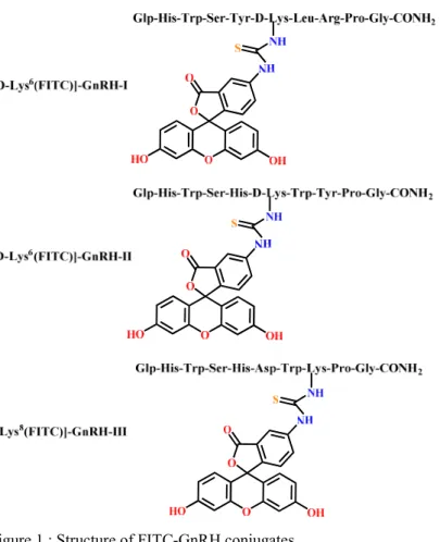
I synthesized three selectively FITC labelled GnRH analogues (Figure 1.). FITC- GnRH conjugates are soluble in PBS and they are stable in cell culture medium.
They have no passive membrane permeability. They have no significant cytotoxic effect on NIH/3T3 cells even at higher (40 µM) concentrations.
Figure 1.: Structure of FITC-GnRH conjugates.
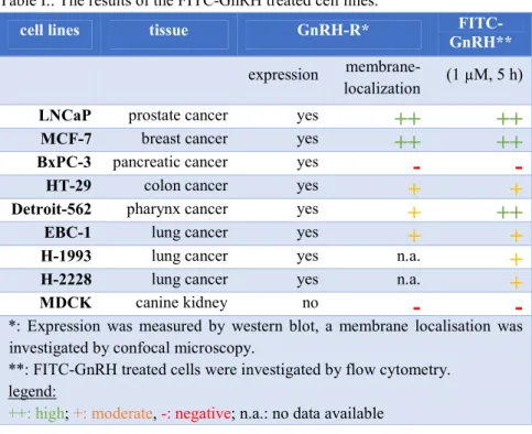
Cellular uptake of FITC-GnRH conjugates and cell surface GnRH-I-R analysis The cellular uptake of FITC-GnRH conjugates were investigated on LNCaP, MCF-7, BxPC-3 and HT-29 cell lines (known GnRH-R expressing cell lines), and on Detroit-562, EBC-1, H-1993, H-2228 and MDCK cell lines (GnRH-R expression previously not investigated). Results are summarized in table I.
Table I.: The results of the FITC-GnRH treated cell lines.
cell lines tissue GnRH-R* FITC-
GnRH**
expression membrane-
localization (1 µM, 5 h)
LNCaP prostate cancer yes
++ ++
MCF-7 breast cancer yes
++ ++
BxPC-3 pancreatic cancer yes
- -
HT-29 colon cancer yes
+ +
Detroit-562 pharynx cancer yes
+ ++
EBC-1 lung cancer yes
+ +
H-1993 lung cancer yes n.a.
+
H-2228 lung cancer yes n.a.
+
MDCK canine kidney no
- -
*: Expression was measured by western blot, a membrane localisation was investigated by confocal microscopy.
**: FITC-GnRH treated cells were investigated by flow cytometry.
legend:
++: high; +: moderate, -: negative; n.a.: no data available
GnRH-R expression was proved on each investigated human cancer cell lines by western blot. However, the cell surface level of GnRH-R shown significant difference between these cell lines.
FITC-GnRH conjugates were detectable in each cell lines at 10 µM. LNCaP, MCF-7 and Detroit-562 showed the highest uptake. There are no significant differences between the efficiency of three conjugates.
Differences between the GnRH analogues were more significant at 1 µM;
furthermore, the different cells preferred different GnRH analogues. The previously not investigated Detroit-562, EBC-1, H-1993 and H-2228 cells proved
to be promising targets, in comparison with the well-known GnRH-target LNCaP, MCF-7 and HT-29 cells. The uptaken FITC-GnRH conjugates were localised mainly in the lysosomes due to the GnRH-R mediated endocytosis. The conjugates were not accumulated in the GnRH-I-R negative MDCK and in the GnRH-I-R non-exhibiting BxPC-3 cells at 1 µM.
These results predict that these analogues have high GnRH-I-R selectivity below 1 µM. Furthermore, we suppose that the uptake of these GnRH peptides could take place not only by the human GnRH-I receptors, at higher concentrations (10 µM or above), but this unknown active transport mechanism should be further examined.
Investigation of BxPC-3 cells revealed a previously not mentioned problem:
cancer types are generally categorized based on their GnRH-R mRNA or protein expression as potential GnRH targets. GnRH-R expression of BxPC-3 cell line was proved by western blot. However, on non-permeabilised BxPC-3 cell surface GnRH-R was not detectable by immunocytochemistry. Subsequent immunocytochemistry method on permeabilised BxPC-3 cells proved the GnRH- R expression, and reveal that GnRH-R does not translocate to the plasma membrane. The permanent lack of membrane GnRH-R on BxPC-3 cells is confirmed by the low cellular uptake of FITC-GnRH.
Design of crizotinib-GnRH conjugates
Increased GnRH-R level on the cell membrane is a necessary condition for effectiveness of crizotinib-GnRH conjugates. Additional condition is that the given cancer cells will be sensitive for crizotinib. From the known molecular targets of crizotinib I choose the c-Met. Thereby from the investigated cell lines I selected the c-Met amplified, and c-Met overexpressing EBC-1 non-small lung cancer cell line to examine the effectiveness of crizotinib analogues and their conjugates.
I took into consideration the structure of the complex formed between crizotinib and c-Met in the design of the crizotinib-GnRH conjugates. The 2-aminopyridine core of crizotinib responsible for the binding to c-Met, furthermore crizotinib contains a pharmacokinetically important piperidin ring, which is located out of the ATP binding pocket and directed to the solvent. Based on these structural properties I selected for conjugation the amino group of piperidine. For this
purpose, the amino group of the 2-aminopyridine core should be selectively protected.
Synthesis and investigation of novel crizotinib analogues
Modification of the piperidin ring of crizotinib, which enables the formation of carbamate or ester bond with the linker was an additional condition in the synthesis of crizotinib analogues. I synthesized six crizotinib analogues. After removal the Boc protecting group these compounds were screened.
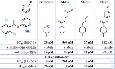
Based on the IC50 values on EBC-1 cells, and on the unique structural properties I selected the crizotinib, MJ55, MJ37 and MJ95 compounds for conjugation (Figure 2.). Each of the four compounds are stable in cell culture medium. MJ37 has the highest solubility, but the MJ95 is nearly insoluble.
Figure 2.: The selected crizotinib analogues for GnRH conjugation.
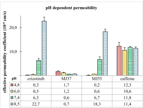
The R-conformer of crizotinib, MJ37 and MJ55 were also synthesized. Crizotinib and MJ55 resulted in very low IC50 values on EBC-1 cells. MJ37 proved to be the most effective in the inhibition of c-Met in vitro, however it resulted in much lower effect on the EBC-1 cells. The very low membrane permeability of MJ37 explained the lower effect on the EBC-1 cells (Figure 3.).
Figure 3.: pH-dependent membrane permeability of crizotinib, MJ37, MJ55 and caffeine, measured by PAMPA.
Synthesis and evaluation of crizotinib-GnRH conjugates
Five crizotinib-GnRH conjugates were synthesized, by using the selectively Boc- protected crizotinib analogues. Conjugates have poor solubility in PBS, their solubility in cell culture medium are better. [Lys8((R)-MJ55)]-GnRH-III has the highest solubility due to the more hydrophilic character of GnRH-III peptide.
Stability of the chemical bond between the agent and the linker determines the selectivity and the efficiency of conjugates. Amide bond containing crizotinib- GnRH-I and carbamate bond containing MJ95-GnRH-I are stabile in cell culture medium, free drugs were not detected after 24 hours. Aromatic ester bond- containing MJ37-GnRH-I and the aliphatic ester bond-containing MJ55-GnRH-I and (R)-MJ55-GnRH-III undergo rapid hydrolysis. After 24 hours technically the full amount of the conjugated drug released.
crizotinib MJ37 MJ55 caffeine
4,8 0,3 1,7 0,2 12,3
6,0 0,5 1,2 0,6 10,6
7,4 6,3 0,6 6,7 11,8
8,5 22,7 0,7 18,3 11,4
0,0 10,0 20,0
effective permeability coefficient (10-6cm/s) pH dependent permeability
Table II.: Crizotinib-GnRH conjugates
conjugate chemical bond formed
with the agent* IC50 (µM)**
bond type stability EBC-1 NIH/3T3 D-Lys6(crizotinib)]-
GnRH-I amide stabile 5,5 >>10
[D-Lys6(MJ37)]-
GnRH-I aromatic ester t1/2=5 h 4,5 >10
[D-Lys6(MJ55)]-
GnRH-I aliphatic ester t1/2=4 h 6,1*10-2 1,6 [Lys8((R)-MJ55)]-
GnRH-III aliphatic ester t1/2=3 h 1,2*10-2 0,8 [D-Lys6(MJ95)]-
GnRH-I carbamate stabile >10 >>10
*: Stability measurement in 10% FBS containing RPMI medium, at 37°C.
**: 50% inhibition of the viability of cells, determined by Cell Titer-Glo®.
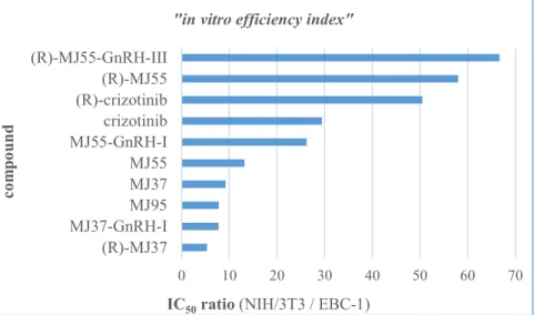
For the better comparison of crizotinib analogues and conjugates their IC50 ratio measured on NIH/3T3 and EBC-1 cells were calculated. These values are defined as „in vitro efficiency index” (in the following: efficiency index) in my PhD thesis (Figure 4.). Efficiency Index do not contain the statistical probability.
Figure 4.: “In vitro efficiency index” of the conjugated and the free crizotinib analogues.
0 10 20 30 40 50 60 70
(R)-MJ55-GnRH-III (R)-MJ55 (R)-crizotinib crizotinib MJ55-GnRH-I MJ55 MJ37 MJ95 MJ37-GnRH-I (R)-MJ37
IC50ratio (NIH/3T3 / EBC-1)
compound
"in vitro efficiency index"
Efficiency index of crizotinib-GnRH-I and MJ95-GnRH-I could not be determined due to their high IC50 values and solubility limits. Both of the conjugated and the free (R)-MJ55 resulted in better efficiency index than the clinically approved crizotinib. However, in the case of (R)-MJ55-GnRH-III the therapeutic benefit from the targeted drug delivery is doubtful due to the fast hydrolysis. Further in vivo studies should be taken into consideration.
“Lysosomal trap-theory”
Investigation of crizotinib-GnRH conjugates revealed a previously not mentioned problem regarding to the GnRH-R targeted drug delivery. This problem has been named as “lysosomal trap-theory” in my PhD thesis.
“Lysosomal trap-theory” was supported by the following results:
Crizotinib-GnRH conjugates could bind to the GnRH-R, which was proved by GnRH-R induced activation of ERK1/2.
Crizotinib-GnRH conjugates have higher IC50 values than the corresponding free analogues.
Crizotinib-GnRH-I and MJ95-GnRH-I do not release the drug in the presence of lysosomal enzymes. However, the GnRH attached crizotinib can effectively inhibit the c-Met kinase in vitro as well. Despite of this phenomenon the effect of these conjugates appears much higher concentration on the EBC-1 cells than the free drug.
Ester bond containing conjugates can release the drug before they reach GnRH-R, thereby these conjugates proved to be more effective and less selective.
The GnRH-III conjugated (R)-MJ55 has higher IC50 value on the EBC-1 cells, than the 1:1 ratio combination of (R)-MJ55 and GnRH-III.
The GnRH-R targeted drug delivery of the most effective c-Met inhibitor MJ37 cannot resolved the problem derived from the poor permeability of MJ37. The low permeability represents a problem for the uptaken MJ37 as well.
Based on literature data, the GnRH conjugates concentrated in the lysosome due to the GnRH-R-mediated endocytosis, which was proved by results occurring in FITC-GnRH as well.
The investigation of the pH-dependent permeability revealed that at the acidic pH of the lysosome the 2-aminopyridine core of crizotinib analogues became ionised, thereby their permeability dramatically decreases.
Based on these results, the lysosomal trap-theory means that the crizotinib analogues uptaken by GnRH-R could reach much harder the ATP-binding pocket of c-Met then the free drug uptaken by passive diffusion. Due to the GnRH-R mediated endocytosis, the major intracellular barrier is the membrane of the lysosomes.
Summary
GnRH analogues are effective targeting peptides which able to deliver anti-cancer agents selectively into highly GnRH-R expressing malignant tumour cells.
Efficiency of the GnRH targeted drug delivery mainly determined by cell type dependent intensity of GnRH-R translocation to the plasma membrane.
Experiments with the crizotinib-GnRH conjugates reveal that the membrane of the lysosome might be a significant barrier to GnRH conjugated drugs to reach their molecular targets. The lysosomotropic 2-aminopyridine core of crizotinib decreases the efficiency of crizotinib-GnRH conjugates even further. The results suggest that GnRH conjugates could enter into cells by GnRH-R independent mechanism at higher concentrations as well, which may be bypassing the lysosome. Among the synthesized conjugates the (R)-MJ55-GnRH-III resulted in the lowest IC50 value on the EBC-1 lung cancer cells. The (R)-MJ55 analogue both in conjugated and in free form resulted in similar efficiency than the clinically approved crizotinib.
5. Conclusions
Conclusions regarding to the results of the novel FITC-GnRH conjugates:
1. Fluorescent intensity of the uptaken [D-Lys6(FITC)]-GnRH-I, [D- Lys6(FITC)]-GnRH-II and [Lys8(FITC)]-GnRH-III is quantifiable in time and concentration dependent manner. Based on their physicochemical properties their appliance is reliable in vitro.
2. GnRH-R expressing cancer cells contain very different levels of GnRH- R on their plasma membrane. Anterograde transport of GnRH might be induced by GnRH analogues also in a cell type dependent manner. Thereby GnRH-R expression is not a reliable predictive biomarker for effective GnRH-based drug delivery. The main advantage of FITC-GnRH conjugates is that the uptaken GnRH is quantifiable, which allow the prediction of the effectiveness of the GnRH based drug targeting in vitro.
3. Investigation of Detroit-562 cell line firstly revealed that certain pharynx cancers could be targeted by GnRH-based drug delivery systems effectively. The enhancement of FITC-GnRH conjugates in the newly investigated EBC-1, H- 1993 and H-2228 lung cancer cell lines carries a significant clinical importance as well.
4. The cellular uptake of GnRH analogues correlated with the level of cell surface GnRH-I-R at 1 µM. These results predict that these analogues have high GnRH-I-R selectivity below 1 µM. Furthermore, we suppose that the uptake of these GnRH peptides could take place not only by the human GnRH-I receptors, especially at higher concentrations (10 µM or above), but this unknown active transport mechanism needs to be further investigated.
Conclusions regarding to the results of the novel crizotinib analogues and crizotinib-GnRH conjugates:
5. Boc-protected crizotinib analogues are suitable for selective GnRH coupling. (R)-MJ55 resulted in similar efficiency as crizotinib on the c-Met amplificated EBC-1 lung cancer cells in vitro. MJ37 proved to be the most effective c-Met inhibitor. However, MJ37 has higher IC50 value on EBC-1 cells due to the lower permeability.
6. The type of the chemical bond formed between the drug and the linker determines the efficiency and the selectivity of crizotinib-GnRH conjugates. The amide and the carbamate bond containing crizotinib-GnRH-I and MJ95-GnRH-I are less effective, because they do not release the conjugated drugs. However, the ester bond containing MJ37-GnRH-I, MJ55-GnRH-I and (R)-MJ55-GnRH-III undergo rapid hydrolysis. The drugs which are released from conjugates before they reach GnRH-R responsible for the lower IC50 value, on the other hand this mechanism reduces the benefit derived from the targeted delivery.
7. Drug delivery into the lysosome due to the GnRH-R-mediated endocytosis plays a crucial rule in the mechanism of action of crizotinib-GnRH conjugates. The ionisation in the lysosome which reduces the membrane permeability is obligated due to the lysosomotropic 2-aminopyridine core of crizotinib. Thereby I hypothesize that crizotinib analogues having a more difficult way to the receptor tyrosine kinases when their cellular uptake is mediated by the GnRH-R.
8. Drugs which could release in the target cell and could penetrate the membrane of the lysosome are needed in order to synthesize more effective GnRH conjugates. If the structure of the drug does not allow this, membrane penetrating sequence inserted between the drug and the GnRH can be an alternative approach. The limited amount of the drug which can be selectively transported into the cancer cells due to the limited amount of GnRH-R should be further examined.
6. Author's own publications
Publications related to the dissertation
Murányi J, Varga A, Gurbi B, Gyulavári P, Mező G, Vántus T
In vitro imaging and quantification of the drug targeting efficiency of fluorescently labeled GnRH analogues
JOURNAL OF VISUALIZED EXPERIMENTS 2017: (121) e55529. (2017)
Murányi J, Gyulavári P, Varga A, Bökönyi G, Tanai H, Vántus T, Pap D, Ludányi K, Mező G, Kéri G
Synthesis, characterization and systematic comparison of FITC-labelled GnRH- I, -II and -III analogues on various tumour cells
JOURNAL OF PEPTIDE SCIENCE 22: (8) pp. 552-560. (2016)