RESEARCH ARTICLE
Eye-tracking-aided characterization
of saccades and antisaccades in SYNE1 ataxia patients: a pilot study
Laszlo Szpisjak1, Gabor Szaraz1, Andras Salamon1, Viola L. Nemeth1, Noemi Szepfalusi1, Gabor Veres1,5, Balint Kincses2, Zoltan Maroti3, Tibor Kalmar3, Malgorzata Rydzanicz4, Rafal Ploski4, Peter Klivenyi1 and Denes Zadori1*
Abstract
Background: SYNE1 ataxia is an autosomal recessive hereditary condition, the main characteristic features of which are gait and limb ataxia and cerebellar dysarthria. Reports have revealed that the clinical phenotype of SYNE1 ataxia is more complex than the first published cases with pure cerebellar signs indicated. The aim of this study was to charac- terize eye movement alterations in the first diagnosed Hungarian SYNE1 ataxia patients.
Results: Saccades and antisaccades were examined with an eye tracker device in 3 SYNE1 (one patient has two frameshift mutations [c.8515_8516insA, p.Met2839Asnfs*53 and c.11594_11595insG, p.Glu3866*] in a compound heterozygous state, whereas two subjects have a splicing variant [c.23146-2A > G] in a homozygous state), 6 Friedreich ataxia (FA) patients and 12 healthy controls. Besides that, detailed clinical phenotyping and comprehensive neuropsy- chological assessment were carried out in all patients with ataxia.
In addition to the characteristic cerebellar alterations, pyramidal signs and polyneuropathy were observed at least in 2 SYNE1 ataxia patients, for which no other underlying reason was found. The eye tracking assessment revealed hypometric saccades in the longer amplitude (18.4°) saccadic paradigm in all SYNE1 patients, whereas 2 out of 3 SYNE1 subjects performed slow saccades as well. In the antisaccade task, higher incorrect ratios of antisaccades were demonstrated in SYNE1 patients compared to healthy controls, showing inverse correlation with working memory test results. The corresponding data of FA patients was dispersed over a wide range, partially overlapping with control data.
Conclusions: The current study draws attention to the presence of eye movement disorders in patients with SYNE1 ataxia and demonstrates that alterations in the antisaccade paradigm may be related to working memory deficits.
Keywords: SYNE1, Ataxia, Genetics, Eye movement, Eye tracking, Saccade
© The Author(s) 2021. This article is licensed under a Creative Commons Attribution 4.0 International License, which permits use, sharing, adaptation, distribution and reproduction in any medium or format, as long as you give appropriate credit to the original author(s) and the source, provide a link to the Creative Commons licence, and indicate if changes were made. The images or other third party material in this article are included in the article’s Creative Commons licence, unless indicated otherwise in a credit line to the material. If material is not included in the article’s Creative Commons licence and your intended use is not permitted by statutory regulation or exceeds the permitted use, you will need to obtain permission directly from the copyright holder. To view a copy of this licence, visit http://creat iveco mmons .org/licen ses/by/4.0/. The Creative Commons Public Domain Dedication waiver (http://creat iveco mmons .org/publi cdoma in/
zero/1.0/) applies to the data made available in this article, unless otherwise stated in a credit line to the data.
Background
Autosomal recessive cerebellar ataxias (ARCA) belong to a continuously expanding group of hereditary neuro- degenerative disorders. Recently, more than 100 genes have been identified which can cause ARCA, including the SYNE1 gene (OMIM 608,441). SYNE1 is one of the largest genes in the human genome, located in 6p25 chromosome and containing 146 exons [1]. This huge
Open Access
*Correspondence: zadori.denes@med.u-szeged.hu
1 Department of Neurology, University of Szeged, Semmelweis u. 6, 6725 Szeged, Hungary
Full list of author information is available at the end of the article
gene encodes a peptide of about 8797 amino acids, known as Nesprin 1 (Nuclear envelope spectrin 1) [1].
It is a member of the spectrin family of proteins and its major function is to link the plasma membrane to the actin cytoskeleton [2]. Nesprin 1 has three domains, including the N-terminal actin binding domain (also called calponin homology domain), multiple spec- trin repeats and the C-terminal KASH domain (also knowns as Klarsicht domain) [1]. In 2007, Gros-Louis et al. reported 26 French-Canadian families from Que- bec, Canada with slowly progressive pure cerebellar hereditary ataxia caused by truncating mutations of the SYNE1 gene. The name of this disease was auto- somal recessive cerebellar ataxia type 1 (ARCA1), also known as spinocerebellar ataxia, autosomal recessive 8 (SCAR8), or recessive ataxia of Beauce [1]. In the following years, SYNE1 ataxia was observed almost exclusively in Quebec, Canada [1, 3]. From 2013, some sporadic cases were reported outside the French-Cana- dian population as well [4–7]. In 2016, Synofzik and Mademan et al. described 33 non-Canadian patients with SYNE1 ataxia from a large multi-center study, which indicated that mutations of SYNE1 gene are much more common causes of ARCA than previously thought [2, 8]. Besides its frequency, the clinical phe- notype was also more complex than the first described, purely cerebellar disease. Most of the newly identified patients had extracerebellar neurological signs, includ- ing upper and lower motoneuron symptoms, and non- neurological abnormalities, including scoliosis, pes cavus or respiratory dysfunction with severe manifesta- tion. Only a small portion of these subjects showed the classical pure cerebellar phenotype [2, 8].
Moreover, mutations of SYNE1 gene have been associated with arthrogryposis multiplex congenita, Emery-Dreifuss muscular dystrophy 4, dilatative and hypertrophic cardiomyopathy, intellectual disability, blepharospasm, autism spectrum disorder and schizo- phrenia [9–16].
After reviewing the clinical phenotype of the previ- ously published 168 SYNE1 ataxia patients, it was noted that detailed characterization of eye movements had not yet been performed, only the occurrence of gaze- evoked nystagmus, slowing of saccades, broken up smooth pursuits, strabismus and square-wave jerks were reported [1–4, 17–19].
In this paper we aimed to characterize the saccadic and antisaccadic eye movements of 3 Hungarian SYNE1 ataxia patients and compare them to the same param- eters of Friedreich ataxia (FA) patients and healthy sub- jects in addition to detailed clinical phenotyping and comprehensive neuropsychological assessment.
Patients and methods Participants
9 patients with unknown cerebellar ataxia and 12 healthy controls (HC) were enrolled in the study. The patients underwent a detailed diagnostic approach including neurological examination, laboratory and radiological investigations to exclude acquired causes of ataxia. Scale for the Assessment and Rating of Ataxia (SARA) scores were recorded in all cases. After obtain- ing written, informed consent, genomic DNA was extracted from peripheral blood leukocytes by standard protocol. First, according to recent guidelines on the management of sporadic ataxias without known sec- ondary etiology [20], the most common repeat expan- sion hereditary ataxias (spinocerebellar ataxia (SCA) 1, 2, 3, 6, 7 and FA) were tested. If these genetic tests did not confirm the diagnosis, new generation sequencing (NGS) was performed.
For proband AT-04, whole exome sequencing (WES) was performed with SureSelectXT Human kit All Exon v7 (Agilent, Agilent Technologies, Santa Clara, CA) according to the manufacturer’s instructions and paired- end sequenced (2 × 100 bp) on HiSeq 1500 (Illumina, San Diego, CA, USA). Prioritized variants were validated in the proband, in the parents of the proband and in his brother by amplicon deep sequencing performed using Nextera XT Kit (Illumina) and sequenced on HiSeq 1500 (Illumina).
For subjects AT-05 and AT-06, a total of 60 ng of genomic DNA was used for library preparation and sequenced with Trusight One clinical exome kit (Illu- mina) on Illumina MiSeq platform. The clinical exome kit covers the coding region of 4813 clinically relevant, disease-associated genes. The 150 bp paired reads were aligned to the GRCh37.75 human reference genome by Burrows Wheel Aligner (BWA v0.7.9a) software. The var- iants were called by Genome Analysis Toolkit Haplotype- Caller (GATK v3.5) best practice; annotated by SnpEff and VariantStudio softwares. Variants were filtered based on severity and frequency against public variant databases, including dbSNP, ClinVar, ExAC, EVS and an in-house clinical exome database of 140 unrelated Hun- garian patients.
Eye tracking Recording system
The system and paradigm that were used are described in a previous study [21]. The assessment was performed in a well-lit room. Subjects sat in front of the monitor and their heads were fixed at a distance 60 cm from the screen. We used a Tobii TX300 eye tracker and tasks were programmed in Psychophysics Toolbox V 3.0.12,
under MatLab. Before every paradigm, a five-points cali- bration was performed.
Saccade task
Subjects accomplished the following visually guided sac- cade task: a black cross appeared at the center of the screen and 1.2–2 s later it jumped to the right or left side of the screen. The background was grey and the distances of displacement of the cross were 9.2° or 18.4°
horizontally. All measurements were repeated 20 times in a pseudorandom order, this means 80 measurements per subject. The participants had to shift their gaze to the new position of the target as fast and accurately as they could. There was a break half-way through the task to prevent subjects tearing and/or tiring.
Antisaccade task
In the antisaccade task, the simple antisaccade paradigm was used [22]. The composition was similar to the visu- ally guided saccade paradigm, however, the participants had to direct their gaze in the opposite direction (e.g. if the target appeared on the left side, they had to look to the right side). We explained explicitly the antisaccade paradigm to the patients before the task and answered their questions. We particularly highlighted for the par- ticipants that the antisaccade task needs more attention.
Before the trial, all patients confirmed that they under- stood the task instructions. Only horizontal movements were recorded, as in the saccade task. There was also a break after the first half of the trial.
Data acquisition and processing
Data recording began when the target jumped to the periphery and stayed there for one second. The record- ing frequency was 300 Hz and both eyes were registered separately. We used a semi-automatic, in-house script to define parameters of saccades, as described in a previ- ous study [21]. The following parameters were measured:
peak velocity, latency, amplitude, gain and duration. In the saccade task, we assessed the main sequence relation- ships of duration versus amplitude and peak velocity ver- sus amplitude using the linear model [23]. Additionally, in the antisaccade paradigm the incorrect ratio of anti- saccades was also examined. It is a quotient showing the incorrectly executed antisaccades, calculated as incor- rect/(incorrect + correct) antisaccades.
Neuropsychological assessment
The enrolled ataxia patients were assessed via cogni- tive examination performed by trained neuropsycholo- gists. The global cognitive performance was measured by Addenbrooke’s Cognitive Examination (ACE) including the Mini-Mental State Examination (MMSE). Executive
function was evaluated by verbal and semantic fluency tests. In addition, working memory and the ability to maintain and manipulate information were estimated by the Backward Digit Span Task (BDST) and the Listening Span Task (LST). The quality of information planning and visuo-constructional and visual organizational abilities were assessed by the Rey Complex Figure Test (RCFT).
Results Patients
The repeat expansion examinations verified the FA diag- nosis of 6 patients. All of them had homozygous GAA repeat expansions in the first intron of the FXN gene. The remaining three patients had negative repeat expansion tests, therefore NGS was performed and it confirmed SYNE1 gene abnormalities. The mean age of FA patients and HC group participants was the same, and the three SYNE1 patients were in a similar age range. The demo- graphic and clinical data of FA and SYNE1 patients and healthy subjects are summarized in Table 1, while the thorough clinical and genetic characteristics of SYNE1 patients are detailed here. AT-04 subject was the second child of Hungarian, non-consanguineous parents. There was no neurological disease in his family. His first symp- tom was gait ataxia at the age of 15 years. He also had delayed puberty in this period. Later, slurred speech also appeared and his gait imbalance progressed. The neuro- logical examination revealed gaze-evoked horizontal nys- tagmus, cerebellar dysarthria, bilateral Babinski sign, gait ataxia and severe lower limb ataxia and mild numbness in the upper extremities. Sometimes stimulus sensitive myoclonic jerks could also be observed. He had strabis- mus and myopia with negative fundoscopy. Electroneu- rography showed mild axonal sensory polyneuropathy.
Currently, the patient requires walking sticks because of the progression of his symptoms. Laboratory examina- tion did not find pathological abnormalities. Brain MRI was performed after sixteen years of disease course and displayed moderate cerebellar atrophy with preserved brainstem and supratentorial structures (Fig. 1a, b).
WES of AT-04 patient revealed a com- pound heterozygote state in SYNE1 gene NM_033071.3:c.8515_8516insA, p.Met2839Asnfs*53 and NM_033071.3:c.11594_11595insG, p.Glu3866* (Fig. 2a).
The c.8515_8516insA variant located in exon 55 out of 146 was inherited from the mother of the proband, while c.11594_11595insG located in exon 71 was inher- ited from the father, and both variants were absent in the healthy brother of the proband. None of the frameshift variants were found in the gnomAD database (www.
gnoma d.broad insti tute.org) and they are predicted to cause the loss of the full-length SYNE1 protein (8750 amino acids).
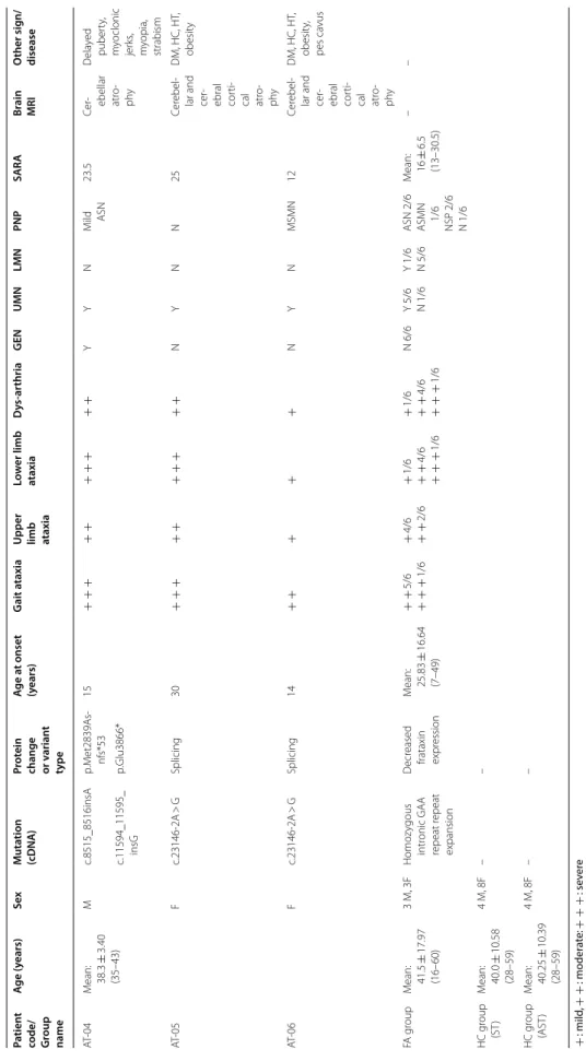
Table 1 Demographic, clinical and genetic data of SYNE1 and FA ataxia patients and healthy controls + : mild, + + : moderate: + + + : severe ASMN axonal sensorimotor polyneuropathy, ASN axonal sensory polyneuropathy, AST antisaccade task, DM diabetes mellitus, F female, GEN gaze-evoked nystagmus, HC hypercholesterolemia, HT hypertension, M male, MSMN mixed sensorimotor polyneuropathy, N not present, NSP not specified polyneuropathy, PNP polyneuropathy, SARA Scale for the Assessment and Rating of Ataxia, ST saccades task, UMN upper motor neuron involvement, Y present
Patient
code/ Group name
Age (years)SexMutation (cDNA)Protein change or v
ariant type
Age at onset (years)Gait ataxia
Upper limb ataxia
Lower limb ataxiaDys-arthriaGENUMNLMNPNPSARABrain MRIOther sign/ disease AT-04Mean: 38.3 ± 3.40 (35–43)
Mc.8515_8516insAp.Met2839As- nfs*5315 + + + + + + + + + +YYNMild ASN23.5Cer- ebellar atro- phy Delayed puberty, myoclonic jerks, myopia, strabism
c.11594_11595_ insGp.Glu3866* AT-05Fc.23146-2A > GSplicing30 + + + + + + + + + +NYNN25Cerebel- lar and cer- ebral corti- cal atro- phy
DM, HC, HT, obesity AT-06Fc.23146-2A > GSplicing14 + + + + +NYNMSMN12Cerebel- lar and cer- ebral corti- cal atro- phy
DM, HC, HT, obesity, pes cavus FA groupMean: 41.5 ± 17.97 (16–60)
3 M, 3FHomozygous intronic GAA repeat repeat expansion Decreased frataxin expression Mean: 25.83 ± 16.64 (7–49) + + 5/6 + + + 1/6 + 4/6 + + 2/6 + 1/6 + + 4/6 + + + 1/6 + 1/6 + + 4/6 + + + 1/6 N 6/6Y 5/6 N 1/6Y 1/6 N 5/6ASN 2/6 ASMN 1/6 NSP 2/6 N 1/6 Mean: 16 ± 6.5 (13–30.5)
–– HC group (ST)Mean: 40.0 ± 10.58 (28–59)
4 M, 8F–– HC group (AST)Mean: 40.25 ± 10.39 (28–59)
4 M, 8F––
The age at onset of AT-05 patient was 30 years and her first complaint was gait ataxia, whereas the first symptom of her sister (patient AT-06) appeared at 14 years of age
and was gait abnormality as well. The neurological exam- ination of both patients revealed cerebellar dysarthria and brisk tendon reflexes with bilateral Babinski signs.
Fig. 1 Brain MRI scans of SYNE1 ataxia patients demonstrated moderate cerebellar atrophy in all subjects and mild cortical atrophy in AT-05 and AT-06 patients. a, b: AT-04 patient; c, d: AT-05 patient; e, f: AT-06 patient; a, c, e: sagittal T1 weighted scans; b, d, f: coronal T1-weighted scans
Fig. 2 Genetic abnormalities and consequent alterations of protein of SYNE1 ataxia patients and their parents. a: SYNE1 gene mutations in AT-04 patient and the parental origin of these variations. b: SYNE1 gene abnormalities in AT-05 and AT-06 subjects and the parental segregation of these mutations. The upper parts of the bars denote the DNA sequence, while the lower parts show the encoded amino acids of the protein. Yellow bars indicate the pathogenic alleles, white bands mark the normal alleles. Red highlights the nucleotide change of the SYNE1 gene. In part (b), the c.23146-2A > G mutation is located in the intron–exon boundary resulting in an abnormal splicing variant
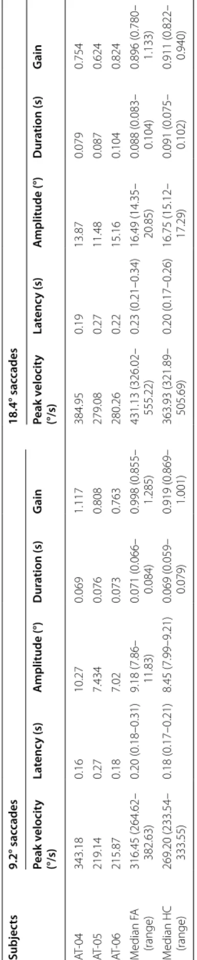
Table 2 Saccade examination in SYNE1 (AT-04–06) and Friedreich ataxia patients and in healthy controls FA Friedreich ataxia, HC healthy control
Subjects9.2° saccades18.4° saccades Peak velocity (°/s)Latency (s)Amplitude (°)Duration (s)GainPeak velocity (°/s)Latency (s)Amplitude (°)Duration (s)Gain AT-04343.180.1610.270.0691.117384.950.1913.870.0790.754 AT-05219.140.277.4340.0760.808279.080.2711.480.0870.624 AT-06215.870.187.020.0730.763280.260.2215.160.1040.824 Median FA (range)316.45 (264.62– 382.63)0.20 (0.18–0.31)9.18 (7.86– 11.83)0.071 (0.066– 0.084)0.998 (0.855– 1.285)431.13 (326.02– 555.22)0.23 (0.21–0.34)16.49 (14.35– 20.85)0.088 (0.083– 0.104)0.896 (0.780– 1.133) Median HC (range)
269.20 (233.54– 333.55)0.18 (0.17–0.21)8.45 (7.99–9.21)0.069 (0.059– 0.079)0.919 (0.869– 1.001)363.93 (321.89– 505.69)0.20 (0.17–0.26)16.75 (15.12– 17.29)0.091 (0.075– 0.102)0.911 (0.822– 0.940)
Truncal ataxia was moderate in the younger patient (AT- 06) and was severe in the elder subject (AT-05). After eleven years of disease course patient AT-05 could only walk with aids. Mild upper limb and moderate lower extremity incoordination developed in the younger sis- ter, whereas her sibling had moderate superior and severe inferior limb ataxia. AT-05 patient has obesity, diabetes mellitus, hypertension and hypercholesterolemia, but ophthalmological and cardiological investigations were normal. AT-06 patient also has the same metabolic dis- orders, moreover, she has an excavated foot and elec- troneurography delineated multifocal sensorimotor mixed type polyneuropathy. The brain MRI showed mod- erate cerebellar and very mild cerebral cortical atrophy in both patients (Fig. 1c–f). Their non-consanguineous par- ents did not suffer from ataxia and the younger patient has two healthy children.
In AT-05 and AT-06 patients the same homozygous NM_182961.3:c.23146-2A > G alteration of the SYNE1 gene was detected. This intronic variant was not found in gnomAD. It causes a TAG–TGG codon change at the Intron 128 – Exon 128 boundary resulting in an abnor- mal splicing variant (Fig. 2b). The presence of these mutations was confirmed by targeted Sanger sequenc- ing. Segregation analysis identified this variant in the het- erozygous state in both parents of the patients.
Eye tracking Saccades
The pooled data of leftward and rightward saccades were analyzed (Table 2). There was not any relevant difference between the three groups of examined subjects in sac- cadic latencies and durations for either the shorter (9.2°) or the longer (18.4°) saccade paradigms. The peak veloci- ties of saccades of AT-05 and AT-06 patients were smaller than the HC subjects and FA patients. However, the peak velocities of the saccades of AT-04 patient were similar to the subjects of HC and FA groups. In the 9.2° saccade task, AT-04 patient demonstrated hypermetric saccadic eye movements, whereas the other two SYNE1 ataxia patients showed hypometric saccades. Nevertheless, in the 18.4° saccade task SYNE1 ataxia subjects performed smaller saccadic amplitudes and gain than the healthy controls with minimal overlap (Fig. 3a). The amplitudes and gain of saccades of FA patients were in a similar range to that of the HC group. Figure 4 displays the main sequence relationships using the linear model. The dura- tion vs. amplitude diagram (Fig. 4a) shows that saccades of SYNE1 ataxia patients are hypometric and their dura- tion is longer than in FA or HC groups. The peak velocity vs. amplitude graph (Fig. 4b) reinforces that the saccades of SYNE1 patients are hypometric and their peak velocity is smaller than in HC or FA groups.
Antisaccades
The pooled data of leftward and rightward antisaccades were evaluated as well (Table 3). There was no remark- able difference between the groups with regard to peak velocities, latencies and durations of antisaccades.
The incorrect ratios were higher in the SYNE1 and FA patients than in the HC group. However, there was a mildly overlapping range in the 9.2° antisaccades within the SYNE1 and HC subjects, whereas this was only minimally detected in the longer antisaccades (Fig. 3b).
Neuropsychological assessment
The neuropsychological assessment of FA and SYNE1 patients are summarized in Table 4. The cognitive per- formance of ataxia patients was compared with the data of age- and education-matched standards in the litera- ture [24–26]. Global cognition was only mildly reduced in two FA patients (AT-11 and AT-20), whereas the other subjects demonstrated normal ACE and MMSE scores. The LST results showed mild abnormalities in all SYNE1 patients and in one FA patient, whereas the BDST results were decreased more prominently in both patient groups. These alterations indicate the impairment of working memory and in the ability to maintain and manipulate information. Surprisingly, the fluency test scores were in the normal range, only AT-04 patient demonstrated a mild deficit in the verbal fluency test. In addition, the RCFT results were equal to the standard outcomes, only AT-05 patient showed a mild impairment.
Discussion
In this paper we describe the clinical phenotype and characteristics of saccades and antisaccades of the first genetically confirmed Hungarian SYNE1 patients caused by novel mutations. The cerebellar symptoms of these patients involved moderate to severe gait and lower limb ataxia and mild to moderate upper limb ataxia and dys- arthria. Extracerebellar involvement was present as well, as all subjects have pyramidal signs and two of the three patients have some types of polyneuropathy. Moreover, AT-04 patient had strabismus, tactile sensitive myoclonic jerks and delayed puberty. In summary, the clinical phe- notype of subjects is not purely cerebellar, in contrast to that described in the first French-Canadian population by Gros-Louis et al. [1], and similar to that of the later reported cases [2, 8]. This symptomatic variability sug- gests that SYNE1 gene plays a broader role in the nor- mal functioning of the nervous and musculoskeletal systems. Consequently, the mutations of this gene can cause symptoms and signs over a large spectrum, but
an obvious genotype–phenotype correlation cannot be established [2].
The eye tracking examination revealed hypometric sac- cades in the 18.4° paradigm in all SYNE1 patients and in two out of three in the 9.2° task. Saccadic dysmetria is a cerebellar symptom and it is a common eye movement abnormality in hereditary ataxias [27]. This is not a spe- cific symptom for any type of inheritable ataxia, but there may be a higher proportion of hypo- or hypermetric saccades, serving as a supporting feature of the disease.
The hypometria of SYNE1 patients at large amplitude stimulus is more pronounced than the well-known mild hypometria in healthy subjects observed at higher target eccentricities [22]. The hypometria of SYNE1 patients is presumably due to the involvement of the cerebel- lar oculomotor vermis and caudal fastigial nucleus [28].
In addition to accuracy, velocity is another impor- tant characteristic of saccades. Previous case reports described slowing of saccades in a portion of SYNE1 Fig. 3 The most characteristic alterations in saccadic and
non-saccadic paradigms in SYNE1 ataxia patients. a: saccadic amplitude of the 18.4 saccade paradigm in the different groups;
b: incorrect ratios of the 18.4 antisaccade task in the investigated subjects; the circles, triangles and squares denote the parameters of healthy controls (HC), Friedreich ataxia patients (FA) and SYNE1 patients, respectively, and the median values are demonstrated as well
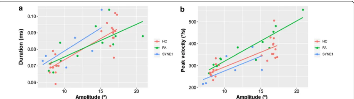
Fig. 4 The main sequence relationships of saccades using the linear model. a: saccadic duration versus amplitude; b saccadic peak velocity versus amplitude; the red, green and blue dots denote the parameters of healthy controls (HC), Friedreich ataxia patients (FA) and SYNE1 subjects, respectively
Table 3 Antisaccade examination in SYNE1 (AT-04–06) and Friedreich ataxia patients and in healthy controls
FA Friedreich ataxia, HC healthy control
Subjects 9.2° antisaccades 18.4° antisaccades
Peak velocity
(°/s) Latency (s) Duration (s) Incorrect
ratio Peak velocity
(°/s) Latency (s) Duration (s) Incorrect ratio
AT-04 261.71 0.28 0.053 0.40 290.23 0.29 0.052 0.27
AT-05 232.76 0.32 0.072 0.64 280.16 0.37 0.097 0.36
AT-06 212.44 0.19 0.077 1.00 221.08 0.41 0.069 0.35
Median FA
(range) 300.87 (237.22–
351.78)
0.28 (0.19–
0.41) 0.067 (0.059–
0.072) 0.62 (0.28–
0.96) 336.57
(253.63–
439.47)
0.32 (0.20–
0.45) 0.083 (0.062–
0.103) 0.33 (0.06–1.00) Median HC
(range) 243.06 (197.67–
313.18)
0.28 (0.23–
0.33) 0.053 (0.046–
0.075) 0.19 (0.08–
0.54) 283.15
(209.63–
382.10)
0.28 (0.24–
0.37) 0.062 (0.049–
0.088) 0.07 (0.00–0.29)
Table 4 Neuropsychological assessment of SYNE1 and Friedreich ataxia patients In ACE and MMSE the lower normal threshold values of the normal population are in the brackets In LST, BDST, verbal fluency, semantic fluency, RCFT copying and RCFT recall the age- and education-matched lower threshold values of the normal population are in the brackets ACE Addenbrooke’s Cognitive Examination, BDST Backward Digit Span Task, LST Listening Span Task, MMSE Mini-Mental State Examination, NA not available, RCFT Rey Complex Figure Test a Mild deficit, bmoderate deficit, csevere deficit (Age- and education-matched standards, and standard deviations of the literature are delineated [23–25]. Mild, moderate and severe deficits mean that the cognitive impairment of the subject is more pronounced than one, two and three standard deviations of the normal standards, respectively)
Patient codeAge (years)Education (years)ACE (93.7 ± 4.3)
MMSE (28.8
± 1.3)LSTBDSTVerbal fluencySemantic fluencyRCFT copyingRCFT recall SYNE1 patients AT-04351489292 (3.38 ± 0.79)a4 (5.88 ± 1.1)a10.5 (17.61 ± 5.42)a14 (17.25 ± 3.96)NANA AT-05431493292.33 (3.38 ± 0.79)a3 (5.88 ± 1.1)b14.5 (17.61 ± 5.42)17 (17.25 ± 3.96)35 (31.1 ± 3.6)15 (23.7 ± 5.2)a AT-06371289292 (3.38 ± 0.79)a2 (5.88 ± 1.1)c16 (17.61 ± 5.42)14 (17.25 ± 3.96)36 (31.1 ± 3.6)21.5 (23.7 ± 5.2) Friedreich ataxia patients AT-082312.593303 (3.45 ± 0.89)4 (5.88 ± 1.1)a14.5 (16.13 ± 5.65)11 (15.84 ± 4.51)36 (31.1 ± 3.6)24 (23.7 ± 5.2) AT-11571687a272.33 (3.11 ± 0.61)a3 (5.34 ± 0.96)b12 (11.02 ± 4.98)16 (13.77 ± 4.05)36 (29.2 ± 4.2)26.5 (15.5 ± 5.5) AT-12601795293 (3.11 ± 0.61)2 (5.34 ± 0.96)c15.5 (11.02 ± 4.98)20 (13.77 ± 4.05)34 (29.2 ± 4.2)27 (15.5 ± 5.5) AT-2016108826a2.66 (3.33 ± 0.59)5 (5.88 ± 0.96)13.5 (13.83 ± 4.31)14 (13.44 ± 3.52)36 (31.1 ± 3.6)26 (23.7 ± 5.2) AT-21591494303 (3.11 ± 0.61)5 (5.34 ± 0.96)15 (11.02 ± 4.98)14 (13.77 ± 4.05)32 (29.2 ± 4.2)17.5 (15.5 ± 5.5) AT-22341596303.3 (3.38 ± 0.79)4 (5.88 ± 1.1)a16.5 (16.13 ± 5.65)21 (15.84 ± 4.51)34 (31.1 ± 3.6)21 (23.7 ± 5.2)
patients, however these observations were based exclu- sively on physical examinations [1–4, 18, 19]. Our find- ings, obtained by fine eye tracking assessment, confirmed the clinical observations of some earlier publications, i.e., a high frequency of slow saccades can be detected in SYNE1 ataxia. This lower saccadic velocity is likely due to brainstem involvement, in particular, the functional loss of pontine saccadic burst generator neurons and omni- pause neurons can explain this observation [23]. Slowing of saccades is a characteristic eye movement abnormal- ity in SCA2 disease. Federighi et al. examined the sac- cadic parameters of seven SCA2 patients at similar target eccentricities (10 and 18°) to those we used in this study and they found more severely reduced peak velocities and delayed saccadic latencies compared to controls than we detected in two of three SYNE1 patients [23]. Presum- ably brainstem impairment is more pronounced in SCA2 than in SYNE1. In addition, saccadic hypometria was not found in SCA2 patients, whereas we observed lower sac- cadic amplitude in SYNE1 patients compared to healthy subjects at the larger stimulus paradigm.
The antisaccade assessment showed higher rates of incorrectly accomplished antisaccades in both FA and SYNE1 patients compared to healthy subjects, whereas the other parameters were in similar ranges. The error rates were higher on the short stimulus amplitude task than on the long amplitude trial. Basically, target eccen- tricities affect gain, latency and peak velocity, whereas its influence on the incorrect ratios is not clear at these amplitudes [29–31]. The higher error rate raises the sus- picion of cognitive impairment, because a strong cor- relation was demonstrated between antisaccades and working memory [32]. The neuropsychological assess- ment revealed that global cognitive performance was normal in SYNE1 patients, whereas executive functions were impaired, especially working memory. The perfor- mance of the examined SYNE1 subjects in BDST and LST paradigms inversely correlated with errors in the antisaccade tasks, i.e., the most severely affected patient in working memory tests (AT-06) had the highest error rate in the antisaccade paradigm (Fig. 5). A similar rela- tionship was not detected in the FA group. Previously published studies indicated higher antisaccadic error rates in other hereditary and idiopathic cerebellar disor- ders, including ataxia with oculomotor apraxia type 1, 2, ataxia telangiectasia, SCA1, 2, 3 and late onset cerebellar ataxia (LOCA) [28, 33–35]. Additionally, Pretegiani et al.
revealed that SCA2 and LOCA patients showed equally poor antisaccade performance irrespective of cortical involvement [33]. Additionally, a thorough investigation of a SCA2 patient cohort confirmed that impaired anti- saccade efficacy was associated with executive test defi- cits, including Stroop interference task and verbal fluency
test [34]. Our findings draw attention to the major role of working memory and inhibitory control in the perfor- mance of antisaccades, and confirm that executive dys- function is a prevalent neuropsychological abnormality in hereditary ataxias as a part of the cerebellar cognitive and affective syndrome [36].
Conclusions
In conclusion, this paper demonstrates the detailed neu- rological assessment of the first Hungarian SYNE1 ataxia patients with novel pathogenic mutations. The eye track- ing investigation detected some interesting alterations regarding both saccades and antisaccades in these sub- jects, including saccadic hypometria and increased error rates for antisaccades. The main weakness of this study is the low case number. Nevertheless, these pilot findings point out the importance of device-aided examination of eye movements in ARCAs. Hopefully in the near future, these parameters can be investigated in a larger number of SYNE1 patients in order to be able to draw statistical conclusions as well.
Abbreviations
ACE: Addenbrooke’s Cognitive Examination (ACE); ARCA : Autosomal recessive cerebellar ataxias; ARCA1: Autosomal recessive cerebellar ataxia type 1; BDST:
Backward Digit Span Task; FA: Friedreich ataxia; HC: Healthy controls; LOCA:
Late onset cerebellar ataxia; LST: Listening Span Task; MMSE: Mini-Mental State Examination; NGS: New generation sequencing; RCFT: Rey Complex Figure Test; SARA : Scale for the Assessment and Rating of Ataxia; SCA: Spinocerebel- lar ataxia; SCAR8: Spinocerebellar ataxia, autosomal recessive 8; WES: Whole exome sequencing.
Fig. 5 The delineation of the possible relationship between working memory test results and the incorrect ratio of antisaccades in ataxia patients. The horizontal axis denotes the mean value of the incorrect ratios of 9.2 and 18.4 antisaccade tasks, whereas the vertical axis indicates the sum of Listening Span Task (LST) and Backward Digit Span Task (BDST) scores; the triangles and squares indicate the data of Friedreich ataxia patients (FA) and SYNE1 patients, respectively, and the regression line is drawn by continuous and dashed lines
Acknowledgements
The linguistic corrections were made by a native English reader, Jennifer Tusz from Canada.
Authors’ contributions
LSZ examined the patients and wrote the manuscript. GSZ and BK performed the eye-tracking examination on the subjects. BK set up the eye-tracking aided device and established the methods of the study. VLN and NSZ performed the neuropsychological examination of the patients. ZM, TK, MR and RP carried out the new generation sequencing of the SYNE1 patients. GV analysed the data and made the diagrams. PK and AS had important recom- mendations to the manuscript. DZ was the major contributor in writing the manuscript. All authors read and approved the final manuscript.
Funding
This work was supported by KTIA Grant No. 13 NAP-A-II/17. Open access fund- ing provided by the University of Szeged (SZTE, Grant Number: 4906).
Availability of data and materials
The datasets used and/or analysed in the current study are available in this paper.
Ethics approval and consent to participate
Written informed consent was obtained from the patients for the publication of this study (Regional Human Biomedical Research Ethics Committee of the University of Szeged, registration number 44/2016). All procedures performed in studies involving human participants were in accordance with the ethical standards of the institutional and/or national research committee and with the 1964 Helsinki Declaration and its later amendments or comparable ethical standards.
Consent for publication Not applicable.
Competing interests
The authors declare that they have no competing interests.
Author details
1 Department of Neurology, University of Szeged, Semmelweis u. 6, 6725 Sze- ged, Hungary. 2 Department of Psychiatry, University of Szeged, Szeged, Hun- gary. 3 Genetic Diagnostic Laboratory, Department of Pediatrics and Pediatric Health Center, University of Szeged, Szeged, Hungary. 4 Department of Medi- cal Genetics, Medical University of Warsaw, Warsaw, Poland. 5 MTA-SZTE Neuro- science Research Group, Szeged, Hungary.
Received: 17 September 2020 Accepted: 13 January 2021
References
1. Gros-Louis F, Dupré N, Dion P, Fox MA, Laurent S, Verreault S, et al. Muta- tions in SYNE1 lead to a newly discovered form of autosomal recessive cerebellar ataxia. Nat Genet. 2007;39:80–5.
2. Synofzik M, Smets K, Mallaret M, Di Bella D, Gallenmüller C, Baets J, et al.
SYNE1 ataxia is common recessive ataxia with major non-cerebellar features: a large multi-centre study. Brain. 2016;139:1378–93.
3. Dupré N, Gros-Louis F, Chrestian N, Verreault S, Brunet D, de Verteuil D, et al. Clinical and genetic study of autosomal recessive cerebellar ataxia type 1. Ann Neurol. 2007;62:93–8.
4. Noreau A, Bourassa CV, Szuto A, Levert A, Dobrzeniecka S, Gauthier J, et al. SYNE1 mutations in autosomal recessive cerebellar ataxia. JAMA Neurol. 2013;70:1296–331.
5. Izumi Y, Miyamoto R, Morino H, Yoshizawa A, Nishinaka K, Udaka F, et al.
Cerebellar ataxia with SYNE1 mutation accompanying motor neuron disease. Neurology. 2013;80:600–1.
6. Fogel BL, Lee H, Deignan JL, Strom SP, Kantarci S, Wang X, et al. Exome sequencing in the clinical diagnosis of sporadic or familial cerebellar ataxia. JAMA Neurol. 2014;71:1237–46.
7. Hamza W, Ali Pacha L, Hamadouche T, Muller J, Drouot N, Ferrat F, et al.
Molecular and clinical study of a cohort of 110 Algerian patients with autosomal recessive ataxia. BMC Med Genet. 2015;16:36.
8. Mademan I, Harmuth F, Giordano I, Timmann D, Magri S, Deconinck T, et al. Multisystemic SYNE1 ataxia: confirming the high frequency and extending the mutational and phenotypic spectrum. Brain. 2016;139:e46.
9. Attali R, Warwar N, Israel A, Gurt I, McNally E, Puckelwartz M, et al. Muta- tion of SYNE-1, encoding an essential component of the nuclear lamina, is responsible for autosomal recessive arthrogryposis. Hum Mol Genet.
2009;18:3462–9.
10. Zhang Q, Bethmann C, Worth NF, Davies JD, Wasner C, Feuer A, et al.
Nesprin-1 and -2 are involved in the pathogenesis of Emery Dreifuss muscular dystrophy and are critical for nuclear envelope integrity. Hum Mol Genet. 2007;16:2816–33.
11. Puckelwartz MJ, Kessler EJ, Kim G, Dewitt MM, Zhang Y, Earley JU, et al.
Nesprin-1 mutations in human and murine cardiomyopathy. J Mol Cell Cardiol. 2010;48:600–8.
12. Schuurs-Hoeijmakers JH, Vulto-van Silfhout AT, Vissers LE, van de Vonder- voort II, van Bon BW, de Ligt J, et al. Identification of pathogenic gene variants in small families with intellectually disabled siblings by exome sequencing. J Med Genet. 2013;50:802–11.
13. Dong H, Luo Y, Fan S, Yin B, Weng C, Peng B. Screening gene mutations in Chinese patients with benign essential blepharospasm. Front Neurol.
2020;10:1387.
14. Yu TW, Chahrour MH, Coulter ME, Jiralerspong S, Okamura-Ikeda K, Ata- man B, et al. Using whole-exome sequencing to identify inherited causes of autism. Neuron. 2013;77:259–73.
15. Griswold AJ, Dueker ND, Van Booven D, Rantus JA, Jaworski JM, Slifer SH, et al. Targeted massively parallel sequencing of autism spectrum disorder-associated genes in a case control cohort reveals rare loss-of- function risk variants. Mol Autism. 2015;6:43.
16. Guipponi M, Santoni FA, Setola V, Gehrig C, Rotharmel M, Cuenca M, et al.
Exome sequencing in 53 sporadic cases of schizophrenia identifies 18 putative candidate genes. PLoS ONE. 2014;9:e112745.
17. Swan L, Cardinal J, Coman D. SYNE1-related autosomal recessive cerebel- lar ataxia, congenital cerebellar hypoplasia, and cognitive impairment.
Clin Pract. 2018;8:1071.
18. Gama MT, Houle G, Noreau A, Dionne-Laporte A, Dion PA, Rouleau GA, et al. SYNE1 mutation cause autosomal-recessive ataxia with retained reflexes in Brazilian patients. Mov Disord. 2016;31:1754–6.
19. Kim JS, Kim AR, Youn J, Lee C, Kim NS, Park WY, et al. Identifying SYNE1 ataxia and extending the mutational spectrum in Korea. Parkinsonism Relat Disord. 2019;58:74–8.
20. De Silva R, Greenfield J, Cook A, Bonney H, Vallortigara J, Hunt B, et al.
Guidelines on the diagnosis of the progressive ataxias. Orphanet J Rare Dis. 2019;14:51.
21. Kincses B, Herak BJ, Szabo N, Bozsik B, Farago P, Kiraly A, et al. Gray matter atrophy to explain subclinical oculomotor deficit in multiple sclerosis.
Front Neurol. 2019;10:589.
22. Evdokimidis I, Tsekou H, Smyrnis N. The mirror antisaccade task: direction- amplitude interaction and spatial accuracy characteristics. Exp Brain Res.
2006;174:304–11.
23. Federighi P, Cevenini G, Dotti MT, Rosini F, Pretegiani E, Federico A, et al.
Differences in saccade dynamics between spinocerebellar ataxia 2 and late-onset cerebellar ataxias. Brain. 2011;134:879–91.
24. Mioshi E, Dawson K, Mitchell J, Arnold R, Hodges JR. The Addenbrooke’s cognitive examination revised (ACE-R): a brief cognitive test battery for dementia screening. Int J Geriatr Psychiatry. 2006;21:1078–85.
25. Marilyn H, Guy P. Sources of age differences on the Rey-Osterrieth com- plex figure test. Clin Neuropsychol. 1998;12:513–24.
26. Bopp KL, Verhaeghen P. Aging and verbal memory span: a meta-analysis.
J Gerontol B Psychol Sci Soc Sci. 2005;60:223–33.
27. Moscovich M, Okun MS, Favilla C, Figueroa KP, Pulst SM, Perlman S, et al.
Clinical evaluation of eye movements in spinocerebellar ataxias: a pro- spective multicenter study. J Neuroophthalmol. 2015;35:16–21.
28. Mariani LL, Rivaud-Péchoux S, Charles P, Ewenczyk C, Meneret A, Monga BB, et al. Comparing ataxias with oculomotor apraxia: a multimodal study of AOA1, AOA2 and AT focusing on video-oculography and alpha-feto- protein. Sci Rep. 2017;7:15284.
29. Fischer B, Weber H. Effects of stimulus conditions on performance of antisaccades in man. Exp Brain Res. 1997;11:191–200.
•fast, convenient online submission
•
thorough peer review by experienced researchers in your field
• rapid publication on acceptance
• support for research data, including large and complex data types
•
gold Open Access which fosters wider collaboration and increased citations maximum visibility for your research: over 100M website views per year
•
At BMC, research is always in progress.
Learn more biomedcentral.com/submissions Ready to submit your research
Ready to submit your research ? Choose BMC and benefit from: ? Choose BMC and benefit from:
30. Dafoe JM, Armstrong IT, Munoz DP. The influence of stimulus direction and eccentricity on pro- and antisaccades in humans. Exp Brain Res.
2007;179:563–70.
31. Smyrnis N, Evdokimidis I, Stefanis NC, Constantinidis TS, Avramopoulos D, Theleritis C, et al. The antisaccade task in a sample of 2006 young males.
II. Effects of task parameters. Exp Brain Res. 2002;147:53–63.
32. Thomas EHX, Rossell SL, Myles JB, Tan EJ, Neill E, Carruthers SP, et al. Work- ing memory and attention influence antisaccade error rate in schizophre- nia. J Int Neuropsychol Soc. 2018;25:1–10.
33. Pretegiani E, Piu P, Rosini F, Federighi P, Serchi V, Tumminelli G, et al. Anti- saccades in cerebellar ataxias reveal a contribution of the cerebellum in executive functions. Front Neurol. 2018;9:274.
34. Rodriguez-Labrada R, Velazquez-Perez L, Aguilera-Rodriguez R, Seifried- Oberschmidt C, Pena-Acosta A, Canales-Ochoa N, et al. Executive deficit
in spinocerebellar ataxia type 2 is related to expanded CAG repeats:
evidence from antisaccadic eye movements. Brain Cog. 2014;91:28–34.
35. Rivaud-Pechoux S, Durr A, Gaymard B, Cancel G, Ploner CJ, Agid Y, et al.
Eye movement abnormalities correlate with genotype in autosomal dominant cerebellar ataxia type I. Ann Neurol. 1998;43:297–302.
36. Schmahmann JD, Sherman JC. The cerebellar cognitive affective syn- drome. Brain. 1998;121:561–79.
Publisher’s Note
Springer Nature remains neutral with regard to jurisdictional claims in pub- lished maps and institutional affiliations.