Full Terms & Conditions of access and use can be found at
http://www.tandfonline.com/action/journalInformation?journalCode=kaup20
ISSN: 1554-8627 (Print) 1554-8635 (Online) Journal homepage: http://www.tandfonline.com/loi/kaup20
Developmentally regulated autophagy is required for eye formation in Drosophila
Viktor Billes, Tibor Kovács, Anna Manzéger, Péter Lőrincz, Sára Szincsák, Ágnes Regős, Péter István Kulcsár, Tamás Korcsmáros, Tamás Lukácsovich, Gyula Hoffmann, Miklós Erdélyi, József Mihály, Krisztina Takács-Vellai, Miklós Sass & Tibor Vellai
To cite this article: Viktor Billes, Tibor Kovács, Anna Manzéger, Péter Lőrincz, Sára Szincsák, Ágnes Regős, Péter István Kulcsár, Tamás Korcsmáros, Tamás Lukácsovich, Gyula Hoffmann, Miklós Erdélyi, József Mihály, Krisztina Takács-Vellai, Miklós Sass & Tibor Vellai (2018):
Developmentally regulated autophagy is required for eye formation in Drosophila, Autophagy, DOI:
10.1080/15548627.2018.1454569
To link to this article: https://doi.org/10.1080/15548627.2018.1454569
View supplementary material
Accepted author version posted online: 25 Jun 2018.
Submit your article to this journal
Article views: 51
View Crossmark data
Accepted Manuscript
1
Publisher: Taylor & Francis & Taylor and Francis Group, LLC Journal: Autophagy
DOI: 10.1080/15548627.2018.1454569
Developmentally regulated autophagy is required for eye formation in Drosophila
Viktor Billes1,*, Tibor Kovács1,*, Anna Manzéger1, Péter Lőrincz2, Sára Szincsák1, Ágnes Regős1, Péter István Kulcsár3, Tamás Korcsmáros1,4,5, Tamás Lukácsovich6, Gyula Hoffmann7, Miklós Erdélyi8, József Mihály8, Krisztina Takács-Vellai9, Miklós
Sass2,#, Tibor Vellai1,#
1Department of Genetics and 2Department of Anatomy, Cell and Developmental Biology, Eötvös Loránd University, Budapest, Hungary; 3Institute of Enzymology, Research Centre for Natural Sciences of the Hungarian Academy of Sciences, Budapest, Hungary; 4Earlham Institute, Norwich Research Park, Norwich, UK; 5Gut Health and Food Safety Programme, Institute of Food Research, Norwich Research Park, Norwich, UK; 6Department of Developmental and Cell Biology; University of California, Irvine, CA, USA; 7Department of Anatomy and Developmental Biology, University of Pécs, Pécs, Hungary; 8Institute of Genetics, Biological Research Centre, Szeged, Hungary; 9Department of Biological Anthropology, Eötvös Loránd University, Budapest, Hungary
*These authors contributed equally to the work.
#Corresponding authors: T.V. (vellai@falco.elte.hu) or M.S. (msass@elte.hu)
Accepted Manuscript
2
Address: Department of Genetics, Eötvös Loránd University, Budapest, Pázmány Péter stny.
1/C, Hungary, H-1117; Tel.: +36-1-372-2500 Ext: 8684; Fax: +36-1-372-2641; E-mail:
vellai@falco.elte.hu
Competing financial interest: The authors declare no competing financial interest.
Author contribution: V.B., T.Kov., A.M., P.L., S.Sz., Á.R., P.I.K., T.L., G.H., M.E., J.M., K.T-V., T.V., and M.S. performed experiments; V.B., T.Kov., A.M., M.E., T.Kor., J.M., K.T- V., T.V., and M.S. designed experiments and analyzed data; V.B., T.Kov., A.M., J.M., K.T- V., T.V. and M.S. wrote the manuscript.
Key words: autophagy, cell death, differentiation, Drosophila, eye development, genetic compensation, HOX, labial, pattern formation, transcriptional control
Abbreviations: αTub84B, α-Tubulin at 84B; Act5C, Actin5C; AO, acridine orange; Atg, autophagy-related; Ato, Atonal; CASP3, caspase 3; Dcr-2; Dicer-2; Dfd, Deformed; DZ, differentiation zone; eGFP, enhanced green fluorescent protein; EM, electron microscopy;
exd, extradenticle; ey, eyeless; FLP, flippase recombinase; FRT, FLP recognition target;
Gal4, gene encoding the yeast transcription activator protein GAL4; GFP, green fluorescent protein; GMR, Glass multimer reporter; Hox, homeobox; hth, homothorax; lab, labial; L3F, L3 feeding larval stage; L3W, L3 wandering larval stage; lf, loss-of-function; MAP1LC3,
Accepted Manuscript
3
microtubule-associated protein 1 light chain 3; MF, morphogenetic furrow; PE, phosphatidylethanolamine; PBS, phosphate-buffered saline; PI3K/PtdIns3K, class III phosphatidylinositol 3-kinase; PZ, proliferation zone; Ref(2)P, refractory to sigma P, RFP, red fluorescent protein; RNAi, RNA interference; RpL32, Ribosomal protein L32; RT-PCR, reverse transcription-coupled polymerase chain reaction; S.D., standard deviation; SQSTM1, Sequestosome-1, Tor, Target of rapamycin; TUNEL, terminal deoxynucleotidyl transferase mediated dUTP nick end labeling assay; UAS, upstream activation sequence; qPCR, quantitative real-time polymerase chain reaction; w, white
The number of words in the abstract: 226
The number of Figures: 10 (multipanel, colored)
Supplementary Materials contain Genotype list, Suppl. Tables (S1, S2), and Suppl.
Figures and Figure Legends (S1 to S31).
Accepted Manuscript
4 Abstract
The compound eye of the fruit fly Drosophila melanogaster is one of the most intensively studied and best understood model organs in the field of developmental genetics. Herein we demonstrate that autophagy, an evolutionarily conserved selfdegradation process of eukaryotic cells, is essential for eye development in this organism. Autophagic structures accumulate in a specific pattern in the developing eye disc, predominantly in the morphogenetic furrow (MF) and differentiation zone. Silencing of several autophagy genes (Atg) in the eye primordium severely affects the morphology of the adult eye through triggering ectopic cell death. In Atg mutant genetic backgrounds however genetic compensatory mechanisms largely rescue autophagic activity in, and thereby normal morphogenesis of, this organ. We also show that in the eye disc the expression of a key autophagy gene, Atg8a, is controlled in a complex manner by the anterior Hox paralog lab (labial), a master regulator of early development. Atg8a transcription is repressed in front of, while activated along, the MF by lab. The amount of autophagic structures then remains elevated behind the moving MF. These results indicate that eye development in Drosophila depends on the cell death-suppressing and differentiating effects of the autophagic process. This novel, developmentally regulated function of autophagy in the morphogenesis of the compound eye may shed light on a more fundamental role for cellular self-digestion in differentiation and organ formation than previously thought.
Accepted Manuscript
5 Introduction
Autophagy (cellular “self-eating”) is a lysosome-mediated self-degradation process of eukaryotic cells. As a main route of eliminating superfluous and damaged cytoplasmic constituents and ensuring macromolecule turnover, autophagy is required for maintaining cellular homeostasis. It also provides energy for the survival of cells under starvation.
Although autophagy primarily functions as a prosurvival mechanism in terminally differentiated cells, under certain physiological and pathological settings it can also promote cell death [1-3]. In mammals, defects in autophagy can lead to accelerated aging and the development of various age-dependent pathologies including neurodegenerative diseases, cancer, diabetes, tissue atrophy and fibrosis, immune deficiency, compromised lipid metabolism, and infection by intracellular microbes [4-10].
During autophagy, parts of the cytoplasm are delivered into the lysosomal compartment for degradation by acidic hydrolases. Depending on the mechanism of delivery, 3 major types of autophagy can be distinguished: macroautophagy, microautophagy and chaperone-mediated autophagy. Macroautophagy (hereafter referred to as autophagy) involves the formation of a double membrane-bound compartment called the phagophore that sequesters the cytoplasmic material destined for degradation. The phagophore matures into an autophagosome, which then fuses with a lysosome, thereby generating a structure called autolysosome where degradation takes place [11-14].
The core mechanism of autophagy involves more than 30 autophagy-related (Atg) proteins, which are evolutionarily conserved from yeast to mammals [15]. Atg proteins are organized into functionally distinct complexes: i) the Atg1 kinase complex for inducing phagophore formation; ii) a class III phosphatidylinositol 3-kinase (PtdIns3K/Vps34) complex for vesicle nucleation; iii) a ubiquitin-like conjugation system for vesicle expansion; and iv) a
Accepted Manuscript
6
recycling complex for recovering utility materials. The ubiquitin-like conjugation system mediates the transient conjugation of Atg8 (whose mammalian orthologs include the MAP1LC3/microtubule-associated protein 1 light chain 3 and the GABARAP/GABA typeA receptor-associated protein families) to a phagophore membrane component, phosphatidylethanolamine (PE).
To date, 2 major developmental functions of autophagy have been uncovered [16,17].
First, it can lead to cell death via, or independently of, apoptosis, thereby removing, for example, larval tissues during metamorphosis in Drosophila [18]. Second, autophagy can selectively degrade specific proteins and organelles to mediate cellular differentiation [17].
However, exploring the function of autophagy in particular developmental events is still in the initial phase. For example, the process plays an important role in spore and fruiting body formation in fungi, and in the life cycle transition of pathogenic protozoans [19-22]. In the nematode Caenorhabditis elegans, autophagic degradation is required for the elimination of paternally distributed mitochondria from [23], and soma-germline separation in, early-stage embryos [24], elongation of the mid-stage embryo [25,26], as well as dauer larva formation [27]. In Drosophila, the process is critical for normal development by degrading larval tissues such as the fat body, salivary gland and midgut [18,28-30], and the removal of paternally delivered mitochondria from the zygote [31]. In chicken, autophagy is necessary for ear development [32]. In mammals, the elimination of maternally distributed gene products from early-stage embryos [33-35] and the embryo-to-neonate transition [36] are mediated by the autophagic process. It is also important in cellular differentiation, such as adipocyte, erythrocyte and lymphocyte maturation [37-39].
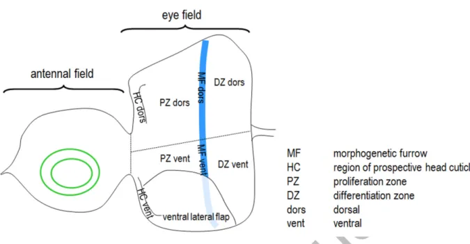
The compound eye of Drosophila, together with antenna, ocelli, head cuticle and palpus, develops from a larval primordium called eye-antennal imaginal disc (Fig. 1) [40,41].
This organ is an epithelial bilayer; one layer is the disc proper, which is built up from
Accepted Manuscript
7
columnar cells and gives rise to the retina, and the other layer called peripodial membrane that is involved in modulating columnar cell fates through emitting signaling cues [42]. Cells of the disc proper divide, grow, and then undergo differentiation into photoreceptors and accessory cells [43]. The border between the proliferating and differentiating cells is marked by the morphogenetic furrow (MF), which migrates from the posterior to anterior direction within the disc [44].
Tor (Target of rapamycin) kinase functions as a main upstream negative regulator of autophagy. Hyperactivation of Tor in the eye primordium leads to a massive reduction in the size of the adult organ and interferes with ommatidial patterning (ommatidia become fused or pitted) [45]. This intervention also delays the progression of MF, and causes disorganization or massive loss of photoreceptor cells [45,46]. Tor inactivation similarly compromises eye development by decreasing the rate of proliferation [47]. These data raise the possibility that autophagy is implicated in normal growth and morphogenesis of this organ. Indeed, silencing of Atg7 behind the MF by a GMR-Gal4 driver was reported to result in a rough eye phenotype with fused and enlarged ommatidia [48]. Conversely, Atg7 loss-of-function (lf) mutant animals are characterized by normal eye morphology [49], and, also using GMR-Gal4, knockdown of the Atg1, Atg4a, Atg5, Atg8a, Atg9, Atg12 or Atg18a genes has no effect on ommatidial structure [48,50]. Furthermore, depletion of Atg1, Atg7, Atg8a and Atg12 proteins, performed at 25°C and without coexpressing Dcr-2 (Dicer-2) that would make gene silencing more efficient, also does not interfere with eye development [51]. Eye morphology likewise remains unaffected by overexpressing a dominant-negative mutant allele of Atg1 [52]. Due to these contradictory data, the role of autophagy in Drosophila eye development remains to be elucidated.
In this study we examined the eye disc-specific accumulation of Atg5 and Atg8a proteins, as well as autophagic structures, and found that while the proteins are detectable
Accepted Manuscript
8
nearly ubiquitously in each part of the organ, but most abundantly in the area of the anteriorly located prospective head cuticle, autophagic compartments display a specific distribution pattern, predominantly accumulating within and behind the MF (the latter corresponds to the differentiation zone; DZ). We further demonstrated that RNA interference (RNAi)-mediated depletion of Atg proteins in the developing eye disc by drivers being active in the MF can severely compromise eye formation. In the affected animals, eye development was completely or partially inhibited as a consequence of ectopic cell death. However, the effect of lf mutations in Atg genes on eye development was largely rescued by genetic compensatory mechanisms involving the action of alternative transcripts, paralogs or maternally deposited factors. We also found that the Hox gene lab (labial) is expressed in front of and along the MF, and that Atg8a expression is strongly influenced in these regions by lab deficiency. These data reveal a novel, developmentally regulated role for autophagy; its cell death-suppressing function is essential for columnar cells in the Drosophila eye primordium to survive, thereby acting as a prerequisite for eye morphogenesis. Since this live-or-die cell fate decision is likely to occur in several cell types during development, autophagy may play a more fundamental role in tissue formation than previously thought.
Results
Autophagic structures accumulate in a specific pattern in the eye primordium
Under normal conditions, autophagy operates at basal levels in terminally differentiated cells to maintain normal macromolecule turnover. During differentiation however when cellular constituents are largely reorganized, the process may exhibit an increased activity in the affected cells. To investigate the potential role of autophagy in Drosophila eye development, we first examined the accumulation pattern of 2 key autophagy proteins, Atg5 and Atg8a, as
Accepted Manuscript
9
well as Atg5- and Atg8a-positive autophagic structures in the eye primordium of wandering L3 stage (L3W) larvae. At this stage the eye disc is divided into 2 major regions by the MF, the anteriorly located proliferation zone (PZ) and the posteriorly located DZ (Fig. 1) [40]. We used an Atg5-specific antibody (Fig. S1) to label Atg5 accumulation in this organ. Atg5 is known to localize to the growing phagophore and remain there until recycling eventually from the autophagosome [53]. Using conventional fluorescent microscopy, the antibody staining revealed abundant Atg5 accumulation in each part of the eye disc, but most obviously in the regions of the prospective head cuticle (indicated by yellow arrows in Figs. 2A and S2).
Semiconfocal and confocal microscopy resolutions however uncovered a relatively large amount of Atg5-positive foci labelling early autophagosomal structures in the MF and DZ, as compared with other areas of the organ (Figs. 2B to B’’’, C to C’’’’ and S7A). Consistent with these data, anti-Atg8a antibody staining performed with confocal microscopy also revealed basal levels of autophagic activity in the antennal field and PZ, but much higher levels in the MF and DZ (Figs. 2D to D’’’ and S3A to A’’’, S4, S7B). It is worth to note that anti-Atg8a antibody staining was also highly specific as the expression of Atg8a-specific double-stranded RNA (dsRNA) almost completely abolished protein accumulation in the eye disc (Fig. S4), and that Atg8a protein, similar to Atg5, was distributed nearly ubiquitously in the eye primordium, most evidently in the prospective head cuticle and MF (see later in this study). Thus, the intensity of Atg5 and Atg8a accumulation did not coincide with the distribution of autophagic structures; while the proteins accumulated nearly ubiquitously in the entire antennal-eye disc, the presence of Atg5- and Atg8a-positive foci (autophagic structures) was mainly concentrated to the regions of MF and DZ. A similar punctuated pattern was detected in these regions when the expression of an UAS-mCherry-Atg8a reporter, which marks phagophores, autophagosomes and autolysosomes, was driven by ey-Gal4(II) in the entire eye disc (Figs. 2E to E’’’ and S3B to B’’’, S5B, S7C). Staining by LysoTracker
Accepted Manuscript
10
Red, which marks acidic compartments including autolysosomes, lysosomes and multivesicular bodies, also revealed a punctuated pattern predominantly behind the MF (Figs.
S6 and S7D). Together, these results point to an unequal distribution for autophagic activity in different parts of the developing eye tissue; autophagic structures predominantly accumulate in the MF and DZ (Figs. 2, S3, and S6, S7). The other parts of the eye field, together with the antennal field, exhibit only basal levels of autophagy. These data suggest that the autophagic process is involved in the differentiation and/or survival of retinal precursor cells.
Downregulation of Atg genes in the eye disc impairs the development of the organ
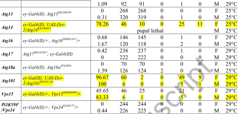
Next, we monitored whether silencing of Atg genes in the eye primordium affects the development of this organ. In previous studies, GMR-Gal4 was used to control the expression of UAS-Atg RNAi constructs in the eye disc [48,50]. However, the activity of this driver was only detectable behind the MF (i.e. within the DZ; Figs. 1A and S5A), and even its own expression disturbs eye development [54]. In addition, the expression of GMR-Gal4 is not restricted exclusively to the eye field [55]. Hence, we used 2 ey-Gal4 drivers, ey-Gal4(II) and ey-Gal4(III), to target gene silencing to a broader area of the eye primordium, including regions in front of, along, and behind the MF (Figs. 1A and S5B, C). Importantly, these drivers per se did not affect eye formation (Table S1 and Fig. 3A, D, F). ey-Gal4(II)- or ey- Gal4(III)-driven silencing of Atg genes led to aberrant eye disc and adult eye morphology ranging from small size through abnormal shape to the complete absence of the organ (Table S1 and Figs. 3B, C, E, F and S8). Each of the major Atg protein complexes was represented in this set of silencing experiments (Fig. 3F). For example, depletion of Atg101 (induction complex) and Atg14 (PtdIns3K complex) with the ey-Gal4(II) driver resulted in aberrant eye morphology with penetrance of 96.67% and 78.26%, respectively. In addition, we silenced
Accepted Manuscript
11
Atg3 (conjugation system) by ey-Gal4(III) (note that Atg3 depletion with ey-Gal4(II) caused the lack of the entire eye disc and pupal lethality; Fig. S8D). Atg3 RNAi/ey-Gal4(III) animals displayed aberrant eye phenotype with penetrance of 93.1% in males and 82.4% in females.
Downregulation of Atg genes by so7-Gal4 driver being active in almost the entire eye field (Fig. S5D) similarly affected eye formation (Fig. S9). These results suggest that the function of Atg genes in front of and/or within the MF is critical for normal eye development, while depletion of Atg proteins in the DZ alone is not sufficient to compromise the morphogenesis of this organ.
Knockdown of certain Atg genes, e.g. Atg3, Atg14 and Atg101, was manifested as abnormal eye development with a relatively high (over 50%) penetrance while silencing of other Atg genes, such as Atg5 and Atg13, did not influence or only slightly affected normal eye formation (Table S1 and Fig. 3F). This may have resulted from the different effectiveness of RNAi constructs we assayed. Indeed, assessing mRNA or protein levels in the eye disc of Atg RNAi animals showed a significant reduction in the level of a given mRNA in those cases where the majority of individuals expressed an aberrant eye phenotype, but no change in samples without an obvious phenotype (Figs. S10 and S11). For example, the corresponding Atg protein levels were not significantly changed in Atg5 RNAi and Atg13 RNAi female samples showing no phenotype in response to knockdown (Table S2 and Fig.
S11). This phenomenon was particularly obvious in case of Atg8a, which was targeted by different RNAi constructs (Fig. S12A). The construct without effect [Atg8a RNAi(V20)] was not capable of lowering the accumulation of Atg8a isoforms, whereas the constructs leading to phenotype [Atg8a RNAi(GD) and Atg8a RNAi(TRiP-1)] considerably reduced their amount, as compared with control (Fig. S12B, C). Consistent with these results, the number of autophagic structures was also significantly reduced in Atg RNAi animals with compromised eye morphology, but not altered in those RNAi samples displaying normal eye
Accepted Manuscript
12
development, as compared with their corresponding ey-Gal4 controls (Figs. 3A’ to A’’’, B’ to B’’’, C’ to C’’’, D’ to D’’’, E’ to E’’’, G and S13).
To further demonstrate the specificity of eye phenotypes caused by Atg gene knockdowns, we could largely rescue normal eye development in Atg RNAi animals by introducing a wild-type copy of the corresponding Atg gene. First, the eye phenotype of Atg8a RNAi and Atg14 RNAi animals was considerably suppressed by an Atg8a reporter transgene (eGFP-Atg8a; see later in the manuscript) and a genomic fragment covering Atg14 (g-Atg14) [56], respectively (Figs. S12D and S14). Then, an extra copy of a genomic fragment (DC352) that covers Atg101 was introduced into Atg101 RNAi animals. DC352 represents a transgenic duplication specific to Atg101 (http://flybase.org/reports/FBab0046578.html), and in a genetic background containing DC352, Atg101 RNAi animals characteristically had normal eye morphology (Fig. S15A). The presence of DC352 also restored autophagic activity to nearly normal levels in Atg101 RNAi eye samples (Fig. S15B).
Some eye selector genes including ey (eyeless), Optix and eya (eyes absent) are expressed in the peripodial membrane, yet with unknown function [57], and ey-Gal4(II) is also active in this part of the eye disc [42]. To examine the possible contribution of autophagy in the peripodial membrane to eye development, we inactivated Atg genes exclusively in this tissue by using c311-Gal4 driver [42], and found no alteration in the eye structure of animals tested (Table S2 and Fig. S16). Thus, autophagy influences eye development in the disc proper only. Together, we conclude that decreasing the activity of Atg genes in front of and within the MF severely interferes with Drosophila eye development.
Genetic compensatory mechanisms largely rescue autophagic activity and normal eye development in Atg loss-of-function mutant animals
Accepted Manuscript
13
We also assessed eye development in Atg lf mutant animals to further confirm the importance of the autophagic process in the formation of the organ. Since mutations in certain Atg genes are known to cause lethality during early development, we analyzed genetic mosaics to determine the size and morphology of adult eye clonally deficient in an Atg protein.
Alternatively, homozygous mutant larvae resulted from the cross of heterozygous parents were monitored. Contrary to previous data reporting almost no effect for mutations in Atg genes on Drosophila eye formation [49], we found that mutational inactivation of Atg17 and Atg1 can seriously affect normal eye morphology. Atg17d130 mutant larvae for instance could exhibit even the complete absence of the eye field, i.e. a phenotype without eyes (Fig. S17A), while Atg17 and Atg1 lf mutant adults occasionally displayed a small eye phenotype (Fig.
S17B, F). However, defects in eye development were detectable at much lower penetrance in these autophagy-defective mutant systems—only in a few animals among many hundreds we examined—than in Atg RNAi animals, in some of which the manifestation of the eye phenotype was almost fully penetrant (Table S1 and Fig. 3F). However, the specificity of eye phenotypes seen in Atg17d130 mutant larvae is supported by the fact that normal morphogenesis of the larval eye disc could be significantly rescued by introducing a transgene that contains the wild-type copy of the gene (Fig. S17C). The fully penetrant lethality of Atg17d130 mutant pupae was also highly suppressed by this transgene; almost half of the transgenic mutants remained alive (Fig. S17D). Furthermore, we observed that in Atg17d130 mutant larvae, unlike control, the htt (huntingtin) gene became strongly overexpressed (Fig.
S17E). Because htt codes for a protein functioning as a scaffold for selective autophagy [58], its hyperactivation in the Atg17d130 mutant background may explain why mutant larvae exhibit defects in eye development with a low penetrance only (Atg17 also acts as a scaffold to recruit other Atg proteins to the phagophore assembly site).
Accepted Manuscript
14
It has been recently revealed that genetic compensation induced by deleterious mutations but not gene knockdowns results in a much milder phenotypic effect in mutant animals, as compared with the corresponding RNAi backgrounds [59]. This prompted us to investigate the mechanisms rescuing normal autophagic functions in Atg mutant systems. We first measured the level of the newly identified 3 Atg8a mRNA isoforms (splice variants), A, B and C, in the eye disc of L3W larvae by semi-quantitative RT-PCR, and found that A is expressed abundantly, B is present only at very low levels, while C is not detectable (Figs.
4A, A’ and S18). We further showed that an Atg8a mutant allele, KG07569, interferes with isoform A only in this organ (Fig. 4A, B), and in the Atg8aKG07569 mutant background, the expression of Atg8a-A ceased, while isoform B became highly activated, as compared with the control (w1118) genetic background (Figs. 4B and S18). In addition, a weak induction of Atg8a-C transcription was also detectable (Figs. 4B and S18). Next, we monitored transcript levels of Atg8b, the sole paralog of Atg8a [60,61], in control versus Atg8aKG07569 mutant samples. The analysis demonstrated the transcriptional activation of Atg8b in response to Atg8a-A deficiency (in control samples Atg8b was not expressed) (Fig. 4C). Another Atg8a-A mutant allele, d4, represents a deletion covering the first exonic sequences, that is present only in splice variant A (Fig. 4A) [62]. Using a primer pair, one member of which is specific to the region that overlaps with deletion d4 and hence expected to produce no amplification product, we could detect Atg8a-A transcript in Atg8ad4 mutant samples (Fig. 4D). Together, these data imply that ectopic expression of splice variants (Atg8a-B and -C) and/or a paralog (Atg8b), as well as a trans-splicing-like mechanism (when 2 primary RNA transcripts are joined and ligated) may rescue some Atg8a-A activities in Atg8a-A lf mutant eye samples.
We observed similar compensatory mechanisms for mutations in Atg18a and Atg4a that also possess a well-defined paralog, Atg18b and Atg4b, respectively. Atg18b became activated in the Atg18aKG07569 mutant background (Figs. 4E and S19), while Atg4b was
Accepted Manuscript
15
upregulated in an Atg4a mutant background, as compared with control eye disc samples (w1118) (Figs. 4F and S20). Consistent with results above, a significant amount of Atg8a- positive autophagic structures was detectable in Atg8aKG07569 mutant, but not in Atg8a RNAi(GD) samples (Fig. 4G to G’’’’). These data indicate that Atg8aKG07569 mutant eye samples are not completely defective for autophagy (indeed, Atg8aKG07569 mutant adults had no defect in eye formation, but nearly half of the Atg8a RNAi(GD) animals exhibited obvious malformations in eye morphology; Table S1 and Fig. 3F). We could also readily identify autophagic structures in eye disc cells clonally deficient in Atg17 or Atg1 function (Figs. 4H to I’).
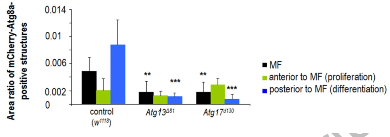
Knockdown of Atg13 and Atg17 had almost no effect on eye development (Table S1 and Fig. 3F). Deletion alleles of Atg13 and Atg17 did also not change (Atg13) or only occasionally altered (Atg17) the morphogenesis of this organ (Fig. S17). This is particularly interesting, as these mutations effectively abolish the transcriptional activity of the corresponding genes in the fat body [63,64]. Analyzing homozygous mutant progeny of heterozygous parents however revealed the presence of both Atg13 transcript and protein in the eye disc of L3W larvae (Figs. S21A to A’’ and S22A, A’). Similar to these results, Atg17- specific mRNA could also be detected in eye disc samples dissected from homozygous Atg17 mutants (Figs. S21B, B’ and S22B). Since both mutations (Atg13∆81 and Atg17d130) represent large deletions covering a significant part of the corresponding coding region, the presence of transcripts (and proteins) could be the consequence of maternally contributed factors. Using a dominant female sterile technique (with the use of ovoD1 dominant negative mutation), we generated homozygous Atg13 mutants with no maternal Atg13 product, and found that animals die prior to the L3W stage (note that homozygous Atg13 mutants with maternal contribution die as pupae) (Fig. S21A’’’). Probably due to these mechanisms, specific transcripts and autophagic structures accumulated, although at lowered levels than in controls,
Accepted Manuscript
16
in Atg13 and Atg17 mutant eye disc samples (Figs. S22 and S23). Together, these results raise the possibility that maternal effect can also rescue autophagic activity in the eye disc of larvae homozygous for certain Atg mutations and derived from heterozygous parents.
To further prove the specificity of genetic compensation eliminating the phenotypic manifestation of Atg lf mutations, we silenced Atg14 in the Atg14∆5.2 mutant genetic background (importantly, Atg14 encodes a single transcript and has no paralog). Atg14 RNAi animals exhibited a compromised eye phenotype with a relatively high penetrance (Fig. 3F and Table S1), while the ∆5.2 mutation [56] did not influence eye morphology (Fig. 5). If genetic compensatory mechanisms rescue normal eye morphology in Atg14∆5.2 mutants, one would expect the suppression of the eye phenotype caused by RNAi treatment in the mutant background (in the mutant, there is no transcript that the RNAi could degrade). Indeed, the presence of the ∆5.2 mutation highly rescued normal eye development in Atg14 RNAi samples (in females, the penetrance of wild-type eye morphology increased from 60% to 95%, in males, it was elevated from 50% to 80%) (Fig. 5).
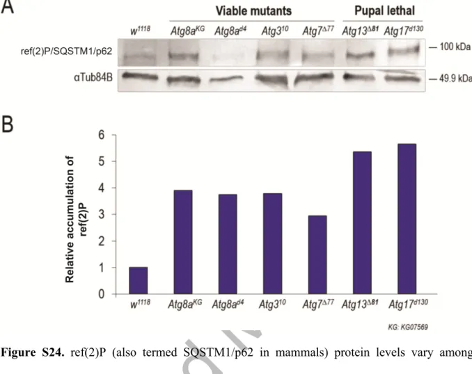
Based on genetic compensation discussed above we postulate that lf mutations in Atg genes do not completely eliminate autophagy functions in the affected tissues, thereby masking the phenotypic manifestation of mutant alleles. In good accordance with this assumption, the level of Ref(2)P/SQSTM1/p62 serving as a substrate for autophagy varied significantly among different Atg mutant animals (Fig. S24). The most significant Ref(2)P accumulation was evident in Atg13 and Atg17 mutant samples, the gross mutant phenotype of which appears to be the most severe (lethal) among those examined (the other mutants are viable). Thus, in the latter samples, autophagy still operates, although at decreased levels as compared with control.
Accepted Manuscript
17
Knockdown of Atg genes in the eye disc triggers apoptotic cell death
Reduced activity of Atg genes in the entire eye disc can retard eye development; the affected animals frequently displayed a small eye or eyeless phenotype (Table S1 and Fig. 3). To address whether these morphological defects result from, at least in part, excessive cell death, we monitored the amount of cells with apoptotic features in normal (control) versus autophagy deficient eye disc samples. We found that samples from animals depleted for Atg3, Atg14 or Atg101 show a much higher number of TUNEL-positive (i.e., fragmented DNA- containing) cells than those derived from the corresponding control [ey-Gal4(II)/+ or ey- Gal4(III)/+] animals (Fig. 6A to E, I). We also performed acridine orange (AO) staining on eye discs of L3W stage larvae to detect acidic compartments, whose accumulation is also a characteristic feature of cells undergoing apoptosis. Samples from Atg3, Atg14 and Atg101 RNAi animals showed increased levels of AO-positive cells, relative to the corresponding controls (ey-Gal4) (Fig. 6A’ to E’, I). The elevated number of TUNEL- and AO-positive cells in Atg RNAi samples was evident both in front of and behind the MF.
Consistent with these data, human cleaved-CASP3/caspase-3-specific antibody staining also revealed elevated amounts of cells showing increased caspase activity in samples dissected from Atg3-, Atg14- and Atg101 RNAi animals, as compared with their corresponding controls (Fig. 6A’’ to E’’, J). This implies increased levels of cell death because this human cleaved-CASP3-specific antibody reveals CASP9-like Dronc activity in Drosophila, at least in part due to generating cleaved Drice and cleaved Cp1 effector caspases [65]. A UAS-Apoliner reporter has previously been developed to effectively detect effector caspase activity in dying apoptotic cells [66]. Using this tool, we observed intense enzymatic activity in samples dissected from certain Atg RNAi animals (Fig. 6F to H, K; in the enlarged part of panels G and H, intense white labeling —that marks cell death events—is visible).
However, contrary to what we found by TUNEL and AO staining, caspase activation was
Accepted Manuscript
18
predominantly detectable in front of the MF (in the PZ and prospective head cuticle). This implies that downregulation of Atg genes in the eye disc triggers at least 2 types of programmed cell death, a caspase-independent and caspase-dependent apoptosis. The former mainly occurs in the DZ, while the latter appears in front of the MF. Alternatively, the elimination of cell corpses is perturbed in the DZ, or the sign of human cleaved-CASP3- specific antibody may reflect apoptosis-independent caspase activity in the PZ [65].
In sum, we conclude that defects in autophagy in the developing eye disc promote apoptotic cell death, and this effect is likely to contribute to reduction in size of the affected adult eye. Inhibiting autophagy in the DZ alone (GMR-Gal4) does not impair eye development. Thus, autophagic activity in front of and/or within the MF is necessary for the survival of columnar cells in the entire eye disc.
The Hox gene lab (labial) is expressed in the disc proper where it modulates the transcription of Atg8a-A
As demonstrated above, the distribution of Atg8a-positive autophagic structures exhibited a specific pattern in the developing eye tissue, locating predominantly in the MF and DZ (Figs.
2D to E’’’ and S3, S7). This observation prompted us to investigate whether autophagy in this tissue is regulated by developmental factors. Transcriptional control of certain Atg genes, including Atg8a, plays an important role in autophagy induction [52,67,68]. Atg8a encodes 3 isoforms, A, B and C, out of which Atg8a-A appears to function primarily in early phases of eye development (Fig. 4A’, B).
To gain insights into the possible mechanisms underlying Atg8a-A regulation during eye development, we searched for conserved binding sites of developmental regulatory
Accepted Manuscript
19
factors in the Atg8a locus (including both regulatory and coding regions), and identified 2 putative conserved binding sites for Hox proteins (Homeobox-containing transcription factors, a subset of homeotic proteins), master regulators of early developmental events. One of these newly identified sites is located in the first intron of Atg8a-A, while the other is located within its 3’ untranslated region (3’ UTR) (Fig. 7A). In close proximity to these Hox regulatory elements, putative binding sites for Hox cofactors including exd (extradenticle) and hth (homothorax) were also identified. The intronic binding site appears to be specific to lab, whereas the 3’ UTR binding site seems to be specific to Dfd (Deformed), but other Hox proteins cannot be excluded (the putative lab binding site is actually similar to an alternative lab consensus binding sequence identified in the regulatory region of the Drosophila gene CG11339) [69,70]. Both lab and Dfd are expressed in the peripodial membrane of the eye- antennal disc [71,72]. To investigate the expression pattern of these Hox genes in more detail, in situ hybridization assays were performed by using antisense lab and Dfd RNA probes.
Specificity of the probes was confirmed by in situ hybridizations which recapitulated the formerly established expression patterns at certain embryonic stages (FlyBase) (Figs. 7B, C and S25A) [73,74]. According to these results, lab was mainly expressed in the MF and in the area from which the head cuticle develops, as well as weak staining was detectable in other parts of the PZ and in the peripodial membrane (Fig. 7D, D’). It is worth to note that this newly identified expression pattern for lab is much wider in this organ than reported previously [71,72]. As strong accumulation of Atg8a-positive autophagic structures was also evident in the MF (Figs. 2D to E’’’ and S3), we propose that Atg8a-A and its potential transcriptional regulator lab share activity domains in the eye disc. We also examined Dfd expression, and found that it is only evident in the peripodial membrane (Fig. S25B, B’), as reported previously [71,72]. This expression domain was further confirmed by analyzing a Dfd-GFP reporter system (Fig. S25C to D’).
Accepted Manuscript
20
To test whether the 2 newly identified conserved Hox binding sites in the Atg8a-A locus are functional in vivo, we generated an eGFP-Atg8a-A reporter construct containing endogenous upstream and downstream regulatory sequences, together with the entire coding region fused with eGFP (Fig. 7E). This construct involves both of the putative Hox binding sites identified in this study. Using site-directed mutagenesis, we further generated 2 mutant versions of the construct. One of them lacks the intronic (i.e., lab-specific) exd-Hox binding site (mutlabeGFP-Atg8a-A), while the other misses the 3’ UTR (i.e. Dfd-specific) exd-Hox binding site (mutHoxeGFP-Atg8a-A) (Fig. 7E’). Importantly, the wild-type reporter was capable of recapitulating the accumulation pattern of Atg8a proteins, obtained by anti-Atg8a antibody staining and using conventional fluorescent microscopy (Fig. 7F, F’). The expression intensity of the mutant reporters—integrated into different genomic environments, the 51C and 58A cytological regions—was significantly elevated in the anterior part of the eye disc, in front of the MF, as compared with the control (non-mutated) construct (Fig. 7G to G’’). To determine precisely the area(s) where lab may repress Atg8a-A expression, we divided the eye disc into 9 parts, and determined mutlabeGFP-Atg8a-A expression levels in these subregions (Figs. 7G’’’ and S26). Quantification of mutlabeGFP-Atg8a-A expression intensity showed that the absence of the putative lab binding site leads to elevated expression in the ventral prospective head cuticle and lateral flap (bars highlighted by red frames in Fig. 7G’’’).
mutHoxeGFP-Atg8a-A expression was also much stronger, mainly in the prospective head cuticle and ventral lateral flap, than the corresponding control (Fig. 7G’’’). Based on these data we propose that these potential Hox binding sites are functional in vivo, and that lab, and perhaps (an)other Hox protein(s), directly regulates Atg8a-A in these regions. As Dfd accumulates in the peripodial membrane but not in the disc proper (Fig. S25B to D’), we focused on the putative lab binding site only in further experiments.
Accepted Manuscript
21
To confirm the inhibitory effect that lab exerts on Atg8a-A expression in front of the MF, we downregulated and overexpressed lab from drivers that are active in this area. Indeed, the former intervention strongly upregulated (Fig. 7H to H’’’’) while the latter inhibited (Fig.
7I to I’’’’) eGFP-Atg8a-A expression. Excessive expression of the reporters was most evident in the region of the ventral head cuticle and lateral flap (yellow arrows in Fig. 7H’ to H’’’).
Thus, lab inhibits Atg8a-A expression in front of the MF, especially in the region of the prospective head cuticle. The inhibitory effect of lab hyperactivation on Atg8a-A expression was abolished when the lab binding site mutant reporter version was examined (Fig. 7I to I’’’), confirming the in vivo functionality of this particular lab binding site.
The expression profile of these reporters highly coincided with the aberrant eye morphology of lab RNAi adult flies. Depletion of lab led to a shift in the ventral head-eye cuticle border in favor of the head cuticle (the white arrow in Fig. 7J’, J’’). This phenotype was often associated with the overgrowth of the head cuticle as well as with the lateral overgrowth of adult eyes (Fig. 7J’’’), morphological features that have been described previously for lab4 mutants [72]. We conclude that lab inhibits the expression of Atg8a-A in the ventral prospective head cuticle and ventral lateral flap via the newly identified putative binding site.
lab upregulates Atg8a-A in the MF
As shown above, autophagic structures abundantly accumulate in the MF and DZ (Figs. 2 and S3, S6, S7). lab mRNA was also readily detectable in the MF, but not in the DZ (Fig. 7D, D’). Upon these data, we investigated how lab influences the transcription of Atg8a-A in the MF. To address this issue we silenced lab by ey-Gal4(III) driver that is active in the MF and DZ (Figs. 1 and S5C). Semi-quantitative PCR experiments revealed highly reduced levels of
Accepted Manuscript
22
Atg8a-A transcript in lab RNAi eye disc samples, as compared with controls (Fig. 8A).
Downregulating lab by GMR-Gal4 driver being active only in the DZ, however, did not alter Atg8a-A transcript levels (lab is not expressed in the DZ; Fig. 7D, D’) (Fig. S27). Next, relative transcript levels of Atg8a-A were determined by quantitative real-time PCR in eye disc samples dissected from L3W larvae with control versus lab RNAi and lab overexpressing genetic backgrounds (Fig. 8B). Data convincingly showed that samples defective for lab contain significantly fewer Atg8a-A transcripts while those hyperactive for lab display higher levels of Atg8a-A transcripts than control ones. Thus, lab activates Atg8a-A expression in the MF. Taken together, we suggest that in the eye primordium, lab has a dual role in the regulation of Atg8a-A activity. First, lab inhibits Atg8a-A transcription in the prospective ventral head cuticle and lateral flap, presumably through a lab-exd-specific binding site we identified in the first intron (Fig. 7G to I’’’’). It is likely that this regulatory interaction plays a role in the determination of the normal head-eye cuticle border (Fig. 7J to J’’’). Second, lab promotes the expression of Atg8a-A in the MF to activate autophagy (Fig. 8A, B). These opposite effects of lab on Atg8a-A activity may be mediated by different Hox cofactors.
lab activates autophagy in the MF and DZ
As shown above, lab increases Atg8a-A expression in the MF (Figs. 8 and S27). This finding raised the intriguing possibility that lab influences eye development, at least in part, through modulating autophagic activity. The overexpression of lab in the eye disc by ey-Gal4(II) driver led to the formation of headless adults. Thus, we overexpressed lab by an eye-specific driver with a weaker activity domain, ey-Gal4(III), and found that this intervention results in small eyes or a phenotype without eyes in the affected adults, with almost a full penetrance (Fig. 9A, B) (in females: no eye, 10.20%; small eye, 89.80%, normal eye, 0%; and in males:
Accepted Manuscript
23
no eye, 18.75%; small eye, 75.00%; normal eye, 6.25%). On the contrary, eye disc-specific silencing of lab resulted in eye overgrowth and also compromised eye development through altering the boundary of the head-eye cuticle (Figs. 7J to J’’’ and 9C, D).
lab overexpression in the eye disc enhanced autophagic activity (Fig. 9A’ to A’’’, B’
to B’’’, E). Conversely, silencing or mutational inactivation of lab in this organ lowered the amount of autophagic structures (Figs. 9C’ to C’’’, D’ to D’’’, F and S28, S31A to A’’).
Indeed, the amount of Atg5- and Atg8-positive structures was significantly increased in lab- hyperactive (Fig. 9B’, B’’, E) but decreased in lab-defective genetic backgrounds (Figs. 9D’, D’’, F and S28A, A’, B, B’, S31A to A’’). Similarly, the number of acidic compartments became higher when lab was overexpressed (Fig. 9B’’’, E), but became smaller when lab was silenced or inactivated (Figs. 9D’’’, F, and S28A’’, B’’). These results indicate that lab induces autophagic activity in the MF and DZ. We hypothesize that this effect of lab in the DZ is likely to be realized in a cell non-autonomous manner (as we could detect no lab transcript in this disc region), probably through stable products of target genes it regulates.
We also studied the complex regulatory relationship between lab and autophagy in the fat body of L3F larvae. In good agreement with data we obtained from the MF, fat body cells clonally defective for lab exhibited very low amounts of LysoTracker-Red-positive (acidic) structures, as compared with control cells (Fig. S29A to A’’). In addition, fat body cells clonally overexpressing lab contained much higher amounts of Atg8a-positive autophagic structures than non-overexpressing control ones (Fig. S29B, B’). Thus, in certain cell types, including columnar cells in the MF and larval fat body cells, lab activates autophagy. The fact that lab induces autophagy in the larval fat body was somehow unexpected since the other Hox proteins were reported to redundantly inhibit developmental autophagy in fat body cells [61]. Thus, lab may be the sole Drosophila Hox paralog that activates the autophagic process in this tissue.
Accepted Manuscript
24
To further distinguish the role of lab in the peripodial membrane from its role in the disc proper (as ey-Gal4 drivers are active in both disc proper and peripodial membrane), we used c311-Gal4 driver [42] to silence lab specifically in the peripodial membrane. This intervention enhanced the amount of acidic compartments in columnar cells (Fig. S30). Since lab knockdown driven by ey-Gal4(II) inhibited autophagy in these cells, it is likely that lab is endogenously active in certain columnar cells where it modulates the autophagic process.
Both overexpression and inactivation of lab in the eye disc cause excessive cell death
As demonstrated above, lab activates autophagy in the MF at least in part through enhancing Atg8a-A expression (Figs. 8A, B, 9B to B’’’, D to D’’’, E, F and S28, S31A to A’’). Then, autophagic activity remains high in the DZ in a cell non-autonomous manner (Fig. 7D, D’).
Since defects in autophagy strongly induced ectopic cell death in this organ (Fig. 6), we asked whether deregulation of lab similarly affects cell survival in the developing eye tissue. We found that depletion of lab leads to a massive elevation in the number of TUNEL-positive nuclei and acidic cell bodies, mainly in the DZ (by 2.84 and 1.53 times, respectively) (Fig.
9C’’’’ to D’’’’’, G). lab deficiency also markedly increased the amount of human cleaved- CASP3-positive cells showing elevated caspase-associated immunoreactivity, but, unlike AO- positive cells, this change was predominantly evident in the PZ and prospective head cuticle (Fig. S31B to B’’, C, C’). lab overexpression similarly increased the number of TUNEL- (8.35 times) and AO-positive (3.49 times) structures (Fig. 9A’’’’, A’’’’’, B’’’’, B’’’’’, G), and the amount of cells with chromatin condensation (Fig. 9H). We conclude that the Hox protein lab, a master regulator of early development, promotes the survival of columnar cells in the eye primordium via, at least in part, fine tuning autophagy. This effect of lab in the DZ may occur indirectly.
Accepted Manuscript
25 Discussion
Under normal cellular settings, autophagy operates at basal levels to maintain the homeostasis and long-term survival of terminally differentiated cells [75]. Cellular stress factors, however, can trigger autophagic activity at both transcriptional and posttranscriptional levels. This response involves various signaling cues and regulatory proteins [76-78]. The autophagic process also becomes activated during numerous developmental events [16,17,34,35]. For example, during Drosophila metamorphosis the degradation of larval tissues is primarily achieved by autophagy [18,30], or at very early stages of mammalian development the elimination of maternally-deposited factors occurs via autophagic degradation [17]. However, little is known about the key regulatory proteins that control the activity of Atg genes in developmental processes. Hox proteins, master regulators of early animal development, modulate autophagy in the Drosophila fat body [61]. This regulatory interaction between Hox factors and autophagy suggests a much stricter developmental control of the autophagic process than was previously assumed.
In this study we demonstrated that autophagic structures accumulate in a specific pattern in the Drosophila eye disc, predominantly in the MF and DZ (Figs. 2, and S3, S6, S7), and that this pattern does not reflect the distribution of 2 key Atg proteins, Atg5 and Atg8a, which, using conventional fluorescent microscopy, were detected nearly ubiquitously in this organ, but most intensely in the area from which the head cuticle develops (Figs. 2A, and 6F, F’). Other parts of the developing eye tissue displayed only basal levels of autophagic structures. Thus, autophagy displays a characteristic spatial activity pattern in the eye disc of L3W larvae, raising the possibility that lysosome-mediated cellular self-degradation contributes to the morphogenesis of this organ.
Accepted Manuscript
26
We further showed that silencing of several Atg genes can seriously compromise the development of the Drosophila compound eye (Table S1 and Figs. 3A to F and S8, S9). In this set of experiments Atg RNAi constructs were driven by ey-Gal4(II), ey-Gal4(III) or so7- Gal4 that have a broad expression domain in the eye primordium (Fig. S5B to D). The effectiveness of RNAi constructs was increased by parallel-expressing Dcr-2 (making RNAi more efficient), and animals were maintained at 29°C, which is the optimum temperature for Gal4 proteins to bind the UAS sequence. The pleiotropic effect of Atg gene knockdowns included severe reduction in organ size (small eye phenotype), even the complete absence of the organ (eyeless phenotype), and alteration in organ shape (aberrant eye morphology). Some of the Atg RNAi constructs we assayed influenced eye growth and morphogenesis with high penetrance, while other constructs proved highly or completely ineffective (Table S1 and Fig.
3F). The former constructs were capable of significantly reducing both the transcriptional and translational activity of the corresponding Atg genes as well as the amount of autophagic structures (Figs. S10, S12 and S13). Contrary to these functional RNAi samples, the latter failed to lower the corresponding protein levels, and were unable to modulate autophagic activity (Figs. S11 to S13). To provide an additional evidence for the specificity of eye phenotypes caused by Atg knockdowns, we rescued normal eye development in Atg8a-, Atg14- and Atg101 RNAi animals by a transgene carrying the wild-type copy of the corresponding Atg gene (Figs. S12D and S14) or a duplication bearing the wild-type copy of Atg101 (Fig. S15). In addition, downregulation of Atg genes specifically in the peripodial membrane did not affect eye morphogenesis (Table S2 and Fig. S16).
Previous studies have detected no obvious defect in adult eye morphology when Atg genes are silenced by GMR-Gal4 driver [48,50]. It is possible that GMR-Gal4 is expressed in less excessive levels in the eye disc than ey-Gal4(II) and ey-Gal4(III) do. Alternatively, the function of autophagy is more significant in the MF and/or PZ where GMR-Gal4 is not active.
Accepted Manuscript
27
We also explored the effect of lf mutations in Atg genes on eye development in this organism. In the literature several studies have reported no influence of autophagy on this developmental paradigm [48-52]. Contrary to these data, we found that mutational inactivation of Atg17 and Atg1 can impede eye formation (Fig. S17). Some of the mutant animals failed to develop the organ. The percentage of eye phenotypes in these mutant backgrounds however was relatively low, appearing only in the minority of animals examined. Lf mutations in other Atg genes did not affect eye formation. It has been recently demonstrated in zebrafish that genetic compensatory mechanisms attenuate the phenotypic effect of deleterious mutations but not gene knockdowns [59]. In accordance with these findings, mutations in Atg4a, Atg8a, and Atg18a led to the activation or upregulation of the corresponding paralogous genes, Atg4b, Atg8b and Atg18b, respectively (Figs. 4A to C and S12F, S19, S20). Moreover, splice variants of Atg8a, A, B and C, identified only recently were expressed in an orchestrated way to compensate their own deficiency; in the eye disc isoform A is active (and B in a lesser extent), and a mutation that specifically inhibits Atg8a-A resulted in the transcriptional activation or upregulation of isoforms B and C (Figs. 4A to F and S12, S18). We also showed the presence of maternally contributed transcripts in homozygous Atg13 and Atg17 mutant samples derived from heterozygous parents (Figs. S21 and S22). Thus, multiple compensatory mechanisms can abolish eye phenotypes in Atg mutant samples. As an evidence, the Atg14∆5.2 mutation, which alone did not affect eye development, strongly suppressed the highly penetrant eye phenotype of Atg14 RNAi animals (the mutation eliminates the transcripts the RNAi could act on) (Fig. 5). The parallel expression of (a) paralog(s) and/or splice variant(s), as well as maternally contributed gene products, each have the potential to rescue autophagic activity in a certain Atg mutant background. In other words, many Atg mutant animals examined so far are not completely defective for autophagy. Indeed, we could readily detect autophagic structures in eye disc
Accepted Manuscript
28
samples dissected from Atg8a, Atg13, Atg17 and Atg1 lf mutants (Figs. 4G to I’ and S23). In the light of these results, the functional analysis of Atg mutant systems requires more attention in future genetic studies on Drosophila and on other models [79].
In Atg RNAi eye disc samples displaying an obvious phenotype, reduction in autophagic activity was accompanied with enhanced amounts of cells with apoptotic features (Fig. 6).
Although mutational inactivation of autophagy is known to trigger apoptosis in mammalian cell lines and nematodes [80,81], one can argue that the increased number of apoptotic cell corpses observed in these autophagy deficient systems is simply a consequence of failure in the heterophagic elimination of dying cells, a process that also requires Atg gene function [82,83]. However, an increase in caspase activity pointed to excessive levels of apoptosis rather than defects in the engulfment of dying cells (Fig. 6A’’ to E’’, F to H, J, K). Both methods (staining with human cleaved-CASP3-specific antibody and using the Apoliner caspase sensor) essentially led to the same observation, i.e. increased levels of apoptotic cell death. This is important because human cleaved-CASP3-specific antibody staining alone could mark positive cells independently of caspase activity [65]. Hence, our present data provide evidence for a role of Atg genes in preventing columnar cells from undergoing apoptosis in the Drosophila eye disc. In clonal analysis of Atg lf mutations with ref(2)P accumulation (also known as SQSTM1 and p62 in mammals) there was no apparent cell death effect. Although the lethal Atg13∆81 and Atg17d130 mutations significantly increased ref(2)P levels (Fig. S24), autophagic activity was still observable in these mutant samples (Fig. S23).
Presumably, this was due to the presence of maternally contributed factors, explaining why the clonal cells contained autophagic structures (Fig. 4H to I’). Alternatively, apoptotic cell death occurred in Atg mutant cell clones but an apoptosis-induced compensatory proliferation mechanism rescued a nearly-normal eye morphology [84].
Accepted Manuscript
29
In mammalian cell cultures, upregulation of the Atg8 ortholog MAP1LC3B by the transcription factor TFEB leads to elevated autophagy [67]. In Drosophila, expression levels of Atg1 and Atg8a are also proportional to autophagic activity [52,68]. Since Atg1 plays a role in brain development in an autophagy-independent manner [85], we focused on the Atg8a genomic region to found potential binding sites for transcription factors that may regulate autophagy during eye morphogenesis. We identified 2 conserved Hox binding sites within the Atg8a coding sequences (Fig. 7A). Furthermore, conserved binding sites for 2 Hox co-factors, Exd and Hth, were also uncovered in the close vicinity of these Hox regulatory elements (Fig.
7A). These sequence data are consistent with a recent finding that Hox proteins including Dfd, Ubx and Abd-B redundantly inhibit autophagy in the fat body to prevent a premature degradation of the organ [61].
By generating an endogenously regulated eGFP-Atg8a-A translational fusion reporter (Fig. 7E) and its 2 mutant derivatives lacking either of the 2 newly identified conserved Hox- Exd binding sites (Fig. 7E’), we revealed that both of these sites are functional in vivo, i.e.
they are responsive to regulatory cues (Fig. 7F to I’’’’). In front of the MF, particularly in the prospective ventral head cuticle, Atg8a-A proteins accumulated at much higher levels in the lab binding site mutant versions than in the corresponding control (Fig. 7G’’’). Thus, the intronic regulatory element may mediate Atg8a-A repression by a specific Hox protein, lab, in the anterior part of the eye disc. In contrast, quantification of Atg8a-A transcript levels in the MF (Fig. 8A, B), together with the analysis of autophagic activity (Figs. 2 and S7), unambiguously showed that lab activates Atg8a-A in this eye disc region. In accordance with these results, lab was also expressed at relatively high levels in the PZ, particularly the prospective head cuticle, and in the MF (Fig. 7D and D’). Consistent with these observations, lab deficiency in the eye disc led to decreased activity of autophagy, while lab hyperactivity elevated the amount of autophagic structures in the MF and DZ (Figs. 9A to D’’’ and S28,
Accepted Manuscript
30
S31). Thus, lab is required for establishing physiological levels of autophagy in the eye disc, most probably by upregulating Atg8a in the MF and downregulating this gene in front of the MF, especially in the regions from which the ventral head cuticle develops. In addition to modulating autophagic activity, dysregulation of lab in the eye disc caused an excess in the amount of columnar cells undergoing apoptosis (Figs. 9A’’’’ to D’’’’’, G, H and S31).
Based on these data we propose a model that lab exerts a dual effect on Atg8a-A expression in the developing eye primordium (Fig. 10). First, lab represses Atg8a-A in the prospective ventral head cuticle. This regulatory interaction may depend on the lab regulatory sequence we identified in the first intron of Atg8a-A (Fig. 7A), and be required for the correct formation of the head-eye cuticle border (Fig. 7J to J’’’). Second, lab activates Atg8a-A expression within the MF (Fig. 8). As a result, autophagic structures are generated abundantly in this eye region (Figs. 2 and S3, S6, S7). As the MF moves anteriorly, autophagy activity remains elevated in the DZ. As lab transcripts were largely undetectable in the latter area (Fig. 7D, D’), autophagic regulation is achieved by factors other than lab. Nevertheless, in the MF and DZ, intense autophagy promotes the survival, and likely differentiation, of photoreceptor and accessory cells (Fig. 10). We propose that lab is critical for normal eye development in Drosophila through supporting survival and differentiation of columnar cells.
Together, these data may shed light into a more prominent role of autophagy in tissue shaping and organ development than previously thought. As autophagy is implicated in several human developmental disorders, such as Vici syndrome and myopathies [5,8-10], findings presented by this study may also provide a better understanding of the mechanisms underlying such pathologies, thereby having significant medical relevance.
Acknowledgements
Accepted Manuscript
31
This work was supported by the Hungarian Scientific Research Found grants NK78012 and K109349 to T.V. and M.S., the Hungarian Brain Research Program (KTIA_NAP_13) and OTKA grant K82039 to J.M, and by the New National Excellence Program of the Ministry of Human Capacities. A part of Drosophila stocks was obtained from the Bloomington Drosophila Stock Center (NIH P40OD018537) and Vienna Drosophila Resource Center (VDRC, www.vdrc.at). Other Drosophila strains and reagents were kindly provided by Barry Dickson, Deborah Hursh, Gábor Juhász, Daniel R. Marenda, Tamás Maruzs and Máté Varga.
qPCR experiments and the evaluation of results were assisted by Mónika Kosztelnik. The authors also thank Regina Preisinger, Beatrix Supauer, Tünde Pénzes, Gabriella Szabados, Judit Botond and Sára Simon for the excellent technical assistance.
Accepted Manuscript
32 Materials and Methods
Fly stocks, genetics and conditions
Drosophila strains were maintained on standard cornmeal-sugar-agar medium. Stocks were obtained from the Bloomington Drosophila Stock Center (referred to as “BL”), the Vienna Drosophila RNAi Center (referred to as “v”) and the Drosophila Genetic Resource Center, Kyoto (referred to as “DGRC”). Other strains were gift from members of the Drosophila research community. We used the following RNAi lines to silence autophagic genes:
Atg1 RNAi (Atg1JF02273, BL26731 and Atg1HMS02750, BL44034) Atg2 RNAi (Atg2HMS01198, BL34719 and Atg2JF02786, BL27706) Atg3 RNAi (Atg3HMS01348, BL34359)
Atg4a RNAi (Atg4aJF03003, BL28367 and Atg4aHMS01482, BL35740)
Atg5 RNAi (Atg5JF02703; BL27551 and Atg5HMS01244, BL34899 and Atg5KK108904, v104461) Atg6 RNAi (Atg6JF02897, BL28060 and Atg6HMS01483, BL35741)
Atg7 RNAi (Atg7JF02787, BL27707 and Atg7HMS01358, BL34369)
Atg8a RNAi (Atg8aGD4654, v43097, Atg8aJF02895, BL28989 and Atg8aHMS01328, BL34340) Atg8b RNAi (Atg8bHMS01245, BL34900)
Atg9 RNAi (Atg9JF02891, BL28055 and Atg9HMS01246, BL34901) Atg10 RNAi (Atg10HMS02026, BL40859)
Atg12 RNAi (Atg12KK111564, v102362 and Atg12HMS01153, BL34675) Atg13 RNAi (Atg13KK100340, v103381 and Atg13HMS02028, BL40861) Atg14 RNAi (Atg14KK100903, v108559)
Atg16 RNAi (Atg16HMS01347, BL34358) Atg17 RNAi (Atg17KK101847, v104864)
Atg18a RNAi (Atg18aJF02898, BL28061 and Atg18aHMS01193, BL34714) Atg101 RNAi (Atg101KK101226, v106176 and Atg101HMS01349, BL34360) Vps15 RNAi (Vps15HMS00908, BL34092 and Vps15GL00085, BL35209)
Vps34/Pi3K59F RNAi (Vps34HMS00261, BL33384 and Vps34GL00175, BL36056) In this study the following Gal4 drivers were used:
ey-Gal4(II) was also obtained from BDSC (w*; P{GAL4-ey.H}, 3-8, BL5534)
ey-Gal4(III) was kindly provided by Barry Dickson (Janelia Research Campus, Ashburn, Virginia, US) GMR-Ga4 (w*; P{GAL4-ninaE.GMR}12, BL1104)
so7-Gal4 (y1 w*; P{so7-GAL4}A/TM6B, BL26810) c311-Gal4 (y1; P{GawB}c311, BL5937)
Ubi-Gal4 (w*; P{Ubi-GAL4.U}2/CyO, BL32551)
UAS-Dcr-2 (w1118; P{UAS-Dcr-2.D}2, BL24650 and w1118; P{UAS-Dcr-2.D}10, Bl24651) was used to enhance the efficiency of long hairpin RNAi constructs.
The following mutant stocks were used: