Proof Central
Dear Author
Please use this PDF proof to check the layout of your proof. If you would like any changes to be made to the layout, you can leave instructions in the online proofing interface. Making your changes directly in the online proofing interface is the quickest, easiest way to correct and submit your proof. Please note that changes made to the proof in the online proofing interface will be added to the proof before publication, but are not reflected in this PDF proof.
If you would prefer to submit your corrections by annotating the PDF proof, please download and
submit an annotatable PDF proof by clicking here and you'll be redirected to our PDF Proofing
system.
Author Query Form
Journal:EUJMI
Article Number:1886.2020.00010
Dear Author,
Please check your proof carefully and mark all corrections at the appropriate place in the proof.
Queries and/or remarks
[Q1] Please confirm that the forename(s) and surname(s) have been identified correctly and please carefully verify the spelling of all authors’names.
[Q2] As per the style, if there are more than six authors, the first six author names should be listed followed by‘et al.’. Hence please provide names offirst six authors followed by‘et al.’for Refs. [15, 35, 37].
[Q3] Please provide a definition for the significance of“bold”values in Table 1.
[Q4] Please note that per journal style,“Author contribution”statement is mandatory. Please check and provide the same.
The incidence of anaerobic bacteria in adult patients with chronic sinusitis: A prospective, single-centre microbiological study
EDIT URB AN
1,2, M ARI O GAJD ACS
3pand ATTILA TORKOS
4 Q11Department of Public Health, Faculty of Medicine, University of Szeged, 6720 Szeged, Dom ter 10., Szeged, Hungary
2Institute for Translational Medicine, Medical School, University of Pecs, 7624 Pecs, Szigetiut 12., Pecs, Hungary
3Department of Pharmacodynamics and Biopharmacy, Faculty of Pharmacy, University of Szeged, 6720 Szeged, E€otv€os utca 6., Szeged, Hungary
4Department of Oto-Rhino-Laryngology and Head-, Neck Surgery, Faculty of Medicine, University of Szeged, Szeged, Hungary
Received: April 14, 2020 • Accepted: April 23, 2020
ABSTRACT
Introduction:Chronic sinusitis caused by anaerobes is a particular concern clinically, because many of the complications are associated with infections caused by these organisms. The aim of this study was to evaluate the incidence of anaerobic bacteria in chronic sinusitis in adults as a part of a prospective microbiological study.Materials and methods:Over a one-year period, aspirations of maxillary sinus secretions and/or ethmoid cavities were derived inn 5 79 adult patients with chronic sinusitis by endoscopy in a tertiary-care teaching hospital in Hungary. The qualitative and quantitative compositions of the total cultivable aerobic and anaerobic bacterial and fungalflora cultured on the samples were compared. Correct anaerobic species level identifications were carried out according to standard methods.Results:Bacteria were recovered for all of the 79 aspirates and the numbers of the significant cultured isolates (with colony forming units≥103) were between 1 and 10. A total of 206 isolates, 106 anaerobic and 100 aerobic or facultative-anaerobic strains were isolated. The most common aerobic bacteria wereStreptococcus pneumoniae(n 5 40), Haemo- philus influenzae(n 5 29), Moraxella catarrhalis (n 5 6), Staphylococcus aureus (n 5 7) and Streptococcus pyogenes(n56). The anaerobic bacteria included black-pigmentedPrevotellaspp.
andPorphyromonasspp. (n527),Actinomycesspp. (n513), Gram-positive anaerobic cocci (n5 16),Fusobacterium spp. (n 519) and Cutibacterium acnes (n5 8).Conclusions:This study il- lustrates the microbial dynamics in which anaerobic and aerobic bacteria prevail and highlights the importance of obtaining cultures from patients with chronic sinusitis for guidance in selection of proper antimicrobial therapy.
KEYWORDS
anaerobic bacteria, chronic sinusitis, prospective, microbiology,Prevotella,Porphyromonas, otolaryngology
INTRODUCTION
Chronic sinusitis (CS) is an inflammatory disorder of the upper airways, which lasts longer than 12 weeks, often causing residual damage to the sinus mucosa, leading to long-term symptoms (according the definition of the International Rhinosinusitis Advisory Board) [1, 2]. Based on literaturefindings, chronic sinusitis is almost always accompanied by concurrent nasal airway inflammation, and is often preceded by symptoms of rhinitis; thus, the term chronic rhinosinusitis (CRS) has evolved to more accurately describe this condition [1]. CRS is a multifactorial morbidity, in which the complex microbiome plays a pathogenic role [2]. It
European Journal of Microbiology and Immunology
DOI:
10.1556/1886.2020.00010
© 2020 The Authors
ORIGINAL RESEARCH PAPER
*Corresponding author.
Tel.:þ36 62 341 330.
E-mail:mariopharma92@gmail.com
EUJMI-2020.00010_proof 16 May 2020 1:35 pm
1 2 3 4 5 6 7 8 9 10 11 12 13 14 15 16 17 18 19 20 21 22 23 24 25 26 27 28 29 30 31 32 33 34 35 36 37 38 39 40 41 42 43 44 45 46 47 48 49 50 51 52 53 54 55 56 57
58 59 60 61 62 63 64 65 66 67 68 69 70 71 72 73 74 75 76 77 78 79 80 81 82 83 84 85 86 87 88 89 90 91 92 93 94 95 96 97 98 99 100 101 102 103 104 105 106 107 108 109 110 111 112 113 114
is a frequent bacterial infection among adults, affecting approximately 5% of the Western population; the overall prevalence of CRS in the United States is 146/1,000 pop- ulation [3]. This involves nearly 30 million US adults annually, accounting for approximately 20 million office visits and 1.2 million hospital visits, making CRS more common than any other chronic condition, and for un- known reasons, the incidence of this disease appears to be increasing. According to the data from the US, the ratio of the recurrence is around 25% and the ratio of therapy- resistant cases of CRS is 10–15%. The European Position Paper on Rhinosinusitis and Nasal Polyps 2007 (EPOS 2007) found that the prevalence of CRS to be around 15– 16% (this is in part, mostly speculative because of the non- uniformity in symptoms criteria and definitions), among which, diagnosis by general practitioners was only around 2–4% [4]. The European prevalence by the EPOS and/
GA(2)LEN epidemiological study criteria was estimated to be 10.9% overall and ranging between 5 and 15% in different countries [4, 5]. Based on the results of the Na- tional Ambulatory Medical Care Survey of the Centers for Disease Control and Prevention (CDC), rhinosinusitis is thefifth most common cause for the prescription of anti- biotics [6]. CRS begins with an inflammation of the mu- cous membranes in the sinuses, the air-filled passages around the nose and throat, leading to mucous stagnation in the sinus, which forms a rich medium for the growth of various pathogens [1–3]. This early stage of sinusitis is often caused by a viral infection, generally lasting up to 10 days, completely resolving in 99% of cases; however, a small number of patients may develop a secondary acute bacterial infection, which is generally caused by aerobic bacteria [1, 2]. The inflammation causes fluid build-up, eventually plugging the sinus cavity and preventing normal mucus drainage. CRS may be caused by infections of the upper respiratory tract —the nose, pharynx, sinuses and throat—but there are some non-infectious triggers, such as allergens, toxins and underlying genetic predisposition.
Approximately 10% of all sinusitis cases are the result of an odontogenic process, with several reports in the literature stating that up to 40% of all sinusitis cases may have an underlying dental pathology [7, 8]. The pathophysiology of this condition is still poorly understood, with multiple environmental, host and microbial factors being impli- cated: allergies are a common cause, and anatomical problems such as a deviated nasal septum can bring on chronic sinusitis, other suspected causes putative patho- logical factors include changes in the microbiota, imbal- ance of the local or systemic immune system, and the presence of moulds or other fungi in the sinuses [9]. There are a lot of the different studies clarifying some microbi- ological aspects of acute and chronic sinusitis, including its pathophysiology, epidemiology, role of bacterial biofilms and more recently, the microbiome of healthy and/or diseased sinuses. The dysbiosis of intramucosal micro- biomes, the presence of biofilms and super-antigens have all been suggested to play a main role in the pathogenesis of CRS: while quantitatively, there are no relevant differences,
there were qualitative differences observed in the compo- sition of the sinus microbiota among healthy and CRS- patients [9]. Defining the nature of the role of the micro- biota in CRS is important because of the associated ther- apeutic implications. Streptococcus pneumoniae, Haemophilus influenzae, Moraxella catarrhalis, Coryne- bacterium spp., Staphylococcus epidermidis and members of the Enterobacterales order have been noted as the pre- dominant aerobic pathogens recovered from patients with sinusitis; however, with the exception of Staphylococcus aureus, the association between any single species and CRS is tenuous [10]. Many of these bacteria can interfere with the overgrowth of potential other pathogens and may play a role in preventing the development of infections. Most cases of CRS are due to acute sinusitis that either is un- treated or does not respond to treatment [11]. However, when sinusitis becomes chronic, these organisms are replaced by a variety of both aerobic and anaerobic bacteria and it has been suggested that anaerobic bacteria play a significant role in the pathogenesis of CRS [12]. This may be the result of the selective pressure of antimicrobial agents, sometimes redundantly used in the management of acute viral sinusitis, that enables resistant anaerobic organisms to survive, and over time, for the development of conditions appropriate for anaerobic growth, which include the reduc- tion in oxygen tension and an increase in acidity within the sinus cavity [11, 12]. CRS caused by anaerobic bacteria is a particular concern clinically because many of very serious complications associated with this condition (spread of infection into the bones of the face, mucocele formation, osteomyelitis, meningitis and/or and brain abscess) are asso- ciated with these microorganisms [8]. Because of the special techniques required for the collection, transport and culture of anaerobes, the availability of reliable data on anaerobic bacteria associated with CRS, especially in adult patients is limited; however, based on various reports, anaerobic path- ogens were recovered in 8–93% of cases [12–14]. The vari- ability in their recovery rate may be due to differences in the methodologies used for sample preparation, transportation, laboratory possibilities of culturing and identification, patient population, different geography and previous surgical and/or antimicrobial therapy.
The evaluation of the pathogenic role of anaerobic bac- teria in the acute exacerbation of CRS is of utmost impor- tance. Establishing the correct microbiological diagnosis of sinusitis is of primary importance, as it can serve as a guide to the choice of adequate antimicrobial therapy. Therefore, the aim of our study was to assess the microbial aetiology of CRS in at a tertiary-care hospital in Hungary over a one-year long period.
MATERIALS AND METHODS
Study design, details of the clinical centre
A prospective study was undertaken to evaluate the patho- genic role of anaerobic bacteria in the acute exacerbation of
2
European Journal of Microbiology and Immunology115 116 117 118 119 120 121 122 123 124 125 126 127 128 129 130 131 132 133 134 135 136 137 138 139 140 141 142 143 144 145 146 147 148 149 150 151 152 153 154 155 156 157 158 159 160 161 162 163 164 165 166 167 168 169 170 171
172 173 174 175 176 177 178 179 180 181 182 183 184 185 186 187 188 189 190 191 192 193 194 195 196 197 198 199 200 201 202 203 204 205 206 207 208 209 210 211 212 213 214 215 216 217 218 219 220 221 222 223 224 225 226 227 228
CRS in our local settings. The Institute of Clinical Micro- biology was the National Reference Laboratory of Human Pathogenic Anaerobic Bacteria in Hungary during the study period. The Institute is a routine diagnostic microbiological laboratory, servicing a 1,820-bed tertiary-care university- teaching hospital in Szeged, Hungary. This Clinical Centre is responsible for the medical care of about 600,000 patients in the southeast region of Hungary (urban and rural popula- tion: around 1.3 million people based on the most recent census data).
Patients, exclusion criteria
During a one-year period, 79 adult patients (45 males, 34 females), ranging in age from 18 to 84 years (mean age: 28.6 years) with CRS (patients corresponding to the following criteria: typical clinical symptoms of sinusitis, i.e. fever, headache, nasal drainage, positive radiographic findings, maxillary sinus and biopsy specimens demonstrating chronic inflammation of the sinus mucosal lining, or clinical and radiologic findings compatible with maxillary sinusitis followed by clinical and radiologic improvement following surgery) were included in the study. Sinusitis was considered chronic if symptoms persisted for≥12 weeks. Patients were excluded from the study if they were immunocompromised, if the previous or current use of antibiotics was known or if the presence of nasal polyps was known.
Cultivation and identification of bacterial isolates
Aspirate samples were obtained by the aspirations of maxillary sinus secretions and/or ethmoid cavities by endoscopy. Specimens were aspirated by use of a syringe, with instillation of non-bacteriostatic saline, if necessary.
Sinus aspirate samples were injected into reduced transport medium (Portagerm Multitransport Medium/bioMerieux, Marcy l’Etoile, France) and sent to the microbiology labo- ratory immediately after collection. All samples were pro- cessed within 1 h of sampling. Samples were suspended in 1 mL of reduced BHI broth (Brain Heart Infusion broth, with a pH adjusted to 7.2; Oxoid, Basingstoke, United Kingdom) and after gentle dispersion these suspensions were diluted (101–106) in pre-reduced BHI broth [8]. The 100
m
L ofeach dilution and 100
m
L of the corresponding undiluted suspension were plated immediately on selective and non- selective media. Columbia agar base (Oxoid, Basingstoke, UK) supplemented with 5% (v/v) cattle blood was used to isolate the total cultivable facultative and aerobic bacterial flora. Samples were also plated on Schaedler agar (bio- Merieux, Marcy l’Etoile, France) containing horse blood 5%v/v, haemin and vitamin K1.For the isolation of anaerobic organisms, these cultures were set up and incubated in an atmosphere of 90% N2, 5% H2and 5% CO2in an anaerobic environment (Concept 400 anaerobic incubator, Biotrace International Plc., UK) for 5–7 days at 37 8C. For the se- lective growth of aerobic Gram-positive cocci and Enter- obacterales, blood agar (Oxoid, Basingstoke, UK) and for the selective growing of Enterobacterales, eosin methylene-blue agar (EMB; bioMerieux, Marcy l’Etoile, France) were
applied, respectively. Fungal isolates were selectively cultured on Sabouroud Dextrose agar (SDA, bioMerieux, Marcy l’Etoile, France).
For aerobic bacteria, the plates were cultured at 378C in a 5% CO2-containing environment for 48 h. The selective agar media for the isolation of Enterobacterales were incu- bated at 378C for 24 h. SDA plates were incubated at 378C in ambient air for 24 h and additionally, at room tempera- ture for a further 5 days. The results from Gram-staining and the atmospheric growth requirements of each colony type were used to determine the additional biochemical tests required to identify the isolates. API 20A, ATB ID 32 ANA (bioMerieux, Marcy l’Etoile, France) tests were used to identify anaerobic bacteria, facultative anaerobic Gram- positive cocci and bacilli. The VITEK 2 Compact ID/AST (bioMerieux, Marcy l’Etoile, France) automated system was used to identify aerobic bacteria and fungi. Identification of anaerobes was performed based on the Wadsworth-KTL Anaerobic Bacteriology Manual, in addition to matrix- assisted laser desorption/ionisation time-of-flight mass spectrometry (MALDI-TOF MS) [8, 15, 16]. The method- ology of sample preparation for mass spectrometry mea- surements was described elsewhere [8, 15]. Mass spectrometry was performed by the Microflex MALDI Bio- typer (Bruker Daltonics Gmbh., Bremen, Germany) in positive linear mode across them/zrange of 2–20 kDa; for each spectrum, 240 laser shots at 60 Hz in groups of 40 shots per sampling area were collected. The MALDI Biotyper RTC 3.1 software (Bruker Daltonics Gmbh., Bremen, Germany) and the MALDI Biotyper Library 3.1 were used during spectrum analysis. We regarded the isolated bacterial strains as significant pathogens, if the bacterial colony count was higher than 103colony forming units (CFU)/mL [8, 15, 16].
Ethical considerations
As a part of this study, data on the affected patients were also collected, which was limited to their demographic characteristics only (age, sex). The study was deemed exempt from ethics review by the Institutional review board and informed consent was not required as data anonymity was maintained.
RESULTS
Significant number of cultivable bacteria and/or fungi were recovered from all of the n5 79 clinical samples received during the study period. Aerobic or facultative anaerobic bacteria were cultured fromn541 samples (51.9%), aerobic and anaerobic mixed flora was cultured in n 5 36 cases (45.6%) and only two patients had anaerobic bacterialflora exclusively (2.5%). A total of 106 anaerobic strains and 100 aerobic-, or facultative anaerobic bacterial strains were iso- lated. The average number of organisms isolated per patient was 2.61 and the number of cultured isolates varied between 1 and 10; the 106 anaerobic strains that belonged to 29 different species were cultured from 36 patients. Only one
European Journal of Microbiology and Immunology
3
229 230 231 232 233 234 235 236 237 238 239 240 241 242 243 244 245 246 247 248 249 250 251 252 253 254 255 256 257 258 259 260 261 262 263 264 265 266 267 268 269 270 271 272 273 274 275 276 277 278 279 280 281 282 283 284 285
286 287 288 289 290 291 292 293 294 295 296 297 298 299 300 301 302 303 304 305 306 307 308 309 310 311 312 313 314 315 316 317 318 319 320 321 322 323 324 325 326 327 328 329 330 331 332 333 334 335 336 337 338 339 340 341 342
EUJMI-2020.00010_proof 16 May 2020 1:35 pm
bacterial strain was isolated in significant colony counts from each of 32 patients (49.4%), 30 of these pathogens were aerobes and only 2 were anaerobes.
The most common isolated aerobic bacteria were S.
pneumoniae(n540; 50.6%),H. influenzae(n529; 36.7%) and M. catarrhalis (n5 6; 7.6%); S. aureus (n5 7; 8.7%) and Streptococcus pyogenes (n 56; 7.6%) strains were also isolated in lower numbers. Some Gram-negative enteric rods were also found in this study, including Klebsiella pneumo- niae, Serratia marcescens, Escherichia coli and Citrobacter spp. (n59; 11.4% altogether). Because these organisms are rarely found in sinus cultures originating from normal in- dividuals, their isolation from these symptomatic patients suggests a potential pathogenic role. Only a few of the pa- tients had significant colony counts for pathogenic yeasts:
n51Candida albicansandn52Candida glabratastrains were isolated from three different patient’s samples.
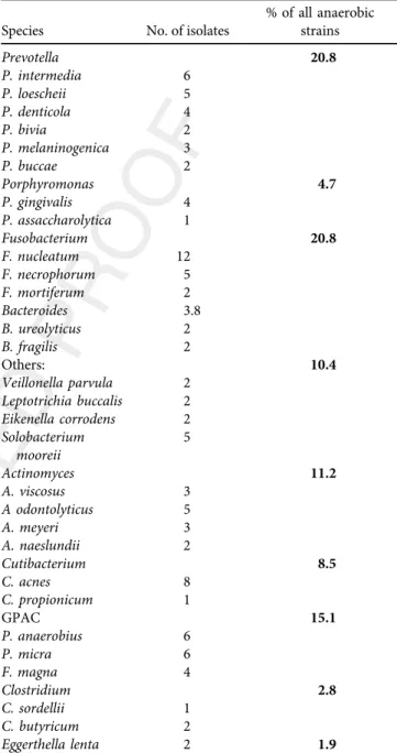
The predominant anaerobic isolates were pigmented Prevotella and Porphyromonas spp. (n 5 27 altogether), Fusobacterium spp. (n 5 19), especially Fusobacterium nucleatum(n512) and numerous Gram-positive anaerobic cocci (GPAC) (n 5 16) (Table 1). The most common anaerobic microorganisms isolated from these samples accounted for 58.8% all of the anaerobic strains in this study.
Unusually high number ofActinomycesspp. strains (n513) were also isolated (12.3%); interestingly one of them was the single cause of the syndrome in a very high colony forming unit count (106 CFU/mL). Typical anaerobic odontopatho- genic strains (e.g.Veillonella parvula, Leptotrichia buccalis, Eikenella corrodensandEggethella lenta) were isolated in the same numbers (n52; 2.6%, respectively). Only four isolates belonged to the genus Bacteroides: n 5 2 of them were Bacteroides fragilisand n5 2 wereBacteroides ureolyticus.
Surprisingly,n53 clostridial strains were also isolated:n51 Clostridium sordelliandn52Clostridiun butyricumisolates, which are not common in this infection, according to the recently published data.
DISCUSSION
In contrast to the well-established roles of microbes in the aetiology of acute sinusitis, the exact roles of the above- mentioned microorganisms (namely Prevotella and Por- phyromonasspp.,Fusobacteriumspp., GPAC,V. parvula,L.
buccalis, E. corrodens, E. lenta, Bacteroides spp. and Clos- tridium spp.) in the aetiology of CRS are uncertain [1, 2].
Various researchers disagree on the microbial aetiology of CRS; some of the disagreement may be explained by the different methodological approaches to the processing of the obtained microbiological samples. Many bacterial organisms have been identified in the sinus tracts of patients with CRS and are reported in the literature, but there is no consensus as to their correct pathogenic role. Despite the exact cause of the inflammation associated with CRS is uncertain, the presence of bacteria within the sinuses has been well docu- mented in different studies [9, 10]. Some of these studies
have examined the bacterial pathogens associated with CRS, but most of these reports did not employ methods for isolation adequate for the recovery of strict anaerobic bac- teria. Studies that have used adequate methods for isolation of anaerobes have demonstrated their prominence in CRS, while those that did not use such methods have failed to recover them. Immunosuppressed patients have episodes of sinusitis caused by the usual agents associated with acute sinusitis in immunocompetent patients and they may also become infected with a broad array of unusual microor- ganisms, including mycobacterial species, fungi and some- times protozoa. According to certain data from the literature, the presence of anaerobic bacteria in CRS in adults is often clinically significant [11]. Initial studies by Frederick and Braude in the 1970s implicated polymicrobial
Table 1.Distribution ofn5106 anaerobic bacterial strains recovered from patients with chronic bacterial sinusitis Q3
Species No. of isolates
% of all anaerobic strains
Prevotella 20.8
P. intermedia 6
P. loescheii 5
P. denticola 4
P. bivia 2
P. melaninogenica 3
P. buccae 2
Porphyromonas 4.7
P. gingivalis 4
P. assaccharolytica 1
Fusobacterium 20.8
F. nucleatum 12
F. necrophorum 5
F. mortiferum 2
Bacteroides 3.8
B. ureolyticus 2
B. fragilis 2
Others: 10.4
Veillonella parvula 2 Leptotrichia buccalis 2 Eikenella corrodens 2 Solobacterium
mooreii
5
Actinomyces 11.2
A. viscosus 3
A odontolyticus 5
A. meyeri 3
A. naeslundii 2
Cutibacterium 8.5
C. acnes 8
C. propionicum 1
GPAC 15.1
P. anaerobius 6
P. micra 6
F. magna 4
Clostridium 2.8
C. sordellii 1
C. butyricum 2
Eggerthella lenta 2 1.9
4
European Journal of Microbiology and Immunology343 344 345 346 347 348 349 350 351 352 353 354 355 356 357 358 359 360 361 362 363 364 365 366 367 368 369 370 371 372 373 374 375 376 377 378 379 380 381 382 383 384 385 386 387 388 389 390 391 392 393 394 395 396 397 398 399
400 401 402 403 404 405 406 407 408 409 410 411 412 413 414 415 416 417 418 419 420 421 422 423 424 425 426 427 428 429 430 431 432 433 434 435 436 437 438 439 440 441 442 443 444 445 446 447 448 449 450 451 452 453 454 455 456
bacterialflora and emphasised the pathogenic importance of different anaerobic species in particular [13].
Previous examinations of sinus puncture aspirates from patients with chronic sinusitis have yielded mixed findings, varying from the absence of anaerobes to anaerobes constituting 56% of all pathogens isolated [13, 14, 17–19].
When adequate sample proceedings and cultivation methods are used, anaerobes can be isolated in more than half of all cases [20]; the leading anaerobic strains were pigmented Prevotella spp., Fusobacterium spp. and GPAC.
Aerobic and anaerobic b-lactamase–producing bacteria (BLPB), such as S. aureus, Haemophilus, Prevotella, Por- phyromonas and Fusobacterium spp. were isolated from more than one-third of patients in different investigations [21–25]. Brook established the microbiological characteris- tics of acute exacerbation of chronic sinusitis (AECS) in an Academic Medical Center compared with chronic sinusitis [24]. He reported 32 patients with chronic sinusitis and 30 patients with AECS and found a total of 81 various isolates (33 aerobic and 48 anaerobic), which were recovered from the 32 cases (2.5 per specimen) with patients of chronic sinusitis. Aerobes alone were recovered in 8 specimens (25%), anaerobes only were isolated in 11 cases (34%), and mixed aerobes and anaerobes were recovered in 13 samples (41%). The predominant aerobic and facultative bacteria were members of Enterobacterales and S. aureus, while predominant anaerobic bacteria were GPAC,Fusobacterium spp., anaerobic Gram-negative bacilli and Cutibacterium acnes[24]. In a study by Erkanet al., a total of 89 isolates (40 aerobic and facultative anaerobes, and 49 anaerobes) were recovered from the 30 patients (3.0 per specimen) with AECS: aerobes were recovered in 8 instances (27%), anaer- obes only in 11 (37%) and mixed aerobes and anaerobes were recovered in 11 cases (37%). The predominant aerobes in his study were S. pneumoniae, Enterobacterales and S.
aureus. This investigation demonstrates that the organisms isolated from patients with AECS were predominantly anaerobic and were similar to those generally recovered in patients with CRS [25]. However, aerobic bacteria that are usually found in acute infections (e.g. S. pneumoniae, H.
influenzae and M. catarrhalis) can also emerge in some of the episodes of AECS [25]. In contrast to these studies, Bhattacharyya et al. found that both anaerobes and aerobic species could be recovered from both diseased and the non- diseased contralateral side of patients with chronic rhinosi- nusitis, casting doubt on the aetiological role of bacteria in CRS; their main finding was that anaerobes are more prevalent in infections secondary to dental problems [26].
Jun Kimet al. investigated the bacteriology and antimicro- bial susceptibility of maxillary sinus aspirates from 81 pa- tients [27]. Aerobes were isolated from 58.0% of the cultures from the middle meatus and from 48.1% of those from the maxillary sinus:S. aureus,H. influenzaeandS. pneumoniae were the most prevalent aerobic pathogens. Anaerobes were only isolated from 8.6% of the cultures from the middle meatus and from 18.5% of the cultures from the maxillary sinus. In this investigation the predominant anaerobic or- ganisms were Prevotella spp. and GPAC in adults, but
interesting, none of these isolates were cultured in children.
A high rate of concordance of the middle meatus and maxillary sinus was noted and monomicrobial infection was most commonly observed [27]. An open-label, multicenter study was performed by Finegold et al. in 2002 to assess culturable bacteriologic findings associated with chronic bacterial maxillary sinusitis in adults [28]. Seventy aerobic (52.2%) and 64 anaerobic (47.8%) pathogens were recovered from clinically evaluable patients at baseline (before ther- apy). The most commonly isolated anaerobic bacteria were Prevotella spp. (31.1%), GPAC (21.9%) andFusobacterium spp. (15.6%), theirfindings consistent with results of other earlier studies. The aerobes most frequently recovered includedStreptococcus spp. (21.4%),H. influenzae (15.7%), Pseudomonas aeruginosa (15.7%), S. aureus and M. catar- rhalis (10.0% each). Recurrences for signs or symptoms of bacterial maxillary sinusitis associated with anaerobes were twice as frequent as were those associated with aerobes when counts of anaerobes were above or equal to 103 CFU/mL [28]. In addition, a pathogenic role forGranulicatellaspp. in chronic sinusitis cases was documented for thefirst time in this study. Brook and Frazier correlated the microbiological findings with the history of sinus surgery in 108 patients with chronic maxillary sinusitis and found a higher rate of isolation ofP. aeruginosaand other Gram-negative bacilli in patients with previous sinus surgery [29, 30]. Anaerobes were, however, isolated significantly more frequently in pa- tients who did not have prior surgery. Brook evaluated the microbiology of 13 chronically infected frontal [30], seven sphenoid [31] and 17 ethmoid sinuses [32]: anaerobic bac- teria were recovered in more than two-thirds of the patients.
In these studies, the predominant anaerobic species included Prevotella, GPAC andFusobacteriumspp., the main aerobic organisms were Gram-negative bacilli (H. influenzae, K.
pneumoniae,E. coliandP. aeruginosa) [30–32]. Nadelet al.
isolated Gram-negative enteric rods more commonly in patient with a history of previous surgery or those who had sinus irrigation, P. aeruginosa was also more frequent in patients who received systemic steroids [21]. Other studies have also noted this shift toward Gram-negative aerobic organisms in patients who had been extensively and repeatedly treated [27, 28, 33]. According to the recent study of Littleet al. the microbiology of odontogenic sinusitis was distinctly different from cases of non-odontogenic sinusitis:
odontogenic-issue sinus infections are generally poly- microbial with obligate anaerobic bacteria predominantly present in cultures, commonly includingGPAC, Prevotella and Fusobacterium spp. [34]. These higher rates of mixed aerobic and anaerobic infections among patients with odontogenic sinusitis have been well documented in the literature [35, 36]. Zirk et al. reviewed 121 cases of odon- togenic sinusitis and noted that 70% demonstrated anaer- obic isolates and 30% aerobes or facultative anaerobes [37].
The variable growth of microbes in samples may also be due to prior exposure of various broad-spectrum antibiotics in patients involved in the studies.
The role of anaerobic bacteria in chronic sinusitis is supported by their ability to induce chronic sinusitis in a
European Journal of Microbiology and Immunology
5
457 458 459 460 461 462 463 464 465 466 467 468 469 470 471 472 473 474 475 476 477 478 479 480 481 482 483 484 485 486 487 488 489 490 491 492 493 494 495 496 497 498 499 500 501 502 503 504 505 506 507 508 509 510 511 512 513
514 515 516 517 518 519 520 521 522 523 524 525 526 527 528 529 530 531 532 533 534 535 536 537 538 539 540 541 542 543 544 545 546 547 548 549 550 551 552 553 554 555 556 557 558 559 560 561 562 563 564 565 566 567 568 569 570
EUJMI-2020.00010_proof 16 May 2020 1:35 pm
rabbit by intra-sinus inoculation ofB. fragilisand the rapid production of serum immunoglobulin G (IgG) antibodies against this organism in the infected animals. In a recent investigation of Jyonouchi et al., the study group induced chronic sinusitis successfully in animal models via intra-si- nus inoculation of a B. fragilis strain [38]. These authors subsequently identified IgG antibodies against the inoculated B. fragilisin the infected rabbits. In addition the other study, the immune response, specific IgG antibodies to 2 anaerobic bacteria (F. nucleatumandPrevotella intermedia) in patients with chronic maxillary sinusitis have been observed [39], so thesefindings further support a pathogenic role for anaer- obes in chronic sinusitis. Antibody levels to these organisms declined in the individuals who responded to therapy and were cured, but did not decline in those who failed treat- ment. In the studies which used appropriate anaerobic cultivation methods and laboratory techniques for identifi- cation, the anaerobic bacteria accounted for 25–56% of the isolates. A recent study using sequencing the species-specific 16S ribosomal DNA fragment for genetic identification of bacteria illustrated the recovery of anaerobes in half of the 18 patients with chronic sinusitis [40].
In our previous study, performed among children after adenoidectomy, the cultivable bacterial composition from nasopharyngeal swabs and from the removed adenoid tissue in the same patient group were compared [41]. The viable bacterial cells (number of colony-forming units) were quantified and the composition of isolated bacteria from both types of samples was also determined in parallel. Our findings showed that the culture results of nasopharyngeal swabs and inner part of the adenoid tissue are in close correlation: polymicrobial aerobic-anaerobicflora was pre- sent in all cases. The predominant aerobic isolates in all two groups were the members of the‘classical triad,’ namelyS.
pneumoniae, H. influenzae and M. catarrhalis. Most com- mon anaerobic strains recovered from the adenoid tissues were Peptostreptococcus spp., Prevotella spp. and Fuso- bacteriumspp. [41].
Our present study of adult CRS patients illustrates the importance of obtaining correct samples from patients with CRS for both aerobic and anaerobic cultures to guide the selection of the proper antimicrobial therapy and to prevent possible life threatening-sequelae. Microbiologic studies of chronic sinusitis often show that the infection is poly- microbial, with the isolation of 1–6 isolates per specimen [21–36]. In this study, the distribution of bacterial number was higher, this number was 1–10 (average: 2.6) and an- aerobes made up 51.5% of the pathogens isolated. Black- pigmented species, includingPrevotellaandPorphyromonas spp., GPAC andFusobacteriumspp. accounted for 63% all of the anaerobic pathogens isolated, a finding consistent with the results of some of the data of the literature which noted a diversity of aerobic and anaerobic bacteria similar to that in our study. Our higher isolation rate ofActinomyces spp. could be attributed to the applied longer incubation period (6–8 days) [42]. The distribution of aerobic and/or facultative anaerobic pathogens in the present investigation was consistent with that seen in some of the other studies of
chronic sinusitis [21–36]. Similar to the data available in the literature,S. pneumoniae(50.6%),H. influenzae(36.7%) and M. catarrhalis(7.6%) were among the most frequently iso- lated aerobic and/or facultative anaerobic pathogens. Isola- tion of Gram-negative enteric rods, includingP. aeruginosa, K. pneumoniae,Proteus mirabilis,Enterobacter spp. and E.
coli were also reported in some other studies [21]. Because these bacteria are rarely isolated from sinus cultures ob- tained from healthy individuals, their recovery from these symptomatic patients suggests their pathogenic role. These organisms may have been selected out following adminis- tration of antimicrobial therapy in patients with chronic sinusitis. Furthermore, consistent with thefindings of other published studies, a wide variety of other aerobes and/or facultative anaerobic pathogens were also recovered (P.
aeruginosa, members of Enterobacterales and fungi). The emergence of new pathogens in all instances, mostly strict anaerobes, generated a polymicrobial infection. This type of infection is one of synergistical nature, and may be more difficult to eradicate with narrow spectrum antimicrobial agents [15, 43]. In such mixed infection, mutual enhance- ment of bacterial growth, and ‘protection’of penicillin-sus- ceptible isolates by beta-lactamase produced by relevant bacteria, may contribute to the chronicity of the infection, and the difficulty in its eradication [15, 16].
CONCLUSIONS
This is the first published account of the detailed microbi- ology of adult-chronic sinusitis in Hungary. The absence of accurate epidemiological data in Hungary on CRS contrasts with the more abundant information on microorganisms, diagnosis and treatment options for these conditions. Our understanding of microorganisms in the paranasal sinus is still incomplete, although there is some association between the viral, fungal and bacterial microorganisms and CRS, the exact nature and importance of the relationship is still un- clear. The microbiology of sinusitis is influenced by the previous antimicrobial therapy, vaccinations, and the pres- ence of the conventional commensal flora, capable of interfering with the growth of pathogens. The microbial flora of chronic sinusitis is affected by previous antibiotic administration, past vaccinations and the presence of normal flora that can suppress the emergence of pathogenic species. In some cases, the baseline chronic sinusitis worsens suddenly or causes new symptoms. This acute exacerbation of chronic sinusitis is often polymicrobial as well, with anaerobic bacteria predominating.
Funding: This research did not receive any specific grant from funding agencies in the public, commercial, or not-for- profit sectors. M.G. was supported by ESCMID’s ‘30 under 30’ Award.
Conflict of interest: The authors declare no conflict of in-Q4 terest, monetary or otherwise.
6
European Journal of Microbiology and Immunology571 572 573 574 575 576 577 578 579 580 581 582 583 584 585 586 587 588 589 590 591 592 593 594 595 596 597 598 599 600 601 602 603 604 605 606 607 608 609 610 611 612 613 614 615 616 617 618 619 620 621 622 623 624 625 626 627
628 629 630 631 632 633 634 635 636 637 638 639 640 641 642 643 644 645 646 647 648 649 650 651 652 653 654 655 656 657 658 659 660 661 662 663 664 665 666 667 668 669 670 671 672 673 674 675 676 677 678 679 680 681 682 683 684
ACKNOWLEDGEMENTS
None.
REFERENCES
1. International Rhinosinusitis Advisory Board. Infectious rhinosi- nusitis in adults: classification, etiology and management. Ear Nose Throat J. 1997;76:5–17.
2. Rosenfeld RM, Piccirillo JF, Chandrasekhar SS, Brook I, Ashok Kumar K, Kramper M, Clinical practice guideline (update): adult sinusitis. Otolaryngol Head Neck Surg. 2015;15:S1–39.
3. National Health Interview Survey. Summary health statistics for US. Adults: National Health Interview Survey; 2012.https://www.
cdc.gov/nchs/data/series/sr_10/sr10_260.pdf (Accessed: 21st of January, 2019).
4. Fokkens W, Lund V, Mullol J, On behalf of the European Po- sition Paper on Rhinosinusitis and Nasal Polyps group.
Europeam Position Paper on RS and NP (EP3OS), Rhinology.
2007;20:1–139.
5. Hastan D, Fokkens WJ, Bachert C, Newson RB, Bislimovska J, Bockelbrink A, Chronic rhinosinusitis in Europe-an under- estimated disease. A GA(2)LEN study. Allergy. 2011;66:1216–23.
6. Centers for Disease Control and Prevention Ambulatory Health Care Data. https://www.cdc.gov/nchs/ahcd/index.htm (Accessed:
21st of January, 2019).
7. Puglisi S, Privitera S, Maiolino L, Serra A, Garotta M, Blandino G, Bacteriological findings and antimicrobial resistance in odonto- genic and non‐odontogenic chronic maxillary sinusitis. J Med Microbiol. 2011;60:1353–9.
8. Gajdacs M, Urban E. The relevance of anaerobic bacteria in brain abscesses: a ten-year retrospective analysis (2008-2017). Infect Dis (London). 2019;51:779–81.
9. Benninger MS, Ferguson BJ, Hadley JA, Hamilos DL, Jacobs M, Kennedy DW, Adult chronic rhinosinusitis: definitions, diagnosis, epidemiology, and pathophysiology. Otolaryngol Head Neck Surg.
2003;129:S1–32.
10. Biel MA, Brown CA, Levinson RM, Garvis GE, Paisner HM, Sigel ME, Evaluation of the microbiology of chronic maxillary sinusitis.
Ann Otol Rhinol Laryngol. 1998;107:942–5.
11. Wald ER. Microbiology of acute and chronic sinusitis in children and adults. Am J Med Sci. 1998;316:13–20.
12. Brook I. Anaerobic infections: diagnosis and management. New York, NY: Informa Healthcare USA, Inc.; 2007.
13. Frederick J, Braude AI. Anaerobic infection of the paranasal si- nuses. N Engl J Med. 1974;290:135–7.
14. Su WY, Liu C, Hung SY, Tsai WF. Bacteriological study in chronic maxillary sinusitis. Laryngoscope. 1983;93:931–4.
15. Summanen P, Baron EJ, Citron DM. Wadsworth anaerobic bacteriology manual. 6th ed. Belmont, CA: Star Publishing; 1993.
Q2
16. Gajdacs M, Spengler G, Urban E. Identification and antimicrobial susceptibility testing of anaerobic bacteria: Rubik’s cube of clinical microbiology? Antibiotics. 2017;6:25.
17. Brook I. Bacteriology of chronic maxillary sinusitis in adults. Ann Otol Rhinol Laryngol. 1998;98:426–8.
18. Karma P, Jokipii L, Sipila P, Luotonen J, Jokipii AM. Bacteria in chronic maxillary sinusitis. Arch Otolaryngol. 1979;105:
386–90.
19. Nash D, Wald E. Sinusitis. Pediatr Rev. 2001;22:111–7.
20. Nord CE. The role of anaerobic bacteria in recurrent episodes of sinusitis and tonsillitis. Clin Infect Dis. 1995;20:1512–24.
21. Nadel DM, Lanza DC, Kennedy DW. Endoscopically guided sinus cultures in normal subjects. Am J Rhinol. 1999;13:87–90.
22. Ramadan HH. What is the bacteriology of chronic sinusitis in adults? Am J Otolaryngol. 1995;16:303–6.
23. Brook I. Microbiology and antimicrobial management of sinusitis.
Otolaryngol Clin North Am. 2004;37:253–66.
24. Brook I. Bacteriology of chronic sinusitis and acute exacerbation of chronic sinusitis. Arch Otolaryngol Head Neck Surg. 2006;132;
1099–101.
25. Erkan M, Aslan T, Ozcan M, Koc N. Bacteriology of antrum in adults with chronic maxillary sinusitis. Laryngoscope. 1994;104:321–4.
26. Bahattacharyya N, Kepnes LJ. The microbiology of recurrent rhi- nosinusitis after endoscopic sinus surgery. Arch Otolaryngol Head Neck Surg. 1999;125:1117–20.
27. Kim HJ, Lee K, Yoo JB, Song JW, Yoon JH. Bacteriologicalfindings and antimicrobial susceptibility in chronic sinusitis with nasal polyp. Acta Oto-laryngologica. 2006;126:489–97.
28. Finegold SM, Flynn MJ, Rose FV, Jousimies-Somer H, Jakielaszek C, McTeague M, Bacteriologic findings associated with chronic bacterial maxillary sinusitis in adults. Clin Infect Dis. 2002;35:
428–33.
29. Brook I, Frazier EH, Foote PA. Microbiology of chronic maxillary sinusitis: comparison between specimens obtained by sinus endoscopy and by surgical drainage. J Med Microbiol. 1997;46:
430–2.
30. Brook I. Bacteriology of acute and chronic frontal sinusitis. Arch Otolaryngol Head Neck Surg. 2002;128:583–5.
31. Brook I. Bacteriology of acute and chronic sphenoid sinusitis. Ann Otol Rhinol Laryngol. 2002;111:1002–4.
32. Brook I. Bacteriology of acute and chronic ethmoid sinusitis. J Clin Microbiol. 2005;43:3479–80.
33. Bolger WE. Gram-negative sinusitis: emerging clinical entity. Am J Rhinol. 1994;8:279–83.
34. Little RE, Long CM, Loehrl TA, Poetker DM. Odontogenic sinus- itis: A review of the current literature. Laryngoscope Investig Otolaryngol. 2018;3:110–4.
35. Saibene AM, Vassena C, Pipolo C Odontogenic and rhinogenic chronic sinusitis: A modern microbiological comparison. Int Forum Allergy Rhinol. 2015;6:41–5.
36. Kuan EC, Suh JD. Systemic and odontogenic etiologies of chronic rhinosinusitis. Otolaryngol Clin N Am. 2017;50:95–111.
37. Zirk M, Dreiseidler T, Pohl M, Odontogenic sinusitis maxillaris: a retrospective study of 121 cases with surgical intervention. J Cra- niomaxillofac Surg. 2017;45:520–5.
38. Jyonouchi H, Sun S, Kennedy CA, Roche AK, Kajander KC, Miller JR, Localized sinus inflammation in a rabbit sinusitis model induced by Bacteroides fragilis is accompanied by rigorous immune responses. Otolaryngol Head Neck Surg. 1999;120:869–75.
39. Brook I, Yocum P. Immune response to Fusobacterium nucleatum and Prevotella intermedia in patients with chronic maxillary sinusitis. Ann Otol Rhinol Laryngol. 1999;108:293–5.
European Journal of Microbiology and Immunology
7
685 686 687 688 689 690 691 692 693 694 695 696 697 698 699 700 701 702 703 704 705 706 707 708 709 710 711 712 713 714 715 716 717 718 719 720 721 722 723 724 725 726 727 728 729 730 731 732 733 734 735 736 737 738 739 740 741
742 743 744 745 746 747 748 749 750 751 752 753 754 755 756 757 758 759 760 761 762 763 764 765 766 767 768 769 770 771 772 773 774 775 776 777 778 779 780 781 782 783 784 785 786 787 788 789 790 791 792 793 794 795 796 797 798
EUJMI-2020.00010_proof 16 May 2020 1:35 pm
40. Stephenson MF, Mfuna L, Dowd SE, Wolcott RD, Barbeau J, Poisson M, Molecular characterization of the polymicrobialflora in chronic rhinosinusitis. J Otolaryngol Head Neck Surg. 2010;39:182–7.
41. Fekete-Szabo G, Berenyi I, Gabriella K, Urban E, Nagy E. Aerobic and anaerobic bacteriology of chronic adenoid disease in children.
Int J Pediatr Otorhinolaryngol. 2010;74:1217–20.
42. Gajdacs M, Urban E, Terhes G. Microbiological and Clinical As- pects of Cervicofacial Actinomyces Infections: an Overview. Dent. J.
2019;7:e85.
43. Gajdacs M, Urban E. Relevance of anaerobic bacteremia in adult patients: a never-ending story? Eur. J. Microbiol. Immunol. 2020;
accepted.
Open Access statement.This is an open-access article distributed under the terms of the Creative Commons Attribution 4.0 International License (https://
creativecommons.org/licenses/by/4.0/), which permits unrestricted use, distribution, and reproduction in any medium, provided the original author and source are credited, a link to the CC License is provided, and changes–if any–are indicated. (SID_1)
8
European Journal of Microbiology and Immunology799 800 801 802 803 804 805 806 807 808 809 810 811 812 813 814 815 816 817 818 819 820 821 822 823 824 825 826 827 828 829 830 831 832 833 834 835 836 837 838 839 840 841 842 843 844 845 846 847 848 849 850 851 852 853 854 855
856 857 858 859 860 861 862 863 864 865 866 867 868 869 870 871 872 873 874 875 876 877 878 879 880 881 882 883 884 885 886 887 888 889 890 891 892 893 894 895 896 897 898 899 900 901 902 903 904 905 906 907 908 909 910 911 912