Accepted Manuscript
Molecularly imprinted polymer based electrochemical sensors for Biopolymers Frieder W. Scheller, Xiaorong Zhang, Aysu Yarman, Ulla Wollenberger, Róbert E.
Gyurcsányi
PII: S2451-9103(18)30231-X
DOI: https://doi.org/10.1016/j.coelec.2018.12.005 Reference: COELEC 352
To appear in: Current Opinion in Electrochemistry
Received Date: 1 November 2018 Accepted Date: 9 December 2018
Please cite this article as: Scheller FW, Zhang X, Yarman A, Wollenberger U, Gyurcsányi RE, Molecularly imprinted polymer based electrochemical sensors for Biopolymers, Current Opinion in Electrochemistry, https://doi.org/10.1016/j.coelec.2018.12.005.
This is a PDF file of an unedited manuscript that has been accepted for publication. As a service to our customers we are providing this early version of the manuscript. The manuscript will undergo copyediting, typesetting, and review of the resulting proof before it is published in its final form. Please note that during the production process errors may be discovered which could affect the content, and all legal disclaimers that apply to the journal pertain.
M AN US CR IP T
AC CE PT ED
Molecularly imprinted polymer based electrochemical sensors for Biopolymers**
Frieder W. Scheller1*,Xiaorong Zhang1, Aysu Yarman1, Ulla Wollenberger1, Róbert E. Gyurcsányi2 1. Institute of Biochemistry and Biology, University of Potsdam, Karl-Liebknecht Str. 24-25, 14476 Potsdam, Germany.
2. Chemical Nanosensors Research Group, Department of Inorganic and Analytical Chemistry, Budapest University of Technology and Economics, Szt. Gellért tér 4, H-1111 Budapest, Hungary.
Frieder W. Scheller: fschell@uni-potsdam.de Xiaorong Zhang: xiaorong.zhang@uni-potsdam.de Aysu Yarman: yarman@uni-potsdam.de
Ulla Wollenberger: uwollen@uni-potsdam.de Róbert E. Gyurcsányi: robertgy@mail.bme.hu
*Corresponding authors: fschell@uni-potsdam.de
Abstract
Electrochemical synthesis and signal generation dominate among the almost 1200 papers published annually on protein imprinted polymers. Such polymers can be easily prepared directly on the electrode surface and the polymer thickness can be precisely adjusted to the size of the target to enable its free exchange. In this architecture the molecularly imprinted polymer (MIP) layer represents only one “separation plate”, thus the selectivity does not reach the values of “bulk” measurements. The binding of target proteins can be detected straightforwardly by their modulating effect on the diffusional permeability of a redox marker through the thin MIP films. However, this generates an “overall apparent” signal which may include non-specific interactions in the polymer layer and at the electrode surface. Certain targets, such as enzymes or redox active proteins enables a more specific direct quantification of their binding to MIPs by in situ determination of the enzyme activity or direct electron transfer, respectively.
** In memory of Professor Emil Palecek
Keywords:
M AN US CR IP T
AC CE PT ED
Electropolymerization Direct electron transfer Redox marker
Epitope imprinting Biomarker
1. Introduction
Highly specific interactions are involved in most essential biological processes, e.g. the antigen-antibody interaction of the immune system, the action of enzymes in substrate conversion and the sequence specific hybridization of nucleic acids. These biomacromolecules are routinely used as specifiers in clinical diagnostics, environmental analysis and food control. In order to overcome some problems of biochemical reagents and to realize low-cost analyses, polymer chemists, biochemists and material scientists develop fully synthetic organic polymers and nucleotide-based aptamers. The synthesis of so-called molecularly imprinted polymers (MIPs) for proteins has been initiated by Mosbach [1,2]. As compared with proteins the number of publications on nucleic acids is relatively small [3–5].
During the synthesis of MIPs monomers are polymerized in the presence of the target molecule, so-called template, which is removed after the formation of a polymeric network.
The removal of the template from the polymer results in the formation of cavities, which mirror the shape of the target molecule. MIPs mimic the binding sites of antibodies by substituting the amino-acid-scaffold for synthetic polymers[6–9]. Whilst enzymes and antibodies are made up by 20 natural amino acids, MIPs can be synthesized from only ONE monomer and even without a cross-linker. This reduction of complexity is a real technological breakthrough. MIPs are more stable under harsh conditions such as high temperature, extreme pH, and organic solvents than antibodies.
2. MIP-Synthesis
Among the almost 1200 papers annually published on MIPs only around 10 percent cover the recognition of biopolymers [10].
The reason for the restricted number of MIPs for high-molecular weight compounds is mostly caused by stability problems of the biomacromolecular templates, e.g. proteins, in the polymerization media. Electropolymerization overcomes several constrictions of radical
M AN US CR IP T
AC CE PT ED
polymerization because it allows for polymer synthesis from aqueous solution under mild conditions. Anodic oxidation of pyrrole, scopoletin, o-phenylenediamine (o-PD), thiophene, p-aminophenylboronic acid and their derivatives in the presence of the target molecule gives ultra-thin MIP-layers directly on the conducting surface of electrodes or chips for quartz crystal microbalance (QCM) and surface plasmon resonance (SPR) [11]. As compared with the chemical MIP-synthesis the spectrum of electropolymerizible monomers is small. Thus the optimization of the monomer/target interaction is restricted.
For the electrosynthesis of MIPs for macromolecular targets, esp. proteins, the following main procedures have been developed (Fig. 1):
(i) In the simplest approach a mixture of functional monomers and macromolecule is polymerized (Fig. 1A). This approach can be also applied to locally electrosynthesize protein- MIPs by the so called microelectrospotting procedure [12].
(ii) Alternatively, the target can be adsorbed at the transducer surface prior polymerization [13] (Fig. 1B). Beside direct adsorption of proteins[14,15], deposition of protein-nanoparticle conjugates can be also used, e.g. by nanosphere lithography [16] to generate surface imprinted polymer layers.
(iii) Oriented binding of the target prior polymerization via site-specific anchors, e.g.
charged self-assembled monolayers (SAMs), boronic acid derivatives [17], aptamers [18] or inhibitors [19], which allows the formation of more uniform cavities in the MIPs (Fig. 1C).
M AN US CR IP T
AC CE PT ED
Fig. 1. Workflow of MIP preparation. A) in one-step by electropolymerization of monomer/template mixture, B) in two-steps that involves the pre-adsorption of the target protein followed by EP of the monomer around the surface-confined targets, C) affinity binding of the target to a self-assembled anchor layer for oriented immobilization of the protein followed by electropolymerization.
Taking advantage of the simpler MIP synthesis and template removal using low-molecular weight compounds, only fragments of the biomacromolecules have been also applied as templates (Fig. 2). In this line exposed peptides (epitopes) [20,21] or protein subunits [22]
have been used as the template in the synthesis of MIPs, which recognize both the epitope and the holo-protein. The concept of using an exposed peptide sequence as the target in the MIP- synthesis -the epitope imprinting approach- has been extended to artificial peptide tags of engineered proteins [23], sugars of glycoproteins [24] and even to chemical labels of macromolecules [25]. Representative examples which have been published within the last three years are presented in section 4.
Fig. 2. Schematic representation of the “epitope” imprinting.
3. Electrochemical readout
Electrochemical approaches allow not only the elegant preparation of MIP-sensors, but they are also powerful tools for the generation of the measuring signal.
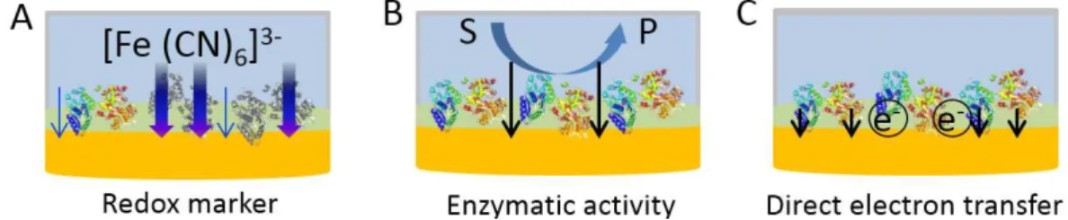
Therefore, fully electronic MIP-sensors, which use electrochemistry for all steps of MIP- synthesis and readout (Fig. 3) are more common among MIP-based protein sensors than SPR, QCM or spectroscopic methods.
(i) The popularity of the electrochemical readout-based MIP sensors is largely due to the simple, cost effective and highly sensitive detection methodology offered by the monitoring the permeability of a small molecular weight redox marker through thin MIP films. The
M AN US CR IP T
AC CE PT ED
simplified model for the generation of the measuring signal assumes that the removal of the protein template generates pathways in the tight MIP layer which allow the permeation of the redox marker to the electrode surface to provide a current signal by its oxidation or reduction.
Rebinding of the target will decrease the current signal by closing these pores and subsequently the pathways to the electrode, thus causing a concentration dependent decrease in the permeation of the redox marker [26]. This methodology applies for insulating MIPs that constrain the redox reaction of the marker species to the electrode surface. The evaluation of the diffusional permeability of the redox active marker can be conveniently followed by many electrochemical techniques, e.g., cyclic voltammetry (CV), square wave voltammetry (SWV), differential pulse voltammetry (DPV) and electrochemical impedance spectroscopy (EIS).
This methodology provides overall a straightforward approach for MIP-based affinity sensors for proteins. It offers also means to characterize each step of the MIP synthesis and the evaluation of the concentration dependence of target-rebinding to the MIP of not only (bio)macromolecular and (nano)particle targets but also of low molecular weight targets.
However, since target rebinding causes only small decreases of the large reference signal after template removal, i.e., signal-off detection methodology, the precision of this approach is inherently problematic. Furthermore, nonspecific adsorption of surface-active constituents of the “real” sample may also influence the current signal.
In spite of the inherent limitations of the method several papers describe MIPs for both small targets and macromolecules with lower limits of detection in the picomolar and even attomolar concentration range (Tab. 1). These publications evaluate either the relative or the absolute decrease of signal suppression in linear or semilogarithmic scales, and generally report two-phasic concentration dependencies without the discussion of the underlying mechanism.
(ii) The analytical performance of MIP-sensors for enzymes can be directly characterized by measuring the enzymatic activity of the biocatalyst bound to the MIP. Using spectroscopic methods, the accumulation of a colored product in the bulk solution was evaluated for trypsin [27], human hemoglobin (Hb) [28] and cytochrome P450 BM3 (P450BM3) [29].
Electrochemical detection of an electroactive product allows the quantification of rebinding directly at the sensor surface. This has been successfully applied for AChE [19], laccase [30], and tyrosinase [31]. This approach is highly sensitive, however, the measuring signal sums up the activity of the enzyme molecules bound to the specific binding sites and that of the non- specifically adsorbed enzyme at the polymer surface.
M AN US CR IP T
AC CE PT ED
Fig. 3. The three main approaches for electrochemical readout of MIP-base electrochemical sensors for proteins: A) the flux of a redox marker is detected at the underlying electrode surface, which is modulated by the protein binding, B) in case of enzyme targets the enzymatic activity is detected through the generation of a redox active product at the electrode surface and C) in case of some redox active proteins the current due to direct electron transfer between the underlying protein and the electrode is measured.
(iii) The most specific electrochemical detection uses direct electron transfer (DET) [32–
34] or bioelectrocatalysis, which is based on DET between underlying electrode and the metalloprotein target. The generation of the catalytic current on addition of the (co)-substrate indicates that the protein reaches the electrode surface and has the “productive orientation” for DET. This approach has been pioneered by Reddy et al. [35] for the catalytic oxygen reduction in the presence of Hb and was transferred to myoglobine [36] and the catalysis of peroxide reduction by MIP-bound Hexameric tytosine-coordinated heme protein (HTHP) [33].
In the following two sections the realization of concepts to uses targets of different complexity from low-molecular weight epitopes via large fragments to biomacromolecules is exemplified by selected papers published within the last three years. The analytical performance of the respective MIP sensors and the potential of the different methods for electrochemical readout of MIPs are compared.
4. MIPs for peptides, proteins, glycoproteins and nucleic acids using epitopes, domains and tags for the MIP-synthesis
The concept of epitope imprinting has been applied for the key player of diabetes-the peptide hormone insulin. A MIP-film for the recognition of insulin was deposited by anodic oxidation of o-PD on top of a SAM of a C-terminal peptide (of not defined length). The measuring signal was generated from the current suppression for the redox marker ferricyanide. The authors report that this epitope-imprinted MIP showed a linear measuring
M AN US CR IP T
AC CE PT ED
range for the “holo”-insulin between 10 fM and 50 pM. They claim “successful application in serum samples” [37].
The carbohydrate antigen 19-9 (CA19-9) is the routinely used biomarker in the diagnosis of pancreatic adenocarcinoma. The epitope approach has been successfully applied for this carbohydrate: The MIP was synthesized using electropolymerization of o-PD in the presence of either the terminal monosaccharide acetylneuraminic acid or the tetra saccharide sialyl Lewis (SLe) on the surface of a glassy carbon electrode (GCE). Rebinding was measured by cyclic voltammetry of the redox marker ferricyanide. The tetra saccharide SLe could be indicated down to 10-13 M and the MIP has a limit of detection (LOD) of 0.028 U/mL for CA19-9 [24].
After the realization of fragment imprinting for IgG [22] the extension of this concept to enzymes has been presented by Jetzschmann et al.[29]. Both the separated domains and the holo P450 BM3 have been bound prior polymer deposition via an N-terminal engineered his6- anchor to the electrode surface. Rebinding after template removal was evaluated by quantifying the suppression of the diffusive permeability of the signal for ferricyanide and by the NADH-dependent reduction of cytochrome c by the reductase domain (BMR). The holoenzyme P450 BM3 was ca. 5.5 times more effectively recognized by the film imprinted with the oxidase domain as compared to the BMR-MIP or the non-imprinted polymer (NIP).
The his6-tagged P450 BM3 binds (30 percent) stronger which shows the additive effect of the interaction with the MIP and the binding to the electrode.
As compared with proteins the number of publications on nucleic acids is very small, due to the fact that hybridization assays represent an alternative difficult to compete with, especially since synthetic analogues of DNA probes, such as peptide nucleic acids, or locked nucleic acids present all advantages in terms of stability that is expected from MIP based receptors. Still DNA MIPs with excellent analytical performances were reported. Thus a MIP for HIV related DNA was prepared by electropolymerizing a solution containing o-PD and 20 µM of 15-mer ssDNA (5-NH2-GGGGGGCCAAGGCCCAGCCCCTCACA-3) on the surface of indium tin oxide (ITO)-electrodes. The template was removed in ethanol/ NaOH mixture.
After rebinding of the template from the sample the MIP-bound ssDNA was hybridized with complementary ssDNA which was conjugated with Europium sulfide nanocrystals. The amount of the HIV-specific DNA was quantified by electrochemiluminescence in the concentration range 3.0 fM to 0.3 nM. The authors do not explain, how the formation of dsDNA which has a larger foot print than the ssDNA target could proceed in the smaller MIP
M AN US CR IP T
AC CE PT ED
cavities and how interaction with only one building block of the polymer (o-PD) could bring about fM affinities [38].
High affinity binding of labeled nucleic acid to a MIP for the low-molecular label was reported by You et al.[25]. A MIP for Rhodamine B (RhB) was prepared by polymerizing methacrylic acid derivatives in the presence of RhB on the surface of a GCE, which was modified with gold nanoparticles and graphene oxide. This MIP recognized RhB modified single stranded DNA with high affinity and allowed the measurement of complementary DNA in the fM-range [25].
5. MIPs for holo-Proteins
The application of the total biopolymer as the target for MIP-synthes is the dominating approach since the intoduction of protein-MIPs. The measuring signal of a MIP sensor for the copper enzyme tyrosinase from mushrooms was generated either by measuring the formation of the oxidation product by the target enzyme or by evaluation of the permeability of the redox marker ferricyanide. It was prepared by electropolymerizing scopoletin or o-PD in the presence of the target protein. The template was removed either by treatment with proteinase K or by alkaline solution. The MIP-sensor has a linear measuring range up to 50 nM of tyrosinase with a limit of detection of 3.97 nM. At saturation of rebinding an imprinting factor of 70 was calculated and the MIP shows good discrimination towards BSA and cytochrome c.
Because both proteins are considerably smaller than tyrosinase, it could be expected that they could simply “fill” the binding pockets and suppress the permeability for the redox marker.
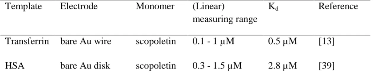
Their smaller effect demonstrates the preference of the interactions between the target and the polymer scaffold [31]. Table 1 presents the analytical parameters of electrosynthesized MIPs which mostly used o-PD or scopoletin as monomers and a redox marker for electrochemical readout.
Tab. 1. MIPs for peptides and proteins prepared by electropolymerization. SPE: screen- printed electrode; CEA: carcinogenic embryonic antigen; RGO: reduced graphene oxide;
FM1: 4-bis(2,2’-bithien-5-yl) methylbenzoic acid glycol ester. nd: not detetermined.
Template Electrode Monomer (Linear)
measuring range
Kd Reference
Transferrin bare Au wire scopoletin 0.1 - 1 µM 0.5 µM [13]
HSA bare Au disk scopoletin 0.3 - 1.5 µM 2.8 µM [39]
M AN US CR IP T
AC CE PT ED
Ferritin bare Au disk scopoletin 0.25 - 0.75 µM nd [39]
Ferritin Carbon- nanotube
phenol 2.3 aM - 227fM 121.8 aM [40]
Troponin T bare Au disk o-PD 0.2 - 21 pM 2.4 pM [41]
Troponin T RGO pyrrole 0.26 - 2.6 pM 0.7 pM [42]
Annexin A3
Carbon-SPE caffeic acid 2.8 pM - 5.5 nM nd [43]
CEA Ag-SPE pyrrole 0.28 - 6.9 fM 32.2 fM [44]
HSA bare Au disk bithiophene derivatives
12 - 300 pM nd [45]
Tyrosinase GCE o-PD 10- 50 nM nd [31]
Oxytocin Au film FM1 0.06 -1 mM nd [46]
Insulin Bare Au o-PD 10 - 500 fM nd [37]
Extraordinary signal amplification was demonstrated for a MIP for epidermal growth factor (EGF). It was prepared by electrochemically initiated polymerization of acrylamide on top of a SAM carrying the immobilized target. Nano-liposomes which were loaded with Cd2+
and decorated with antibodies against EGF were applied for signal amplification. The measuring signal was generated by potentiometric stripping analysis of the liberated Cd2+
ions. The measuring range extended from 0.005 to 5.000 pg/mL [47].
Recently Sun et al. [34] reported about the readout by DET of MIP–bound Hb. The MIP was deposited on top of the electrode which was modified with Fe3O4@SiO2 nanoparticles.
The current signal in the CVs however, are far too cathodic for native Hb, thus the evaluation is questionable. From the analytical point of view binding assays like MIPs cannot compete with simple spectroscopic measurements, e.g. with Drabkin’s method for Hb and signal amplification (as described in [48]) is not required for measurements in the mM-concentration range.
6. Conclusions
Application of MIP-sensors in real samples is still a challenge and the spectrum of targets is still considerably smaller than that of commercially available immunoassays [49,50]. Several protein MIPs have been tested in artificial urine or spiked semi-synthetic plasma and
M AN US CR IP T
AC CE PT ED
measurements in real samples have been claimed. However, measurements by “binding”
sensors in blood are complicated by the presence of highly abundant proteins, e.g. serum albumin, in the g/L region whilst protein marker for heart failures and cancer are typically in the mg/L to ng/L range. Therefore, selectivity coefficients above 1.000 are required whilst MIPs which are synthesized from one or two monomers typically possess vales below 100.
This drawback of electrosynthesized MIPs can be partially compensated by the combination with specific anchors, e.g. boronic acid derivatives [17] or aptamers [18].
Acknowledgments
The authors gratefully acknowledge financial support from Deutsche Forschungsgemeinschaft (DFG) within the framework of the German Excellence Initiative Unicat (EXC 314) and ERA-Chemistry (2014, 61133; NKFIH OTKA NN117637).
References
[1] M. Glad, O. Norrlöw, B. Sellergren, N. Siegbahn, K. Mosbach, Use of silane monomers for molecular imprinting and enzyme entrapment in polysiloxane-coated porous silica, J. Chromatogr. A. 347 (1985) 11–23. doi:10.1016/S0021-
9673(01)95465-2.
[2] M. Kempe, M. Glad, K. Mosbach, An approach towards surface imprinting using the enzyme ribonuclease A, J. Mol. Recognit. 8 (1995) 35–39.
doi:10.1002/jmr.300080106.
[3] S. Li, S. Cao, M.J. Whitcombe, S.A. Piletsky, Size matters: Challenges in imprinting macromolecules, Prog. Polym. Sci. 39 (2014) 145–163.
doi:10.1016/J.PROGPOLYMSCI.2013.10.002. **Very general overview on MIPs from low moleculat to macromolecular analytes
[4] V. Ratautaite, S.N. Topkaya, L. Mikoliunaite, M. Ozsoz, Y. Oztekin, A.
Ramanaviciene, A. Ramanavicius, Molecularly Imprinted Polypyrrole for DNA Determination, Electroanalysis. 25 (2013) 1169–1177. doi:10.1002/elan.201300063.
[5] O. Slinchenko, A. Rachkov, H. Miyachi, M. Ogiso, N. Minoura, Imprinted polymer layer for recognizing double-stranded DNA, Biosens. Bioelectron. 20 (2004) 1091–
1097. doi:10.1016/j.bios.2004.06.027.
[6] G. Wulff, A. Sarhan, Use of Polymers with Enzyme-Analogous Structures for the Resolution of Racemates, Angew. Chem. Int. Ed. Engl. 11 (1972) 341–344.
https://ci.nii.ac.jp/naid/10006961230/ (accessed August 6, 2018).
[7] O. Hayden, P.A. Lieberzeit, D. Blaas, F.L. Dickert, Artificial Antibodies for Bioanalyte Detection—Sensing Viruses and Proteins, Adv. Funct. Mater. 16 (2006) 1269–1278.
doi:10.1002/adfm.200500626.
M AN US CR IP T
AC CE PT ED
[8] D.R. Kryscio, M.Q. Fleming, N.A. Peppas, Protein Conformational Studies for Macromolecularly Imprinted Polymers, Macromol. Biosci. 12 (2012) 1137–1144.
doi:10.1002/mabi.201200068.
[9] N.W. Turner, C.W. Jeans, K.R. Brain, C.J. Allender, V. Hlady, D.W. Britt, From 3D to 2D: A Review of the Molecular Imprinting of Proteins, Biotechnol. Prog. 22 (2006) 1474–1489. doi:10.1021/bp060122g.
[10] M.J. Whitcombe, N. Kirsch, I.A. Nicholls, Molecular imprinting science and technology: a survey of the literature for the years 2004-2011, J. Mol. Recognit. 27 (2014) 297–401. doi:10.1002/jmr.2347.
[11] J. Erdőssy, V. Horváth, A. Yarman, F.W. Scheller, R.E. Gyurcsányi,
Electrosynthesized molecularly imprinted polymers for protein recognition, TrAC Trends Anal. Chem. 79 (2016) 179–190. doi:10.1016/J.TRAC.2015.12.018.
[12] M. Bosserdt, J. Erdőssy, G. Lautner, J. Witt, K. Köhler, N. Gajovic-Eichelmann, A.
Yarman, G. Wittstock, F.W. Scheller, R.E. Gyurcsányi, Microelectrospotting as a new method for electrosynthesis of surface-imprinted polymer microarrays for protein recognition, Biosens. Bioelectron. 73 (2015) 123–129. doi:10.1016/j.bios.2015.05.049.
[13] X. Zhang, A. Yarman, J. Erdossy, S. Katz, I. Zebger, K.J. Jetzschmann, Z. Altintas, U.
Wollenberger, R.E. Gyurcsányi, F.W. Scheller, Electrosynthesized MIPs for transferrin: Plastibodies or nano-filters?, Biosens. Bioelectron. 105 (2018) 29–35.
doi:10.1016/J.BIOS.2018.01.011.
[14] A. Menaker, V. Syritski, J. Reut, A. Öpik, V. Horváth, R.E. Gyurcsányi,
Electrosynthesized Surface-Imprinted Conducting Polymer Microrods for Selective Protein Recognition, Adv. Mater. 21 (2009) 2271–2275. doi:10.1002/adma.200803597.
[15] G. Lautner, J. Kaev, J. Reut, A. Öpik, J. Rappich, V. Syritski, R.E. Gyurcsányi, Selective Artificial Receptors Based on Micropatterned Surface-Imprinted Polymers for Label-Free Detection of Proteins by SPR Imaging, Adv. Funct. Mater. 21 (2011) 591–597. doi:10.1002/adfm.201001753.
[16] J. Bognár, J. Szűcs, Z. Dorkó, V. Horváth, R.E. Gyurcsányi, Nanosphere Lithography as a Versatile Method to Generate Surface-Imprinted Polymer Films for Selective Protein Recognition, Adv. Funct. Mater. 23 (2013) 4703–4709.
doi:10.1002/adfm.201300113.
[17] S. Wang, J. Ye, Z. Bie, Z. Liu, Affinity-tunable specific recognition of glycoproteins via boronate affinity-based controllable oriented surface imprinting, Chem. Sci. 5 (2014) 1135–1140. doi:10.1039/c3sc52986j.
[18] P. Jolly, V. Tamboli, R.L. Harniman, P. Estrela, C.J. Allender, J.L. Bowen, Aptamer–
MIP hybrid receptor for highly sensitive electrochemical detection of prostate specific antigen, Biosens. Bioelectron. 75 (2016) 188–195. doi:10.1016/j.bios.2015.08.043.
[19] K.J. Jetzschmann, G. Jágerszki, D. Dechtrirat, A. Yarman, N. Gajovic-Eichelmann, H.- D. Gilsing, B. Schulz, R.E. Gyurcsányi, F.W. Scheller, Vectorially Imprinted Hybrid Nanofilm for Acetylcholinesterase Recognition, Adv. Funct. Mater. 25 (2015) 5178–
5183. doi:10.1002/adfm.201501900.
[20] A. Rachkov, N. Minoura, Recognition of oxytocin and oxytocin-related peptides in aqueous media using a molecularly imprinted polymer synthesized by the epitope
M AN US CR IP T
AC CE PT ED
approach., J. Chromatogr. A. 889 (2000) 111–118.
http://www.ncbi.nlm.nih.gov/pubmed/10985543 (accessed October 15, 2018).
[21] M. Singh, N. Gupta, R. Raghuwanshi, Epitope Imprinting Approach to Monitor Diseases, J. Mol. Genet. Med. 11 (2017) 1–6. doi:10.4172/1747-0862.1000270.
*General presentation of the epitope aproach for protein MIPs
[22] G. Ertürk, L. Uzun, M.A. Tümer, R. Say, A. Denizli, Fab fragments imprinted SPR biosensor for real-time human immunoglobulin G detection, Biosens. Bioelectron. 28 (2011) 97–104. doi:10.1016/j.bios.2011.07.004.
[23] S. Li, K. Yang, J. Liu, B. Jiang, L. Zhang, Y. Zhang, Surface-Imprinted Nanoparticles Prepared with a His-Tag-Anchored Epitope as the Template, Anal. Chem. 87 (2015) 4617–4620. doi:10.1021/ac5047246.
[24] J. Li, X. Ma, M. Li, Y. Zhang, Does polysaccharide is an idea template selection for glycosyl imprinting?, Biosens. Bioelectron. 99 (2018) 438–442.
doi:10.1016/j.bios.2017.08.004. *Important demonstration of the epitope approach for carbohydrates
[25] M. You, S. Yang, W. Tang, F. Zhang, P. He, Molecularly imprinted polymers-based electrochemical DNA biosensor for the determination of BRCA-1 amplified by SiO2@Ag, Biosens. Bioelectron. 112 (2018) 72–78. doi:10.1016/j.bios.2018.04.038.
*Binding of labeled dsDNA to a MIP for the label
[26] Z. Iskierko, P.S. Sharma, K. Bartold, A. Pietrzyk-Le, K. Noworyta, W. Kutner, Molecularly imprinted polymers for separating and sensing of macromolecular compounds and microorganisms, Biotechnol. Adv. 34 (2016) 30–46.
doi:10.1016/j.biotechadv.2015.12.002.
[27] G. Ertürk, M. Hedström, B. Mattiasson, A sensitive and real-time assay of trypsin by using molecular imprinting-based capacitive biosensor, Biosens. Bioelectron. 86 (2016) 557–565. doi:10.1016/j.bios.2016.07.046.
[28] A. Bossi, S.A. Piletsky, E. V. Piletska, P.G. Righetti, A.P.F. Turner, Surface-Grafted Molecularly Imprinted Polymers for Protein Recognition, Anal. Chem. 73 (2001) 5281–5286. doi:10.1021/AC0006526.
[29] K.J. Jetzschmann, A. Yarman, L. Rustam, P. Kielb, V.B. Urlacher, A. Fischer, I.M.
Weidinger, U. Wollenberger, F.W. Scheller, Molecular LEGO by domain-imprinting of cytochrome P450 BM3, Colloids Surfaces B Biointerfaces. 164 (2018) 240–246.
doi:10.1016/j.colsurfb.2018.01.047. *First example for a domain specific MIP for an enzyme
[30] A. Yarman, Electrosynthesized Molecularly Imprinted Polymer for Laccase Using the Inactivated Enzyme as the Target, Bull. Korean Chem. Soc. 39 (2018) 483–488.
doi:10.1002/bkcs.11413.
[31] A. Yarman, Development of a molecularly imprinted polymer-based electrochemical sensor for tyrosinase, Turkish J. Chem. 42 (2018) 346–354. doi:10.3906/kim-1708-68.
[32] M. Bosserdt, N. Gajovic-Eichelman, F.W. Scheller, Modulation of direct electron transfer of cytochrome c by use of a molecularly imprinted thin film, Anal. Bioanal.
Chem. 405 (2013) 6437–6444. doi:10.1007/s00216-013-7009-8.
M AN US CR IP T
AC CE PT ED
[33] L. Peng, A. Yarman, K.J. Jetzschmann, J.-H. Jeoung, D. Schad, H. Dobbek, U.
Wollenberger, F.W. Scheller, Molecularly Imprinted Electropolymer for a Hexameric Heme Protein with Direct Electron Transfer and Peroxide Electrocatalysis, Sensors. 16 (2016) 272. doi:10.3390/s16030272.
[34] B. Sun, X. Ni, Y. Cao, G. Cao, Electrochemical sensor based on magnetic molecularly imprinted nanoparticles modified magnetic electrode for determination of Hb, Biosens.
Bioelectron. 91 (2017) 354–358. doi:10.1016/J.BIOS.2016.12.056.
[35] S.M. Reddy, G. Sette, Q. Phan, Electrochemical probing of selective haemoglobin binding in hydrogel-based molecularly imprinted polymers, Electrochim. Acta. 56 (2011) 9203–9208. doi:10.1016/J.ELECTACTA.2011.07.132.
[36] V. V. Shumyantseva, T. V. Bulko, L. V. Sigolaeva, A. V. Kuzikov, A.I. Archakov, Electrosynthesis and binding properties of molecularly imprinted poly-o-
phenylenediamine for selective recognition and direct electrochemical detection of myoglobin, Biosens. Bioelectron. 86 (2016) 330–336. doi:10.1016/j.bios.2016.05.101.
[37] C. Zhao, X. Ma, J. Li, An Insulin Molecularly Imprinted Electrochemical Sensor Based on Epitope Imprinting, Chinese J. Anal. Chem. 45 (2017) 1360–1366.
doi:10.1016/S1872-2040(17)61039-9.
[38] B. Babamiri, A. Salimi, R. Hallaj, A molecularly imprinted electrochemiluminescence sensor for ultrasensitive HIV-1 gene detection using EuS nanocrystals as luminophore, Biosens. Bioelectron. 117 (2018) 332–339. doi:10.1016/j.bios.2018.06.003.
[39] Z. Stojanovic, J. Erdőssy, K. Keltai, F.W. Scheller, R.E. Gyurcsányi,
Electrosynthesized molecularly imprinted polyscopoletin nanofilms for human serum albumin detection, Anal. Chim. Acta. 977 (2017) 1–9. doi:10.1016/j.aca.2017.04.043.
[40] D. Cai, L. Ren, H. Zhao, C. Xu, L. Zhang, Y. Yu, H. Wang, Y. Lan, M.F. Roberts, J.H.
Chuang, M.J. Naughton, Z. Ren, T.C. Chiles, A molecular-imprint nanosensor for ultrasensitive detection of proteins., Nat. Nanotechnol. 5 (2010) 597–601.
doi:10.1038/nnano.2010.114.
[41] N. Karimian, M. Vagin, M.H.A. Zavar, M. Chamsaz, A.P.F. Turner, A. Tiwari, An ultrasensitive molecularly-imprinted human cardiac troponin sensor, Biosens.
Bioelectron. 50 (2013) 492–498. doi:10.1016/j.bios.2013.07.013.
[42] B.V.M. Silva, B.A.G. Rodríguez, G.F. Sales, M.D.P.T. Sotomayor, R.F. Dutra, An ultrasensitive human cardiac troponin T graphene screen-printed electrode based on electropolymerized-molecularly imprinted conducting polymer, Biosens. Bioelectron.
77 (2016) 978–985. doi:10.1016/j.bios.2015.10.068.
[43] T.S.C.R. Rebelo, C.M. Pereira, M.G.F. Sales, J.P. Noronha, F. Silva, Protein Imprinted Material electrochemical sensor for determination of Annexin A3 in biological
samples, Electrochim. Acta. 190 (2016) 887–893. doi:10.1016/j.electacta.2015.12.214.
[44] F.T.C. Moreira, M.J.M.S. Ferreira, J.R.T. Puga, M.G.F. Sales, Screen-printed electrode produced by printed-circuit board technology. Application to cancer biomarker
detection by means of plastic antibody as sensing material, Sensors Actuators B Chem.
223 (2016) 927–935. doi:10.1016/j.snb.2015.09.157.
[45] M. Cieplak, K. Szwabinska, M. Sosnowska, B.K.C. Chandra, P. Borowicz, K.
Noworyta, F. D’Souza, W. Kutner, Selective electrochemical sensing of human serum
M AN US CR IP T
AC CE PT ED
albumin by semi-covalent molecular imprinting, Biosens. Bioelectron. 74 (2015) 960–
966. doi:10.1016/j.bios.2015.07.061.
[46] P.S. Sharma, Z. Iskierko, K. Noworyta, M. Cieplak, P. Borowicz, W. Lisowski, F.
D’Souza, W. Kutner, Synthesis and application of a “plastic antibody” in electrochemical microfluidic platform for oxytocin determination, Biosens.
Bioelectron. 100 (2018) 251–258. doi:10.1016/J.BIOS.2017.09.009.
[47] M. Johari-Ahar, P. Karami, M. Ghanei, A. Afkhami, H. Bagheri, Development of a molecularly imprinted polymer tailored on disposable screen-printed electrodes for dual detection of EGFR and VEGF using nano-liposomal amplification strategy, Biosens. Bioelectron. 107 (2018) 26–33. doi:10.1016/j.bios.2018.02.005. *Highly efficient signal amplification by combination of MIP with nano-liposomes
[48] Y. Li, Y. Li, M. Hong, Q. Bin, Z. Lin, Z. Lin, Z. Cai, G. Chen, Highly sensitive protein molecularly imprinted electro-chemical sensor based on gold microdendrites electrode and prussian blue mediatedamplification, Biosens. Bioelectron. 42 (2013) 612–617.
doi:10.1016/j.bios.2012.10.069.
[49] K.J. Jetzschmann, X. Zhang, A. Yarman, U. Wollenberger, F.W. Scheller, Label-Free MIP Sensors for Protein Biomarkers, in: Label-Free Biosensing, Springer, Cham, 2017:
pp. 291–321. doi:10.1007/5346_2017_3. *Overview on MIPs for protein biomarkers [50] O.S. Ahmad, T.S. Bedwell, C. Esen, A. Garcia-Cruz, S.A. Piletsky, Molecularly
Imprinted Polymers in Electrochemical and Optical Sensors, Trends Biotechnol.
(2018). doi:10.1016/j.tibtech.2018.08.009.
M AN US CR IP T
AC CE PT ED
Highlights
1. MIPs mimic efficiently the function of antibodies
2. Electrochemical synthesis and signal generation dominate among the MIP-sensors
3. Number of analytes is still smaller than that of commercially available immunoassays
4. Specificity of MIPs can be improved by integration of boronic acid derivatives or aptamers