Metagenomic etiology and therapy in pediatric gastrointestinal inflammation
PhD thesis
Dorottya Nagy-Szakal MD
Semmelweis University
Doctoral School of Clinical Medicine
Consultant: Dr. Vannay Ádám MD, PhD Official reviewers: Dr. Bene László MD, PhD
Dr. Müllner Katalin MD, PhD
Head of the Final Examination Committee: Dr. Szabó András MD, PhD Members of the Final Examination Committee: Dr. Hegyi Péter MD, PhD
Dr. Miheller Pál MD, PhD
Budapest
2017
“Research is what I am doing when I don’t know what I am doing.”
Wernher von Braun
Table of Contents
List of Abbreviations ... 5
1 Introduction ... 8
1.1 Nutritional developmental origins of disease hypothesis ... 8
1.2 What biologic systems can be modulated by the nutrition? ... 8
1.3 The Human Microbiome Project ... 9
1.4 The microbiome matures during pediatric development ... 10
1.5 The microbiome is nutritionally responsive ... 11
1.6 The human microbiome as an active metabolizer ... 12
1.7 Rapid microbiome responses to marked diet changes ... 13
1.8 The microbiome possesses compositional stability ... 14
1.9 The microbiome possesses metabolic stability ... 14
1.10 The dynamically changing microbiota throughout life stages ... 14
1.11 The role of host immunity and infections in the modulation of gut microbiota ... 14
1.12 Postnatal (pediatric) nutritional exposures affect intestinal inflammation and associate with IBD susceptibility ... 16
1.13 Cellulose supplementation early in life ameliorates acute colitis in adult mice ... 17
1.14 Loss of omega-6 fatty acid induced pediatric obesity protects against acute murine colitis ... 18
1.15 Dysbiosis-associated gastrointestinal disorders and future therapeutic options ... 18
1.16 The manipulation of microbiota as a possible therapeutic option in pediatric GI disorders ... 19
1.17 CDIF and FMT ... 20
1.18 IBD and FMT ... 20
1.19 CDIF and IBD association ... 22
1.20 Other GI disorders and FMT ... 22
1.21 Non-GI disorders and FMT ... 22
2 OBJECTIVES ... 23
3.1 Animals, diets and experimental design ... 24
3.1.1 Cellulose supplementation study ... 24
3.1.2 High fat supplementation study ... 26
3.2 Dextran sulfate sodium exposure ... 26
3.3 Tissue collection and histological analysis ... 27
3.4 Microbiota transplantation in mice ... 27
3.5 DNA extraction and analysis of microbiome ... 27
3.6 Flow cytometry ... 29
3.7 In vivo chemokine blocking ... 29
3.8 Human samples and Enzyme-linked Immunosorbent Assay (ELISA) ... 29
3.9 FMT protocol ... 30
3.9.1 Subjects with recurrent CDIF ... 30
3.9.2 Subjects with UC ... 31
3.9.3 Donor recruitment ... 32
3.9.4 FMT preparation ... 32
3.9.5 FMT treatment protocol in CDIF patients ... 33
3.9.6 FMT treatment protocol in UC patients ... 33
3.9.7 Sample collection ... 34
3.10 Human microbial data analysis ... 35
3.11 RNA sequencing and analysis ... 35
3.12 Histology ... 36
3.13 Additional statistical analysis ... 36
4 RESULTS ... 37
4.1 Cellulose supplementation studies in mice ... 37
4.1.1 Increased severity of DSS colitis on a synthetic, low fiber diet ... 37
4.1.2 Transient trophic and anticolitic effects of cellulose supplementation ... 38
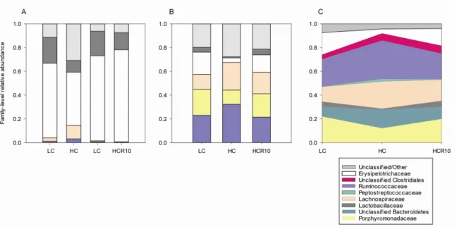
4.1.3 Microbial diversity and composition changes following cellulose supplementation ... 41
4.2 ω-6 fat diet reversal and its effect in murine studies ... 46
4.2.1 Reversal from ω-6 fat diet protected against colitis ... 46
4.2.2 Protection against colitis was ω-6 and reversal dependent ... 47
4.2.3 ω6-reversal did not affect other models of colitis ... 48
4.2.4 The gut microbiome of ω6-reversed mice transmitted the colitis suppression . 48 4.2.5 The microbiome of ω6-reversed mice segregated from control ... 49
4.2.6 Cxcr5+ CD4+ T cells were suppressed in ω6-reversed mice ... 50
4.2.7 Cxcr5 ligand B-lymphocyte chemo-attractant (or Cxcl13) modulated colitis susceptibility ... 50
4.2.8 CXCL13 levels in the plasma of IBD patients ... 50
4.3 Clinical results of FMT ... 52
4.3.1 FMT treatment outcomes of CDIF patients ... 52
4.3.2 Clinical outcomes of patients with ulcerative colitis ... 52
4.3.3 Adverse events monitoring ... 57
4.4 Microbial results of FMT ... 58
4.4.1 Increased alpha-diversity following FMT in CDIF patients ... 59
4.4.2 Taxonomical changes upon FMT in CDIF ... 59
4.4.3 Microbial analyses of UC patients ... 60
4.4.4 Microbiome shifts upon FMT ... 61
4.4.5 Prolonged microbiome effects of serial FMT in two patients ... 62
4.4.6 Remarkable bacterial composition changes in pediatric CDIF after a single FMT ... 64
4.4.7 Normalization of microbiome composition (beta-diversity) upon FMT in CDIF patients ... 66
4.4.8 Microbiome shifts in CDIF following FMT are more pronounced than in UC 67 4.5 Mucosal transcriptome and epithelial cell proliferation changes after the FMT series ... 67
5 CONCLUSIONS ... 70
5.1 Cellulose supplementation early in life ameliorates acute colitis in adult mice ... 70 5.2 Loss of omega-6 fatty acid induced pediatric obesity protects against acute murine
5.3 The manipulation of microbiota as a possible therapeutic option in pediatric
gastrointestinal disorders ... 75
6 KEYNOTES ... 79
7 SUMMARY ... 80
8 ÖSSZEFOGLALÁS ... 81
9 REFERENCES ... 82
10 PUBLICATIONS ... 94
10.1 Related publications of the author ... 94
10.2 Additional unrelated publications of the author ... 95
10.3 Book chapters of the author ... 97
11 ACKNOWLEDGMENTS ... 98
List of Abbreviations
6MP: 6-mercaptopurine ab: antibody
BMI: body mass index
CAP: College of American Pathologists CD: Crohn’s disease
CD: cluster differentiation
CDIF: Clostridium difficile infection CLA: conjugated lineolic acid
CLAI: Clinical Laboratory Improvement Amendment CXCR: C-X-C motif ligand chemokine receptor D: donor
DAB: diaminobenzidine tetrahydrochloride DCs: dendritic cells
DSS: dextran sulfate sodium Dx: diagnosis
E: ethnicity
EIA: enzyme immunoassay F: female
FFAR2: free fatty acid receptor 2 FMT: fecal microbiota transplantation FOS: fructooligosaccharides
G: gender
GABA: gamma-aminobutyric acid GALTs: gut-associated lymphoid tissues GF: germfree
GIT: gastrointestinal tract GLP: Good Laboratory Practice GOS: galactooligosaccharides
GPR43: G-protein coupled receptor 43 H: Hispanic
HC: high cellulose (12.5% cellulose) HE: hematoxylin-eosin
HF (or ω6): high fat (ω6) diet (40% caloric content corn oil/linoleic acid) HMP: Human Microbiome Project
IBD: inflammatory bowel disease IBS: irritable bowel syndrome ID: identification
IL: interleukin IFX: infliximab
IND: Investigational New Drug IRB: Institutional Review Board LC: low cellulose (2.5% cellulose) M: male
MGB axis: microbiota-gut-brain axis MLNs: mesenteric lymph nodes NEC: necrotizing enterocolitis
NF-κB: nuclear factor kappa-light chain enhancer of activated B cells NH: non-Hispanic
NIH: National Institute of Health OTUs: operational taxonomic units
P21/30/80/90/120, 30/80/90/120 days postnatal age PBL: peripheral blood
PBMCs: peripheral blood mononuclear cells PBS: phosphate-buffered solution
PCoA: principal coordinate analysis
PCR: polymerase chain reaction PRED: prednisone
PSA: polysaccharide A Pt: patient
qMRI: quantitative magnetic resonance imaging R: race
R10 and R40: 10 or 40 days of reversal following high cellulose/ high ω6 diet SCFAs: short chain fatty acids
SOPs: Standard Operating Procedures
TCMC: Texas Children’s Microbiome Center TNF-α: tumor necrosis factor α
Tx: therapy
UC: ulcerative colitis W: White
y: years
1 Introduction
1.1 Nutritional developmental origins of disease hypothesis
There is a critical developmental period when nutrition may modify the predisposition to diseases later in life. The hypothesis “Developmental Origins of Health and Diseases” was established in the late 80’s by Barker.(1) The retrospective studies showed that infant mortality (undernutrition in utero and low birth weight) permanently changes body metabolism and leads to an increased cardiovascular and metabolic disease risk later in life. The possible mechanism behind the hypothesis is programming. This process describes the mechanism whereby stimuli at a critical period of development may have long-lasting and irreversible effect on the body structure or function.
1.2 What biologic systems can be modulated by the nutrition?
Biological systems are most likely to respond to environmental stimuli when they are in flux. Developmental maturation and modification is a window opportunity to alter the biological systems. By definition, the biological system has to be responsive for environmental factors and nutritional stimuli. The biological system has to stay stable once adjusted to external stimuli. The biological system needs to be penetrant, and convey phenotype effects.
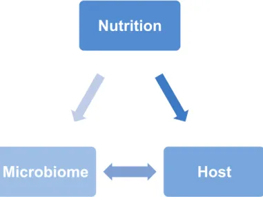
In my studies, we focused on the MICROBIOME, and how such a biological system may be modulated by the nutrition and to characterize the effect on host by these alterations.
1.3 The Human Microbiome Project
The Human Microbiome Project (2) was established in 2008 as a five-year project with a total budget of $115 million funded by the National Institute of Health (3) to characterize the human bacteria composition associated with health and disease.
The human microbiome is a pool of all microorganisms living in association with our body. The microorganisms contain ecological communities of commensal, symbiotic and pathogenic organisms. The number of bacteria is ten times more than the number of human cells; and the compounded genes are thousand times more in the bacteria than presented in the human genome.
An individual’s microbiome is varying depending on genetic predisposition, the mode of delivery (natural birth or cesarean section), age and different environmental factors such as dietary and nutritional intake, physical activity, use of different drugs, and lifestyle.
The homeostasis of gut microbiome is an important key to balance health. On the other hand, dysbiosis (alteration of abundance in bacterial taxa compared to healthy individuals) is associated with diseases and could lead to different disorders. Gut bacteria have a tremendous number of roles, including (1) modifying energy balance and metabolism, and (2) controlling pathogen colonization and resistance. The bacteria performing these roles – and the mechanisms they use – are actively being uncovered.
The improving diagnostic technique took over the culture-based procedures and provided comprehensive characterization of the microbial community. The advanced high- throughput technologies have changed the global view on microbiome dramatically.
Metagenomics is the study of genetic materials recovered directly from environmental samples.
The process starts with the extraction of DNA by the biological sample, followed by amplification and sequencing of 16S ribosomal RNA genes. The 16S rRNA gene allows taxonomic identification from species to phyla level. The identification is made by the comparison to a reference-based library. The advanced bioinformatics techniques provided unique opportunity to map the microbial community and give microbial information about
Focusing on the intestinal microbiome, one large step forward came with the completion of the HMP. Being catalogued the different bacteria species from stools of 242 healthy individuals.(4; 5) The HMP has taught us that the gastrointestinal (GI) microbiome contains around 15,000 to 36,000 bacterial species, and specifically enriched in Bacteroidetes and Firmicutes. These two phyla cover nearly 90% of bacteria in the human gut microbiome. Proteobacteria, Actinobacteria, Fusobacteria and Verrucomicrobia are also presented in the human gut microbiome. However, there is a highly remarkable diversity between individuals at a lower level of taxonomy such as species or strain.
1.4 The microbiome matures during pediatric development
The GI microbiome changes during development. In the beginning, the fetal GI tract (6) is sterile. Colonization of the GIT begins immediately at birth and is directly influenced by mode of delivery, with infants delivered by Cesarean section having delayed colonization and decreased colonization with Bacteroides fragilis as well as Lactobacillus- and Bifidobacterium-like bacteria.(7) Controversial studies showed that the placenta harbors a unique microbiome.(8) First-pass meconium samples of newborns show extremely limited relative abundance of bacteria.(9; 10) The gut is rapidly colonized thereafter. After the first week of life, the microbiome appears to stabilize but varies depending on how the infant is fed. Breast-fed infants are colonized mostly by Bifidobacteria and Ruminococci, and lesser quantities of facultative anaerobes (such as Streptococci, Staphylococci, Enterococci, Lactobacilli and Enterobacteria).(11; 12) Formula-fed infants are colonized by a larger proportion of Firmicutes, Bacteroides and Proteobacteria. Recent studies have shown that formulas supplemented with prebiotics can increase the amount of Bifidobacteria and Lactobacilli in formula fed infants to similar levels found in breast-fed newborns.(13) In infancy, the microbiome continues to change, but by age 2 or 3, an “adult-like” microflora is established. However, a recent study from our collaborators provided evidence that healthy children and adult microbiome remarkably differs in composition and function.(14) Adult microbiome contains more Bacteroides species, while the relative abundance of Bifidobacterium species, Faecalibacterium species
and Lachnospiraceae are increased in children. This study proposes that microbiome may undergo a more prolonged development and more responsible for environmental effects.
1.5 The microbiome is nutritionally responsive
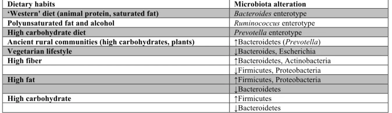
Is it really true that ‘We are what we eat’? From a gut microbiome perspective, what we eat does indeed determine what bacteria we have more so than nationality, age, gender or body mass index (BMI) (Table 1). Humans can generally be divided into three
‘enterotypes’ based on the genera of bacteria in their gut.(15)
Table 1 Nutrition-associated modifications of the microbiota composition.
Dietary habits Microbiota alteration
‘Western' diet (animal protein, saturated fat) Bacteroides enterotype Polyunsaturated fat and alcohol Ruminococcus enterotype
High carbohydrate diet Prevotella enterotype
Ancient rural communities (high carbohydrates, plants) ↑Bacteroidetes (Prevotella)
Vegetarian lifestyle ↓Bacteroides, Escherichia
High fiber ↑Bacteroidetes, Actinobacteria
↓Firmicutes, Proteobacteria
High fat ↑Firmicutes, Proteobacteria
↓Bacteroidetes
High carbohydrate ↑Firmicutes
↓Bacteroidetes
Humans consuming more modern diet riches in animal protein and saturated fat – the
“Western diet” – harbor the Bacteroides enterotype. In contrast, those who consume high amounts of carbohydrates, similar to those from ancient rural communities, have the Prevotella enterotype. Finally, those with high polyunsaturated fat and alcohol intake belong to the Ruminococcus-dominated enterotype. The functional capacity of the human gut microbiome can be mapped to the enterotypes. For instance, the Bacteroides enterotype is characterized by genes involved in biotin, riboflavin, panthothenate and ascorbate biosynthesis were enriched in the Bacteriodes enterotype, associated with genes encoding enzymes involved in the degradation and fermentation of these nutrients. The Prevotella enterotype is enriched in thiamine and folate biosynthesis genes, while the Ruminococcus enterotype consists of heme biosynthesis.
1.6 The human microbiome as an active metabolizer
Gut microbiota are not passive responders to their host’s diet; rather they metabolize a number of substances and function as a nutritive source for the host. For example, bacteria ferment complex carbohydrates into short chain fatty acids (SCFAs). SCFAs in turn boost the human metabolism level; increase energy intake and the bioavailability of toxins. Increased SCFAs decrease colonic pH, inhibit the growth of pathogens, and these nutritional components are the main energy source of colonic epithelial cells. All three SCFAs are reported to enhance colonic blood flow, to stimulate the proliferation of normal crypt cells. By increasing the frequency of the smooth muscle contractions of the colonic mucosa, acetate and propionate leads to an enhanced colonic motility. Butyrate is the main source (about 60 to 70%) of energy for the colonic epithelial cells. Butyrate also influences cell growth and differentiation. Propionate is associated with lipid metabolism
Additionally, pharmaceutical industries are exploiting the nutritive role of SCFAs, and provide prebiotics. Prebiotics are nonviable nutritional components that modify the colonic microbiota conferring benefits for the host health. These components escape digestion in the upper GIT and reach the colon, where they are fermented by the gut microbiota. Inulin-derived fructooligosaccharides (16) and galactooligosaccharides (GOS) change the composition and activation of the microbiota, especially lead to an increase in the amount of Bifidobacteria and Lactobacilli. These products have significant health benefits, including the influence of laxation, mineral absorption, lipid metabolism, and potential anticancer and anti-inflammatory effects.
Microbes also generate a number of essential vitamins for the host. For example, Bacteroides fragilis is known to produce Vitamin K2, therefore a key component in bone and vascular health (increases bone mineral density and modulate the blood coagulation).
Another nutrient, Vitamin B12 (cobalamin) is produced by Lactobacillus reiteri, and important for growth and the development of the nervous system. The absence of this bacteria and its product decreases the risk of different neurological and hematological disorders. Other micronutrients, such as biotin, folate, thiamin, riboflavin and pyridoxine (produced by Bifidobacterium), are not only reinforcing the immune system, but also have epigenetic effects to regulate cell proliferation. Choline metabolites are related to
Faecalibacterium prausnitzii and Bifidobacterium, and linked to glucose and lipid homeostasis, involved in obesity, diabetes and cardiovascular diseases.
Malnutrition and obesity exemplify the relationship between the gut microbiota and human nutrition.(17; 18) Undernutrition (kwashiorkor or marasmus) is linked to an altered microbiota with overall reduced gene content. Malnourished children had increased in Lactobacillus and Bifidobacterium species and decreased in Bacteriodales with nutritional therapy. They also showed signs of a more robust microbiome, including stools with reduced saccharides and an increase of carbohydrate, amino acid, nucleotide and fatty acid metabolism products. . Additionally, antibiotic supplemented therapeutic food can increase the percentage of weight gain and decrease the mortality than therapeutic food only.(19) According to the randomized, double-blinded, placebo-controlled trial, the recovery rate of severe acute malnutrition was 88.7% in the amoxicillin-complemented group and 90.9% in the cefdinir-complemented group, while placebo control reached only an 85.1% recovery rate. The mortality rate changed respectively (4.8% in the amoxicillin group, 4.1% in the cefdinir group and an increased 7.4% in the placebo group).
The obesity-associated microbiota changes are better characterized. Obese individuals have a significantly less diverse microbiome compared to non-obese controls. They are dominated by Actinobacteria (75%) and Firmicutes (25%) phyla, compared to Bacteroidetes (40%) in controls.
1.7 Rapid microbiome responses to marked diet changes
Changes in diet may lead to alteration in gut enterotypes. For instance, individuals switching from the Western diet to a vegetarian lifestyle showed decreased amount of Bacteroides and the related genus Escherichia. These changes in turn influence overall bowel health. Increased endotoxin linked to Bacteroides and Escherichia increases systemic inflammation, whereas high-fiber diets promoting Prevotella correlate with increased bowel transit and production of SCFAs that stimulate intestinal cells.
Supporting the central role of diet in shaping the microbiome, radical dietary change alters
fat/low fiber diet to a high fiber/low fat diet led to detectable changes within 24 hours.(20) Both the composition of bacteria and bacterial gene expression patterns changed, indicating an acute shift in how the microbiome metabolized nutrients. However, only long-term diet changes may lead to actual switches among the three enterotypes.
1.8 The microbiome possesses compositional stability
The species composition may be highly variable among different people. However, within the same person, the gut microbiome composition varies little over time compared to other body sites such as oral cavity, skin and hair.(21)
1.9 The microbiome possesses metabolic stability
The second core principle is that even though microbial taxa may vary among individuals, the metabolic pathways catalyzed by the bacteria remain stable.(5) Hence, current techniques focused on identifying specific bacteria, such as shotgun sequencing of bacterial genomic DNA, may misrepresent the actual metabolic capacity of a microbiome.
Instead, much larger scale systems biological approaches are needed in the future to confirm the conclusions of the Human Microbiome Project Consortium in respect to the high similarity in metabolic patterns representative of different microbial habitats of the human body.
1.10 The dynamically changing microbiota throughout life stages
The gut microbiome loses richness with age, correlating with altered dietary habits, chronic illnesses, and increased use of medications.(22)
1.11 The role of host immunity and infections in the modulation of gut microbiota Gut microbes must maintain the barrier between host and all the environmental challenges present in the intestine. Stratification and compartmentalization is occurred to decrease the exposure of external stimuli and increase the efficacy of the immunity.(23)
The microbiota actively interacts with the innate and adaptive immune system. The infant gut microbiome affects the maturation of the host immune system and carefully adjust it in early days of life. The microbiota contributes to develop the gut-associated lymphoid tissues (GALTs). The GALTs are immune structures, which participate in lymphocyte functions and have an important role in tolerance or inflammation. The main function of the microbiota-induced development of the intestinal immune system is the promoted T helper 17 and T helper 1 cells (activated by the lamina propria dendritic cells [DCs] and macrophages) and the stimulated intestinal epithelial cells and DCs promoting IgA- producing B cells and plasma cell differentiation. (24; 25)
The decision-making process of the immune system (recognition of pathogenic or non-pathogenic bacteria) is orchestrated by the host-microbial interaction. The pathogenic bacteria will be destructed and eliminated, while the host intestine will tolerate the beneficial bacteria. The finely balanced interplay between the healthy beneficial microbiota and the exogenous and potentially harmful pathogens maintains the intestine homeostasis.
This process is based on metabolites cellular and soluble elements associated to the microbiota and sensed by the immune system. These metabolites can be uniquely produced by the microbes (such as SCFAs), or evaluated (e.g. retinoic acid), or metabolized by the microbes (such as the host-presented bile acids).(18; 26) These specific metabolites and their activities -can present the current microbial composition and host-microbial communication.
Microbiota can produce nutritional components -short chain fatty acids (SCFAs), conjugated lineolic acid (CLA), polysaccharide A (PSA) gamma-aminobutyric acid (GABA) and histamine- which affect the host immune system via direct or indirect mechanisms. As an immunological standpoint, the end products of carbohydrate fermentation (by SCFAs) can bind to a G-protein coupled receptor 43 (GPR43) or free fatty acid receptor 2 (FFAR2); and affect inflammatory responses. (27) The receptor-binding mechanism leads to an over-expression of interleukin (IL)-10, and a decreased expression of IL-12 in the human monocytes. This interaction is a great example for the diet-GI
tumor necrosis factor α (TNF-α), IL-8 and inhibited nuclear factor kappa-light chain enhancer of activated B cells (NF-κB) pathway leading to an enhanced anti-inflammatory response.
Microbiota can also generate CLA, which are produced mostly by Bifidobacterium breve, B. longum, Lactobacillus and Propionibacterium and modulate the immune system with its anti-inflammatory properties. PSA (produced by Bacteroides fragilis) is also characterized by its immunomodulatory and anti-inflammatory activities. The neurotransmitters (GABA and histamine) affect the central nervous system produced by various microorganisms, especially Lactobacillus brevis.(28)
1.12 Postnatal (29) nutritional exposures affect intestinal inflammation and associate with IBD susceptibility
Inflammatory bowel diseases (IBDs) including Crohn’s disease (CD) and ulcerative colitis (UC), have been recognized as disorders whereby environmentally sensitive developmental factors may play an important etiologic role (30). The peak incidence of IBD is in young adulthood. Therefore, there is a prolonged developmental period from conception to young adulthood for environmental influences to critically impact biological systems relevant for IBD pathogenesis (31). Important elements of IBD pathology are thought to be the intestinal microbiome, the gut mucosa, and the mucosa associated immune system (30; 32). Our research group recently shown in a mouse model that maternal supplementation of 4 micronutrients could significantly modify offspring colitis susceptibility in association with colonic mucosal gene expression and microbiome alterations (33; 34). However, the same diet did not induce obvious phenotype changes with respect to intestinal inflammation when given during pediatric development. These findings underscored that pertinent nutritional factors can exert persistent colitis modifying effects during critical periods of mammalian maturation.
1.13 Cellulose supplementation early in life ameliorates acute colitis in adult mice As for IBD, the decreased consumption of dietary fibers has been recognized to potentially play an etiologic role (35). The rising incidence of IBD in the developed world associating with a decrease in dietary fiber intake was emphasized by Burkitt during the early 70’s (36). In agreement with this observation, a recent study highlighted the importance of different nutritional habits and gut microbial diversity in children from two different continents and cultures (European and a rural African village) (37). The authors called the attention to the reduction of fiber intake and the associated decrease of microbial richness in European children, which may be relevant in respect to gastrointestinal diseases.
Indirect epidemiologic data supports these conclusions where high fiber and fruit consumption are associated with a decreased risk of CD and high vegetable intake is associated with a decreased risk of UC (35).
Cellulose is an insoluble fiber and an abundant component of a vegetarian diet since it is present in most plant tissues (38). It has a proliferative effect on the colon, mostly in the distal areas in rodent models (39). The colonic trophic effects of cellulose do not depend on microbial fermentation (40), as opposed to the case of other fibers, such as guar gum (41). Although cellulose may be fermented in the colon, the secondary production of SCFAs is limited (42). Despite the limited fermentation of cellulose, it has been shown to substantially modify colonic microbial composition (43). These results implicate it as a potential prebiotic (non-digestible carbohydrate that favors the growth of desirable microflora in the large bowel). Since cellulose is a major component of vegetable and fruit fibers, the indirect nutritional epidemiologic data already discussed (35; 37) would support it as an important factor in IBD pathogenesis. However, the effect of cellulose on mammalian colonic mucosal microbiome (which may be more relevant for IBD pathogenesis than luminal bacteria (44; 45)), or colitis susceptibility has not yet been investigated especially from the developmental origins perspective.
Therefore, in this study, we examined the direct and prolonged effects of pediatric cellulose supplementation on large intestinal growth, chemically induced colitis susceptibility, and colonic mucosal microbial community composition in mice.
1.14 Loss of omega-6 fatty acid induced pediatric obesity protects against acute murine colitis
A large-scale prospective nutritional study linked ω-6 fatty acid consumption to the development of UC.(46) However, clear results from human epidemiologic observations (even in prospective and well controlled) are very difficult to obtain for obvious ethical and technical considerations.(47) Therefore, the timing and nature of environmental factors critical to IBD development have remained largely unknown, as have the molecular mechanisms, which may uncover the environmental contribution to disease induction.(48) Environmental influences may trigger critical pathogenic changes at any time within an individual prior to the onset of disease, which can occur during a broad age range from fertilization to adulthood in case of IBD. Here, we examined the complex effects of transient high ω-6 fat diet consumption during pediatric development on the young adult metagenome, and immune system in a murine model of IBD.
1.15 Dysbiosis-associated gastrointestinal disorders and future therapeutic options The commensal microbiota is a major communicator of dietary modification towards the intestinal immune system of the host and can rapidly change its composition upon nutritional challenges. Disease-associated microbiome changes can be seen in different GI disorders. For example, dysbiosis is found in IBDs. IBD is linked to a reduced proportion of Bacteroidetes and Firmicutes (with an increased Firmicutes/Bacteroidetes ratio) and an increased proportion of Proteobacteria compared to healthy individuals.(49) Colitis is also associated with a decreased abundance of Faecalibacterium prausnitzii, which is characterized by a notable anti-inflammatory effect.(50) Premature colonization of the intestine and other trigger factors together can lead to necrotizing enterocolitis (NEC), which is characterized by an increased abundance of Proteobacteria and Gammaproteobacteria and a decrease in Bacteroidetes and Firmicutes.(51) The key role is the pathomechanism of NEC is supported by the fact that probiotic supplemented breast milk can decrease the chance of developing the disease.(52) Irritable bowel syndrome (IBS) is a functional bowel disorder described with abdominal pain and alternating defecation habit. Pediatric IBS subtypes are distinguished by microbial composition differences. IBS
is associated with an increased abundance of Gammaproteobacteria and pain frequency is linked to a greater proportion of Alistipes genus.(53)
Reduction in microbial diversity or richness is also related to the development of intestinal diseases (e.g., IBDs, NEC). However, alteration of diet does not specifically mean beneficial effect on the colon. Monotonous dietary intake may decrease mammalian vulnerability against intestinal inflammation in association with microbiota separation. (54) In conclusion, the understand of the unique human gut microbiome and its association with disorders will may help developing personalized medicine and therapeutics in the future.
However, results of the microbiome studies are based mostly on findings from pharmacologically treated patients. Treatments highly modify microbiome, and more significantly than the disease itself. (55; 56) For example, enteric dysbiosis in untreated IBD patients appears to be rather modest and provided unexpected findings in recent pediatric studies.(57)
Microbial communities with different metabolic profiles on drugs will may modify the current treatment strategies. (58) Microbiome can modify drug action, metabolism and toxicity in the human gut and liver. Gut-associated microbes alter drug metabolism by modulating the drug-metabolizing enzymes activity, or change their level. Microbes also can directly produce enzymes that activate or degrade drug metabolites, or compete with molecules affecting xenobiotic metabolism. In the future, pharmacomicrobiomics will may be taken into consideration with pathologic and pharmacokinetic characteristics of individuals.
1.16 The manipulation of microbiota as a possible therapeutic option in pediatric GI disorders
The restoration of human microbial homeostasis may be highly beneficial for GI disorders, where dysbiosis present. The most complex bacteriotherapy is fecal microbiota transplantation (FMT). FMT is the process of stool transplantation from a healthy individual into a recipient. The history of FMT goes back to the 4th Century, when Ge Hong
pseudomembranous colitis by fecal retention enema.(60) Immediate recovery and resolution of symptoms was observed within days in all patients. As our current knowledge, FMT is safe and highly effective for the treatment of recurrent Clostridium difficile infection (CDIF) in patients without complicating clinical conditions. Besides recurrent CDIF, there is an emerging interest to use FMT as a novel therapeutic option in patients with other GI disorders, such as IBD or other diseases.
1.17 CDIF and FMT
CDIF is emerging, despite the increased efforts towards prevention, improving diagnosis and therapy.(61) CDIF is the most commonly reported nosocomial pathogen in the United States (US). Epidemiologic data suggest that the CDIF-associated severe diarrhea is continuously increasing with a rise in hospitalization, morbidity and mortality.(62; 63) The latest reports showed CDIF to cause more than $1.5 billion excess medical costs annually in the US. (64) The incidence of pediatric CDIF is also increasing.(65) Recurrence of CDIF is 20-30%, which increases upon repeated infections in spite of new antibiotic regimens.(66) FMT has been shown to be the most effective treatment to date for CDIF.(67) The largest systemic review including 844 patients (76%
diagnosed with CDIF) undergoing FMT showed 90.7% cure rates.(68) Similar cure rates have been shown for children,(69; 70) but microbiome analyses were performed only in a few cases.(71)
1.18 IBD and FMT
UC and CD are the two forms of IBD that affects about 50% of the estimated 1.8 million people suffering from IBD in the US.(72) About 20% of IBD cases present in children, where the disease frequently (~40%) becomes refractory to conventional medical therapy within 10 years of diagnosis, leading to colectomy.(73) FMT is an emerging unconventional treatment for UC.(74; 75) Almost half (46%) of adult UC patients would consider FMT as therapeutic option (76), and both adult and pediatric patients wish to have it available as a treatment modality.(77) A recent meta-analysis evaluated the efficacy of
FMT as a treatment option for IBD patients.(78) Eighteen studies were included with 122 patients (79 UC and 39 CD). The 9 cohort studies, 8 case studies and 1 randomized controlled trial demonstrated an overall 45% clinical remission over varying length of follow up time. According to disease severity, 37% of patients had active disease, 29% had moderate-to-severe disease, and 23% had mild disease. However, the review did not specify any correlation between FMT efficacy and IBD disease activity.
A pediatric trial tested 5 daily FMT in mild-to-moderate UC, where 6 out of 9 children (67%) achieved and maintained clinical remission 4 weeks after the last treatment.(79) However, a phase I pediatric study showed negative results following a single FMT via nasogastric tube.(80) The first randomized, placebo controlled trial of FMT as treatment for active UC was presented as an abstract at the Digestive Disease Week conference in 2014. Moayyedi et al. compared administration of FMT versus water-enema for 6 consecutive weekly treatments. They found no difference in the primary outcome of clinical remission after 6 weeks. However, 16 of the 27 patients in the active arm reported subjective improvement, and were allowed to continue receiving weekly FMT for an additional 6-12 weeks. With the extended therapy, 33% of patients achieved clinical remission.(81) Their consecutive publication indicated that FMT induces remission in a greater proportion of UC patients than placebo. Additionally, patients with a less than 1- year history of UC responded better to FMT than those with more prolonged disease course prior to the intervention.(82) Interestingly, another recently published placebo controlled trial did not find a clinically significant benefit from FMT in adult UC patients.(83) This protocol differed significantly from Moayyedi, et al. by administering only 2 FMT treatments within 3 weeks by naso-duodenal delivery and they used autologous stool as placebo. The different outcomes from the two controlled studies may be related to the difference between the placebo used, the mode of delivery, the number of FMT given in addition to differences in recipient disease activity at the time of FMT initiation.
1.19 CDIF and IBD association
Interestingly, Clostridium difficile is an opportunistic pathogen that presents at high rates in patients with underlying chronic diseases and long-term hospitalization. For instance, the prevalence of CDIF at disease onset was 8.1% in a pediatric population with underlying IBD at Texas State, USA.(84) This prevalence is much higher than the general population.
1.20 Other GI disorders and FMT
There is an increasing attention on the association between intestinal microbiome, obesity and obesity-related metabolic disorders. The presented altered microbiome is associated with low-grade inflammation of the gut.
Researchers described dysbiosis in patients with irritable bowel syndrome. The composition of microbiota represented the symptoms associated with the disease (constipation vs. diarrhea type IBS). (85) Additionally, 70% of the patients experienced improvement of their symptoms following FMT.(86) Furthermore, long-term resolution was seen almost half of the patients.
The strong association between diet, host metabolism and gut microbiome demonstrates homeostasis and misbalance in cases with metabolic disorders. The transplantation of microbiome composition from a lean subject to an obese improved insulin resistance by the increased presence of SCFA-producing bacteria.(87)
1.21 Non-GI disorders and FMT
There is an increasing interest in the role of intestinal dysbiosis and non- gastrointestinal disorders, and to treat patients with FMT. The gut microbiota regulates intestinal function, the immune and nervous systems of the intestine, and influence the brain by the microbiota-gut-brain (MGB) axis. This concept opens the door for a possibly novel therapeutic option for the treatment of neuropsychiatric disorders, chronic fatigue disease and autism.
2 OBJECTIVES
1. There is a direct link between nutrition, microbiome and host response.
2. Postnatal exposure to different nutrients (such as cellulose or fat) has transient or persistent effect on intestinal homeostasis and predispose to the development for intestinal inflammation.
3. Complex bacteriotherapy, such as FMT provides treatment and/or the resolution of symptoms for patients suffering in CDIF and IBD.
Figure 1 The Nutrition-Microbiome-Host Triangle.
Nutrition
Host
Microbiome
3 METHODS
3.1 Animals, diets and experimental design 3.1.1 Cellulose supplementation study
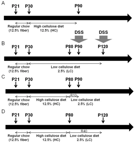
Our initial observations were made on C57BL/6J male mice (Jackson Laboratories, Bar Harbor, ME, USA) receiving (regular chow [12.5% dietary fiber]; 2920X, Harlan- Teklad, Madison, WI, USA) or synthetic low cellulose (2.5% cellulose, LC; #102460, Dyets Inc., Bethlehem, PA, USA) from 30 to 90 days of age. In the consecutive experiments, 21-day old (postnatal days 21, P21) C57BL/6J male mice were provided free access to regular chow (12.5% dietary fiber) within the same room of our animal facility for 9 days. At P30, the mice were randomly allocated to receive synthetic low cellulose or high cellulose (12.5% cellulose, HC; #102532) diet for 50 days (Table 2).
Table 2 The composition of the synthetic low cellulose (LC: 2.5% cellulose) and the high cellulose (HC: 12.5% cellulose) diets.
LC HC LC HC
Ingredient kcal/gm grams/kg kcal/kg
Casein 3.58 200 716
DL-Methionine 4 3 12
Sucrose 4 341.46 1366
Cornstarch 3.6 242.5 172.5 873 621
Dyetrose 3.8 90 60 342 228
Corn Oil 9 51 459
Cellulose 0 25 125 0
Mineral Mix #200000 0.47 35 16.45
Vitamin Mix #300050 3.92 10 39.2
Choline Bitartrate 0 2 0
Ethoxyquin 0 0.04 0
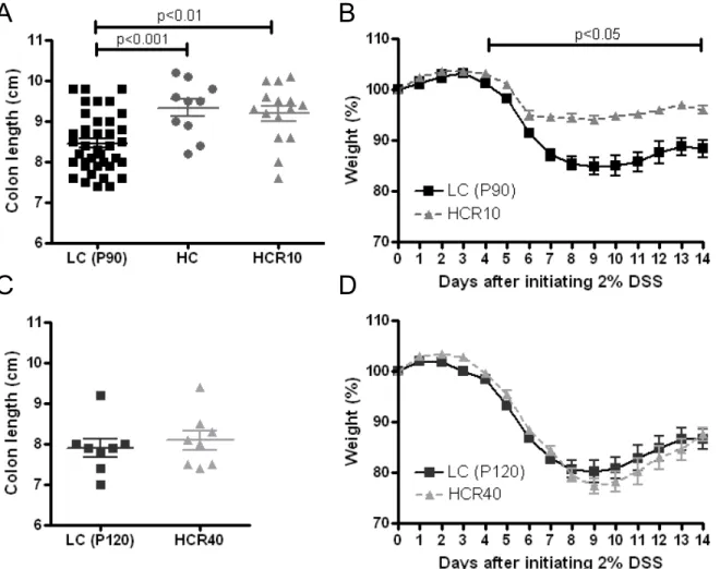
Conventional chows regularly contain ~12.5% fiber (including cellulose). At P80, the HC animals were reversed to control (low cellulose) diet for 10 (HCR10 = P90), or 40 days (HCR40 = P120) (Figure 2). For the microbiome studies, 10 animals in two cages were used for each dietary group. Those were allocated to receive either low cellulose (control, LC, 10 animals in 2 cages), or high cellulose diets (HC, 10 animals in 2 cages). At P80, 2 animals from each cage were crossed between the groups to eliminate cage bias.
Five control animals at P80 and 5 at P90 were euthanized for tissue collection. There was no significant separation between these groups based upon a principal coordinates analysis
(PCoA) of 16S rRNA sequence information; therefore, these were grouped together as a single control. As for the high cellulose group, 2 animals were crossed between the two cages at P80. One cage was continued on high cellulose diet until P90 (HC), and the other cage was reversed to the control diet (low cellulose, LC) until P90 (HCR10), when those were euthanized for tissue collection. The discovery cohorts included 3-3 mice in two independent studies (P90 LC and HC; and P90 LC and HCR10) with the same experimental conditions.
Figure 2 Schematic description of the cellulose feeding protocols. A: 12.5% high cellulose (HC) group without reversal for the purposes of colonic length and microbiota analysis. B:
2.5% low cellulose (LC) group. C: 10-day reversal group (HCR10). D: 40-day reversal group (HCR40). For the B, C and D groups DSS was administered at postnatal day 90 (P90) and P120, respectively. (DSS: dextran sulfate sodium; R10 and R40: 10 or 40 days of reversal following high cellulose diet; P21/30/80/90/120, 30/80/90/120 days postnatal age).
3.1.2 High fat supplementation study
P21 male C57BL/6 mice received standard rodent diet (2920X) until P30. Then, mice were randomly assigned to artificial diets: low 12% caloric content corn oil/linoleic acid: control diet, C (DYET# 102460); or to high ω-6 (40% caloric content corn oil/linoleic acid: ω6 or HF) (DYET# 102459) from P30 to P80. Following this 50-day period, the animals were reversed to C for 10 (ω6-R10) or 40 (ω6-R40) days. High milk fat, high cholesterol (MF) diet (DYET# 112734) was used with the same feeding protocol to examine fat-dependent effects. Fat to body weight ratio was interrogated on P90 and P120 by quantitative magnetic resonance imaging (qMRI). 6-week-old Swiss-Webster germfree (GF) mice were derived by cesarean section under GF conditions at Taconic Farms Inc.
(SWGF; Hudson, NY, USA) and delivered with specific GF shipping for the microbiota transfer experiments.
All mice were housed in our specific pathogen free animal facility during the experiment. All applicable institutional and governmental regulations concerning the ethical use of animals were followed. The protocol was approved by the Institutional Animal Care and Use Committee of Baylor College of Medicine.
3.2 Dextran sulfate sodium exposure
Susceptibility to colitis was tested by administering 2% (wt/vol) (for the synthetic chow experiments) or 3% (for the original regular chow vs. synthetic chow observations) dextran sulfate sodium (DSS; MW=36000-50000, MP Biomedicals, LLC, Solon, OH, USA) in the drinking water at P90 or P120 ad libitum for 5 days followed by regular water for an additional 9 days. DSS of this molecular weight induces diffuse colitis from cecum to distal large bowel (88). The animals were weighed daily and colonic length measurements were performed at the end of the experiments following CO2 asphyxiation.
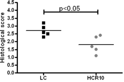
Weight loss during DSS administration in mice has been shown to correlate well with molecular and histological outcome measures of colitis severity (27; 31; 89). Therefore, we decided to follow weight loss, colon length, and histological severity of intestinal inflammation as the primary outcomes measuring colitis severity in our DSS experiments.
3.3 Tissue collection and histological analysis
At the end of the feeding periods, mice were sacrificed by CO2 asphyxiation between 11:00 AM and 2:00 PM without any previous food restriction. The colons were placed on ice, transected longitudinally, cleansed from feces, washed with ice cold normal saline, followed by the collection of colonic mucosa with a microscope slide (90) (excluding the cecum). The mucosal scrapings were flash frozen on dry ice, and stored at - 80oC as earlier described (31).
For histological analysis, additional colonic samples were transected and processed for standard hematoxylin-eosin (HE) staining following fixation in 10% formaldehyde.
Histological severity of intestinal inflammation was determined by a blinded pathologist based upon tissue damage grade and the presence of inflammatory cells. (54)
Morphometric analyses (crypt length, surface area) were performed with Olympus DP2-BSW program on transected proximal (post-cecal) colonic specimens.
3.4 Microbiota transplantation in mice
Cecal contents were pooled from ω6-R40 or C120 littermates. Cecal extracts were suspended in phosphate-buffered solution (PBS, 2.5 ml per cecum) and were administered orally by gavage (0.1 ml per mouse) immediately to sterilely packed 6-week-old SWGF male GF mice (Taconic, Hudson, NY, USA) as described by Vijay-Kumar et al.(91) Transplanted mice were fed with control diet for 5 days, and then 3% of DSS was given to induce acute colitis. Body weight was measured daily. The animals were euthanized and tissues were collected as above.
3.5 DNA extraction and analysis of microbiome
Colonic mucosal samples were submitted to the Texas Children’s Microbiome Center (TCMC, Houston, TX, USA) for DNA extraction and sequencing. Community DNA was extracted from each specimen using the PowerSoil DNA isolation kit (Mo Bio Laboratories, Carlsbad, CA, USA), following the HMP modifications (2) to the
1000 spectrophotometer (NanoDrop, Wilmington, DE, USA) and Qubit fluorometer (Life Technologies Corporation, Carlsbad, CA, USA). Barcoded universal primers 357F (5’- CCTACGGGAGGCAGCAG-3’) and 926R (5’-CCGTCAATTCMTTTRAGT-3’) were utilized to amplify the V3-V5 region of the 16S rRNA gene. The discovery study utilized a different set of PCR primers (28F [5’-GAGTTTGATCNTGGCTCAG-3’] and 519R [5’- GTNTTACNGCGGCKGCTG-3’] amplifying the V1-V3 region of the 16S rRNA). Each library construct was then processed and purified for 454 sequencing. Sequencing was performed on the Roche GS FLX 454 sequencer (454 Life Sciences, Branford, CT, USA).
Sequence data were parsed by barcode and quality filtered using QIIME (version 1.3.0) (92), as implemented in the Genboree Microbiome Toolset (93). Sequences shorter than 200 bp lengths, having average quality scores less than 20, harboring ambiguous base calls, or having mismatches to their barcode or sequencing primer were excluded from further analysis. Both the barcodes and sequencing primers were trimmed away, and the remaining sequences from the control and treatment groups were pooled and assigned to operational taxonomic units (OTUs) at a similarity cut off of 97% using Cd-hit (94). The data set was screened for potential chimeras using the ChimeraSlayer algorithm (95), and all potential chimeras were excluded from downstream analysis. Identities were assigned to each OTU using the Ribosomal Database Project Classifier (96).
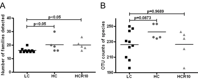
Prior to the calculation of diversity metrics or comparison across treatments, the sequence libraries were randomly subsampled to achieve even sampling depth (subsampled depth = 3634 sequences per library). All results presented here were determined using our subsampled sequence libraries. Community similarity was evaluated across treatments among treatments using principle coordinates analysis (PCoA) of OTU data. PCoA plots were generated using weighted Unifrac distances, as calculated in QIIME.
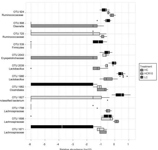
Sequence libraries were also evaluated for potential differences in composition using two-tailed Mann-Whitney U-tests and false discovery rate correction for multiple comparisons in Genespring GX (version 12.0) (Agilent Technologies, Santa Clara, CA, USA). A stratified approach was used, first identifying OTUs that differed significantly with respect to the treatment groups and controls, then exploring these differences further in
the context of the R10 and R40 treatment, with the goal of identifying changes that may be transient or longer-lasting.
3.6 Flow cytometry
To conduct lymphocyte population analysis, mesenteric lymph nodes (MLNs) and spleens (SPLs) underwent mechanical disruption, erythrocyte lysis and preparation for flow cytometry as described previously.(97) Lymphocytes were labeled for the markers cluster differentiation (CD) 4 and C-X-C motif ligand chemokine receptor (CXCR) 5 (BD Biosciences, San Diego, CA, USA). Flow cytometry was performed using a FACSCanto instrument (BD Biosciences, San Diego, CA, USA), and the data was analyzed using FlowJo software (Treestar, Ashland, OR, USA).
3.7 In vivo chemokine blocking
C57BL/6J mice were treated with 0.2 mg of neutralizing C-X-C motif ligand chemokine (CXCL) 13 antibody (ab) (#MAB470; R&D Systems) or isotype control ab (#MAB006; R&D Systems, Minneapolis, MN, USA). Both mouse abs were delivered by intraperitoneal injection in 1 mg/ml concentration dissolved in PBS. To examine the effect of CXCL13 ab on DSS-induced colitis model, abs were administered on the first, third and fifth day (3 times) after initiating 3% DSS.
3.8 Human samples and Enzyme-linked Immunosorbent Assay (ELISA)
33 pediatric patients were recruited prior to endoscopy following informed consent though the institutional review board (IRB) approved tissue bank of the Pediatric Inflammatory Bowel Disease Consortium Registry at the Baylor College of Medicine (H- 17654). Sera of 12 UC, 11 CD and 10 healthy controls (C) were used for quantification of circulating CXCL13 levels. Pediatric controls included children who underwent colonoscopic evaluation for diagnoses of hematochezia, diarrhea, or abdominal pain, but
Preparation Tube with Sodium Citrate was used to isolate peripheral blood mononuclear cells (PBMCs) from peripheral blood (PBL). PBLs were centrifuged at 1000x g for 10 min and stored at -80oC. QuiAmp DNA mini kit (Qiagen, Valencia, CA) was utilized on the isolated PBMCs to retract DNA. Plasma concentrations of CXCL13 were quantified by Quantikine ELISA methodology according to the manufacturer’s instructions (#DCX130;
R&D Systems, Minneapolis, MN, USA). All samples were measured in duplicate. Color intensity of the assay was measured by a standard ELISA microplate reader. Color intensity correlates with the amount of bound CXCL13. Quantikine kit standards were used for generation of standard curves.
3.9 FMT protocol
3.9.1 Subjects with recurrent CDIF
Ten pediatric patients (5 females and 5 males; 2-16 years of age [y], average 9.3 y) with recurrent CDIF received FMT under an IRB approved protocol (Table 3). The experimental nature of FMT was highlighted during consenting.
Table 3 Characterization of patients received fecal microbiota transplantation. Patients highlighted had no major underlying diseases and were evaluated for further analysis.
Pt G R E Age (y) Significant complicating disease Prior CDIF treatment
1 M W NH 15 ulcerative colitis vancomycin
2 F W NH 16 vancomycin and metronidazole
3 F W H 15 cerebral palsy vancomycin
4 M W NH 4 vancomycin
5 M W NH 2 vancomycin
6 F W NH 2 heart transplant vancomycin
7 M W NH 6 Crohn’s disease Vancomycin
8 F W NH 2 infantile spasm vancomycin
9 M W NH 15 vancomycin
10 F W NH 16 mild ulcerative colitis vancomycin
(E: ethnicity; F: female; G: gender; H: Hispanic; M: male; NH: non-Hispanic; Pt: patient;
R: race; W: White; y: years)
Three patients had IBD (2 UC and 1 CD), 1 had heart transplant, and 2 had significant neurologic impairment as underlying conditions (Table 3). All patients received at least 1 course of metronidazole (10-14 days) and vancomycin orally. They all had recurrent/ongoing symptoms in spite of at least 2 courses of antibiotics. Eight out of ten patients received vancomycin until one day prior to FMT; one patient had finished vancomycin therapy 2 weeks before FMT, and one 3 weeks prior to FMT.
3.9.2 Subjects with UC
Subjects were recruited from the patients treated by the Pediatric Gastroenterology, Hepatology, and Nutrition Section at Baylor College of Medicine/Texas Children’s Hospital. Only patients whose clinical, endoscopic and histologic findings supported the diagnosis of UC were recruited. Only steroid, thiopurine, or biologic agent dependent patients were included following informed consent (i.e. “immunotherapy” dependent).
Enrollees had to test negative for Clostridium difficile toxin by polymerase chain reaction (PCR), or enzyme immunoassay (EIA), and agree to withdraw all medications prior to and during the trial. They also had to agree to a pre-treatment surgical consultation and acknowledge the potential need for colectomy, if disease exacerbations cannot be controlled by conventional medical therapy.
We analyzed the clinical and microbial data of 3 patients from our first pilot study (Table 4) and 6 patients from our second Investigational New Drug (IND)-linked phase 1 clinical trial (Table 5).
Table 4 Patient characteristics and clinical outcomes after sequential FMT from our first pilot study.
Patients (Age, Gender)
Disease Behavior
Mayo Score Tx
after Dx
Mayo Score FMT
#
Remissio n during FMT (in Days)
Mayo
Score Remission after last Medication (Days)
Remission after last FMT (Days)
Tx following Flare
At Dx Before
FMT After
FMT
1 (16y M) Pancolitis 2 IFX 0 30 65/70
(93%) 0 261 126 IFX
2 (15y M) Pancolitis 2 6MP 1 25 58/58
(100%) 0 159 80 PRED
FMT 3 (14y F) Pancolitis 3 PRED 0 22 36/36
(100%) 0 105 79
PRED FMT Colectomy
(Dx: diagnosis; F: female; IFX: infliximab; M:male; 6MP: 6-mercaptopurine; PRED:
prednisone; Tx: therapy)
Table 5 Patient characteristics, premedication and donor characterics in our IND phase 1 clinical study.
Study
ID G R E Age
(y) Treatment
prior to FMT FMT
specimens Age at
diagnosis (y) Time between diagnosis and first FMT (months)
P001* M W NH 16 Prednisone D001 13 41 (*29)
P002 F W NH 18 Prednisone D002 17 7
P003# F W H 15 Prednisone D001 13 18 (#6)
P004 M W NH 20 Prednisone D001 16 43
P005 M W NH 17 Prednisone D001 17 3
P006 M W NH 13 Prednisone D001 11 22
D001 M A/I NH 37 D002 F A/I NH 30
(D: donor; E: ethnicity; F: female; G: gender; H: Hispanic; ID: identification; IFX:
infliximab; M: male; 6MP: 6-mercaptopurine; NH: non-Hispanic; PRED; prednisone; Pt:
patient; R: race; W: White; y:years) 3.9.3 Donor recruitment
Healthy adult stool donors (between 18 and 45 years of age) were recruited by the research staff following informed consent. Donors were asked to volunteer for the screening (pass a health questionnaire, serologic and stool tests and regularly supply stool samples according to the study protocol.
3.9.4 FMT preparation
The stool preparations were performed in the TCMC. This facility operates under Good Laboratory Practice (GLP) and is part of the clinical enterprise in the Department of
Pathology, accredited by the College of American Pathologists (CAP) and certified by Clinical Laboratory Improvement Amendment (CLIA). Standard Operating Procedures (SOPs) on fecal specimen preparation and for decontamination procedures for biosafety cabinets and equipment were followed before and after fecal preparation. Freshly collected stool specimens from the healthy adult donor (within 2 hours of passing) were delivered on ice for processing. Specimens were aliquoted to ~50 g aliquots, and cold sterile normal saline solution (NSS) was added prior to homogenization in a strainer bag with 500-µm pore size (Seward Laboratory Systems Inc., Port Saint Lucie, FL) using the Smasher Laboratory Blender/Homogenizer (AES CHEMUNEX Inc., Cranbury, NJ). Sterile glycerol was added to filtered homogenized stool specimens containing the fecal microbiome at a final concentration of 10% according to Hamilton et al.(98) Stool preparations were immediately stored at -80˚C until transplantation or analysis. All stool preparations were labeled with an expiration date 8 weeks from the date of preparation. At each FMT treatment, fecal preparations were rapidly thawed at 35˚C in a water bath and used within 15 minutes. Sterile NSS was used as a diluent to reach the final volume of 250 ml from 50mg of original stool prior to delivery.
3.9.5 FMT treatment protocol in CDIF patients
Patients received filtered, frozen-thawed fecal preparation from a single, screened, standardized (D002, 9 patients), or a self-designated donor (D005-father of patient 6) through colonoscopy, followed by enema or nasogastric consecutive FMT, if clinically indicated. The donor screening and process was approved by the US Food and Drug Administration and published previously.(99) The CDIF patients received the therapy only one time (or a second time if there was no clinical improvement in their condition.
3.9.6 FMT treatment protocol in UC patients
UC patients received a sequential therapy of more FMTs:
Initial colonoscopy and FMT treatment (Day 1): At the time of colonoscopy, an assessment
obtained from the rectosigmoid and cecum in an ascending fashion for routine histopathology and research purposes. Following mucosal sampling, subjects underwent FMT with 250 ml of thawed stool preparation, 1/3 of which was endoscopically administered into the terminal ileum and 2/3 into the right colon as targeted site as found feasible by Brandt and colleagues.(100)
Subsequent FMT Treatments: The duration of FMT therapy was planned to be 12 weeks.
Days 2 through 14: Subjects came to the ambulatory clinic daily for clinical symptom evaluation and fecal retention enema administration (60-250 ml rectally [as tolerated] with retention for at least 30 minutes).
Days 15 through 28: Enemas were given 3 times a week on weeks 3 and 4 of the protocol.
Days 29-84 (2 to 3 months): Enemas were given weekly for a total of 3-8 weeks (less than 8 secondary to the cessation of the protocol according to the FDA mandate).
During the IND study (P001-P006, this protocol does not involve the first 3 patients from the pilot study), monthly enema was provided up to a year.
As supportive care, patients were allowed to take 4 mg (2 tablets of over the counter Imodium) loperamide by mouth 15-30 minutes prior to enema treatments to help retain the preparation. This dose of loperamide is appropriate for the age group. Loperamide is over- the-counter and FDA approved for the treatment of inflammatory bowel disease associated diarrhea.
Response and progression was monitored by PUCAI during the protocol. The clinical symptoms survey was performed prior to each enema delivery and a disease progression table was recorded for each enrolled patient.
3.9.7 Sample collection
Stool samples of CDIF patients were collected the day prior FMT and 8-9 weeks following treatment on a follow-up visit. Select patients provided additional samples.
Samples from UC patients were collected on the day prior to FMT, two weeks after the last weekly enema, and at 6 months after the initiation of FMT.
![Figure 4 Increased severity of colitis and decreased colonic lengths in P90 mice fed by a synthetic diet [low cellulose (2.5%, LC) as fiber] compared to regular chow (12.5% dietary](https://thumb-eu.123doks.com/thumbv2/9dokorg/1373393.112681/39.918.139.787.530.807/figure-increased-severity-colitis-decreased-synthetic-cellulose-compared.webp)