FAO Fisheries and Aquaculture Circular
ISSN 2070-6065
FIELD GUIDE TO WARMWATER FISH DISEASES IN CENTRAL AND EASTERN EUROPE, THE CAUCASUS AND CENTRAL ASIA
FIELD GUIDE TO WARMWATER FISH DISEASES IN CENTRAL AND EASTERN EUROPE, THE CAUCASUS AND CENTRAL ASIA
By
Kálmán Molnár1, Csaba Székely1 and Mária Láng2
1Institute for Veterinary Medical Research, Centre for Agricultural Research, Hungarian Academy of Sciences, Budapest, Hungary
2 National Food Chain Safety Office – Veterinary Diagnostic Directorate, Budapest, Hungary
FOOD AND AGRICULTURE ORGANIZATION OF THE UNITED NATIONS Ankara, 2019
The designations employed and the presentation of material in this information product do not imply the expression of any opinion whatsoever on the part of the Food and Agriculture Organization of the United Nations (FAO) concerning the legal or development status of any country, territory, city or area or of its authorities, or concerning the delimitation of its frontiers or boundaries. The mention of specific companies or products of manufacturers, whether or not these have been patented, does not imply that these have been endorsed or recommended by FAO in preference to others of a similar nature that are not mentioned.
The views expressed in this information product are those of the author(s) and do not necessarily reflect the views or policies of FAO.
ISBN 978-92-5-131489-0
© FAO, 2019
Some rights reserved. This work is made available under the Creative Commons Attribution-NonCommercial-ShareAlike 3.0 IGO licence (CC BY-NC-SA 3.0 IGO; https://creativecommons.org/licenses/by-nc-sa/3.0/igo/legalcode/legalcode).
Under the terms of this licence, this work may be copied, redistributed and adapted for non-commercial purposes, provided that the work is appropriately cited. In any use of this work, there should be no suggestion that FAO endorses any specific organization, products or services.
The use of the FAO logo is not permitted. If the work is adapted, then it must be licensed under the same or equivalent Creative Commons licence. If a translation of this work is created, it must include the following disclaimer along with the required citation: “This translation was not created by the Food and Agriculture Organization of the United Nations (FAO). FAO is not responsible for the content or accuracy of this translation. The original [Language] edition shall be the authoritative edition.”
Disputes arising under the licence that cannot be settled amicably will be resolved by mediation and arbitration as described in Article 8 of the licence except as otherwise provided herein. The applicable mediation rules will be the mediation rules of the World Intellectual Property Organization http://www.wipo.int/amc/en/mediation/rules and any arbitration will be conducted in accordance with the Arbitration Rules of the United Nations Commission on International Trade Law (UNCITRAL).
Third-party materials. Users wishing to reuse material from this work that is attributed to a third party, such as tables, figures or images, are responsible for determining whether permission is needed for that reuse and for obtaining permission from the copyright holder. The risk of claims resulting from infringement of any third-party-owned component in the work rests solely with the user.
Sales, rights and licensing. FAO information products are available on the FAO website (www.fao.org/publications) and can be purchased through publications-sales@fao.org. Requests for commercial use should be submitted via: www.fao.org/contact-us/licence-request. Queries regarding rights and licensing should be submitted to: copyright@fao.org.
Preparation of this document
Recognizing that diseases cause severe damage to aquaculture production and fisheries of Central and Eastern Europe, the Caucasus and Central Asia, the Food and Agriculture Organisation of the United Nations prepared a comprehensive document that details the diseases that influence the outcomes of warmwater fish production in the region. The authors of this book are experts in diagnosis, prevention and treatment fish diseases in Hungary, as well as having extensive knowledge of fish diseases in the countries of Central and Eastern Europe, Caucusus and Central Asia. The main goal of this study is to provide information and easy to follow instructions / diagrams on identification ofthe diseases of the most commonly cultured cyprinid species (carps and their relatives) for the relevant region. However, the diseases of other cultured warmwater species (e.g. predators such as northern pike and wels catfish) are also detailed. This will be most beneficial to extension agents and research institutes of the targeted region in supporting aquaculture development in their respective communities.
The preparation of this publication was initiated by Mr Raymon Van Anrooy, Fishery Officer and funded through the FAO Sub Regional Office for Central Asia External editing was provided by Dr James Richard Arthur, FAO Consultant (English grammar and technical editing) and Nadav Davidovich, Fish Health Veterinary Officer, Ministry of Agriculture and Rural Development, Israel. Final editing and publication was facilitated by Dr Melba Reantaso, Fishery Resources Officer (Aquaculture), FAO; Dr Victoria Chomo, Senior Fishery and Aquaculture Officer and Ms Eva Kovaks, Fish Production Expert from the FAO Regional Office for Europe and Central Asia, Budapest and Dr Atilla Ozdemir, Aquaculture Expert from the FAO Subregional Office for Central Asia, Ankara.
Abstract
Due to the recent rapid development of freshwater aquaculture in the Caucasus Region, many new and previously known fish diseases have appeared. One of the most prominent features of the region’s aquaculture is that it is mostly based on the rearing of cyprinids, mainly the common carp (Cyprinus carpio), as well as a few other predatory fish species. As a result, this book focuses on the diseases that affect these and other important warmwater fish species.
Although this field guide covers the diseases of warmwater fish of Central and Eastern Europe, the Caucasus and Central Asia, it also draws upon the extensive knowledge base available for the countries of Central Europe and the former Soviet Union, as well as recent research findings from the Islamic Republic of Iran and from Turkey. The major warmwater fish species cultured in the region and their health status are discussed, and two major categories of disease are recognized: biotic and abiotic diseases. Although there are numerous biotic diseases, abiotic factors (e.g. lack of oxygen, temperature, feeding mistakes) remain the main cause of losses in aquaculture. The best practices for the field and laboratory examination of disease outbreaks are reviewed, and the importance of accurate and detailed data recording emphasized.
Prevention as a key factor in avoiding the spread of disease is highlighted, and actions to prevent the spread of diseases between farms, regions, countries and continents are discussed.
Possible methods for the treatment of each disease are reviewed; unfortunately, the chemicals available for use in aquaculture are now rather limited, as many of them are hazardous to both the environment and human health. Of the viral diseases discussed, spring viraemia of carp (SVC) and koi herpesvirus (KHV) pose the greatest threats to the world’s carp populations. Of the bacterial diseases, ulcer disease is still the main problem in carp culture, while among the parasites, Ichthyophthirius multifiliis, the cause of white spot disease, is among the most important. Exotic parasites such as various Thelohanellus species, as well as tapeworms belonging to the genera Bothriocephalus and Khawia, are responsible for a considerable amount of damage. Some diseases of unknown aetiology are also discussed.
Key Words: Central and Eastern Europe, the Caucasus and Central Asia; Aquaculture; Fish Diseases; Identification; Prevention; Treatment
Contents
Preparation of this document ... iii
Abstract ...iv
Acknowledgements ... viii
Abbreviations and acronyms ... ix
Glossary ...x
1. INTRODUCTION ... 1
1.1 Fish species included ... 2
1.2 Status of fish health in the region ... 3
1.3 Guide to users ... 4
2. NATURE AND TYPES OF FISH DISEASE ... 5
2.1 Biotic fish diseases ... 5
2.3 Abiotic fish diseases ... 9
2.4 Diseases of unknown aetiology ... 10
2.5 Tumours ... 10
3. FIELD INSPECTION OF FISH HEALTH ... 11
3.1 Examinations on site ... 11
3.2 Taking and sending samples for laboratory examinations ... 15
4. PREVENTING THE SPREAD OF FISH DISEASES ... 18
4.1 Administrative measures for preventing the spread of diseases between continents, watersheds and farms ... 18
4.2 Practical measures for preventing the spread of diseases between continents, watersheds and farms ... 18
4.3 Practical measures for preventing the outbreak and spread of diseases within a farm ... 19
5. TREATMENT OF FISH DISEASES ... 20
5.1 Bath treatments ... 20
5.2 Oral treatments ... 21
5.3 Injection ... 22
6. DISEASES CAUSED BY VIRUSES... 24
6.1 Spring viraemia of carp ... 24
6.2 Haemorrhagic disease of grass carp ... 25
6.3 Carp pox ... 26
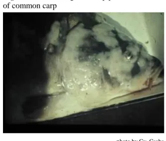
6.4 Koi herpesvirus disease ... 26
6.5 Herpes viral haematopoietic necrosis of goldfish ... 28
6.6 Herpes virus infections of silurids ... 28
6.7 Pike fry rhabdovirus disease ... 29
6.8 European catfish virus infection of the brown bullhead ... 29
7. DISEASES CAUSED BY BACTERIA ... 30
7.1 Carp erythrodermatitis ... 30
7.2 Infectious dropsy (septicaemia) of carp ... 31
7.3 Flexibacteriosis or columnaris disease ... 31
7.4 Mucophilosis or epitheliocystis disease of common carp ... 32
7.5 Fish tuberculosis ... 33
8. DISEASES CAUSED BY FUNGI AND ALGAE ... 34
8.1 Saprolegniosis (dermatomycosis) ... 34
8.2 Putrefaction of gills (branchiomycosis) ... 35
8.3 Infections caused by Dermocystidium spp. ... 36
8.4 Toxicosis caused by algae ... 36
8.5 Algal bloom ... 37
9. DISEASES CAUSED BY FLAGELLATED PROTOZOANS ... 38
9.1 Veil disease or ichthyobodonosis (costiosis) ... 38
9.2 Gill cryptobiosis ... 39
9.3 Sleeping disease of fish ... 39
9.4 Spironucleosis ... 40
10. DISEASES CAUSED BY CILIATED PROTOZOANS ... 41
10.1 Chilodonellosis ... 41
10.2 Trichodinosis ... 42
10.3 Apiosomosis ... 43
10.4 White spot disease (Ichthyophthiriosis)... 43
10.5 Balantidiosis ... 45
10.6 Capriniana infection ... 46
11. DISEASES CAUSED BY COCCIDIANS ... 47
11.1 Diffuse coccidiosis of common carp ... 47
11.2 Coccidiosis of silver and bighead carps... 48
11.3 Nodular coccidiosis of common carp ... 48
12. DISEASES CAUSED BY MYXOSPOREANS... 50
12.1 Swimbladder inflammation (SBI) of common carp ... 51
12.2 Gill sphaerosporosis of common carp ... 52
12.3 Myxobolus cyprini infection of the muscle of common carp ... 52
12.4 Myxobolus pavlovskii infection of silver and bighead carps ... 53
12.5 Thelohanellus nikolskii infection of common carp ... 53
12.6 Thelohanellus hovorkai infection of common carp ... 54
13. DISEASES CAUSED BY MONOGENEANS (GILL WORMS) ... 55
13.1 Gill disease caused by Dactylogyrids ... 55
13.2 Gyrodactylus infection ... 59
13.3 Diplozoon infection of cyprinids ... 59
14. DISEASES CAUSED BY TAPEWORMS (CESTODES) ... 60
14.1 Infection with Bothriocephalus acheilognathi ... 60
14.2 Infection of common carp with Khawia sinensis ... 61
14.3 Infection of common carp with Atractolytocestus huronensis ... 62
14.4 Ligulosis ... 63
14.5 Other tapeworm infections ... 64
15. DISEASES CAUSED BY PARASITIC FLUKES (DIGENEANS) ... 65
15.1 Sanguinicolosis of common carp ... 65
15.2 Diplostomosis of cyprinids ... 66
15.3 Blackspot disease ... 67
15.4 Tetracotylosis ... 67
15.5 Other metacercarial infections ... 68
16. DISEASES CAUSED BY ROUNDWORMS (NEMATODES) ... 69
17. DISEASES CAUSED BY SPINY-HEADED WORMS (ACANTHOCEPHALANS) ... 70
18. DISEASES CAUSED BY LEECHES ... 71
19. DISEASES CAUSED BY PARASITIC LARVAE OF BIVALVE MOLLUSCS (GLOCHIDIA) ... 72
20. DISEASES CAUSED BY CRUSTACEANS... 73
20.1 Ergasilus sieboldi infection ... 73
20.2 Other ergasilid infections ... 74
20.3 Lernaeosis ... 75
20.4 Fish lice (Argulosis) ... 76
21. DISEASES INDUCED BY THE PHYSICAL AND CHEMICAL QUALITIES OF WATER ... 77
21.1 Diseases caused by unfavourable water temperature ... 77
21.2 Problems in oxygen supply ... 77
21.3 Gas-bubble disease (GBD) ... 78
22. POISONINGS OF FISH ... 79
22.1 Poisonings of industrial origin ... 79
22.2 Poisonings of agricultural origin ... 79
22.3 Poisonings of aquatic habitat origin ... 80
22.4 Enteric inflammation caused by feeds ... 80
23. DISEASES OF UNKNOWN AETIOLOGY... 82
23.1 Winter skin disease of common carp ... 82
23.2 Gill necrosis of common carp ... 82
24. ZOONOTIC DISEASES... 84
Annex 1. Diagnosis of fish diseases by changes found on the body and in the organs ... 85
Annex 2. Fish Health-related International Recommendations, Regulations and Guidelines for Measurements ... 87
Annex 3. Chemicals, Drugs and Antibiotics used to Prevent and Treat Fish Diseases ... 91
Annex 4. Recommended Reading ... 97
Acknowledgements
The authors would like to thank András Woynarovich, FAO Aquaculture Consultant for his valuable help during the preparation of this document. They also thank György Csaba, DVM for providing some of the photographs.
Special thanks are given to Dr Richard Arthur for his professional review of the manuscript, who, besides editorial corrections, enriched the text with his comments, and Mr Raymon Van Anrooy, FAO Fisheries and Aquaculture Officer, who initiated the development of this field guide.
Abbreviations and a cronyms
BW Body weight
CCA Caucasus and Central Asia CE Carp erythrodermatitis CEE Central and Eastern Europe CK Carp kidney (cells)
CyHV1 Cyprinid herpesvirus1 CyHV2 Cyprinid herpesvirus2 CyHV3 Cyprinid herpesvirus3 DNA Deoxyribonucleic acid ECV European catfish virus
EPC Epithelioma papulosum cyprinid (cells)
EU European Union
GBD Gas-bubble disease GCRV Grass carp reovirus GPS Global positioning system
HVHN Herpes viral haematopoietic necrosis KHVD Koi herpesvirus disease
OIE World Organisation for Animal Health PCR Polymerase chain reaction (examination) PFRD Pike fry rhabdovirus disease
RNA Ribonucleic acid
SBI Swimbladder inflammation SVC Spring viraemia of carp
TAADs Transboundary aquatic animal diseases
Glossary
Actinospore A life-cycle stage of Myxosporea that develops in an oligochaete alternative host
Adhesion An abnormal union of surfaces due to inflammation or injury Aetiology The cause or causes of a disease
Alternative host A host in which a second, equally ranked phase of a parasite’s development takes place
Annelid worm A segmented worm of the Phylum Annelida, which includes earthworms, lugworms, rag worms and leeches
Anorexia An abnormal thinness of a vertebrate due to lack of appetite Anoxia Absence or deficiency of oxygen
Ascites The accumulation of fluid in the peritoneal cavity causing abdominal swelling (also referred to as dropsy or oedema)
Aseptate hyphal Non-segmented filaments (hyphae) of fungi strands
Benthos A collective name for organisms living on or in the pond bottom Branchiura A subclass of crustaceans, commonly known as "fish lice"
Cachexia Weakness and decline of body condition due to severe chronic illness Catarrhal Referring to the excessive production of mucus
Cercaria A larval stage of digenetic trematodes that is produced by asexual reproduction within a sporocyst or redia
Ciliate A protozoan bearing hair-like peripheral organelles. (see also cilium) Cilium A short, microscopic, hair-like vibrating structure on the surface of
certain cells (plural: cilia)
Clubbing A change in the structure of the gills in which, due to proliferation of epithelioid cells, the ends of neighbouring lamellae grow together, the gill lamellae disappearing from the damaged filaments
Coccidia Members of the Subclass Coccidia
Collagenous tissue Tissue comprised of any of a group of insoluble fibrous proteins that constitute the main structural component of animal connective tissue Commensalism An association between two organisms in which one benefits while the
other neither benefits nor is harmed
Copepod A member of the Order Copepoda, a large group of tiny aquatic crustaceans which are important members of the zooplankton and which includes many parasitic forms
Copepodite A developmental stage of parasitic copepods in which their structure resembles that of free-living copepods
Coracidium The initial, ciliated larval stage of some cestodes Cornea The transparent layer forming on the front of the eye
Cyclopoid A developmental stage of parasitic copepods in which their structure resembles members of the free-living genus Cyclops
Cytostoma The mouth-like structure of some protozoans
Dactylogyrid A monogenean worm belonging to the Family Dactylogyridae Desquamation Separation of scales or laminae from any surface
Digenean fluke A parasitic worm belonging to the Subclass Digenea, members of which require from two to four hosts to complete their life cycle Digenic Referring to a developmental cycle in which at least two hosts (a final
and an intermediate host) are needed to complete the life cycle
Dropsy The accumulation of fluid in the peritoneal cavity causing abdominal swelling (also referred to as ascites or oedema)
Ectoparasite A parasite living on the body surface of its host Endoparasite A parasite living inside the body or organs of its host Enteritis Inflammation of the intestine
Epidermal Referring to the surface epithelium of the skin of an animal
Epithelium The cellular tissue covering surfaces, forming glands and lining most cavities of the body. It consists of one or more layers of cells with only little intercellular material.
Epitheloid cell A type of histiocyte which participates in reparation Erythrodermatitis Inflammation of the skin with associated redness
Eurythermal Referring to the ability to tolerate, survive and grow within a wide range of temperature
Eutrophic Referring to a waterbody that is rich in plant nutrients and so
supporting increased growth of plants in general, and phytoplankton in particular
Exophthalmia Abnormal protrusion of the eyeballs (commonly referred to as
"popeye") (also exophthalmos)
Facultative Occurring optionally in response to circumstances rather than by nature Family A principal taxonomic category below an order and above a genus Fingerling A young fish of about 10–20 cm in length and 20–50 g in weight,
which in some temperate areas is also termed a "one-summer-old fish"
Fission Reproduction of a cell or organ by dividing into two or more new cells or organelles
Flagellate A member of the Phylum Mastigophora, a group of flagellated protozoans, some of which are parasitic
Fry The developmental stage of fish that starts when larvae gulp air and finishes when all organs are developed (or in case of the ovary and testes, when development is initiated). In the case of warmwater fish
species, this life stage lasts about 20–40 days, depending on water temperature.
Gamete A mature haploid female or male germ cell which is able to unite with one of the opposite sex in sexual reproduction and form a zygote (a fertilized female germ cell)
Gametogonic Stages in the process during which cells undergo meiosis to form stages gametes
Gene A unit of heredity determining the characteristics of the progeny through the sequence of DNA, and which is part of the chromosome
Genus A principal taxonomic category that is below family and above species.
The first part of the scientific (or Latin) name of species refers to the name of the genus, which always starts with a capital letter. (plural:
genera).
Gill arch The U-shaped cartilaginous structure that supports the gill filaments Gill filaments The filamentous parts of the gill, also called the primary lamellae Gill lamellae The subdivision of the gill filaments in which most respiratory and
excretory changes take place
Granuloma A mass of granulated connective tissue, typically produced in response to infection, inflammation or a foreign substance (plural: granulomas or granulomata)
Granulomatosis The formation of multiple granulomas
Gyrodactylid A type of monogenean worm belonging to Family Gyrodactylidae Haematopoietic Referring to the process of haematopoiesis, during which blood cells
are produced
Haematopoietic A type of tissue which takes part in the formation of blood cells tissue
Haemorrhage Heavy bleeding
Histopathology The branch of medicine dealing with tissue changes caused by a disease
Hydropic A type of degeneration affected by dropsy (oedema) degeneration
Hyperaemia Excess of blood in an organ or part of the body
Hyperplasia Increase in volume of a tissue or organ caused by the growth of new cells
Hypertrophy Increase in the volume of a tissue or organ produced entirely by the enlargement of existing cells
Hypha A filament composing the mycelium of a fungus (plural: hyphae) Hypotonic Referring to a lower osmotic pressure than a given fluid, or the state of
an abnormally low muscle tone
Inflammation A specific tissue response to injury evidenced by vascular dilatation Interlamellar Located between two gill lamellae
Intermediate host An animal in which an early developmental stage of a parasite takes place
Lamella A thin layer, membrane or tissue; gill plates on gill filaments serving for gas exchange (plural: lamellae)
Lordosis Curvature of the spine with abnormal concavity of the back Macrogametes The larger, female gametes
Macrophage A large phagocytic cell, which engulfs and absorbs bacteria or other small particles. It is found in stationary form in the tissue as a mobile white blood cell, especially in infections.
Meiosis Meiotic division, a type of cell division that results in daughter cells having half the number of chromosomes of the parent cell
Melano- An accumulation of pigmented macrophage cells macrophage centre
Merogony A series of stages in the life cycle of certain protozoans (Subphylum Apicomplexa) that forms merozoites and involves asexual reproduction
by multiple fission
Meront A uninucleate or multinucleate parent cell of certain protozoans (Subphylum Apicomplexa) that forms merozoites by a process of multiple fissions
Metacercaria A stage between the cercaria and the adult in the life cycle of digenetic trematodes, usually encysted and quiescent (a stage of inactivity) (plural: metacercariae)
Metazoans All organisms which are built up from more than one cell
Microgametes The smaller, male gametes of a heterogamous organism (see also macrogametes)
Miracidium The ciliated first larval stage of digenetic trematodes (plural: miracidia) Monogenean A member of the Class Monogenea, a group of parasitic flukes
requiring only one host to complete their life cycle Monogenetic A type of development without intermediate hosts Mucosa The membrane that lines the gastrointestinal tract Mycelium The filamentary part of a fungus
Myofibrils Bundles of contractile filaments that are arranged in parallel groups in the cytoplasm of striated muscle cells
Myxospores Spore stages of myxosporidians developing in fish hosts Nauplius The first larval stage of a parasitic copepod
Necrosis Death of most or all cells of an organ due to a disease (adj.: necrotic) Nematode A roundworm or threadworm belonging to the Phylum
Nematoda which has a slender, unsegmented cylindrical body Neoplasm A new and abnormal growth of tissue in a part of a body
Oocyst A cell in the ovary that undergoes meiosis to form ova (plural of ovum), which are the mature female reproductive cells. From this, after meiosis and fertilization by a male sex cell, develops the embryo.
Papillomatous Referring to a process resembling a papilloma
Pathogen A virus, bacterium or other organism which causes disease Pathogenesis The way a disease develops
Pathogenicity Having the ability to cause disease
Periciliated Referring to the whole body being covered by cilia
Peritonitis Inflammation of the peritoneum, typically caused by bacterial infection Petechia A small haemorrhagic spot on the skin, mucous membrane, etc.
Photosynthesis The process by which green plants (with the help of light and chlorophyll) produce their cells (their organic materials) from inorganic materials such as minerals and carbon dioxide. During this process they consume carbon dioxide and produce oxygen. The opposite of
assimilation is dissimilation, when in the dark, plants respire, consuming oxygen and producing carbon dioxide.
Plasmodium A form within the life cycle of some simple organisms that consist of a mass of protoplasm containing many nuclei. In myxosporeans, this is a life-cycle stage in which spores develop. (plural: plasmodia)
Plerocercoid The larval stage of a cestode which develops from a procercoid, usually showing little differentiation
Poikilotherm An organism whose temperature depends on and is equal to the temperature of its environment
Procercoid The larval stage of a cestode which develops from a coracidium;
usually having a posterior cercomer Proliferation Multiplication of cells
Propria A tissue layer below the epithelium Protozoans Single-celled microscopic animals
Sclerotized Hardened by conversion into sclerotin (dead thickened skin), which is a solid structural protein of, for example, the cuticle of insects
Scolex The anterior end of a tapeworm (plural: scolices)
Scraping A sample taken from the skin or gills by scraping with a scalpel blade or a glass microscope slide and examined as a wet-mount or stained preparation using a compound microscope
Septicaemia A morbid condition due to the presence and reproduction of pathogenic bacteria in the blood
Serosa The issue of the serous membrane which produces serum
Spore A minute, typically single-celled reproductive unit of lower plants and protozoans, which is capable of giving rise to a new individual without sexual fusion
Sporocyst The larval stage of a digenetic trematode developing after infection of the tissue of its molluscan intermediate host (typically a snail), and having a sack-like form
Sporogony Multiple fission of a zygote (which is also called a sporont) Sporozoite A motile spore-like stage in the life cycle of sporozoans Sporulated stage The life-cycle stage of coccidians forming the spore or spores Sporulation The formation of spores
Stenothermal Referring to the ability to tolerate, survive and grow within only a narrow range of temperature
Strobila The body of an adult tapeworm behind the scolex and neck, consisting of a series of similar proglottids or segments (plural: strobilae)
Subepithelium A tissue layer below the propria layer Submucosa A tissue layer below the mucosa
Surveillance A systematic series of investigations of a given population of aquatic animals to detect the occurrence of disease for control purposes, and which may involve testing samples of a population Tentacle A slender whip-like organ of some protozoans
Tomite A multiplying stage of Ichthyophthirius developing inside the tomont
Tomont The stage in the life cycle of Ichthyophthirius in which tomites develop
Toxicosis Poisoning
Trophont The growing stage of Ichthyophthirius infecting under the superficial epithelium of fish
Ulcer An open sore on the internal or external surface of a body caused by broken skin or mucous membrane, and which cannot heal
Vegetative stage An asexual reproductive stage of protozoans (also termed merogonic stage)
Viraemia The presence of viruses in the blood
Viviparous Referring to those organisms that give birth to living young which are developed in the mother
1. INTRODUCTION
According to the most recently available data from the Food and Agriculture Organization of the United Nations (FAO, 2018), in 2016, global production of finfish, crustaceans, molluscs and other aquatic animals (excluding aquatic mammals and reptiles) rose to about 171 million tonnes, with aquaculture contributing about half of this total (47 percent) and more than half (about 64 percent) of the value (USD 232 billion). While production from capture fisheries has remained more or less unchanged for several decades, production from aquaculture has continued to increase, with the sector continuing to grow faster than other major food production sectors (average annual growth of 5.8 percent during the period 2000–2016). In 2016, global aquaculture produced some 80.0 million tonnes of foodfish and 30.1 million tonnes of aquatic plants, as well as 37 900 tonnes of non-food products. Farmed foodfish production included 54.1 million tonnes of finfish, 17.1 million tonnes of molluscs, 7.9 million tonnes of crustaceans and 938 500 tonnes of other aquatic animals (FAO, 2018). Clearly, if fish production is to keep pace with the future growth of the human population, this additional supply will have to come mainly from increased aquaculture production.
The geographical area covered by this field guide includes Central and Eastern Europe (CEE), which is comprised of 20 countries (Albania, Belarus, Bosnia and Herzegovina, Bulgaria, Croatia, Czech Republic, Estonia, Hungary, Latvia, Lithuania, Moldova, Montenegro, Poland, Romania, Russian Federation, Serbia, Slovakia, Slovenia, the former Yugoslav Republic of Macedonia and Ukraine) and the Caucasus Central Asia (CCA), which includes the five countries of Central Asia (Kazakhstan, Kyrgyzstan, Tajikistan, Turkmenistan and Uzbekistan) and the four countries of the Caucasus (Armenia, Azerbaijan, Georgia and Turkey).
Although none of these countries are among the world’s top aquaculture-producing nations, warmwater finfish, mainly cyprinids (various carps) have been cultured in ponds for many centuries and are a traditionally and regionally important food item that is destined primarily for domestic consumption. More recently, other warmwater fish species, including catfishes (both native and introduced), northern pike, eels and perch have become locally important. In the rearing of warmwater finfish, the CEE and CCA countries still mainly utilize traditional pond-culture methods. There is thus a significant potential to increase fish production by using non-traditional methods (e.g. aeration, recirculating systems) that will allow faster growth with higher stocking densities. The most recently available FAO data (for 2016) shows the total freshwater fish production for the 29 countries covered by this field guide to be 485 174 tonnes, with cyprinids contributing 57 percent to this total (276 983 tonnes). Freshwater aquaculture production for the CEE countries was 323 559 tonnes during this period, with cyprinids contributing 74 percent (240 542 tonnes), while for the CCA countries, total freshwater aquaculture production was 161 615 tonnes, with cyprinids (36 441 tonnes) contributing 22.6 percent.
Disease in aquatic animals has long been recognized as a result of the interaction between the host (i.e. the aquaculture stock), its environment (e.g. the fish pond) and the pathogen, disease only occurring when these components overlap in a suitable manner. An intimate understanding of the the cultured species, the culture environment and the specific pathogen is thus critical to accurate disease diagnosis and treatment. The interaction of these three major players is shown in the accompanying"Szneisko circle”.
Globally, diseases caused mainly by viruses and a few bacteria (see Bondad-Reantaso et al.
2005, OIE 2017a) are a major constraint to aquaculture production, causing billions of dollars of losses due to mortalities and decreased growth of cultured fish, shellfish and molluscs. As described in this field guide, two of the ten transboundary aquatic animal diseases (TAADs) of finfish that are listed as reportable to the OIE cause problems in warmwater fish culture in the CEE and CCA countries (i.e. spring viraemia of carp (SVC) and koi herpesvirus (KHV)).
However this field guide also presents information on many other diseases, of both biological and environmental origin, that cause mortalities, poor growth and other problems in warmwater fish culture.
Recognizing that diseases cause severe damage to aquaculture production and fisheries of the CEE and CCA countries, the authors have provided a comprehensive document that details the diseases that impact warmwater fish production in the region. The main goal of this field guide is thus to provide information and guidance that will assist fish health specialists, veterinarians and aquaculturists to identify, treat and prevent the diseases of the warmwater fish species (primarily the carps and their relatives) that are most widely cultured in the CEE and CCA countries.
1.1 Fish species included
According to one of the widely used practical classifications of freshwater fish species, there are coldwater, warmwater and tropical fish. Coldwater fish species are usually stenothermal, and live in waters where the mean monthly temperature does not exceed 20 oC1. Tropical fish species are also stenothermal but require a relatively high water temperature (24–34 oC) and do not survive in waters where the temperature is constantly below 15–18 oC. Warmwater fish species are eurythermal, and hence tolerate a wide range of water temperature between 2 and 32 oC. However, all species have an optimal range of water temperature in which they grow and propagate the best.
1Source: http://pubs.usgs.gov/wri/wri984249/pdf/6ecological.web.pdf.
Typical culture systems for coldwater fish species are intensive tank and cage culture, while warmwater fish species are usually raised in pond polyculture systems.
In the countries and regions of Central and Eastern Europe (CEE) and the Caucasus and Central Asia (CCA), warmwater fish species include not only large cyprinids such as common carp and Chinese major carps, but other smaller carps and predators such as northern pike, pikeperch, wels catfish and brown bullhead. These all live in the same natural or manmade waters or are grown together in pond polyculture.
Examples of some of the typical species discussed in this field guide are given in Box 1.
1.2 Status of fish health in the region
Before the 1950s, in CEE and CCA countries, fish health services for warmwater fish species were based on knowledge obtained and disseminated by German specialists, while a branch of fish health science (fish parasitology) was dominated by Soviet, Polish and Czech scientists.
The pathogenicity of most parasitic protozoans and helminths was well studied, but only a little was known about bacterial pathogens, and nothing about the viral diseases of carps.
The 1950s brought a general change, when wild common carp from the Amur River and Chinese major carps were introduced to CCA and CEE. Together with these fish species, several new pathogens were also introduced.
In the 1970s, a significant step was taken when the complex nature of infectious dropsy of common carp was revealed by separating this disease into spring viraemia of carp (SVC) and ulcer disease. In CCA countries, successes relied mostly on scientists of the former Soviet Union, who published several books on the parasites of fish in the former Union of the Soviet Socialist Republics (USSR).
Today, the fish health services in both the CEE and CCA countries are organized in two different ways. In some of the countries, fish health services are run by veterinarians specialized in fish disease, while in other countries, prevention and treatment of fish diseases is coordinated by specialized fishery and aquaculture engineers or biologists. Both options have advantages and disadvantages. In general, however, fish health services are under the veterinary services, regardless of the academic qualifications of those who provide it.
Box 1. Fish families and their typical species discussed in this field guide
Esocidae (pikes): Northern pike (Esox lucius)
Cyprinidae (carps): Common and koi carps (Cyprinus carpio); Chinese major carps, such as silver carp (Hypophthalmichthys molitrix), bighead carp (H. nobilis) and grass carp (Ctenopharyngodon idella); freshwater bream (Abramis brama); goldfish (Carassius
auratus); Crucian carp (C. carassius); gibel carp (C. auratus gibelio); roach (Rutilus rutilus); ide (Leuciscus idus) and tench (Tinca tinca)
Siluridae (catfishes): Wels catfish (Silurus glanis)
Ictaluridae (catfishes): Brown bullhead (Ameiurus nebulosus)
Anguillidae (eels): European eel (Anguilla anguilla)
Percidae (perches): Pikeperch (Sander lucioperca), Volga pikeperch (S. volgensis)
Due to the banning of residues of many chemicals and medications in fish and fisheries products by the European Union (EU), today only a few effective chemicals and medications are available for the treatment of diseases of foodfish. As a consequence, the number of chemicals and drugs allowed, licensed and legally used by the fish health services in the EU is very small. Many drugs which were previously widely used in European fish culture have been banned due to their carcinogenic effects (e.g. malachite green) or because of the lack of license (e.g. some of the organophosphates). Nevertheless, these drugs are also mentioned in this field guide, because in several countries fish (e.g. goldfish and koi carp) are cultured on a wide scale for non-nutritional purposes and hence, for these ornamental species, there are no restrictions on the use of substances banned for use on foodfish.
1.3 Guide to users
The structure of this field guide is the following:
This publication aims to provide a concise guide for field personnel which, in addition to being a first reading on the subject, is also envisaged to serve as a technical book to which both unspecialized and specialized veterinarians can refer to in case they have to act and provide professional assistance.
A Glossary providing definitions for many of the scientific and specialized veterinary terms used in this field guide is given at the front of this document.
Section 1 provides a brief introduction to the field guide.
Section 2 presents a short general introduction to the nature and main types of fish disease. This section aims to help readers find their way in the “maze” of fish diseases presented in Sections 6 to 24.
In Section 3, the main aspects and protocols for field health inspection of fish are discussed and practical guidelines as to where, when and what should be done during routine and emergency fish health site inspections are given. This section is supported with relevant additional information presented in Annex 1.
In Section 4, some administrative and practical measures of fish disease prevention are discussed. This section is supported by Annex 2, in which information and international regulations as presented by the World Organisation for Animal Health (OIE) can be found.
Section 5 presents a concise inventory of the different methods of fish disease
treatment, and is supported by Annex 3, which provides information on the chemicals, drugs and antibiotics used as disinfectants and curatives for warmwater fish diseases.
Sections 6 to 24, which are arranged by major taxonomic groups, present detailed information on the all common diseases of warmwater fish mentioned in Section 2, including, for each disease, information, as appropriate, on its causative agent, the host species infected, optimal epizootic temperature, routes of transmission, pathology, clinical signs and methods for pathogen detection, prevention and treatment.
Annex 1 presents information of the clinical signs of fish diseases based on macroscopic changes seen in the body and organs.
Annex 2 presents fish health-related international recommendations, regulations and guidelines for measurements.
Annex 3 presents a summary of the chemicals, drugs and antibiotics used for the prevention and treatment of warmwater fish diseases.
Annex 4 provides a list of sources and recommended literature for further reading.
2. NATURE AND TYPES OF FISH DISEASE
One of the practical classifications of fish diseases divides them according to their cause, which can be either biotic or abiotic. Biotic fish diseases derive from living organisms, while abiotic diseases do not involve or derive from living organisms, but are related to factors such as water quality, the presence of poisonous materials or management problems, including incorrect feeding. As illustrated in Figure 1, these two main groups of disease have a complex interrelationship, both in natural waters and fish farms.
Figure 1. Practical grouping of fish diseases
Biotic fish diseases Abiotic fish diseases Fish diseases of unknown aetiology
In addition to the above-mentioned groups, there are also fish diseases of unknown aetiology, which may have either a biotic, an abiotic or a combined cause. As well as these three main groups of fish disease, there is a fourth group, tumours. These are also discussed in this field guide.
There are a few diseases which can be transmitted to humans from fish. These zoonoses are discussed in Section 24.
2.1 Biotic fish diseases
There is a wide range of organisms which cause fish disease when conditions are favourable for their development. These organisms can be categorized based on whether they are viruses, bacteria, fungi, plants or animals, as summarized below and discussed in detail in Sections 6 to 20. Biotic fish diseases are caused by the following major groups:
Viruses
Bacteria
Fungi and algae
Parasites
o Protozoans o Myxosporeans o Parasitic worms o Molluscs (glochidia) o Crustaceans
2.1.1 Diseases caused by viruses
Viruses are extremely minute (maximum 300 nm) infectious agents which cannot survive and multiply outside the cells of the host organism. Although viruses are not considered as living organisms, they are biological systems because they have DNA and RNA. Therefore, as well as for thematic and didactical reasons, they are discussed with the group of biotic agents responsible for causing diseases in fish.
Unlike numerous diseases caused by viruses in coldwater fish, especially in salmonids, the number of viral diseases known in warmwater fish species is relatively small. Nevertheless, some of these are regarded as important pathogens. Table 3 provides a list of the most frequent diseases caused by viruses, while Chapter 6.1 presents their descriptions.
2.2.2 Diseases caused by bacteria
There are not too many fully described diseases of warmwater fish caused by bacteria. These are listed below and are discussed in detail in Section 7. The most frequent diseases caused by bacteria are:
Ulcer disease (carp erythrodermatitis)
Infectious dropsy (septicaemia) of carp
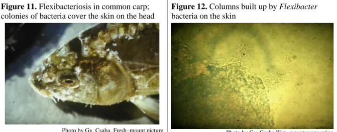
Flexibacteriosis or columnaris disease
Mucophilosis or epitheliocystis disease of common carp
Fish tuberculosis
2.2.3 Diseases caused by fungi and algae
Fungal organisms are typically facultative pathogens that infect fish with low resistance. These are listed below, while their characteristics are described in Section 8.
In a strict sense, algae do not belong among the pathogenic organisms infecting fish; however, they may cause massive fish mortality for two reasons: they produce toxic materials or when they bloom, the oxygen content of water can be dangerously reduced. The most frequent diseases of warmwater fish caused by fungi and algae are as follows:
Diseases caused by fungi
o Saprolegniosis (dermatomycosis)
o Putrefaction of the gills (branchiomycosis) o Infections caused by Dermocystidium spp.
Diseases caused by algae o Toxicosis
o Algal bloom
2.2.4 Diseases caused by parasites
Many fish diseases are caused by parasitic organisms. The concept of parasitism is rather extensive; in its wide meaning, all living organisms (viruses, bacteria, fungi, animals) which live in, feed from, and damage another organism are considered parasites. However, in its practical sense, only organisms, belonging to protozoans and metazoans, are real parasites. The main groups of fish disease caused by parasites are given below:
Diseases caused by protozoans (flagellates, ciliates, apicomplexans)
Diseases caused by myxosporeans
Diseases caused by parasitic worms (helminths): monogeneans, cestodes, digeneans, nematodes, acanthocephalans, leeches)
Diseases caused by parasitic larval molluscs
Diseases caused by crustaceans
Some parasites live their entire lives in or on the same host, while others have more complex life cycles. Besides the final host in which they mature, they may have one or more intermediate hosts, in which they grow during their subsequent developmental stages. Of these hosts, the main or final host is that organism in which they reach sexual maturity.
2.2.5 Diseases caused by protozoans
Protozoan parasites, described in Sections 9 to 11 are single-celled microscopic animals, and can be flagellates (Section 9), ciliates (Section 10) or apicomplexans (Section (11). Some of them are obligate parasites of fish, which means they cannot live without a fish host. Others are facultative parasites, because they can survive without a host; however, these also frequently cause infections and disease in fish. Most of the flagellate and ciliate species belong to this latter group. These parasites grow on the fish body, causing changes in the fins, skin and gills, and frequently causing the death of fish. The majority of flagellates and ciliates infecting fish are ectoparasites. As they harm the epithelial surface of the gills and skin, they cause similar damage and clinical signs. For these reasons, the methods for their prevention and treatment are also similar; hence, they are often mentioned using the common name
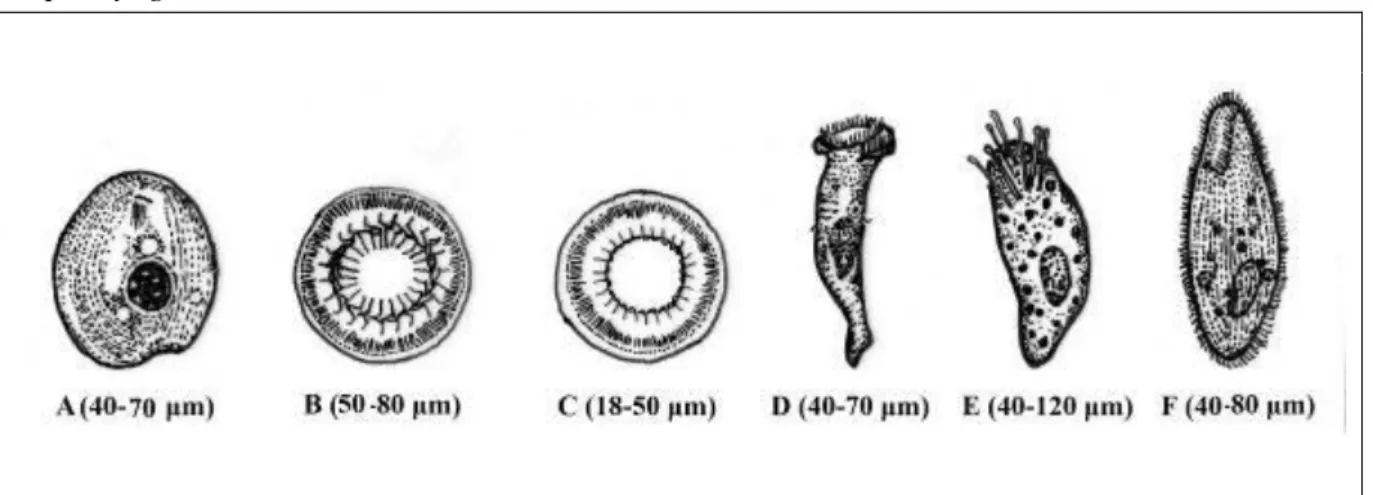
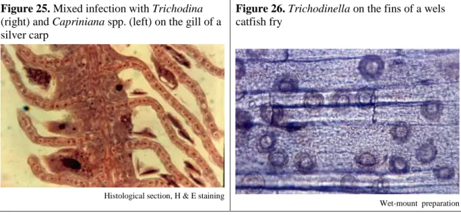
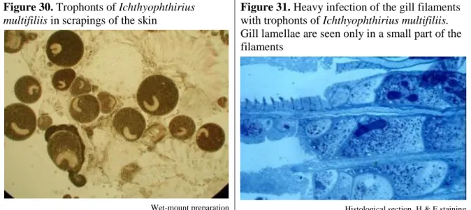
“ectoparasitic protozoans”. White spot disease is often incorrectly grouped among the diseases caused by ectoparasitic protozoans. However, the ciliate that causes this disease lives under the epithelium, and its life cycle and pathogenicity also differ from that of ectoparasitic protozoans, and so the methods for its prevention and treatment are also different. A list of the most frequently encountered diseases of warmwater fish caused by protozoan parasites is given below:
Diseases caused by flagellates
o Veil disease or ichthyobodonosis (costiosis) o Gill cryptobiosis
o Sleeping disease of fish o Spironucleosis
Diseases caused by ciliates o Chilodonellosis o Trichodinosis o Apiosomosis
o White spot disease (ichthyophthiriosis) o Balantidiosis
o Capriniana infection
Diseases caused by apicomplexans (coccidians) o Diffuse coccidiosis of common carp o Coccidiosis of silver and bighead carps o Nodular coccidiosis of common carp
2.2.6 Diseases caused by myxosporeans
The myxosporeans presented in Section 12 are common and pathogenic parasites of warmwater fish. For a long time, they were regarded as protozoan parasites, but new research has proved
that they are metazoan organisms. The development of all myxosporeans is complex: one of their developmental stages is in fish, and another one is in oligochaetes (annelid worms). They form two types of spores, namely myxospores in fish and actinospores in oligochaetes.
Actinospores infect fish, while myxospores infect oligochaetes. The most important diseases of warmwater fish caused by myxosporeans are as follows:
Swimbladder inflammation (SBI) of common carp
Gill sphaerosporosis of common carp
Myxobolus cyprini infection of common carp
Myxobolus pavlovskii infection of silver and bighead carps
Thelohanellus nikolskii infection of common carp
Thelohanellus hovorkai infection of common carp
2.2.7 Diseases caused by parasitic worms (helminths)
The parasitic worms (helminths) described in Sections 13 to 18 are the most common and pathogenic parasites of fish. Some of them are ectoparasites, while others are endoparasites.
Some infect fish in their adult stage; others, however, are parasites of aquatic birds and mammals and fish serve only as intermediate hosts for their developmental stages. Most of the known parasitic worms of fish belong to the Phylum Platyhelminthes: the Monogenea (Section 13), Cestoda (tapeworms) (Section 14) and the Trematoda (digenean flukes) (Section 15), but members of the Nematoda (roundworms) (Section 16), Acanthocephala (spiny-headed worms) (Section 17) and Hirudinea (leeches) (Section 18) are also common parasites of fish. Some of these worms are extremely pathogenic for fish. Of them, the monogenean gill worms, which include many host-specific species, are the best known pathogens of cyprinid fish, but tapeworms living in the intestine, as well as the larval stages of flukes can also cause mortality in fish stocks and economic losses for farmers. The most important diseases of warmwater fish caused by parasitic worms are given below:
Diseases caused by gill worms (monogeneans)
o Gill disease of common carp fry caused by Dactylogyrus vastator o Gill disease of common carp caused by Dactylogyrus extensus o Dactylogyrus infection of Chinese major carps
o Gill disease of wels catfish caused by Thaparocleidus vistulensis o Gyrodactylus infection
o Diplozoon infection of cyprinids
Diseases caused by parasitic tapeworms (cestodes) o Infection with Bothriocephalus acheilognathi o Infection of common carp with Khawia sinensis
o Infection of common carp with Atractolytocestus huronensis o Ligulosis
o Other tapeworm infections
Diseases caused by parasitic flukes (trematodes) o Sanguinicolosis of common carp
o Diplostomosis of cyprinids o Blackspot disease
o Tetracotylosis
o Other metacercarial infections
Diseases caused by parasitic roundworms (nematodes)
Diseases caused by parasitic thorny-headed worms (acanthocephalans)
Diseases caused by leeches (Hirudinea)
2.2.8 Diseases caused by parasitic larvae of molluscs (glochidia)
Certain species of freshwater bivalve mollusc use fish as hosts for developing their larvae, which are called glochidia. At first, female freshwater mussels incubate their fertilized eggs within their shells. However, after the incubation period, they release the glochidia into the water. These young larvae attach to the fins or gills of fish and remain parasitic for one or more months, while the young mussels develop. Although fish are able to sustain low levels of glochidial invasion without apparent harm, heavy infestations, especially in the gills of young fish, can cause injury and even death. Infection with glochidia is especially fatal in newly stocked fry populations. These parasitic larvae of bivalve molluscs are discussed in Section 19.
2.2.9 Diseases caused by crustaceans
The majority of aquatic crustaceans are free-living organisms, which means that they live independently, and not as parasites. However, some species have developed a parasitic lifestyle or a close association with fish. Many of them are responsible for disease, especially of farmed fish. There are a few dozen crustaceans that infect warmwater cultured fish, of which the most pathogenic species are those described in Section 20. These parasites cause severe economic losses in cyprinids and cultured predatory fish such as northern pike, pikeperch and wels catfish. Besides fingerlings, they also infect older fish, causing severe weight loss and death.
The most common diseases of warmwater fish caused by crustacean parasites are as follows:
Infection by Ergasilus sieboldi
Other ergasilid infections
Lernaeosis
Infection by fish lice (argulosis)
2.3 Abiotic fish diseases
At present, there is an ever-increasing knowledge on diseases caused by viruses, bacteria, fungi and parasitic organisms. Still, both in natural waters and pond polyculture of carps, much more harm is caused by environmental factors, such as oxygen shortage, low and high water temperature, accumulation of poisons in the water, and by human activities, including unsuitable or poorly implemented fish production technologies, incorrect nutrition and rough handling. These abiotic diseases of warmwater fish are discussed in Sections 20 to 22 and include the following:
Diseases induced by the physical and chemical qualities of water o Diseases caused by unfavourable water temperature o Problems in oxygen supply
o Gas-bubble disease (GBD)
Poisonings of fish
o Poisonings of industrial origin o Poisonings of agricultural origin
o Poisonings of aquatic habitat origin o Enteric inflammation caused by feeds
2.4 Diseases of unknown aetiology
Research on fish diseases is a fast-developing branch of animal pathology. While in the middle of the past century, we had only a scant knowledge of viral diseases, today dozens of these agents are known, first of all the pathogens of salmonids. On the other hand, swimbladder inflammation of common carp proved to be due to a myxosporean infection, although earlier it was thought to be a bacterial disease. Despite the great progress achieved in diagnosing fish diseases, the aetiology of some economically important fish diseases is still unknown. These are presented in Section 23 and are as follows:
Winter skin disease of common carp
Gill necrosis of common carp
2.5 Tumours
Tumours are widely reported in many families of fish. Fortunately, they are relatively rare in cultured cyprinids. This can be attributed to the fact that the rearing periods in culture systems do not allow the development of tumours, which characteristically occur in older fish.
Consequently, tumours appear more frequently in wild fish populations and in ornamental fish such as goldfish and koi carp.
Of the neoplasms found in fish from natural waters, epidermal hyperplasia in the fins and skin of the common barbel (Barbus barbus) seems to be the most common.
3. FIELD INSPECTION OF FISH HEALTH
The diagnosis of fish diseases is based on proper observation, sampling and examination of both the fish and their habitat. The best practice is when both on-site and laboratory examinations are performed.
3.1 Examinations on site
The steps of field examination are as follows:
Inspection of the waterbody
Sampling of fish
Examination of fish
Collection of data and other information
3.1.1 Inspection of the waterbody
The examination of fish starts with the inspection of the affected waterbody and the behaviour of fish. Unlike healthy fish, sick animals neither feed nor hide. They slowly swim up to the oxygen-rich locations, such as the water surface or inflow. Dead and severely sick fish often drift with the currents or float in unusual positions and jerk irregularly. A description of a healthy fish is given in Box 2.
It is important to note that dead fish initially sink to the bottom, rising to the water surface only after a couple of days. The length of time during which dead fish remain on the bottom depends on the water temperature. In colder water, dead fish can appear on the surface after even a week, while in warmer water this happens faster, within two or three days.
3.1.2 Sampling of fish
After studying the behaviour of the stock, fish samples should be taken from different parts of the waterbody. In waters where fish are fed, samples should be captured at feeding spots. Here healthy or relatively healthier specimens can be found. Examination of samples taken at inflows and outflows, together with fish captured at feeding points, will provide a more reliable picture about the stage and range of the illness.
Box 2. Appearance of a healthy fish
The body is covered with a thin layer of mucus and is free of wounds, ulcers and parasites.
The scales fit tightly into the dermis and their colour is characteristic for the species and age.
The eyes are white and the pupils are black.
The eye reflex is responsive; when the fish is turned, its eyes also move. The cornea of the eye is tight, shiny and reflective.
The back is fleshy and rounded.
The gill covers are undamaged. The gill rakers and filaments are also intact and free of wounds and parasites. They are covered with a thin layer of mucus and their colour is a deep red.
The fins are also undamaged and free of wounds and parasites.
3.1.3 Examination of fish
The examination of fish on site includes a close observation of the appearance of the fish, its body parts, and after dissection, its organs.
3.1.3.1 Observation of the body
The appearance of fish that differ from healthy individuals (see Box 2) allows the establishing of a first diagnosis. The most frequent changes in the shape, colour and intactness of the body are as follows:
The body condition can be estimated from examination of the dorsal surface of the fish. An emaciated fish, viewed from this perspective, resembles the blade of a knife. Abnormal thinness may indicate many various sicknesses, including inflammation of the digestive tract or a high number of worms.
The body surface, as well as the volume and quality of mucus on it, gives useful information about the possible cause of death. Poison provokes the skin to produce excess mucus, while with some diseases such as saprolegniosis (see Section 8.1) or winter skin disease of common carp (Section 23.1), mucus disappears from the skin.
Loss of scales may be due to mechanical injury. However, scale loss also frequently occurs during bacterial and fungal infections; this is very common in Chinese major carps, whose scales are less resistant. With some diseases such as spring viraemia of carp (SVC) (Section 6.1) and carp erythrodermatitis (ulcer disease) (Section7.1), fluid accumulates in the scale pockets, causing the scales to protrude.
Wounds, ulcers and parasites on the body and fins are evident signs of a problem.
Often, parasites such as leeches, fish lice, larval flukes (metacercariae) and white spot disease (ichthyophthiriosis) (Section 10.4) can be observed on the skin, but hyphae of the fungus Saprolegnia and proliferation of the epithelium due to fish pox are also clinical signs of changes. External injuries on fish suggest mechanical effects; however, they can also develop due to infection of the epithelium by pathogens such as Saprolegnia (Section 8.1) or ulcer disease (Section 7.1). Less frequently, ulcers on the skin and black spots (metacercarial infections) on the fin rays call for attention.
An unusually large belly can be due to viral or bacterial infection. Enlargement of organs, accumulation of secretions in the abdominal cavity (ascites), or the presence of parasites large in size and/or number can also cause an unusually large belly.
This can also be a clinical sign indicating an accumulation of sera in the abdominal cavity due to infectious dropsy, swimbladder inflammation or abdominal metacercariae (Section 15.4), but the presence of large cestode plerocercoids (Ligula spp.) (Section 14.4) produces similar clinical signs. Enlargement of the belly can also be due to either a severe infection with tapeworms in the gut or because of the accumulation of gas in the intestines caused by bacterial infection.
In the latter case, fish often float belly up at the surface of the water, as they are unable to submerge.
The protrusion of the anus indicates a possible inflammation, as well as being a sign of fluid accumulation in the digestive tract.
Rough handling, as well different parasites can cause broken fins. As fins regenerate rather quickly, such wounds indicate recent problems.
The mouth and gill slits are usually closed, but in the case of suffocation they remain open. After death, the gills become pale and the tissues lose their structure.
The presence of excess mucus on the gills suggests poisoning or suffocation, while tissue erosion is a clinical sign of gill necrosis and putrefaction.
Crippled or dwarfed fish are rare in nature. A deformed spine can be observed mostly in fish that have developed under unsuitable conditions during their larval stage.
Whether the fish is dead or alive can be judged by the presence or absence of the eye reflex.
Stiff eyes are evidence of death. The white outer layer of the eyeball (sclera) of a recently dead healthy fish is tight and shining, while the sclera of a dead diseased fish is sunken, dim and wrinkled. In normal cases, the colour of the pupil is black but, for example, in the case of a severe infection by eye flukes (Section 15.2), it becomes white. Due to various parasitic infections, goggled, protruded eyes can often be observed.
3.1.3.2 Observation of the organs of fish
Before dissection, fish should be killed swiftly, as specified in the relevant regulations of the given country. The initial steps of dissection to expose the internal organs are illustrated in Figure 2.
Figure 2. Initial steps in the dissection of a fish
1 2
3 4
1) The fish is killed by cutting through the backbone. 2–3) A cut starts from the head and the body is opened by a long cut in the medium line of the belly towards the anus. 4) Afterwards, a curved cut is made on the dorsal side in order to expose the internal organs. 5) Some of the internal organs:
gills (A), heart (B), kidney (C), swimbladder (D) and liver (E).
5 Fresh preparation pictures
Sera may be discharged and/or parasites may be observed upon opening up the fish;
these are obvious signs of a potential health problem.
In the gut, the mucous membrane of the intestine of a healthy fish is a light rose colour and its surface is flat. Inflammatory changes or thickening of the wall may indicate a bacterial infection.
Adhesion of the viscera and peritoneum may also indicate bacterial infection. Large worms (cestodes and trematodes) may occur in the gut in large numbers. Some parasites, such as plerocercoids of the cestode Ligula or the nematode Philometra, hundreds of Tetracotyle metacercariae or large numbers of pseudocysts containing spores of Myxobolus draw the attention to infections that are already well established.
The swimbladder is usually transparent and has no thick walls. Inflammatory changes in the swimbladder suggest sphaerosporosis or bacterial infections.
The heart of a fish that is being necropsied is typically empty. However, in suffocated fish it may be filled with coagulated blood.
A highly swollen gall bladder is a sign of starvation or of an illness of the digestive system.
Normally, the kidneys are brownish-red in colour and easy to tear. However, bacterial (Aeromonas) infection degenerates them, and blood fluke infection (sanguinicolosis) causes them to become oedematous.
The liver of a healthy fish is light brown, while the spleen is a dark-red elongated organ. When infected, these organs will become swollen.
The gills and gill cavity can be best studied by cutting off the gill covers (see Figure 2). For detailed examinations, the gill rakers (cartilaginous gill arches) should be separated.
A table providing guidance for diagnosing fish diseases is found in Annex 1.
3.1.3.3 Collection of data and information
Collecting data and other information on background and the measures that have already been taken is the third step in on-site actions. This includes finding out:
Whether there were similar earlier precedents and the date when mass mortality started.
The number of dead fish.
Whether and what actions have been taken so far.
Studying past management actions and situations on site gives useful information and data on:
type and parameters of the waterbody;
water supply (its source and whether there is any source of pollution or poisoning);
physical (temperature, transparency/turbidity, colour), chemical (pH, oxygen content) and biological (aquatic weeds, number and size of the different fish species)
parameters and qualities of the waterbody;
dates of last stockings;
quantity and quality of feeds given if the fish stock was fed;
applied fishery and/or aquaculture management techniques;
location of nearby industrial and agricultural plants; and
possible agricultural sprayings in the vicinity.
3.2 Taking and sending samples for laboratory examinations
In cases where there is no specialized professional on the field, or if an on-site veterinary inspection does not result in a diagnosis, fish should be sent to a laboratory. In the laboratory, dissection and parasitological, bacteriological, virological and histological examinations will