Polluted Waters and the Contamination of Fish
A. GU£UN
Institut Pasteur, Paris, France
I. Introduction 481 II. Enteric Bacteriophages in Water 484
A. Bacteriophages as Indicators of the Presence of Pathogenic Bac-
teria in Water 485 B. Water Phages of Different Sizes 488
C. The Role of Bacteriophages in the Purification of Waters 491
III. Enteric Bacteria and Bacteriophages in Fish 492
References 495
I. Introduction
If it were true that the bacterial flora of a fish reflected the bacterio- logical conditions in the waters from which it originated, then the fish could be considered as an indication of the sanitary conditions of these waters. Because fish cover great distances in different waters, the bacterio- logical examination of their intestinal contents supplements in a good way the direct sampling of the waters. This interpretation immediately raises a number of questions of an ecological and physiological nature.
Besides the fact that bacteria have a certain distribution pattern in waters of varying depths depending on the distance from outflowing contaminating rivers, it is essential to know if fish are subject to a contamination by the Enterobacteriaceae and if this group as a whole may constitute part of the bacterial flora of fish and, if not, how long these bacteria may survive in the digestive tract.
Enterobacteriaceae common to fish, or accidentally living in the di- gestive system for a prolonged period, may not furnish useful informa- tion regarding the actual sanitary state of the waters. The presence of these bacteria merely indicates that a contamination has taken place, but perhaps long ago, and has no relationship to conditions in the waters under study. Brown (1917), studying fish captured in nonpolluted waters, established the presence of Escherichia colt and Welchia per- fringens, which could be interpreted in this way.
The publications from the last sixty years concerning the bacteriology of fish have either been inspired by sanitary precautions: by a desire to investigate the origin of bacterial contamination of commercial fishes
481
or by the study of the problem of polluted waters. The presence of coli- forms in a water is generally interpreted as a sign of fecal contamination and has been the prime object for study. A few investigations deal with Es- cherichia paracoli or Proteus but rarely Salmonella or Welchia perfringens.
In 1899 Hoag isolated EschericHia colt from the intestines of sea- water fish. Whipple (see Johnson, 1904) did not find this organism
in 75 samples of sea-water fish from nonpolluted waters. Amyot (1901) obtained positive results with 23 samples. Kister and later Mallanah (un
published results, referred to by Fromme, 1910), the former on the basis of 21 samples and the latter with 32 fish samples, reached identical results. Houston (1903, 1904) found 13% containing E. coli and drew the conclusion that this was due to a contamination of the water through sewage. Eyre et al. (1904) considered E. coli as a normal in
habitant of fish intestine. His material was collected two metric miles from the coast. Johnson (1904) observed E. coli 47 times out of 67 and assumed that fish carried this organism from contaminated waters out into clean waters. Bettencourt and Borges (1908b) indicated two cases of colif orms out of 17 fish. Fromme (1910) found 18 out of 33 fish contaminated. Browne (1917) studied fish living in nonpolluted waters and established that 59% harbored E. coli and Welchia per- fringens. Hunter (1922) isolated E. coli and Aerobacter areogenes from the intestines of salmon. Stutzer (1926) considered E. coli and B. paracoli as part of the normal flora of fish intestines. Fellers (1926) showed that the digestive system of salmon contained no E. coli. Reed and Spence (1929) found 70% of haddock containing Proteus, but E. coli was extremely rare. Stewart (1932), too, was unable to detect E. coli in haddock and found very few Aerobacter. Gibbons (1934a, b) indicated the presence of Proteus in 2 cases out of 43; only once was Eberthella met with. He did not consider the colif orms to be normal inhabitants of the digestive system of sea-water fish. When they were found, it was in fish which were contaminated by polluted water. Grif
fith and Fuller (1936) concluded, basing their studies on commercial fishes, that E. coli was presumably an external contaminant. Markoff (1939-1940) believes that the presence in fish viscera of bacteria belong
ing to the coli-typhus group shows that the fish have been staying for some time in polluted water. Sooner or later they get rid of these micro
organisms. Snow and Beard (1939), studying 1838 bacterial cultures isolated from salmon, found E. coli in 0.1% of the cases. Wood (1940) found no coliforms in the different fish of Australian waters. Nor did ZoBell (1941, 1946) find this type of bacteria in studying 387 fishes and 961 samples of sea water; 5 cases of E. coli were observed in fish caught close to the coast. The Aerobacter are more frequent (13%) than the
other coliforms ( 6 % ) . According to ZoBell, it is not a habitual flora in fish but always an introduced flora which may persist for a considerable length of time. Fredericq (1950) studied the intestine of various marine animals in the bay of Villefranche and established the presence of Enterobacteriaceae in 37 fish. Guelin (1952b) isolated bacteria and enteric bacteriophages in fish captured in waters of the littoral zone exclusively. Margollis (1952) reports the presence of E. colt and Proteus in pikes intestine. Lodenkemper (1952) concludes that salmonellae in fish play the principal part in food poisonings. Venkatraman and Sreenivasan (1953) found Paracolobacterium in fresh-water fish. Floyd and Jones (1954), studying 376 lots of fish, revealed the presence of Salmonella in 11% of the lots. Jadin et al. (1957) discussed the possible transmission of Shigelh and Salmonella by fish.
An analysis of all these observations reveals contradictory results in a few cases. Prior to 1926, particularly, authors frequently reported the isolation of enteric microorganisms. On the other hand, almost as many found none. Eyre et al. (1904) and Stutzer (1924) considered the enterobacteria as normal inhabitants in fish. Houston (1903), John- son (1904), Fellers (1926), Gibbons (1934a, b ) , Markoff (1939-1940), ZoBell (1941), Guelin (1952b) and Jadin et al. (1957) reached the con- clusion that fish became contaminated by the Enterobacteriaceae when exposed to polluted waters.
These contradictory results are explained by the absence of coordina- tion in the study and by the development of our knowledge on Escherichia and Esch. like bacteria. As a matter of fact, the relative frequency of Aeromonas (Buttiaux, personal communication) in certain species of fish should not be overlooked. These germs, when a study of their cilial apparatus is neglected, are generally identified as Paracolobacterium or Cloaca. This also applies to the numerous strains of unpigmented Ferratia to which Ewing et al (1959) justly have drawn attention. Therefore, different organisms have been studied and with varying methods, and on different species and from localities which frequently were very poorly defined. But as a whole the results show the accidental character of the presence of Enterobacteriaceae in fish.
In my personal studies of fish in 1951-1952, besides investigating the Enterobacteriaceae, I also executed observations on the intestinal bac- teriophages. Since Dumas (1920) first established the presence of coli- dysenteric bacteriophages in the water of the river Seine, studies of bacteriophages in water have become more and more common. In France the practical application of bacteriophagic research to waters was the topic of a special government circular of November 24, 1954
(Nevot, 1957; Parvis and Tarantini, 1958).
II. Enteric Bacteriophages in Water
In 1891, Siemens presented a hypothesis that bacteria were sus
ceptible to infectious diseases caused by extremely small organisms.
Gamaleia (1899) was the first to announce the lysis of bacteria and the ability of this lysate to dissolve a freshly prepared culture. Twort
(1915) gave a detailed description of a transmissible lysis of staphylo- cocci and supposed that the causative agent was a filterable virus. His ob
servations, as well as those of Gamaleia, did not attract due attention and were not pursued. It was d'Herelle (1917) who, after having ob
tained a lysis of the Shiga bacillus by Bacteriophagum intestinale (an agent which can be filtered), initiated a serious study of this agent. These classic investigations by d'Herelle were rapidly followed by similar studies by other authors and contribute the basis of our present knowledge of bacteriophages. Their more recent definition as "bacterial viruses"
refers to the extremely small dimensions of their body (from 15 to 100 ιημ in diameter) and to their ability to develop exclusively in living bacteria and there exhibit their growth phase. The bacteriophages at
tach themselves to the surface of the bacteria and soon afterwards start to increase in number, the more so as the bacteria gradually keep multi
plying. But young bacteria which are attacked undergo lysis and the whole culture becomes completely transparent. Bacteriophages are found wherever bacteria are present; there are numerous varieties corresponding to a great number of bacterial species. Kriss (1959) re
vealed the presence of bacteriophages active on marine bacteria in the Black Sea. They have been isolated in vitro and in the waters down to a depth of 2,000 meters. (Kriss and Rukina, 1947; Spencer, 1955).
In polluted waters, they are always found (Dumas, 1920; d'Herelle, 1921; Hauduroy, 1923; Arloing and Chavanne, 1925; Arloing and Sempe, 1926a, b; Monteiro, 1926; Fabry, 1928; Morin and Guillerm, 1928; Gilde- meister and Watanabe, 1931; Robic, 1937; Dienert et al, 1934; Pana- yotatou, 1935; Klemparskaja, 1940; Rita, 1940; Vaccaro et al, 1941).
Bacteriophages may remain for long periods in river or sea waters without losing their specific activity against bacteria.
Assuming that bacteriophages are constant companions of bacteria and that their number vary according to the number of bacteria present, they can be looked upon as evidence of water pollution (Arnold, 1925; Fortunato, 1929; Renaux, 1929; Pasricha and Gupta, 1931; Vagedes,
1932; Dionert, 1934; Vagedes and Gildemeister, 1934; Couture, 1936;
Ciuca and Cocioba, 1937; Cocioba, 1937, 1941; Rhodes and Ludlam, 1939; Abdelrachman, 1943; Guelin, 1943, 1948a, 1955; Guolin and Le Bris, 1947; Gernez-Rieux et al, 1949; Buttiaux, 1949, 1951; Buczowski
and Buczowska, 1951; Alva et al, 1951; Leclerc, 1956, 1957, 1959; Parvis and Tarantini, 1958; Monnet et al, 1958; Nicolle, 1959; Carlucci and Pramer, 1960d). Buttiaux considers the presence of "fecal bacteriophages"
(according to his terminology) as a proof of dangerous contamination of water. The fecal pollution of waters at a distance of 1,000 meters from the sewage outflow in the estuary of the Ranee (Atlantic) has been brought into evidence by the presence of these bacteriophages (Buttiaux, personal information, 1958). Verde (1958) published a complete survey on this subject. Penna (1959) tested 138 water samples, comparing the bacteriological method with the bacteriophage procedure. See also Chapter 13.
A. BACTERIOPHAGES AS INDICATORS OF THE PRESENCE OF PATHOGENIC BACTERIA IN WATER
It is still an open question if sea-water as such exerts a direct bactericidal effect without the aid of bacteriophages (ZoBell, 1936;
Krassilnikov, 1938; Johannesson, 1957). Several good reviews on this question are available (Greenberg, 1956; Carlucci and Pramer, 1959).
Although several enteric bacteria may create a health hazard when spread in sea-water, it is nevertheless well established that the rate of their disappearance is far greater that what could be expected from dilution alone. Undoubtedly the salinity and pH of sea-water do not favor the direct survival of E. coli (Carlucci and Pramer, 1960b), unless protected by organic pollution or certain mineral salts. This evidently is a causal factor of some significance. Heat-labile substances of an antibiotic nature have been repeatedly found in sea-water (ZoBell, 1936; Krassilnikov, 1938). Furthermore, marine microorganisms have been shown to produce such substances (Rosenfeld and ZoBell, 1947).
In a recent study, however, no antibiotics were found exerting any particular influence on E. coli (Carlucci and Pramer, 1960c). This is in contradistinction with the findings of Johannesson (1957) who reported a clear bactericidal effect of sea water on E. coli. He indicated that the active agent was inorganic and identical to iodine. Evidently it is a question of definition if this inorganic compound should be listed as an antibiotic or merely a bactericidal agent. Coli bacteria could be protected against its killing action by small amounts of cysteine. Naturally this latter compound readily appears in polluted water or sewage effluents.
Among the so-called fecal bacteriophages isolated from water a large number bear evidence of a common fecal contamination. They can easily multiply at the expense of various enteric bacteria. Being very polyvalent they are of less interest from the point of view of water sanitation. Specific bacteriophages multiplying only at the expense of
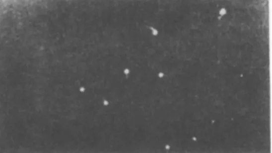
one special kind of bacteria can be used for detecting pathogenic microorganisms in water. Isolation of such typhi or perfringens phages from water can, for instance, justify the conclusion that these waters, at a given moment, were contaminated by typhic bacteria or by Welchia perfringens. The Vi bacteriophage, specifically active on Salmonella Vi (Scholtens, 1936; Sertic and Boulgakov, 1936; Craigie and Brandon, 1936), clearly reveals the presence of these bacteria (see Fig. 1).
The study of typical bacteriophages for the detection of SalmoneUa in waters has been recommended by Guelin and Le Bris (1947). Guelin (1948b) describes the method for isolating the bacteriophages Vi from waters. Gernez-Rieux et al. (1949) points out the importance of searching
FIG. 1. Typhic bacteriophages of type Vi (after Guintini et al, 1953).
for these phages for the demonstration of Eberthellae. Morzycki et al.
(1952) conclude in a study of the Vistula waters stretching over 427 kilometers, that the Vi bacteriophage can be regarded as a specific or- ganism indicating the presence of typhic bacilli in the waters; the re- sistance of the phage in industrial waters (harmful to E. colt) makes it more valuable. Its human origin, shown by Morzycka and Georgiades (1952), gives it all the advantages in a study of water hygiene. This was confirmed by Alva et al (1951), Leclerc (1956, 1957, 1959), Parvis and Tarantini (1958).
During recent years, Timakov and Goldfarb (1956a, b ) , Goldfarb (1957, 1959), and Goldfarb and Erchov (1958) developed a bacterio- phagic method for the detection of SalmoneUa and dysenteric bacteria in water. This method is not characterized by the kind of bacterio- phages being employed but rather by the susceptibility of the various bacteria occurring in the water, increasing the proportion of specific standard phages. The bacteriophagic method is many thousand times more sensible than any direct bacteriological analysis (Timakov and Goldfarb, 1956b; Goldfarb and Ostrovskaja, 1957; Goldfarb and Kouznetzova, 1957; Abdouzamatov, 1959).
The presence of phages in water does not, however, automatically indicate how long it has been contaminated. The disappearance of the typhus bacilli is faster than that of the phage Vi (Guelin, 1950a;
Buttiaux, 1951; Emilianowicz, 1952). Even if the prolonged sojourn of the bacteriophage Vi in water does not destroy its specificity toward the typhic strains (Guelin, 1948b, 1950a; Goldfarb and Ostrovskaja, 1957), its concentration gradually diminishes, particularly during the first few days. Factors such as temperature, solar radiation, the pH of the water, and the mineral content have a direct effect in this regard.
So it is the quantity of isolated phages which makes it possible to estab
lish approximately how old the contamination of the water is. When
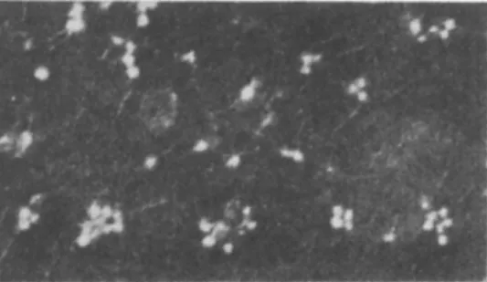
FIG. 2. Bacteriophages of Welchia perfringens (after Elford et al., 1953).
the specific bacteriophages are found in large quantities this indicates a rather recent pollution; on the contrary, their repeated isolation in small numbers indicates an older contamination.
Comparative studies in the Mediterranean, the Seine, and the Thames show that the bacteriophage count varies according to its type. Under the given conditions and when a typhic bacteriophage can be identified in 0.1-0.01 ml3 water, it can be concluded that it is present in the water in large quantity. The same is true with the coli-phage isolated from 0.001-0.0001 ml3, as well as with the phage of W. perfringens found in 1 ml3 (Fig. 2). Differences in quantities of isolated phages found in one and the same water are most likely to run parallel with the frequency of the corresponding bacteria with which the water is polluted. E. coli generally occurs in larger quantities than W. per- fringens. S. typhi is rarely isolated (Buttiaux, 1951; Gevaudan et al., 1958).
The study of a Vi bacteriophage, the presence of which indicates pol
lution by Salmonella typhi, requires the use of standard strains rich in Vi antigen and hardly sensitive to bacteriophages of non-Vi type. Such strains may facilitate the isolation of specific phages by eliminating those
to which they are not sensitive. The strains are chosen by means of two tests: (1) sensitivity of these strains to the action of various bacterio- phages, and (2) study of new bacteriophages isolated with these strains
(Guelin, 1950b).
The strain should be lysed exclusively by standard phages belong- ing to the same type. On the other hand, new phages, isolated by means of this strain, should in their turn not be active on bacteria other than those belonging to this particular species and type. The question could be raised, however, whether a strain which is sensitive exclusively to Vi-type phage would for any length of time maintain its specificity.
Having a strain specific only for Vi-type phages does not exclude the possibility of some day seeing it become susceptible to bacterio- phages of non-Vi type. For this reason the standard strain has to be constantly checked. Before isolating the various Vi phages, it is preferable to have a series of typhi-Vi strains with highly specific charac- teristics. It is easy to realize the importance of determining phage- types in bacterial species by establishing differences in their sensitiv- ity to the bacteriophages. The method called "phage typing" makes it possible to collect a number of types for each region. This method, for which the principles are laid down by Craigie and Yen
(1938), has been applied in France by Nicolle (see bibliography in Nicolle, 1957). In regard to the strains of W. perfringens which are not always of animal origin, Beerens and Delcourte (1958a, b) developed a test of obvious importance as it allowed the differentiation of fecal W. perfnngens from the soil strains. This test is based upon the study of the survival of vegetative forms in a solution of 20% sodium chloride.
B. WATER-PHAGES OF DIFFERENT SIZES
The variation in the size of the bacteriophages is reflected in the dimensions of the plaques formed by these corpuscles; the diameter of the plaques increases in proportion to the diminishing phage size (Elford and Andrewes, 1932).
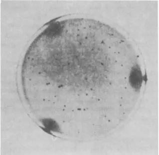
We had the opportunity to study Salmonella bacteriophages produc- ing large or small plaques, isolated from various waters. While the for- mer were specific, active exclusively on one species, the latter were poly- valent, often attacking numerous bacteria of the coli-typhi and dys- enteric groups (Guelin, 1952a, 1955b; Leclerc, 1957). Thus those small plaques phages can sometimes regenerate in other strains than the typhic ones. The pollution of waters by Salmonella is certain only in the case of isolation of Vi large plaques phages. Small plaques phages have first to be tested as to their specificity (see Figs. 3 and 4).
The occurrence of large and small plaques phages does not seem to be related to certain geographical locations. They are found in various waters, as in the Seine and the Thames rivers, and the Mediterranean Sea. On the other hand, their distribution changes with the season, which indicates a possible effect of day length. The appear
ance of small plaques phages coincides with the lengthiest and sun
niest days of the summer. Large plaques phages then become less and less numerous but reappear during the winter.
The varying sensitivity to light, exhibited by bacteriophages of various dimensions has been studied by Guolin (1942), Rouyer and Guelin (1942), and Wahl (1946). Their investigations were preceded by those of Wollman and Lacassagne (1940), who established the dif
ferent resistance of large and small bacteriophages to X-ray.
Daylight seems to be to a great extent responsible for the disappearing of typhic large plaques phages (Guelin, 1955b). Studies on the photo- sensitivity of bacteriophages in open basins, containing 700 liters of pond water and placed in a park, confirmed the laboratory findings.
The extreme sensitivity to light of the large plaques phages perhaps offers an explanation of the intermittent occurrence of phages of differ
ent size in the Seine and the Thames rivers, and the Mediterranean Sea (ob
served during the years 1952-1953). During the short daylight season of the winter, the disappearance of large plaques phages is delayed.
Their number prevents the isolation of other bacteriophages because the greater dimensions of their plaques masks the smaller ones. It is possible that the simultaneous presence of unequal quantities of bacteriophages of varying sizes in a growing culture inhibits the development of one type at the cost of another. The greater light in
tensity during the summer season is accompanied by a decline in quantity of large plaques phages. Their presence no longer inter
feres with the development of small plaques phages. The number increases in proportion to the disappearance of large-plaque bacterio
phages. This is only a hypothesis to explain the seasonal variations which have been found in the distribution of Vi large and small plaques phages at the surface of polluted waters. The question remains whether the bacteriophages of various sizes differ in their photosensitiv- ity. Our observations apply exclusively to water close to the surface.
Buttiaux (1951) recommends a sampling of water for bacteriophagic work at a greater depth. It is possible that deeper water layers, less accessible to solar radiation, contain phages of different sizes during any season. In the summer at the Suresnes dam of the Seine the author demonstrated the presence of numerous large plaques phages at a
FIG. 3. Small plaques formed by the typhic bacteriophages on Salmonella typhi (after Nicolle et al, 1953).
FIG. 4. Large plaques formed by typhic bacteriophages on SalmoneUa typhi (after Nicolle et al., 1953).
depth of 3 meters, whereas the surface water contained only small plaques phages.
C. THE ROLE OF BACTERIOPHAGES IN THE PURIFICATION OF WATERS
It remains to examine the possible role of bacteriophages in the self- purification of waters. The destructive power of bacteriophages as regards bacteria attracted, from the very beginning, the attention of numerous workers. It has been suggested that the early observa- tions of Hankin (1896) of the bactericidal eifect of the waters of the Jumna and Ganges against cholera vibrios could possibly be
ascribed to bacteriophages (Flu, 1923, 1941; Bilouet, 1926; Bujanowski, 1929; Beckwith and Rose, 1930; Guidice, 1931; Nyberg, 1931; Petrilli, 1940; Georgiades, 1952). On the contrary, Ware and Mellon (1956) never observed bacteriophagy, not even in sewage effluents.
It is, however, difficult to conceive the existence in a river of an im- portant bacteriolysis identical to that observed in the laboratory. Such a phenomenon requires a multiplication of enteric bacteria to a degree difficult to attain in waters lacking sufficient nutritive constituents.
Nevertheless, a minor quantity of organic material dissolved in the water may, under certain conditions, permit bacterial growth (Bigger, 1937) and a corresponding increase in bacteriophages (Guelin, 1945a, b ) . Thus, observation has been made of a multiplication of paradysenteric bacteria in a solution of peptone (1:70,000) invisible to the naked eye but attaining tenfold increases or more. In such a culture, the phages multiply and cause lysis of the bacteria (Guelin and Lepine, unpub- lished ). It could, therefore, be expected that under favorable conditions a bacterial lysis by the phages might take place on a water surface. There is, however, little likelihood that such a lysis could in an efficient way aid the self-cleansing of waters, at least in nontropical waters.
The purificating role of bacteriophages can be shown further if the fact is taken into account that these phages are adsorbed on the surface of the pathogenic bacteria in the water. The introduction of such micro- organisms, already contaminated by a phage capable of interfering with their multiplication, would imply a subsequent destruction of in- troduced bacteria.
Our observations with a microbial agent of gas gangrene confirm the active role of bacteriophages in the field of water hygiene, and also of Wehhia perfringens, carrying phages in the water, were not destroyed even when a nonnutritive environment prevented multiplication. It was, however, sufficient to introduce these bacteria into a richer environ- ment to obtain their destruction. They did not develop colonies at all or
in smaller numbers than control colonies without phages (Guelin, 1950c). Guinea pigs injected with such bacteria showed no signs of sickness, while control animals, injected with bacteria not previously attacked by bacteriophages, showed all symptoms of the classic gas gangrene. The bacteria carrying phages were probably destroyed at the moment when they started to multiply in the muscle tissue of the guinea pigs (Guelin, 1955a).
The presence of enteric bacteriophages in natural waters may there- fore indicate a contamination of these waters by the corresponding bacteria. It may also reveal the presence of pathogenic germs in the water, e.g. in the case of the typhus bacilli or W. perfringens. The research technique for bacteriophages is simple and rapid. Even small quantities of phages in waters diluted up to a million times are detect- able. All these results permit considering the bacteriophagic method as complementary to the colimetric one. In the field of water sanitation the use of bacteriophages is not limited to the study of polluted waters. They indirectly participate in the self-purification of waters;
a practical application of this discovery might be envisaged.
III. Enteric Bacteria and Bacteriophages in Fish
We have tried to establish a relationship between the bacteriological status of the digestive tract of fish and the sanitary conditions of the waters. Intestinal bacteria and bacteriophages in the viscera of 75 fish were investigated along with an identification of their occurrence in the corresponding waters. This study was carried out during the months of August and September at the Arago laboratory in the Mediterranean (Guelin, 1952b).
The fish were caught by dragnet or bownet in the immediate proxim- ity of the coasts (Bay of Banyuls and that of Port-Vendres), or by trawling a few miles from the coast, at depths between 60 and 100 meters. Catching at a certain depth was necessary to obtain specimens which had not been in contact with coastal waters, and also to avoid taking fish coming from superficial layers of water, possibly con- taminated accidentally by passing ships or in other ways.
Each sample was tested as to the presence of conforms, of W. per- fringens, and of bacteriophages, active on these microorganisms. The count of the total number of germs was established each time. In the water samples, the presence of enteric bacteriophages was de- termined. In order to isolate and to identify these bacteria, we ap- plied the methods employed in the laboratory of Professor Buttiaux at the Pasteur Institute in Lille (Buttiaux, 1951, 1958; Buttiaux et dl.,
1956). The search for bacteriophages wras carried out by applying the technique developed in our laboratory.
Seventy-five specimens of fish were examined. They belonged to the following species: Blennius palmicornis Cuv. et Val., Blennius ocel·
tois L., Crenilabrus roissali Risso, Gadus capelanus Risso, Gobius sp., Gobius capito Cuv. et Val., Gobius quadrimaculatus Cuv. et Val., Julius giofredi Risso, Julius vulgaris Cuv. et Val., Merluccius vulgaris Flem., Mullus barbatus L., Pagellus erythrinus Cuv. et Val., Serranus cabriUa L., Serranus hepatus L., Smarts maurii Delav., Smarts vulgaris, TrigL· aspera Vivi., Trigla cuculus Risso., TrigL· lineata Will., Zeus faber L.
The fish samples can be classified into two groups: one comprising 30 samples collected from the high seas and 45 from the littoral zone.
In the viscera of fish caught in the high sea we never found bacteria belonging to the intestinal group such as E. colt or W. perfringens, nor colt or perfringens bacteriophages. In two cases we did isolate a germ, reducing sulfites. Presumably this may be ascribed to a secondary contamination. In the majority of cases, the agar and even the broth, both containing lactose, remained sterile after inoculation with the contents of the intestines. These facts do not indicate an absolute steril- ity of the viscera of the specimens taken in the high seas. A microscopic investigation of their intestinal contents always showed the presence of fine gram-negative rods and of gram-positive cocci. The isolation of these halophilic bacteria requires the use of special media and have been of no interest to us in our work, as it was aimed at a study of bacteria belonging to the Enterobacteriaceae.
The results obtained with fish from the coastal area gave a different picture. The inoculated plates exhibited an abundant development of aerobic and anaerobic germs up to a count of 106 per milliliter. The culture on the Vincent substrate was exuberant, sometimes with a pro- duction of indole. The lactose substrates often fermented with a pro- duction of gas. Numerous bacteria producing hydrogen sulfide, as well as proteolytic species, were found. Anaerobic germs producing hydrogen sulfide were found in 23 samples out of a total of 45. Prevot (1948) indicated that in addition to Welchia perfringens, 21 species of the ChstHdium group are capable of reducing mineral sulfites. As a matter of fact, only 2 of the 23 colonies isolated belonged to W. perfringens.
The rarity of this species explains the negative outcome of the search for perfringens bacteriophages. In 45 fish specimens from the littoral zone, E. colt was isolated in 8 cases, E. intermedium twice, Aerobacter 5 times, and Proteus and B. paracoli once each. Coli bacteriophages were found only in 3 cases.
So we find that in distant waters, which do not carry either coli-typhi
bacteriophages or bacteriophages active on W. perfringens, fish do not contain enteric bacteria in their digestive tracts. On the other hand, in coastal waters where the bacteriophages of SalmoneUa and of W. perfringens are constantly isolated, the viscera of fish contain an abundant flora, in which, however, the enteric bacteria of man and those of the warm-blooded animals are not common. So the enteric bacteria do not seem to represent the normal flora of fish which confirms the findings of certain authors quoted above. The presence of enterobacteria in a fish may be explained by its being contaminated, and thus reflects a pollution of the waters which the fish has been frequenting.
The rarity of E. colt and of coliphages in contaminated fish from the littoral zone leads one to believe that these germs are not adapted to the digestive tract. Buttiaux had already expressed doubts as to the chances of survival of enteric bacteria of man in the viscera of fish.
Studies were, therefore, started with an experimental contamination of fish viscera. All these experiments were executed at the Laboratory for Marine Biology at Plymouth, England. The collected material was analyzed at the Pasteur Institute in Paris. In the course of these ex- periments, 61 samples of Ctenolabrus rupestris, of average size, were contaminated with either E. coli 16, or with coliphage 36. Previous examination of some specimens of Ctenolabrus rupestris, as well as of the waters used in these experiments, eliminated the possibility of the presence of E. coli and of coliphages other than the experimental con- tamination. The fish were then placed in clean water for observation.
According to the results, the contamination of fish by E. coli and by coliphages is not of long duration. Five hours after the contamination the bacteria and the bacteriophages were isolated from the viscera of the fish directly without any previous special handling. Twenty-four hours later, the isolation of the germs already required an incubation of the intestinal juice. On the sixth day after incubation, it was possible to identify E. coli in merely 60% and the phages in 80% of the cases. On the seventh day, no fish contained E. coli any longer. The bacteriophages disappeared between the eighth and twelfth day, although the water of the aquarium contained small quantities of them (Guelin, 1954).
These observations indicate the incapacity of Escherichia coli 16 and coliphage 36 to adapt themselves to the digestive tract of Cteno- labrus rupestris. It would be necessary to repeat these experiments with other species of fish and other enteric bacteria. The brief survival period of intestinal bacteria of man and warm-blooded animals in the fish digestive tract does, however, offer a basis for demonstrating the pol- lution in waters, where fish are caught.
An entirely different problem is whether such polluting micro
organisms subsequently may influence the spoilage pattern of con
taminated fish. Some observations of this nature are discussed in Chapters 15, 16 and 27.
REFERENCES
Abdelrachman, A. (1943). Can we use Shiga bacteriophage as indicator for a faecal pollution of water? Antonie van Leeuwenhoek J. Microbiol. Serol. 9, 143-151.
Abdusamatov, M. (1959). Etude comparee de la methode bacteriophagique avec la methode bacteriologique pour le diagnostic de la fievre thypho'ide chez les malades et les convalescents. /. Microbiol. Epidemiol. Immunobiol. (U.S.S.R.) 30, No. 11, 59-64.
Alva, P. S., Rendon, C, and Perez, A. J. (1951). Los ultra-virus bacteriofagicos en el control sanitario de las aguas potables de la planta de la Atarjea di Lima y distritos. Anales fac. farm, y bioquim. 1, 5-18.
Amyot, J. (1901). Is the colon bacillus a normal habitant of intestines of fishes?
Am. J. Public Health 27, 400; quoted after Fromme, 1910.
Arloing, F., and Chavanne (1925). Proprietes empechantes des eaux de l'lsere a Tegard de diverses cultures microbiennes. Compt. rend. soc. biol. 92, 257-259.
Arloing, F., and Sempe (1926a). Pouvoir anti-microbien lytique d'eaux fluviales ou marines frangaises et etrangeres. Role possible du bacteriophage. Compt.
rend. soc. biol. 94, 191-194.
Arloing, F., and Sempe (1926b). Recherches sur la lyse microbienne avec des eaux d'Extreme-Orient. Compt. rend. soc. biol. 94, 428-429.
Arnold, L. (1925). The significance of bacteriophage in surface water. Am. J.
Public Health 15, 950-952.
Beckwith, T., and Rose, E. J. (1930). The bacteriophage content of sewage and its action upon bacterial organisms. /. Bacteriol. 20, 151-159.
Beerens, H., and Delcourte, F. (1958a). Caractere differentiel entre Clostridium perfringens fecale et tellurique. Ann. inst. Pasteur 95, 739-740.
Beerens, H., and Delcourte, F. (1958b). Action des sels mineraux dans la differentiation des souches telluriques et fecales de Clostridium perfringens.
Ann. inst. Pasteur 95, 740-742.
Bettencourt, A., and Borges, A. (1908a). Recherche sur le B. coli des vertebres Inferieurs et des cereales. Arch. real. inst. bacteriol. Camara Pestana 2, 221-243.
Bettencourt, A., and Borges, A. (1908b). Peut-on distinguer le Coli-bacille de Thomme de celui des animaux au moyen de la fixation du complement. Arch.
real. inst. bacteriol. Camara Pestana 2, 245-270.
Bigger, J. W. (1937). The growth of coliform bacilli in water. /. Pathol. Bacteriol.
44, 167-211.
Bilouet, V. (1926). fipuration spontanee des eaux de riviere et lyse transmissible.
Compt. rend. soc. biol. 94, 708-710.
Browne, W. (1917). The presence of the coli and B. welchii groups in the in
testinal tract of fish Stenomus chrysops. J. Bacteriol. 2, 417-422.
Buczowski, Z., and Buczowska, Z. (1951). The essay of using bacteriophage for bacteriological estimation of water (in Polish). Med. DoSwiadczalna i Mikrobiol.
3, 329-441; quoted after Parvis and Tarantini, 1958.
Bujanowski, D. (1929). Der Bakteriophage im Wasser des Donflusses. Zentr.
Bahteriol. ParasUenk. Abt. I Orig. 110, 120-125.
Buttiaux, R. (1949). L'analyse bacteriologique des eaux de consommation. Don- nees recentes sur leur technique. Interpretation des resultats par l'hygieniste.
Ann. biol. din. (Paris) 7, 331-338.
Buttiaux, R. (1951). "I/analyse bacteriologique des eaux de consommation."
Flammarion, Paris.
Buttiaux, R. (1958). Surveillance et controle des eaux d'alimentation. III. La standardisation des methodes d'analyse bacteriologique de l'eau. Rev. hyg. et med. sociale 6, 170-192.
Buttiaux, R., Semaille, J., and Pierens, Y. (1956). Identification des Escherichiae des eaux. Test d'Eijkman et production d'indole a 44°C. Test I.M.V.I.C. Ann.
inst. Pasteur Lille 8, 137-145.
Carlucci, A. F., and Pramer, D. (1959). Factors affecting the survival of bacteria in sea water. Appl. Microbiol. 7, 388-392.
Carlucci, A. F., and Pramer, D. (1960a). An evaluation of factors affecting the survival of Escherichia colt in sea water. I. Experimental procedures. Appl.
Microbiol. 8, 243-247.
Carlucci, A. F., and Pramer, D. (1960b). An evaluation of factors affecting the survival of Escherichia coli in sea water. II. Salinity, pH, and nutrients. Appl.
Microbiol. 8, 247-250.
Carlucci, A. F., and Pramer, D. (1960c). An evaluation of factors affecting the survival of Escherichia coli in sea water. III. Antibiotics. Appl. Microbiol. 8 ( 4 ) , 251-254.
Carlucci, A. F., and Pramer, D. (1960d). An evaluation of factors affecting the survival of Escherichia coli in sea water. III. Antibiotics. Appl. Microbiol. 8, 8, 254-256.
Ciuca, M., and Cocioba, J. (1937). Valoarea epidemiologica a decelarii bac- terioagului in apa. Al. II-lea. Congr. Natl. de Boale Contagioase Chisinau. 3 pp.
Cocioba, J. (1937). Prezenta bacteriofaguli (principiul litie) in ape Valoarea acestuia pentru apreciere a gradului de poluare al apelor. Teza de doctorat.
(Ph.D. Thesis.) Tiparul Romanese, Bucaresti.
Cocioba, J. (1941). Presence du bacteriophage dans les eaux, son importance epidemiologique. Arch, roumaines pathol. exptl. microbiol. 11, 437-461.
Couture, E. (1936). Sur la valeur de la presence d'un principe bacteriophage dans les eaux comme element d'appreciation de leur qualite. Rev. hyg. et med.
prevent. 58, 371-377.
Craigie, J., and Brandon, K. F. (1936). Bacteriophage specific for the O-resistant V forms of B. typhosus. J. Pathol. Bacteriol. 43, 233-248.
Craigie, J., and Yen, C. H. (1938). The demonstration of types of B. typhosus by means of preparations of type II Vi-phage. Can. J. Public Health 29, 448- 463.
d'Herelle, F. (1917). Sur un microbe invisible antagoniste des bacilles dysen- teriques. Compt. rend. acad. sei. 165, 373-375.
d'Herelle, F. (1921). "Le bacteriophage, son role dans l'immunite," p. 277.
Masson, Paris.
Dienert, F. (1934). De l'utilite de la recherche du bacteriophage dans les eaux.
Bull. acad. natl. med. (Paris) 112, 611-613.
Dienert, F., Etrillard, P., and Lambert, M. (1934). Sur la recherche du bacterio- phage dans les eaux. Compt. rend. acad. set. 199, 102-104.
Dumas, J. (1920). Sur la presence du bacteriophage dans l'intestin sain dans la terre et dans l'eau. Compt. rend. soc. biol. 83, 1314-1315.
Elford, W. J., and Andrewes, C. H. (1932). The sizes of different bacteriophages.
Brit. J. Exptl. Pathol 13, 446-456.
Elford, W. J., Guelin, A. M., Hotchin, J. E., and Challice, C. E. (1953). Le phe- nomene de la bacteriophagie chez les anaerobies (Welchia perfringens). Ann.
inst. Pasteur 84, 319-327.
Emilianowicz, W. (1953). Comparison of the vitality of bacteriophages and Bacterium typhi under the environment of nat-iral waters. (In Polish.) Med.
Doswiadczalna i Mikrobiol. 5, 351.
Ewing, V. H., Davis, B. R., and Reavis, R. W. (1959). "C.D.C. Laboratory Manual," 19 pp.
Eyre, J., Durch, M., and Edin, F. (1904). On the distribution of Bacillus coli in nature. Lancet 1, 648-649.
Fabry, P. (1928). Le bacteriophage de d'Herelle dans les eaux alimentaires.
Rev. hug. et mid. prevent, δθ, 667-671.
Fellers, C. R. (1926). Bacteriological investigation on raw salmon spoilage.
Univ. Wash. Publs. Fisheries 1, 157-188; quoted after Griffiths and Fuller (1936).
Floyd, T. M., and Jones, G. B. (1954). Isolation of ShigelL· and Salmonella organisms from Nil fish. Am. J. Trop. Med. Hyg. 3, 475-480.
Flu, P. C. (1923). Der Bakteriofage und die Selbstreinigung des Wassers. Zentr.
Bakteriol. Parasitenk. Abt. II 59, 317-321.
Flu, P. C. (1941). Are bacteriophages significant for the self-purification of surface water? Antonie van Leeuwenhoek J. Microbiol. Serol. 7, 39-60/
Fortunato, L. (1929). II fenomeno litico del d'Herelle in rapporto alia flora batterica delle acque di mare. Zentr. Bakteriol. Parasitenk. Abt. I Ref. 92, 383.
Fredericq, P. (1950). Etude de la flore microbienne intestinale de divers animaux marins. Com.pt. rend. soc. biol. 144, 984-985.
Fromme, W. (1910). Über die Beurteilung des Colibakterienbefundes im Trink
wasser nebst Bemerkungen über den Nachweis und das Vorkommen der Coli- bazillen. Z. Hyg. Infektionskrankh. 65, 251-304.
Gamaleia, N. F. (1899). Bakteriolysine-Bakterienzerstörende Fermente. Zentr.
Bakteriol. Parasitenk. Abt. I Orig. 26, 661-663.
Georgiades, J. (1952). Investigations concerning the influence exerted by bio
logical purification of sewage upon the latter's content of the specific anti-Vi bacteriophage. (In Polish.) Bull. Inst. Marine and Trop. Med. Gdansk Poland 4, 91-95.
Gernez-Rieux, C , Buttiaux, R., and Muchemble, G. (1949). Interet epidemio- logique de la recherche des bacteriophages dans les eaux. Hidrobiologia 1, 105-114.
Gevaudan, P., Aldighieri, R., and Gay, R. (1958). De la survie des E. coli des coliformes et de W. perfringens dans le milieu marin. Ret;, pathol. gen. et physiol. clin. 25-28.
Gibbons, N. E. (1934a). The slime and intestinal flora of some marine fishes.
Contribs. Can. Biol. and Fisheries [N.S.] 8, 275-290; quoted after ZoBell (1946).
Gibbons, N. E. (1934b). Lactose fermenting bacteria from the intestinal contents of marine fish. Contribs. Can. Biol. and Fisheries [N.S.] 8, 291-300.
Gildemeister, E., and Watanabe, H. (1931). Untersuchungen über das Vorkommen von Bakteriophagen in Oberflächenwasser. Zentr. Bakteriol. Parasitenk. Abt. I Orig. 122, 556-575.
Goldfarb, D. M. (1957). Detection through the bacteriophage-method of dysentery bacilli in water, contaminated experimentally. (In Russian.) /. Microbiol.
Epidemiol Immunobiol. (U.S.S.R.) 28, No. 1, 26-29.
Goldfarb, D. M. (1959). Major aspects of current problems referring to bacterio- phages. (In Russian.) Vestnik Akad. Med. Nauk. S.S.S.R. No. 3, 29-37.
Goldfarb, D. M., and Erchov, F. J. (1958). Modification of the bacteriophage method through studies on free bacteriophages. (In Russian.) /. Microbiol.
Epidemiol Immunobiol (U.S.S.R.) 29, No. 12, 30-34.
Goldfarb, D. M., and Kouznetzova, V. N. (1957). Studies on the diagnosis of dysentery with the bacteriophage method. (In Russian.) /. Microbiol. Epi- demiol. Immunobiol. (U.S.S.R.) 28, No. 8, 90-94.
Goldfarb, D. M., and Ostrovskaja, Z. S. (1957). Detection of Salmonella in water with the aid of the bacteriophage method. (In Russian.) /. Microbiol.
Epidemiol. Immunobiol. (U.S.S.R.) 28, No. 5, 17-24.
Greenberg, A. E. (1956). Survival of enteric organisms in sea water. Public Health Repts. (U.S.) 71(1), 77-86.
Griffiths, F. P., and Fuller, J. E. (1936). Detection and significance of Escherichia colt in commercial fish and fillets. Am. J. Public Health 26, 259-264.
Guelin, A. (1942). Action des rayons lumineux sur le bacteriophage. Ann. inst.
Pasteur 68, 245-248.
Guelin, A. (1943). Recherche sur les bacteriophages de l'eau de la Marne. Ann.
inst. Pasteur 69, 219-229.
Guelin,. A. (1945a). Augmentation progressive du titre bacteriophagique en l'absence de multiplication microbienne appreciable, dans certains echantillons autoclaves d'eau de riviere. Ann. inst. Pasteur 71, 61-64.
Guelin, A. (1945b). Comportement du bacteriophage au cours de son developpe- ment dans l'eau de Seine autoclavee en l'absence de multiplication bacterienne.
Ann. inst. Pasteur 71, 303-306.
Guelin, A. (1948a). Etude quantitative du bacteriophage de la mer. Ann. inst.
Pasteur 74, 104-112.
Guelin, A. (1948b). Etudes des bacteriophages Typhiques Vi dans les eaux. Ann.
inst. Pasteur 75, 485-496.
Guelin, A. (1950a). La survie du bacille Typhique Vi et de son bacteriophage dans l'eau. Ann. inst. Pasteur 78, 78-82.
Guelin, A. (1950b). Sur le choix des souches etalons pour la detection du bacille typhique dans les eaux. Ann. inst. Pasteur 79, 186-191.
Guelin, A. (1950c). Sur la presence du bacteriophage Welchia perfringens dans les eaux et son role dans l'epuration des eaux stagnantes. Ann. inst. Pasteur 79, 447-453.
Guelin, A. (1952a). Application de la recherche des bacteriophages a l'etude des eaux polluees. I. La survie des Enterobacteriacees dans les eaux. II. Bacterio
phages des eaux a grandes et k petites plages. Ann. inst. Pasteur 82, 78-89.
Guelin, A. (1952b). Bacteriophage et enterobacteries chez les poissons de mer et le probteme des eaux polluees. Ann. inst. Pasteur 83, 46-56.
Guelin, A. (1954). La contamination des poissons et le probleme des eaux polluees.
Ann. inst. Pasteur 86, 303-308.
Guelin, A. (1955a). Isolement et £tude des bacteriophages actifs sur les anaerobies.
Leur role dans revolution du phlegmon experimental. Ann. inst. Pasteur 88, 289-298.
Guelin, A. (1955b). Bacteriophages typhiques a grandes et a petites plages des- eaux polluees et leur photosensibilite. Ann. inst. Pasteur 88, 576-590.
Guelin, A., and Le Bris, J. (1947). Indications fournies par le bacteriophage sur l'etat sanitaire des eaux. Ann. inst. Pasteur 73, 508-511.
Guidice, A. (1931). Potere litico e autodepurazione dell' acqua delle laguna Veneta. Zentr. Bakteriol. Parasitenk. Abt. I Ref. 102, 44-45.
Guintini, J., Edlinger, E., and Nicolle, P. (1953). Etude de quelques bacterio- phages typhiques Vi. Morphologie des corpuscules au microscope electronique.
Aspect des plages et thermosensibilite. Ann. inst. Pasteur 84, 787-793.
Hankin, M. E. (1896). L'aotion bactericide des eaux de la Jumna et du Gange sur le microbe du cholera. Ann. inst. Pasteur 10, 511-523.
Hauduroy, P. (1923). Recherche sur le bacteriophage de d'Herelle (presence du principe dans l'eau de mer). Bull. inst. oceanog. Monaco, No. 433, 1-12.
Hoag (1899). (Quoted after Fromme, 1910).
Houston, A. C. (1903-1904). Report on the bacteriological examination of (1) the normal stools of healthy persons, (2) the intestinal contents of the sea-fowl and fish, (3) certain of our public water supplies. Suppl. to 33rd Annual Report of the Local Government Board containing Report of the Medical Officer.
(Quoted after Browne, 1917).
Hunter, A. C. (1922). The sources and characteristics of the bacteria in decom- posing salmon. /. Bacteriol. 7, 85-109.
Jadin, J., Resseler, J., and van Looy, G. (1957). Presence de Shigella et de Sal- monella chez les poissons et dans les eaux des grands lacs du Congo Beige et de Ruanda-Urundi. Bull. acad. roy. mid. 22, 85-96.
Johannesson, J. K. (1957). Nature of the bactericidal agent in sea-water. Nature 180, 285-286.
Johnson, G. (1904). Isolation of Bacillus coli communis from the alimentary tract of fish and the significance thereof. /. Infectious Diseases 1, 348-354.
Klemparskaja, N. N. (1940). Bacteriophage in Wässern Samarkands. Zentr.
Bakteriol. Parasitenk. Abt. I Ref. 136, 479.
Krassilnikov, N. A. (1938). The bactericidal action of sea water. (In Russian with English summary.) Mikrobiologiya 7, 329-334.
Kriss, A. E. (1959). Bacteriophages at the sea bottom. In "Deep-Sea Micro- biology" (In Russian.), pp. 258-261. Acad. Sei. U.S.S.R., Moscow.
Kriss, A. E., and Rukina, E. A. (1947). Bacteriophages in the sea. (In Russian.) Doklady Akad. Nauk S.S.S.R. 57, 833-836.
Leclerc, H. (1956). Recherches bacteriophagiques dans les eaux de la ville de Hanoi (Etude speciale des bacteriophages S. typhi et des bacteriophages anti- bacille de Whitmore). These Doct, Univ. Nancy.
Leclerc, H. (1957). Les bacteriophages typhiques dans les eaux de Hanoi. Ann.
inst. Pasteur 92, 369-379.
Leclerc, H. (1959). Les bacteriophages dans les eaux polluees. Problemes pratiques. Interet epidέmiologique. Bull, assoc. diplomas microbiol. fac. pharm.
Nancy No. 74, 8-20.
Lodenkemper, H. (1952). Über die Bedeutung der Fische für die Pathologie des Menschen. Z. Hyg. Infektionskrankh. 134, 53-62.
Margollis, L. (1952). Ascorbic bacteria in the intestines and slime of pike (Esox lucius) in Lake Monroe, Quebec. Rev. can. biol. 11, 20-48.
Markoff, W. (1939-1940). Zum Problem der Seefischfäulnis. Zentr. Bakteriol Parasitenk. Abt. II 101, 151-171.
Monnet, P., Le Tellier, H. L., and Drevon, A. (1958). La recherche du bacterio
phage dans les eaux par la technique du chloroforme. Rev. hyg. et mid. sociale 6, 221-224.
Monteiro, J. L. (1926). Presence du bacteriophage dans Teau des rivieres de Säo Paulo. Compt. rend. soc. biol. 95, 994-996.
Morin, H. G. S., and Guillerm, J. (1928). Principe lytique anti-Shiga de certaines eaux de Cochinchine. Compt. rend. soc. biol. 98, 575-576.
Morzycka, M., and Georgiades, J. (1952). Investigations on a specific anti-Vi- bacteriophage in the faeces of experimental and domestic animals. (In Polish.) Bull. Inst. Marine and Trop. Med. Gdansk Poland 4 ( 4 ) , 399-407.
Morzycki, J., Morzycka, M., and Georgiades, J. (1952). Bacteriological and bacteriophage examinations of the water of the Vistula from Warsaw to the mouth of the river. (In Polish.) Bull. Inst. Marine and Trop. Med. Gdansk Pohnd 4, 395-398.
Nevot, A. (1957). Examen bacteriologique des eaux. In "Precis de Bacteriologie medicale" (P. Gastinel, ed.), pp. 1165-1177. Masson, Paris.
Nicolle, P. (1957). Les bacteriophages et la lysotypie. Rev. jrang. etudes clin.
et biol 2, 929-961.
Nicolle, P. (1959). Interet de la recherche et de rutilisation des bacteriophages en bacteriologie clinique et en epidemiologie. Pathol. et biol. Semaine hop. 7, 2127-2130.
Nicolle, P., Hamon, Y., and Edlinger, E. (1953). Aspects theoriques et pratiques de la lysotypie des bacilles thyphiques et paratyphiques B. Biol. med. (Paris) 42, 437-518.
Nyberg, C. (1931). Über Nachweis und Gewinnung von Bakteriophagen. Zentr.
Bakteriol. Parasitenk. Abt. I Orig. 122, 270-276.
Panayotatou, A. (1935). Sur les recherches du bacteriophage dans les eaux du Nil. Bull. acad. natl. mad. (Paris) 114, 493^501.
Parvis, D., and Tarantini, F. (1958). Presenza e significato epidemiologico dei batteriofagi antitifici Vi ed anticoli in acquae di varia natura. Boll. ist. siero- terap. müan. 37, 289-302.
Pasricha, C. L., and Gupta, S. K. (1931). Seasonal variations of cholera bacterio
phage in natural waters and in man in Calcutta during the year 1930. Indian Med. Gaz. 66, 543-549.
Penna, R. (1959). Importanza dei fagi nel giudizio di potabilita di un'acqua in relazione agli altri indici microbiologici di inquinabilita. Riv. ital. igiene 19, 51-86.
Petrilli, (1940). Importanza della presenza del Bacteriophage per la de- purazione del acquo di mare con divers grado di inquinamento. Igiene mod.
(Parma) 6, 207-225.
Prevot, A. R. (1948). Recherche sur la reduction des Sulfates et des Sulfites mineraux par les bacteries anaerobies. Ann. inst. Pasteur 75, 571-575.
Reed, G. B., and Spence, C. M. (1929). The intestinal and slime flora of the haddock, a preliminary report. Contribs. Can. Biol. and Fisheries [N.S.] 4, 257-264; quoted after ZoBell (1946).
Renaux, M. E. (1929). La presence des principes lytiques (bacteriophages) dans une eau destinee a la consommation est-elle de nature a le faire suspectee? Bull.
acad. roy. med. Belg. 432-444.
Rhodes, A. J., and Ludlam, M. B. (1939). The significance of the occurrence of bacteriophage for dysentery bacilli in water supplies. /. Hyg. 39, 658-663.
Rita, G. (1940). Sul comportamento del Bacteriofago nelle acque de Tevere.
Boll. ist. sieroterap. mifon. 19, 27-33.
Robic, J. (1937). Sur la recherche des bacteriophages dans les eaux de Madagas- car. Bull. soc. pathol. exotique 30, 325-327.
Rosenfeld, W. D., and ZoBell, C. E. (1947). Antibiotic production by marine microorganisms. /. Bacteriol. 54, 393-398.
Rouyer, M., and Guelin, A. (1942). Sensibilite relative a la lumiere de divers bacteriophages. Ann. inst. Pasteur 68, 485-488.
Ruska, H. v. (1943). Ergebnisse der Bakteriophagenforschung und ihre Deutung nach morphologischen Befunden. Ergeh. Hyg. Bakteriol. Immunitätsforsch u.
Exp. Therap. 25, 437-498.
Scholtens, R. T. (1936). The resistance developed against bacteriophage. /. Hyg.
36, 452-455.
Sertic, V., and Boulgakov, N. A. (1936). Sur la sensibilite des souches d'Eber- thella typhi au bacteriophage, en relation avec les caracteres antigeniques.
Compt. rend. soc. biol. 122, 35-37.
Siemens, W. von (1891). Lebenserinnerungen; quoted after Ruska, 1943, p. 439.
Snow, J. E., and Beard, P. J. (1939). Studies on bacterial flora of North Pacific salmon. Food Research 4, 563-585.
Spencer, R. (1955). A marine bacteriophage. Nature 175, 690-691.
Stewart, M. (1932). The bacterial flora of the slime and intestinal contents of the haddock (Gadus aeglefinus). J. Marine Biol. Assoc. United Kingdom 18, 35-50;
quoted after ZoBell, (1946).
Stutzer, M. I. (1926). Darmbakterien der Kaltblüter. Zentr. Bakteriol. Parasitenk.
Abt. II 66, 344-354.
Timakov, V. D., and Goldfarb, D. M. (1956a). Experimental studies on the de- tection of typhus or dysentery bacilli with the aid of the bacteriophagic method.
(In Russian.) /. Microbiol. Epidemiol. Immunobiol. (U.S.S.R.) 27, No. 10, 3-9.
Timakov, V. D., and Goldfarb, D. M. (1956b). Bacteriophages and the problem of detecting pathologenic bacteria. Vestnik Akad. Med. Nauk S.S.S.R. No. 2, 28-34.
Twort, F . W. (1915). An investigation on the nature of ultra-microscopic viruses.
Lancet 2, 1241-1243.
Vaccaro, H., Matus, M. P., and Cooper, T. (1941). El bacteriofago en las aquas del rio Mapocho. Rev. chilena hig. y med. prevent. 4, 81-92.
Vagedes, K. (1932). Die Bedeutung des Lysin-(coliphagen) Nachweises für die gesundheitliche Beurteilung des Wassers. Zentr. Bakteriol. Parasitenk. Abt. I Orig. 126, 287-296.
Vagedes, K., and Gildemeister, E. (1934). Vergleichende Untersuchungen über den Nachweis von Bakteriophagen in Wasserproben. Zentr. Bakteriol. Parasitenk.
Abt. I Orig. 131, 414-419.
Venkataraman, R., and Sreenivasan, A. (1953). The bacteriology of fresh-water fish. Ind. J. Med. Research 41, 385-392.
Verde, B. (1958). II batteriofago nei riguardi di alcuni problemi dell'igiene. Acta Med. ItaL Mal. e Parassit. 13, 204-221.
Wahl, R. (1946). Quelques precisions au sujet de Faction de la lumiere sur les bacteriophages. Ann. inst. Pasteur 72, 284-287.
Ware, G. C., and Mellon, M. A. (1956). Some observations on the coli/coliphage relationship in sewage. 54, 99-101.
Wollman, E., and Lacassagne, A. (1940). Recherches sur le phenomene de Twort-d'Herelle VI. Mem. Evaluation des dimensions des bacteriophages au moyen des rayons X. Ann. inst. Pasteur 64, 5-39.
Wood, E. J. F. (1940). Studies on the marketing of fresh fish in eastern Australia, Part I. Field observation and quantitative bacterial resultats. Australia Com- monwealth Council Sei. Ind. Research Tarn. No. 100, 1-92; quoted after ZoBell,
1946.
ZoBell, C. E. (1936). Bactericidal action of sea water. Proc. Soc. Exptl. Biol. Med.
34, 113-116.
ZoBell, C. E. (1941). The occurrence of coliform bacteria in oceanic water.
/. Bacteriol. 42, 284.
ZoBell, C. E. (1946). "Marine Microbiology." Chronica Botanica, Waltham, Massachusetts.


