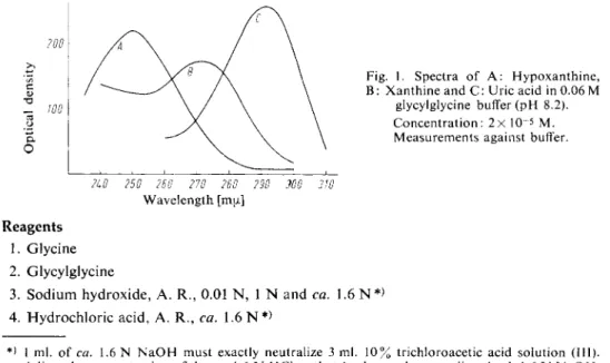
Fig. 1. Spectra of A : Hypoxanthine, B: Xanthine and C: Uric acid in 0.06 M
glycylglycine buffer (pH 8.2).
Concentration: 2x 10~
5
M.
Measurements against buffer.
2L0 250 260 270 260 290 WO 310 Wavelength [mu.]
Reagents 1. Glycine 2. Glycylglycine
3. Sodium hydroxide, A. R., 0.01 N, 1 N and ca. 1.6 N*) 4. Hydrochloric acid, A. R., ca. 1.6 N*)
*) 1 ml. of ca. 1.6 N N a O H must exactly neutralize 3 ml. 10% trichloroacetic acid solution (III).
Adjust the concentration of the ca. 1.6 N HC1 so that 1 ml. exactly neutralizes 1 ml. 1.6 N N a O H . 1) H. M. Kalckar, J. biol. Chemistry 167, 429 [1947].
2) S. Jergensen and H. E. Poulsen, Acta Pharmacol, toxicol. / / , 223 [1955].
3) K. Ro, Biochem. J. 14, 361 [1931].
4
) P. Plesner and H. M. Kalckar, in D. Glick: Methods of Biochemical Analysis. Interscience, N e w York 1956, Vol. Ill, p. 103.
Hypoxanthine and Xanthine
Soren Jorgensen Principle
The method described here was developed by Kalckar
l
\ Xanthine oxidase oxidizes hypoxanthine and xanthine to uric acid:
( l a ) Hypoxanthine + H2O + O2 xanthine + H2O2 ( l b ) Xanthine + H
2
0 + 02
• uric acid + H2
02
The uric acid formed is specifically oxidized to allantoin by uricase:
(2) Uric acid + 0
2
+ 2 H2
0 • allantoin + H2
02
+ C 02
This reaction is accompanied by a decrease in the absorption at 293 mu, due to the oxidation of the uric acid. The decrease in optical density at this wavelength is proportional to the xanthine and hypo
xanthine content of the s a m p l e
2
) .
If the sample contains uric acid, then a preliminary incubation with uricase is necessary. Before the start of the determination of hypoxanthine and xanthine the uricase must be completely destroyed (addition of alkali to the solution to give pH 1 1 )
3
) .
Hypoxanthine can be determined in the presence of xanthine if the optical density changes are mea
sured at two wavelengths
4
). The decrease in optical density at 250 mu. after the addition of xanthine oxidase is proportional to the amount of hypoxanthine, and the decrease of optical density at 293 mu.
after the addition of uricase gives the sum of hypoxanthine + xanthine (refer to Fig. 1). Blood and urine often absorb so strongly at 250 mu, that this determination by difference is impossible.
5. Trichloroacetic acid 6. Hypoxanthine
commercial preparation, see p. 1022.
7. Xanthine
commercial preparation, see p. 1033.
8. Uric acid
commercial preparation, see p. 1031.
9. Xanthine oxidase
prepared from cream according to
5
*. Outline of the procedure, see Appendix, p. 499.
10. Uricase
prepared from pig liver according to
6
*. Commercial preparation, see p. 1000.
Purity of the e n z y m e preparations
The purity of the enzymes prepared according to
5
* or
6
*, or of the commercially available prepara
tions described on p. 1000. satisfies the requirements.
Preparation of Solutions
I. Glycine buffer (0.6 M; pH 9.3):
Dissolve 4.48 g. glycine in 40 ml. distilled water, add 19.7 ml. 1 N NaOH and dilute to 100 ml. with distilled water. Check the pH with a glass electrode.
II. Glycylglycine buffer (0.6 M; pH 8.2):
Dissolve 3.17 g. glycylglycine in 5 ml. 1 N NaOH, add 20 ml. distilled water, check the pH (glass electrode) and dilute to 40 ml. with distilled water.
III. Trichloroacetic acid (10% w/v):
Dissolve 10 g. trichloroacetic acid in distilled water and make up to 100 ml.
IV. Hypoxanthine standard solution (2x 1 0 -3
M):
Dissolve 27.2 mg. hypoxanthine in 100 ml. 0.01 N NaOH.
V. Xanthine standard solution (2x 10~
3 M):
Dissolve 30.4 mg. xanthine in 100 ml. 0.01 N NaOH.
VI. Uric acid standard solution (2 x 10~
3 M):
Dissolve 33.6 mg. uric acid in 100 ml. 0.01 N NaOH.
VII. Xanthine oxidase
If necessary, dilute the phosphate gel eluate obtained according to 5
* or commercial preparations with 0.2 M phosphate buffer (pH 7.4) so that 0.01 ml. of the enzyme solu
tion increases the optical density at 293 mji. by 0.010/min., when it is added to a mixture of 0.03 ml. hypoxanthine standard solution (IV) and 2.97 ml. 0.06 M glycyl
glycine buffer (solution II diluted 1:10 with distilled water).
VIII. Uricase
Dissolve the contents of an ampoule of the commercially available preparation " Leo " **
in 0.25 ml. 0.06 M glycine buffer (solution I diluted 1:10 with distilled water). Or if necessary dilute the enzyme preparation obtained according to
6
* with 0.06 M glycine
** Pharmaceutical Products, Copenhagen, Denmark.
5* H. Klenow, Arch. Biochem. Biophysics 58, 276 [1955].
*) C. G. Holmberg, Biochem. J. 33, 1901 [1939].
buffer so that 0.01 ml. of the enzyme solution causes a decrease in optical density at 293 my* of 0.005 to 0.010/min. when it is added to a mixture of 0.03 ml. uric acid standard solution (VI) and 2.97 ml. 0.06 M glycine buffer.
Stability of the s o l u t i o n s
Store the buffers at 4 ° C and add 0.5 ml. chloroform/100 ml. Store the xanthine oxidase in the frozen state in small portions corresponding to the daily requirements. Keep the solution at 4 ° C during a series o f measurements. Store the uricase suspension at 4 ° C .
Procedure
Experimental material
Allow blood (4—5 ml.) to flow from the vein into a short test tube containing 1 —2 drops of heparin solution (39 mg. heparin/ml. distilled water = 5000 International Units/ml.).
Deproteinize immediately. If plasma is to be analysed, centrifuge immediately, since the breakdown of the ATP contained in the blood cells, which begins shortly after collecting the blood, leads to formation of hypoxanthine both in the blood cells
7
* and in the plasma. For this reason serum can not be analysed.
Urine need not be deproteinized. Dilute the urine according to the extent of the diuresis.
With a diuresis of 1 ml./min. add 4 ml. distilled water to 0.3 ml. urine. Analyse the dilute urine.
D e p r o t e i n i z a t i o n
Suck up 2 ml. of the sample with a syringe and squirt in a thin but powerful stream into 2 ml. 10% trichloroacetic acid solution (III). Centrifuge, mix 2 ml. of the supernatant with 3 ml. distilled water and 0.2 ml. 1.6 N NaOH. Analyse the mixture.
Preliminary e n z y m a t i c reaction
Pipette into 10 ml. volumetric flasks:
deproteinized mixture or dilute urine.
Adjust the pH to 9.3 (indicator paper) with 0.3—0.4 ml. glycine buffer (solution I), mix in
0.01 —0.02 ml. uricase suspension (VIII)
and allow to stand for at least 1 hour at room temperature.
Destroy the uricase activity with 1.0ml. 1.6 N NaOH**,
allow to stand for 15 min., neutralize with 1.0 ml. 1.6 N HC1*>
and adjust the pH to 8.2 (indicator paper) with ca. 0.6 ml. glycylglycine buffer (solution II).
Mix in
0.02 ml. xanthine oxidase solution (VII)
*) See footnote on p. 495.
7)
S. Jorgensen and H. E. Poulsen, Acta Pharmacol, toxicol. 77, 287 [1955].
and allow to stand for 2 hours at room temperature. Adjust the pH to ca. 9 (indicator paper) with 0.05 to 0.10 ml. 1.6 N NaOH,
add
1.0 ml. glycine buffer (solution 1)
(the pH should now be 9.3, check with indicator paper) and dilute to 10 ml. with distilled water.
D e t e r m i n a t i o n of uric a c i d
8
) — Spectrophotometric m e a s u r e m e n t s
Wavelength: 293 mu.; light path: 1 cm.; final volume: 3.01 ml. Measure against a control cuvette.
Pipette into the experimental and control cuvettes:
3.0 ml. of the preliminary enzymatic reaction mixture.
Set the spectrophotometer so that the optical density (293 mu) of the control cuvette is 0.1, 0.2 or 0.3, according to the magnitude of the optical density change expected in the experimen
tal cuvette after the addition of the uricase. Usually an optical density of 0.1 is sufficient.
Read the optical density Ei of the experimental cuvette and mix in 0.01 ml. uricase suspension (VIII)
with a small plastic rod flattened at one end. Read the optical density 4 to 5 times within 60—80 sec, plot the values against time and extrapolate to zero time to obtain the optical density E 2 .
Calculations
AE x 10 . , u - / i - • • • i- •
— — - = u.moles hypoxanthine + xanthine/preliminary enzymatic incubation mixture where
A E = E
{
- E2
10 = volume of the sample after the preliminary enzymatic reaction [ml.]
12.5 = extinction coefficient of uric acid at 293 mu [cm.
2
/u.mole]
With 0.3 ml. of urine, to obtain the [jimoles of hypoxanthine - f xanthine/ml. sample it is necessary to multiply by ~ . With samples which have to be deproteinized the values obtained by the above formula correspond to the hypoxanthine + xanthine content/ ml. sample.
Normal Values
Plasma: 5 to 10 u.moles hypoxanthine + xanthine/1000 ml. or 0.1 to 0.3 mg./lOO ml. If the blood is not treated immediately after collection, then the values increase 50 to 100-fold (see "Experimental material").
Urine: The hypoxanthine + xanthine concentration is Vio to V20 of the uric acid concentration.
The hypoxanthine + xanthine content does not alter if the urine sample is stored for 2 weeks at 4°C.
Specificity
Uricase is specific for uric a c i d
9
) . In contrast, xanthine oxidase catalyses the oxidation o f at least 30 aldehydes, ketones and p u r i n e s
1 0
-
1 1
) . Hypoxanthine and xanthine can therefore only be deter
mined specifically if the uric acid formed by the action of xanthine oxidase on both purines is measured.
8) For a detailed description, see: E. Praetorius and H. E. Poulsen, Scand. J. clin. Lab. Invest. 5, 273 [1953].
9) D. Keilin and E. F. Hartree, Proc. Roy. Soc. [London], Ser. B. 7 / 9 , 114 [1936].
^ V. H. Booth, Biochem. J. 32, 494 [1938].
ii> H.J. Coombs, Biochem. J. 27, 1259 [1927].
Appendix
Isolation of x a n t h i n e o x i d a s e
5
)
R e m o v e the cream from fresh milk while it is still warmer than 25° C. Store the cream overnight at 0 ° C and next day churn. A d d to the buttermilk 0.6 volumes of 0.2 M Na2HPC>4 solution and to every 1000 ml. of this mixture add 600 mg. trypsin. Incubate for 3 hours at 35°C. While stirring, slowly add 0.5 volumes o f water saturated n-butanol, centrifuge the mixture for 15 min. at 10000 g, decant the aqueous phase and to every 100 ml. add 47 ml.
(NH4)2S04
solution saturated at 0°C.After 15 min. centrifuge and discard the precipitated protein. T o every 100 ml. o f the supernatant add 47 ml.
(NH4)2S04
solution saturated at 0 ° C , after 1 hour centrifuge, discard the supernatant and dissolve the precipitate in distilled water to give 8 mg. protein/ml. Centrifuge, to each 1 ml. of the supernatant add 1.2 ml. calcium phosphate g e l1 2
* (16 mg. dry weight/ml.) and centrifuge. Elute the gel three times with 1 ml. portions of 0.2 M phosphate buffer (pH 7.4) and once with 1 ml. 0.3 M
K2HPO4
solution (the volumes of the solutions used for elution should be related to the volume of the enzyme solution before addition of the gel). Combine the eluates and use as the enzyme preparation.*
2
> D. Keilin and E. F. Hartree, Proc. Roy. Soc. [London], Ser. B. 124, 397 [1938].