Article
Ingol and Ingenol-Type Diterpenes from Euphorbia trigona Miller with Keratinocyte Inhibitory Activity
Reham Hammadi1, Norbert Kúsz1 , Csilla Zsuzsanna Dávid1, Zoltán Behány2, LászlóPapp3, Lajos Kemény2 , Judit Hohmann1,4, Lóránt Lakatos2,5,* and Andrea Vasas1,*
Citation: Hammadi, R.; Kúsz, N.;
Dávid, C.Z.; Behány, Z.; Papp, L.;
Kemény, L.; Hohmann, J.; Lakatos, L.;
Vasas, A. Ingol and Ingenol-Type Diterpenes fromEuphorbia trigona Miller with Keratinocyte Inhibitory Activity.Plants2021,10, 1206.
https://doi.org/10.3390/
plants10061206
Academic Editor: Salvatore Antonino Raccuia
Received: 26 April 2021 Accepted: 8 June 2021 Published: 14 June 2021
Publisher’s Note:MDPI stays neutral with regard to jurisdictional claims in published maps and institutional affil- iations.
Copyright: © 2021 by the authors.
Licensee MDPI, Basel, Switzerland.
This article is an open access article distributed under the terms and conditions of the Creative Commons Attribution (CC BY) license (https://
creativecommons.org/licenses/by/
4.0/).
1 Department of Pharmacognosy, Interdisciplinary Excellence Centre, University of Szeged, Eötvös u. 6, 6720 Szeged, Hungary; reham.hammadi@pharmacognosy.hu (R.H.); kusznorbert@gmail.com (N.K.);
davidzsuzsanna88@gmail.com (C.Z.D.); hohmann.judit@szte.hu (J.H.)
2 Department of Dermatology and Allergology, University of Szeged, Korányi fasor 6, 6720 Szeged, Hungary;
behany.zoltan@med.u-szeged.hu (Z.B.); kemeny.lajos@med.u-szeged.hu (L.K.)
3 Botanical Garden, Eötvös Loránd University, Illés u. 25, 1083 Budapest, Hungary; papplaca@gmail.com
4 Interdisciplinary Centre of Natural Products, University of Szeged, Eötvös u. 6, 6720 Szeged, Hungary
5 Photo- and Chronobiology Group Eötvös Loránd Research Network (ELKH), Institute of Plant Biology, Biological Research Center Szeged, Temesvári krt. 62, 6726 Szeged, Hungary
* Correspondence: lakatos.lorant@brc.hu (L.L.); vasas.andrea@szte.hu (A.V.); Tel.: +36-62546451 (A.V.)
Abstract:Ingenol mebutate, isolated fromEuphorbia peplus, is an ingenane-type diterpenoid, primarily used for the topical treatment of actinic keratosis, a premalignant skin condition. The aim of our work was to investigate otherEuphorbiaspecies to find structurally similar diterpenes that can be used as alternatives to ingenol mebutate. Pharmacological investigation ofEuphorbia candelabrum,Euphorbia cotinifolia,Euphorbia ramipressa, andEuphorbia trigonarevealed the potent keratinocyte (HPV-Ker cell line) inhibitory activity of these spurge species. From the methanolic extract of the aerial parts of Euphorbia trigonaMiller, the most active species, five ingol (1–5) and four ingenane-type diterpenoids (6–9) were isolated by various chromatographic separation techniques, including open column chromatography, vacuum liquid chromatography, thin-layer chromatography, and high-performance liquid chromatography. The structures of the compounds were determined by NMR spectroscopic analysis and by comparison of the assignations with the literature data. The cytotoxic activity of the compounds against keratinocytes was tested in vitro by using ingenol mebutate as a positive control. Among the isolated compounds, two ingenane derivatives (6and7) exhibited remarkably stronger cytotoxic activity (IC50values 0.39µM and 0.32µM, respectively) on keratinocytes than ingenol mebutate (IC50value 0.84µM). These compounds could serve as starting materials for further investigations to find alternatives to Picato®(with active substance ingenol mebutate), which was withdrawn from marketing authorization in the European Union.
Keywords:Euphorbiaceae;Euphorbia trigona; ingol esters; ingenol diterpenes; keratinocyte; cytotoxi- city; actinic keratosis
1. Introduction
Actinic keratosis (AK) is a skin disorder with erythematous plaques that are frequently found on sun-damaged skin [1]. AK is more likely to occur in men than women and is more prevalent in older adults [2,3]. The main causative agent of AK is excessive UV radiation, which can lead to destructive inflammatory processes, and ultimately to substantial structural damage to cell DNA and membrane lipids [4].
AK is considered to be an epidermal dysplasia, in which keratinocytes in the stratum basale and stratum spinosum have lost polarity, and appear disordered with pleomorphic nuclei. The stratum corneum shows parakeratosis and hyperkeratosis, most probably due to abnormal keratinocyte development [5]. In many AK patients, inflammatory infiltra- tion of lymphocytes and plasma cells in the dermis can also be observed [6]. Although
Plants2021,10, 1206. https://doi.org/10.3390/plants10061206 https://www.mdpi.com/journal/plants
spontaneous regression of AK is very common (>50%) [7], the condition must be taken seriously, since it can develop into malignant lesions, such as squamous cell carcinoma (SCC), the second most common form of skin cancer, and basal cell carcinoma (BCC). These conditions predominantly affect light-skinned populations [6]. Treatment options for AK include destructive therapies (e.g., surgery, cryotherapy, dermabrasion, and photodynamic therapy (PDT)), topical medications (e.g., topical fluorouracil, imiquimod, ingenol mebu- tate, and diclofenac), and field ablation treatments (chemical peels and laser resurfacing) [8].
Self-directed treatments are preferred by patients, but are time-consuming, as they need to be applied for weeks or months before producing clinical results. Although many options are available, the search for new and more effective agents of AK is in progress.
Ingenol mebutate (Picato®) is the active ingredient of the sap ofEuphorbia peplus[9,10], a plant that has long been used in Australian folk medicine for the treatment of skin cancers. It is offered with a short treatment schedule (2–3 days), providing an effective and sustained clearance of AK lesions with a predictable onset and short duration of local skin responses [11]. Ingenol mebutate acts on keratinocytes in two different ways: (1) induces the cell death of aberrant keratinocytes, and (2) induces a lesion-directed immune response that is mediated, at least partially, by the enzyme family protein kinase C (PKC) [11].
The ingenol-mebutate-induced cell death is mediated through the PKCδ/mitogen-activated protein kinase (MEK)/extracellular signal-regulated kinase (ERK) pathway. PKCδ is a key mediator in cell differentiation and the inhibition of proliferation in various tissues.
Ingenol mebutate directly binds to PKCδand leads to its phosphorylation, which activates MEK/ERK signaling, resulting in decreased viability and cell proliferation [12].
The topical application of ingenol mebutate causes rapid cell death via necrosis.
Subsequently, the concentration of TNF-αand IL-8 promptly increases, which leads to the recruitment and infiltration of neutrophils into the inflamed region, followed by the activation of an apoptotic cell death [13]. Unfortunately, Picato is no longer authorized in the EU, as the European Medicinal Agency has concluded that the medicine may increase the risk of skin cancer and that its risks outweigh its benefits. Searching for new analogues of ingenol mebutate with a higher potency and a more favorable side effect spectrum seems to be a rational aim of phytochemical researchers.
Most of the plants belonging to the genusEuphorbia(family Euphorbiaceae) contain a milky irritant latex and accumulate different types of diterpenoid esters, characteristic of the family [14,15]. In recent decades, numerous pharmacological investigations into Euphor- biaceae plants focused on the authentication of their traditional use in cancerous conditions and confirmed the efficacy of various diterpenes as promising antitumor agents [15,16].
However, no publication can be found in the literature about the keratinocyte inhibitory activity of theEuphorbiaspecies.
In a continuation of our work dealing with the isolation of biologically active diter- penoids, different extracts, prepared fromE. candelabrumTrémaux ex Kotschy,E. cotinifolia (L.) Millsp.,E. ramipressaCroisat, andE. trigonaMill., were tested for their keratinocyte inhibitory activities. E. candelabrumandE. cotinifolia were reported to contain ingenol derivatives [17]. AlthoughE. ramipressawas not investigated from the phytochemical and pharmacological perspectives, as it belongs to the same section (Euphorbiasect. Eu- phorbia) asE. candelabrumand other ingenol derivative-containing species (e.g.,E. nivulia, E. antiquorum,E. kamerunica), it is worthy of investigation [18]. Based on the results of the pharmacological assay,E. trigonaMill. was chosen for further examination.E. trigonais native to Central Africa, where it is commonly planted around villages as a ceremonial plant [19]. In Japan and the United States, it is cultivated as an indoor ornamental plant [20].
In the traditional medicinal use ofE. trigona, usually, some drops of the latex are added to palm wine for the treatment of severe cases of constipation or epileptic attacks. It is also used as an arrow and fish poison [19]. The plant is also well recognised in India, as it has been used in Ayurveda to combat infections (e.g., in the urinary tract) and to alleviate the symptoms inflammation [21].
Previous studies have revealed the presence of ingol and ingenol esters in the latex of E. trigonathat are responsible for its strong skin-irritating and pesticidal properties [20];
while lectins, another important class of phytoconstituents described from Euphorbia lat- ices, have been reported to possess potent erythrocyte agglutinating ability [19]. The latex also contains compounds acting as antimicrobial agents in the urinary tract by attenuating the virulence of the pathogens and facilitating the elimination of them via stimulating the host’s immune system [21,22]. Interestingly, the extracts ofE. trigonadisplayed immunos- timulant activity as well [23].
Previously, ingol- and ingenane-type diterpenoids were isolated from the latex ofE.
trigona[20]. Moreover, the presence of triterpenoids was reported from the stems of the plant [24].
The aim of our work was to isolate the diterpenoids from the plant and to investigate their effects on keratinocytes in order to find potential lead compounds for the effective treatment of AK.
2. Results and Discussion
The main risk factor of induction of AK is UV irradiation. The UVB component of sunlight damages DNA, which might cause mutations in p53 and other tumor suppressor genes [25]. Therefore, as an in vitro model of AK, we used the HPV-Ker cell line which contains the E6 oncoprotein of the human papillomavirus, leading to the degradation of the p53 tumor suppressor [26].
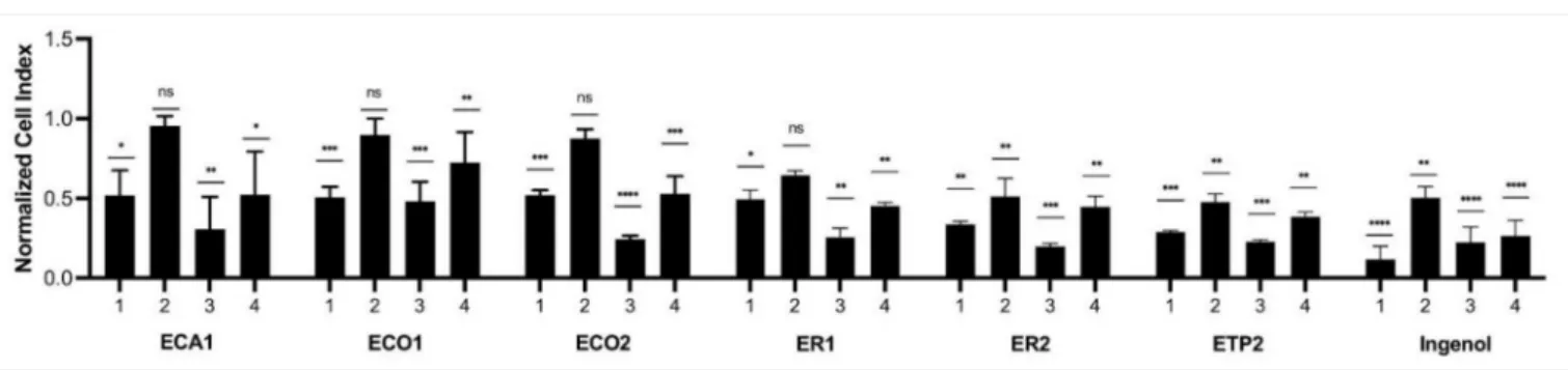
The extracts, prepared fromE. candelabrum,E. cotinifolia,E. ramipressa, andE. trigona, were tested at concentrations of 5 and 0.5 µg/mL for their inhibitory activity against keratinocytes (Figure1). Ingenol mebutate administered at 5µg/mL for 24 h exerted the strongest cytotoxic effect, and after a 48 h treatment, a lower cytotoxicity was measured.
The treatment of keratinocytes with ingenol mebutate at a 0.5µg/mL concentration resulted in a weaker, but still significant, cytotoxic effect after 24 h and, interestingly, after 48 h, the inhibitory activity was comparable to the treatment when 5µg/mL was used for 48 h.
The extracts with a 5µg/mL concentration were applied for 24 h, and ET1 and ET2 had similar, but lower, cytotoxic activity than that of ingenol mebutate. Interestingly, the 48 h treatment of cells with 5µg/mL extracts ECA1, ECO2, ER1, ER2, ET1, and ET2 showed a very similar cytotoxic property as ingenol mebutate. Cytotoxic activity was the lowest when the extracts were used at a 0.5µg/mL concentration for 24 h, but in the case of ER2, ET1, and ET2, cytotoxic activity was significant compared to the control and quite similar to that of ingenol mebutate. After administration of the extracts at a 0.5µg/mL concentration for 48 h, only ET1 displayed cytotoxicity comparable to that of ingenol mebutate. Based on these results, then-hexane extract ofE. trigona(ET1) could be considered the most promising one for further investigation.
Figure 1.Inhibitory activity of differentEuphorbiaextracts against keratinocytes. ECA1:E. candelabrum n-hexane extract, ECO1:E. cotinifolia n-hexane extract, ECO2:E. cotinifoliaCHCl3extract, ER1:E. ramipressa n-hexane extract, ER2:E. ramipressa CHCl3extract, ET1:E. trigona n-hexane extract, ET2: E. trigonaCHCl3extract; 1: 5µg/mL, 24 h, 2: 0.5µg/mL, 24 h, 3:
5µg/mL, 48 h, 4: 0.5µg/mL, 48 h; *p< 0.05; **p< 0.01; ***p< 0.001; ****p< 0.0001.
Previous investigations into the plants resulted in the isolation of various diterpenoids, such as 3-O-propionyl-20-O-(S)-(20-methyl)butyryl-ingenol, 20-O-isobutyryl-ingenol, 3- O-propionyl-20-O-isobutyryl-ingenol, and 3,20-O-di-isobutyryl-ingenol from the leaves ofE. cotinifolia[27], and 17-acetoxyingenol 3 angelate 5,20-diacetate and 17-acetoxy-20- deoxyingenol 3-angelate, from the latex ofE. trigona[20].
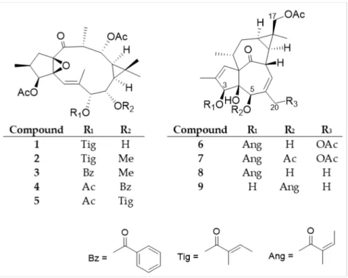
Among the investigated plants, the most activeE. trigona was chosen for further phytochemical and pharmacological investigation. Chromatographic separation of the methanolic extract, prepared from the fresh aerial parts of E. trigona by open column chromatography using polyamide and vacuum liquid chromatography on silica gel, as well as by preparative TLC, and normal and reversed-phase HPLC, yielded nine diterpenoids (1–9), including five ingol (1–5) and four ingenol (6–9) esters (Figure2).
Figure 2.Structures of the isolated diterpenoids (1–9).
The structures of the compounds were determined by comparison of their spectro- scopic data to those of reported literature values (Supplemetary Materials Figures S1–S16).
Diterpenoid esters1–9were described earlier from only a few plant species. Ingol 3,12- diacetate 7-tigliate (1) was previously isolated fromE. hermentiana[28], 8-O-methyl-ingol 3,12-diacetate 7-tigliate (2) fromE. acrurensis[29],E. hermentiana[28], andE. kamerunica[30], 8-O-methyl-ingol 3,12-diacetate 7-benzoate (3) fromE. antiquorum[31],E. hermentiana[28], andE. kamerunica[30], ingol 3,7,12-triacetate 8-benzoate (4) fromE. antiquorum[31],E. nivu- lia[16,32]E. hermentiana[28], andE. kamerunica[33], and ingol 3,7,12-triacetate 8-tigliate (5) from E. antiquorum[31], andE. kamerunica[33]. The ingenane-type compound 17- acetoxyingenol 3-angelate 20-acetate (6) was previously reported fromE. canariensis[34]
andE. hermentiana[35], 17-acetoxyingenol 3 angelate 5,20-diacetate (7) from E. hermen- tiana[35],E. kamerunica[30],E. royleana[36], andE. trigona[20], 17-acetoxy-20-deoxyingenol 3-angelate (8) fromE. acrurensis[29],E. hermentiana[35] andE. trigona[20], and 17-acetoxy- 20-deoxyingenol 5-angelate (9) fromE. hermentiana[35]. None of the compounds were evaluated for keratinocyte viability modifying activity.
According to the classification of Pax and Hoffmann,E. antiquorumandE. royleana belong to the Section Euphorbium Bentham, Subsection Diacanthium Boiss., and Series Trigonae Berger (V), whileE. canariensis,E. candelabrum, andE. kamerunicabelong to the Series Polygonae Berger (VI) of the same Section and Subsection [37]. Therefore, botanically they are very close to each other, and it is not surprising that they contain structurally
similar diterpenoids, as was proven by Evans and Kinghorn [38]. Moreover, all the species are succulent and originate from Africa.
After the discovery of ingenol mebutate as an effective drug for the treatment of AK, a number of semisynthetic derivatives were produced or synthesized with full regiocontrol from ingenol [11,39].
Since our compounds, especially the ingenol-derivatives, are structurally very similar to ingenol mebutate (Figure3), we were interested in whether they have any effect on the viability of keratinocytes. The HPV-Ker cell line was treated with the isolated ingol- and ingenol-type diterpenoids with a concentration range of 5×10⁻⁹–5×10⁻⁴M, the viability was measured with the xCELLigence System RTCA HT (Agilent Technologies, Santa Clara, CA, USA) instrument for 72 h, and data at 24 and 48 h were used for calculations (Table1).
IC50values were calculated based on the sigmoidal dose–response formula.
Figure 3.The structure of ingenol mebutate.
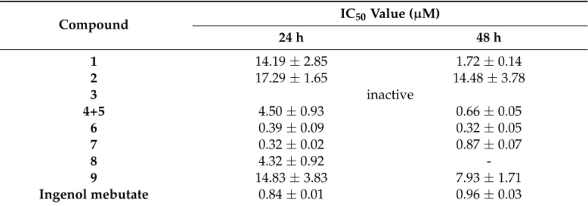
Table 1.IC50values (µM±SD) of the ingol- and ingenane-type diterpenoids (1–9).
Compound IC50Value (µM)
24 h 48 h
1 14.19±2.85 1.72±0.14
2 17.29±1.65 14.48±3.78
3 inactive
4+5 4.50±0.93 0.66±0.05
6 0.39±0.09 0.32±0.05
7 0.32±0.02 0.87±0.07
8 4.32±0.92 -
9 14.83±3.83 7.93±1.71
Ingenol mebutate 0.84±0.01 0.96±0.03
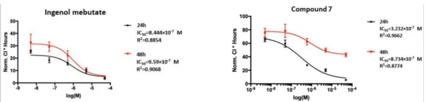
Ingenol mebutate, which was used as a positive control, showed IC50 values of 0.84 and 0.96µM at 24 and 48 h of treatment (Figure4), respectively. For the ingenol- type compound, 17-acetoxy-20-deoxyingenol 5-angelate (9), about one and two order of magnitude higher IC50 values, were recorded after 24 and 48 h of treatment (14.83 and 7.93µM) than for ingenol mebutate (Supplementary Materials Figure S17). Strikingly, the IC50values of the ingenane-type diterpenoids, 17-acetoxyingenol 3-angelate 20-acetate (6) and 17-acetoxyingenol 3 angelate 5,20-diacetate (7), were the same order of magnitude as ingenol mebutate (Figure4and Figure S1). Moreover, the IC50values of6(0.39µM and 0.32µM) and7(0.32µM and 0.87µM) were slightly lower on the HPV-Ker cell line than that of ingenol mebutate (0.84µM and 0.96µM).
Figure 4. Real-time cell analysis (RTCA) measurement of CI (cell index) values of HPV-Ker cells treated with ingenol mebutate and compound7. Normalized CI * hours values were plotted as a function of concentration of the indicated diterpenoid (logM).
In the case of the ingol-type diterpenoids1,2and4+5, the measured IC50values were one or two orders of magnitude higher than that of ingenol mebutate, except for4+5at 48 h (0.66µM) (Table1).
Based on the pharmacological results, structure–activity relationship (SAR) investi- gations could also be performed. The ingenol derivatives (6–9) are structurally close to ingenol mebutate. The main difference between ingenol mebutate and the isolated com- pounds is the presence of an acetoxy group at C-17 in6–9instead of a 17-methyl group in ingenol mebutate. In the less active 17-acetoxy-20-deoxyingenol 5-angelate (9), the angeloyl group at C-3, and hydroxy group at C-5, are replaced compared to ingenol mebutate and 6–8; therefore, it was concluded that the presence of the angeloyl group at C-3 seems to be essential for the cytotoxic activity. Since compound8differs from ingenol mebutate in only the presence of an acetoxy group at C-17, and its activity was lower, the acetoxy group alone, presumably, is not able to increase the activity. In the case of the most active com- pounds6and7, one (at C-20 in6) or two (at C-5 and C-20 in7) additional acetyl groups are attached to the diterpenoid core; therefore, acetylation of the molecule results in increased cytotoxic activity. This is in accordance with the previously determined SAR statement that the carbonyl moieties of the ester groups are essential for the desired biological effects and the activation of PKC, which likely happens through interaction with Gly23 NH in the C1 domain [11]. Moreover, besides the activation of PKCδ, ingenol mebutate was found to reduce the expression of PKCα, which is the PKC isoform responsible for the promotion of cell survival [40]. Thus, further studies are required to evaluate the beneficial effect of our compounds that might result in more effective isoform-specific regulation.
In summary, among the ingol- and ingenol-type diterpenoids tested in our work, the ingenol-type 17-acetoxyingenol 3-angelate 20-acetate (6) and 17-acetoxyingenol 3 ange- late 5,20-diacetate (7) showed stronger cytotoxic activity on keratinocytes after 24 and 48 h of administration than ingenol mebutate.
3. Materials and Methods
3.1. General Experimental Procedures
NMR spectra were recorded in MeOD and DMSO-d6on a Bruker Avance DRX 500 spectrometer at 500 MHz (1H) and 125 MHz (13C). The signals of the deuterated solvents were chosen as references. The chemical shift values (δ) were given in ppm and coupling constants (J) in Hz. Two-dimensional (2D) experiments were performed with standard Bruker software. In the1H–1H COSY, HSQC, and HMBC experiments, gradient-enhanced versions were used. Column chromatography (CC) was performed on polyamide (MP Biomedicals Germany GmbH). Normal phase vacuum liquid chromatography (VLC) was carried out on silica gel (Kieselgel 60 GF254, 15µm, Merck, Darmstadt, Germany). Thin- layer chromatography was performed on Kieselgel 60 RP-18 F254 and Kieselgel 60 F254
(Merck). Spots on UV active silica gel were detected under UV light (245 nm and 336 nm) and made visible with vanillin sulphuric acid and heating at 105◦C for 2 min. The high- performance liquid chromatography (HPLC) separation was carried out on a Waters HPLC
(Waters 600 controller, Waters 600 pump, and Waters 2998 photodiode array detector), using normal (LiChrospher Si 100 (250×4 mm, 5µm, Merck, Darmstadt, Germany) and RP (LiChrospher RP-18 (5µm, 250×4 mm, Merck, Darmstadt, Germany) columns. In the case of the gradient elution, the mobile phase consisted of cyclohexane (solvent A) and EtOAc (solvent B) with a flow rate of 1.5 mL/min at normal phase separation. The initial mobile phase composition was maintained at 80% solvent A for 1 min, changed linearly to 60% (1–10 min), then changed linearly to 80% (10–10.5 min) and held at 80% for 1 min (10.5–
11.5 min), then followed by changing to 100% mobile phase A within 30 s (11.5–12 min) and kept 3 min (12–15 min) for the chromatography column equilibrium. In the case of the RP column, the mobile phase consists of MeOH (solvent A) and H2O (solvent B). The initial mobile phase composition was maintained at 80% solvent A for 1 min, changed linearly to 60% (1–10 min), then changed linearly to 80% (10–10.5 min) and held at 80% for 1 min (10.5–11.5 min), followed by a change to 100% mobile phase A within 30 s (11.5–12 min) and maintained for 3 min (12–15 min) for the chromatography column equilibrium. The flow rate was 1 mL/min and the injection volume was 25µL. The data were acquired and processed with the Empower software.
All solvents used for CC were of at least analytical grade (VWR Ltd., Szeged, Hungary).
Ultra-pure water was prepared with a Milli-Q water purification system (Merck, Darmstadt, Germany ).
3.2. Plant Materials
The aerial parts ofE. candelabrum,E. cotinifolia,E. ramipressa, andE. trigona(100 g, each) were collected in the Botanical Garden of the University of Szeged and were identified by AnikóNémeth (director of the Botanical Garden). The aerial parts (5.7 kg, fresh weight) ofE. trigonaMill. were collected in June 2018, in the Botanical Garden of Eötvös Loránd University, Hungary, and were identified by one of the authors (L.P., Botanical Garden, Eötvös Loránd University, 1083 Budapest, Hungary). A voucher specimen (No. 891) was deposited at the Department of Pharmacognosy, University of Szeged, Szeged, Hungary.
3.3. Extraction and Isolation
The fresh plant materials ofE. candelabrum,E. cotinifolia,E. ramipressa, andE. trigona (100 g, each) were extracted with methanol (3 × 500 mL, each) at room temperature.
The extracts were concentrated in vacuo, and then dissolved in MeOH–H2O 1:1 (150 mL, each), and partitioned withn-hexane and CHCl3(3×150 mL, each). Then-hexane and CHCl3extracts were evaporated to dryness and used for pharmacological investigation.
The fresh aerial parts ofE. trigona(5.7 kg) were blended and percolated with methanol (30 L) at room temperature. The methanol extract was concentrated in vacuo, and then dissolved in MeOH–H2O 1:1, and partitioned withn-hexane, CHCl3, EtOAc, and BuOH to obtain five fractions (n-hexane, CHCl3, EtOAc, BuOH, and aqueous residue). The fractions were monitored by normal phase thin-layer chromatography (TLC). After UV detection, the plates were sprayed with concentrated sulfuric acid and heated at 105◦C for 2 min.
Based on the TLC determination, it could be observed that diterpenes were accumulated in then-hexane fraction.
n-Hexane fraction (26.5 g) was separated by polyamide column chromatography with a MeOH–H2O gradient system (3:2, 4:1, and 1:0) to obtain three fractions (P1-3).
Fraction P1 (10.0 g) was then separated by vacuum liquid chromatography (VLC) on silica gel with a gradient system of cyclohexane–EtOAc–EtOH (99:1:0–1:1:1; 500 mL of each).
TLC determination and combination of the fractions afforded 20 main fractions (P1/1- 20). Fractions P1/7 (164.8 mg), P1/8 (326.7 mg), P1/9 (160.9 mg), and P1/12 (96.8 mg) were purified by VLC on NP silica gel with a cyclohexane–EtOAc gradient solvent system (95:5:0–6:3:0.5) to yield combined fractions P1/7/1-4, P1/8/1-4, P1/9/1-4, and P1/12/1-6, respectively. Fractions P1/7/2 (89 mg), P1/8/3 (189 mg), P1/8/4 (161 mg), and P1/9/4 (82 mg) were further purified by preparative TLC on NP silica gel with toluene–acetone 8:2 as the mobile phase, and then by NP-HPLC with a gradient system of cyclohexane–EtOAc
(flow rate 1.5 mL/min) to yield compounds:4+5(13.3 mg) from P1/7/2;2(45.7 mg) from P1/8/3;3(1.4 mg) from P1/8/4;7(6.8 mg);9(4.8 mg) from P1/9/4. Compound1(12.1 mg) was isolated from P1/9/4 by preparative TLC using toluene–acetone 8:2 as the mobile phase. Purification of P/1/12/4 (9.6 mg) by preparative TLC (NP, toluene–acetone 8:2) resulted in the isolation of compound8(1.9 mg). Fraction P/1/12/6 (12 mg) was separated by preparative TLC with toluene–acetone 7:3, and then by RP-HPLC with a gradient system of MeOH–H2O (flow rate 1 mL/min) to obtain compound6(6.5 mg).
3.4. Keratinocyte Inhibitory Activity Investigation
The immortalized human keratinocyte cell line (HPV-Ker) was cultured in Keratinocyte- SFM supplemented with 5 ng/mL of recombinant epidermal growth factor, 50 mg/mL of bovine pituitary extract, and antibiotic/antimycotic solution in a CO2thermostat at 37◦C.
Viability Test
The effect of the extracts and the purified compounds on HPV-Ker cells was investi- gated by real-time monitoring with the xCELLigence System RTCA HT Instrument (ACEA Biosciences, San Diego, CA, USA). 8×10³HPV-Ker cells were seeded in 96-well E-plates and keratinocytes were allowed to attach to the bottom of the wells and grow for 24 h.
Cells were then treated with the indicated extract or substance for another 72 h. Real-time measurements of impedance were monitored every 15 min, then data at 24 and 48 h were used for calculations. The half-maximal inhibitory concentration (IC50) values for the com- pounds were calculated and GraphPad Prism 9.0 was used to plot results. All experiments were performed in duplicate, in three independent repeats.
4. Conclusions
The extracts of fourEuphorbiaspecies, namelyE. candelabrum,E. cotinifolia,E. rami- pressa, andE. trigona, were tested for their antiproliferative effect against keratinocytes.
All species proved to be active. Among them,E. trigona, the most active, was chosen for further investigation. From the aerial parts of the plant, nine diterpenoids (1–9) were isolated, including five ingol (1–5) and four ingenol esters (6–9). 17-Acetoxyingenol 3- angelate 5,20-diacetate (7) and 17-acetoxy-20-deoxyingenol 3-angelate (8) were previously identified from the plant, while the others were described from otherEuphorbiaspecies.
The diterpenoid profile ofE. trigonawas found to be very similar to that ofE. hermentiana, confirming that these two species are chemotaxonomically closely related. The isolated compounds1–9 have been tested for their keratinocyte inhibitory activity for the first time. Two ingenanes (6and7) demonstrated strong keratinocyte inhibitory activity in vitro which was comparable to that of ingenol mebutate, the drug used in the treatment of actinic keratosis. None of the ingol-type esters exerted considerable activity compared to ingenol mebutate.
The mechanism of action of ingenol mebutate is complex; it involves a combination of necrotic and immunostimulating effects. Previous studies established that the apoptotic properties of ingenol mebutate are based on the activation of PKCδand its translocation from the cytoplasm into the nuclear membrane. Furthermore, the reduction in the expres- sion of PKCαalso plays a role in the effect of the compound. The interaction of ingenoids with PKC is critically dependent on their type of ester decoration and requires a combina- tion of optimal hydrogen bonding and hydrophobic contacts for high potency [41]. To date, only limited information exists on the structure–activity relationships of ingenol esters as PKC ligands, and in the previous studies, the influence of 17-O-acyl groups present in6 and7was not studied.
17-Acetoxyingenol 3-angelate 20-acetate (6) and 17-acetoxyingenol 3 angelate 5,20- diacetate (7) could serve as promising compounds in the search for novel therapeutic agents for actinic keratosis, as they are structurally very similar to ingenol mebutate and possess higher keratinocyte inhibitory activity. This higher activity is probably due to the acetoxy group(s), as it was previously observed that the carbonyl moieties of the ester groups
are essential for the activation of PKC, and therefore, the biological effect. Based on the pharmacological screening study, the other investigatedEuphorbiaspecies (E. candelabrum, E. cotinifolia, andE. ramipressa) are also worthy of phytochemical and pharmacological investigation.
Supplementary Materials:The following are available online athttps://www.mdpi.com/article/
10.3390/plants10061206/s1. Figures S1–S16: 1H and13C JMOD NMR spectra of compounds1–9.
Figure S17: RTCA (real-time cell analysis) measurement of CI (cell index) values of HPV-Ker cells treated with compounds1,2,4+5,6,and9. Normalized CI * hours values were plotted as a function of concentration of the indicated diterpenoid (logM).
Author Contributions:Conceptualization, A.V. and L.L.; methodology, A.V. and L.L.; investigation, L.P., R.H., Z.B. and C.Z.D.; writing—original draft preparation, R.H. and N.K.; writing—review and editing, A.V. and L.L.; supervision, L.K. and J.H. All authors have read and agreed to the published version of the manuscript.
Funding: This research was funded by the Economic Development and Innovation Operative Programme GINOP-2.3.2-15-2016-00012, GINOP-2.3.2-15-2016-00015 and GINOP-2.3.2-15-2016-00032 andÚNKP-20-4 (co-author N.K.)—New National Excellence Program of the Ministry for Innovation and Technology from the source of the National Research, Development and Innovation Funds.
Institutional Review Board Statement:Not applicable.
Informed Consent Statement:Not applicable.
Acknowledgments:The authors would like to thank AnikóNémeth (director of the Botanical Garden of University of Szeged) for providing plant samples and for the identification of the plants.
Conflicts of Interest:The authors declare no conflict of interest. The funders had no role in the design of the study; in the collection, analyses, or interpretation of data; in the writing of the manuscript, or in the decision to publish the results.
References
1. De Oliveira, E.C.V.; Da Motta, V.R.V.; Pantoja, P.C.; De O Ilha, C.S.; Magalhães, R.F.; Galadari, H.; Leonardi, G.R. Actinic keratosis—Review for clinical practice.Int. J. Dermatol.2019,58, 400–407. [CrossRef] [PubMed]
2. Costa, C.; Scalvenzi, M.; Ayala, F.; Fabbrocini, G.; Monfrecola, G. How to treat actinic keratosis? An update.J. Dermatol. Case Rep.
2015,9, 29–35. [CrossRef] [PubMed]
3. Dodds, A.; Chia, A.; Shumack, S. Actinic keratosis: Rationale and management. Dermatol. Ther. 2014,4, 11–31. [CrossRef]
[PubMed]
4. Zhao, B.; He, Y.Y. Recent advances in the prevention and treatment of skin cancer using photodynamic therapy. Expert Rev.
Anticancer Ther.2010,10, 1797–1809. [CrossRef]
5. Bickers, D.R.; Lim, H.W.; Margolis, D.; Weinstock, M.A.; Goodman, C.; Faulkner, E.; Gould, C.; Gemmen, E.; Dall, T. The burden of skin diseases: 2004: A joint project of the American Academy of Dermatology Association and the Society for Investigative Dermatology.J. Am. Acad. Dermatol.2006,55, 490–500. [CrossRef]
6. Siegel, J.A.; Korgavkar, K.; Weinstock, M.A. Current perspective on actinic keratosis: A review.Br. J. Dermatol.2017,177, 350–358.
[CrossRef]
7. Marks, R.; Foley, P.; Goodman, G. Spontaneous remission of solar keratoses: The case for conservative management. Br. J.
Dermatol.1986,115, 649–655. [CrossRef]
8. Gupta, A.K.; Paquet, M.; Villanueva, E.; Brintnell, W. Interventions for actinic keratoses.Cochrane Database Syst. Rev.2012,12, CD004415. [CrossRef]
9. Hohmann, J.; Evanics, F.; Berta, L.; Bartók, T. Diterpenoids fromEuphorbia peplus.Planta Med.2000,66, 291–294. [CrossRef]
10. Ogbourne, S.M.; Suhrbier, A.; Jones, B.; Cozzi, S.J.; Boyle, G.M.; Morris, M.; McAlpine, D.; Johns, J.; Scott, T.M.; Sutherland, K.P.;
et al. Antitumor activity of 3-ingenyl angelate: Plasma membrane and mitochondrial disruption and necrotic cell death.Cancer Res.2004,64, 2833–2839. [CrossRef]
11. Liang, X.; Grue-Sørensen, G.; Månsson, K.; Vedsø, P.; Soor, A.; Stahlhut, M.; Bertelsen, M.; Engell, K.M.; Högberg, T. Syntheses, biological evaluation and SAR of ingenol mebutate analogues for treatment of actinic keratosis and non-melanoma skin cancer.
Bioorg. Med. Chem. Lett.2013,23, 5624–5629. [CrossRef] [PubMed]
12. Freiberger, S.N.; Cheng, P.F.; Iotzova-Weiss, G.; Neu, J.; Liu, Q.; Dziunycz, P.; Zibert, J.R.; Dummer, R.; Skak, K.; Levesque, M.P.;
et al. Ingenol mebutate signals via PKC/MEK/ERK in keratinocytes and induces interleukin decoy receptors IL1R2 and IL13RA2.
Mol. Cancer Ther.2015,14, 2132–2142. [CrossRef] [PubMed]
13. Hui, A.; Markowitz, O. Ingenol mebutate for the treatment of actinic keratosis.Curr. Dermatol. Rep.2013,2, 172–176. [CrossRef]
14. Scholz, A. Euphorbiaceae. InSyllabus der Planzenfamilien; Engler, A., Ed.; Berlin-Nikolassee, Gebrüder Bornträger: Berlin, Germany, 1964; Volume 2, pp. 255–261.
15. Vasas, A.; Hohmann, J. Euphorbia diterpenes: Isolation, structure, biological activity, and synthesis (2008−2012).Chem. Rev.2014, 114, 8579–8612. [CrossRef]
16. Ravikanth, V.; Reddy, V.L.N.; Rao, T.P.; Diwan, P.V.; Ramakrishna, S.; Venkateswarlu, Y. Macrocyclic diterpenes fromEuphorbia nivulia.Phytochemistry2002,59, 331–335. [CrossRef]
17. Evans, F.J.; Taylor, S.E. Pro-inflammatory, tumour-promoting and antitumour diterpenes of the plant families Euphorbiaceae and Thymeleaceae. InProgress in the Chemistry of Organic Natural Products; Herz, W., Grisebach, H., Kirby, G.W., Eds.; Springer: New York, NY, USA, 1983; Volume 44, pp. 1–99.
18. Dorsey, B. Phylogenetics and Morphological Evolution ofEuphorbiasubgenusEuphorbia. Ph.D. Thesis, University of Michigan, Michigan, MI, USA, 2013.
19. Schmelzer, G.H.; Gurib-Fakim, A.Plant Resources of Tropical Africa, Medicinal Plants; PROTA Foundation: Wageningen, The Nether- lands, 2008; Volume 11, p. 260.
20. Tada, M.; Seki, H. Toxic diterpernes fromEuphorbia trigona(Saiunkaku: Indoor foliage plant in Japan).Agric. Biol. Chem.1989,53, 425–430.
21. Upadhyay, M.; Nashikkar, N.; Begde, D.; Bundale, S.; Pise, M.; Rudra, J.; Upadhyay, A. Study of antimicrobial, antioxidant and antiquourm sensing properties ofEuphorbia trigona.Global J. Res. Med. Plants Indigen. Med.2013,2, 630–641.
22. Nashikkar, N.; Begde, D.; Bundale, S.; Pise, M.; Rudra, J.; Upadhyay, A. Inhibition of swarming motility, biofilm formation and virulence factor expression of urinary pathogens byEuphorbia trigonalatex extracts.Int. J. Pharm. Res.2011,2, 558–566.
23. Nashikkar, N.; Begde, D.; Bundale, S.; Mashitha, P.; Rudra, J.; Upadhyay, A. Evaluation of the immunomodulatory properties of Euphorbia trigona—An in vitro study.IJIPLS2012,2, 88–105.
24. Anjaneyulu, V.; Rao, G.S.; Connolly, J.D. Occurrence of 24-epimers of cycloart-25-ene-3β,24-diols in the stems ofEuphorbia trigona.
Phytochemistry1985,24, 1610–1612. [CrossRef]
25. Reinehr, C.P.H.; Bakos, R.M. Actinic keratoses: Review of clinical, dermoscopic, and therapeutic aspects.An. Bras. Dermatol.2019, 94, 637–657. [CrossRef] [PubMed]
26. Tax, G.; Urbán, E.; Palotás, Z.; Puskás, R.; Kónya, Z.; Bíró, T.; Kemény, L.; Szabó, K. Propionic acid produced byPropionibacterium acnesstrains contributes to their pathogenicity.Acta Derm Venereol.2016,96, 43–49. [CrossRef]
27. Hirota, M.H.; Yoshinori, O.O.; Koshimizu, K. New ingenol-esters as piscicidal constituents ofEuphorbia cotinifoliaL.Agric. Biol.
Chem.1980,44, 1351–1356. [CrossRef]
28. Lin, L.J.; Kinghorn, A.D. 8-methoxyingol esters from the latex ofEuphorbia hermentiana. Phytochemistry1983,22, 2795–2799.
[CrossRef]
29. Marco, J.A.; Sanz-Cervera, J.F.; Ropero, F.J.; Checa, J.; Fraga, B.M. Ingenane and lathyrane diterpenes from the latex ofEuphorbia acrurensis.Phytochemistry1998,49, 1095–1099. [CrossRef]
30. Connolly, J.D.; Fakunle, C.O.; Rycroft, D.S. Five ingol esters and a 17-hydroxyingenol ester from the latex ofEuphorbia kamerunica.
Assignment of esters using13C NMR methods.Tetrahedron Lett.1984,25, 3773–3776. [CrossRef]
31. Yin, Z.Y.; Dai, Y.; Hua, P.; Sun, Z.J.; Cheng, Y.F.; Yuan, S.H. Discovery of diverse diterpenoid scaffolds fromEuphorbia antiquorum and their activity againat RANKL-induced osteoclastogenesis.Bioorg. Chem.2019,92, 103292. [CrossRef] [PubMed]
32. Ravikanth, V.; Reddy, V.L.N.; Reddy, A.V.; Diwan, P.V.; Venkateswarlu, Y. Diterpenes from the latex ofEuphorbia nivulia.Biochem.
Syst. Ecol.2003,31, 447–449. [CrossRef]
33. Abo, K.; Evans, F.J. The composition of a mixture of ingol esters fromEuphorbia kamerunica. Planta Med. 1981,43, 392–395.
[CrossRef]
34. Marco, J.A.; Sanz-Cervera, J.F.; Yuste, A. Ingenane and lathyrane diterpenes from the latex ofEuphorbia canariensis.Phytochemistry 1997,45, 563–570. [CrossRef]
35. Lin, L.J.; Marshall, G.T.; Kinghorn, A.D. The dermatitis-producing constituents ofEuphorbia hermentianalatex.J. Nat. Prod.1983, 46, 723–731. [CrossRef] [PubMed]
36. Li, X.L.; Li, Y.; Wang, S.F.; Zhao, Y.L.; Liu, K.C.; Wang, X.M.; Yang, Y.P. Ingol and ingenol diterpenes from the aerial parts of Euphorbia royleanaand their antiangiogenic activities.J. Nat. Prod.2009,72, 1001–1005. [CrossRef]
37. Pax, F.; Hoffmann, H. Euphorbiaceae. InDie Natürlichen Pflanzenfamilien, 2nd ed.; Eiiglcr, A., Prantl, K., Eds.; Engelmann: Leipzig, Germany, 1931; p. 19C.
38. Evans, F.J.; Kinghorn, A.D. A comparative phytochemical study of the diterpenes of some species of the generaEuphorbiaand Elaeophorbia(Euphorbiaceae).Bot. J. Linn. Soc.1977,74, 23–35. [CrossRef]
39. Grue-Sorensen, G.; Liang, X.; Mansson, K.; Vedso, P.; Dahl, S.M.; Soor, A.; Stahlhut, M.; Bertelsen, M.; Engell, K.M.; Högberg, T.
Synthesis, biological evaluation and SAR of 3-benzoates of ingenol for treatment of actinic keratosis and non-melanoma skin cancer.Bioorg. Med. Chem. Lett.2014,24, 54–60. [CrossRef] [PubMed]
40. Ghoul, A.; Serova, M.; Astorgues-Xerri, L.; Bieche, I.; Bousquet, G.; Varna, M.; Vidaud, M.; Phillips, E.; Weill, S.; Benhadji, K.A.;
et al. Epithelial-to-mesenchymal transition and resistance to ingenol 3-angelate, a novel protein kinase C modulator, in colon cancer cells.Cancer Res.2009,69, 4260–4269. [CrossRef] [PubMed]
41. Appendino, G. Ingenane diterpenoids.Prog. Chem. Org. Nat. Prod.2016,102, 1–90. [PubMed]