International Journal of
Molecular Sciences
Article
CRK5 Protein Kinase Contributes to the Progression of Embryogenesis of Arabidopsis thaliana
Abu Imran Baba1,2,† , IldikóValkai1,†, Nitin M. Labhane3, Lilla Koczka4,5,
Norbert Andrási1,2,Éva Klement6, Zsuzsanna Darula6 , Katalin F. Medzihradszky6, LászlóSzabados1, Attila Fehér1,5 , Gábor Rigó1,* andÁgnes Csépl ˝o1,*
1 Institute of Plant Biology, Biological Research Centre, 6726 Szeged, Hungary; baba.abuimran@brc.hu (A.I.B.);
ildiko.valkai@gmail.com (I.V.); andrasi.norbert@brc.hu (N.A.); szabados.laszlo@brc.hu (L.S.);
feher.attila@brc.hu (A.F.)
2 Doctoral School in Biology, Faculty of Science and Informatics, University of Szeged, 6720 Szeged, Hungary
3 Department of Botany, Bhavan’s College Andheri West, Mumbai 400058, India; nitin.labhane@bhavans.ac.in
4 Developmental and Cell Biology of Plants, CEITEC Masaryk University, 62500 Brno, Czech Republic;
lilla.koczka@ceitec.muni.cz
5 Department of Plant Biology, University of Szeged, 52. Középfasor, H-6726 Szeged, Hungary
6 Proteomics Research Group, Biological Research Centre, 6726 Szeged, Hungary; klement.eva@brc.hu (É.K.);
darula.zsuzsanna@brc.hu (Z.D.); medzihradszky.katalin@brc.hu (K.F.M.)
* Correspondence: rigo.gabor@brc.hu (G.R.); cseplo.agnes@brc.hu (Á.C.); Tel.:+36-62-599-703 (G.R. &Á.C.)
† These authors contributed equally to this work.
Received: 31 October 2019; Accepted: 30 November 2019; Published: 4 December 2019
Abstract: The fine tuning of hormone (e.g., auxin and gibberellin) levels and hormone signaling is required for maintaining normal embryogenesis. Embryo polarity, for example, is ensured by the directional movement of auxin that is controlled by various types of auxin transporters.
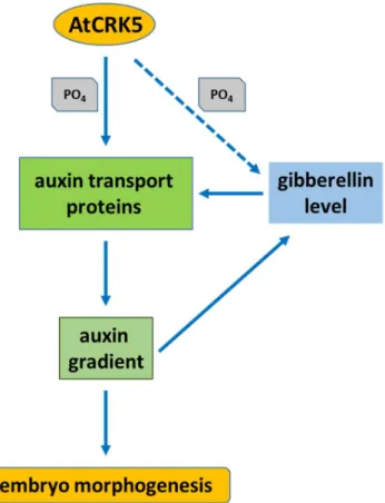
Here, we present pieces of evidence for the auxin-gibberellic acid (GA) hormonal crosstalk during embryo development and the regulatory role of theArabidopsis thalianaCalcium-Dependent Protein Kinase-Related Kinase 5 (AtCRK5) in this regard. It is pointed out that the embryogenesis of the Atcrk5-1mutant is delayed in comparison to the wild type. This delay is accompanied with a decrease in the levels of GA and auxin, as well as the abundance of the polar auxin transport (PAT) proteins PIN1, PIN4, and PIN7 in the mutant embryos. We have previously showed that AtCRK5 can regulate the PIN2 and PIN3 proteins either directly by phosphorylation or indirectly affecting the GA level during the root gravitropic and hypocotyl hook bending responses. In this manuscript, we provide evidence that the AtCRK5 protein kinase can in vitro phosphorylate the hydrophilic loops of additional PIN proteins that are important for embryogenesis. We propose that AtCRK5 can govern embryo development in Arabidopsis through the fine tuning of auxin-GA level and the accumulation of certain polar auxin transport proteins.
Keywords: auxin gradient; polar auxin transport (PAT) proteins; GA3; embryogenesis;Arabidopsis thaliana; Calcium-Dependent Protein Kinase-Related Kinase (CRK)
1. Introduction
Embryogenesis is the first stage of the plant life cycle, which initiates with fertilization and zygote development and it terminates with the maturation of the embryo. The three phases of embryogenesis are the proembryo stage, the early, and the late embryogenesis. Embryo polarity is determined during the proembryo stage with asymmetric division of the zygote [1], while morphogenesis characterizes early embryogenesis from the globular to the heart stage [2]. The late embryogenesis is marked by maturation (expansion of the cotyledons and the axis of the embryo without cell divisions) and ends in
Int. J. Mol. Sci.2019,20, 6120; doi:10.3390/ijms20246120 www.mdpi.com/journal/ijms
Int. J. Mol. Sci.2019,20, 6120 2 of 23
dormancy [3]. Maturation also leads to embryo desiccation and the accumulation of storage nutrients in the cotyledons of Arabidopsis [3,4].
Several hormones control plant growth and development. Amongst them, auxin (indole-acetic acid; IAA) is fundamental during the whole plant lifespan [5–8] and it has a pivotal role in the determination of embryo structure and size at the beginning of seed development [4,9–12]. Plants have a peculiar way of performing the directional cell-to-cell auxin transport that is required for the generation of a polarized embryonic axis determining the body plan of the adult organism [4]. In Arabidopsis, the PIN-FORMED (PIN) efflux transporters, the members of the AUX1/LIKE-AUX1 (AUX/LAX) auxin influx protein family, and the ABCB transporter superfamily (PGP proteins), PIN-Like transporters (PILS), and WALLS ARE THIN 1 (WAT1) transport auxin [13–20]. The polar subcellular localization of some of these transporters, especially the PINs, ensures the directional movement of auxin [21]. Amongst the eight PIN proteins found in Arabidopsis, the PIN1, PIN3, PIN4, and PIN7 are expressed during embryogenesis [4,10,18,22,23]. Maternally produced auxin contributes to the earliest phase of embryo development in Arabidopsis [23]. Until the 16-cell stage, PIN7 is located in the apical membranes of the suspensor cells, ensuring auxin transport from the suspensor to the pro-embryo, while PIN1 is localized in the pro-embyo in a non-polarized manner. At the 32-cell-stage, auxin is produced in the apical part of the globular embryo and changing PIN7 polarity (facing to the basal membrane of the suspensor cells) and polarizing PIN1 location to the basal membrane of embryo cells reverses the route of auxin. Auxin accumulates in a PIN1- and PIN4-dependent manner in the hypophysis (the uppermost cell of the suspensor) specifying the root pole [4,10,18]. PIN3 activity was observed during embryogenesis at the basal pole of the heart stage embryo [10]. The PIN4 protein was found to locate at the descendants of the hypophysis and at provascular initials [10]. The important role of the auxin influx carrier AUX1 and LIKE-AUX1 (LAX1) proteins in the formation of embryo shoot and root poles was also confirmed [17,24]. AUX1 was localized in the central cells of a 32-cell-stage embryo, together with LAX2 [17]. Patterning defects in the upper region, as well as in the future root pole, were detected in aux1 lax1 lax2 triple-mutant embryos [17]. Members of the AUX1/LAX family were shown to be redundantly required for correct cell organization in the radicle tip of mature embryos [24]. These data indicate that the auxin efflux and influx carriers collaborate for regulating cell specification, and both types of transporters are important for well-balanced auxin transport that is responsible for embryo polarity determination and normal embryo development [17].
The gibberellins (GAs) are other key hormonal regulators of embryogenesis, in addition to auxin [25–29]. Several data support the crosstalk between gibberellin and auxin, including the regulation of the auxin transport machinery [30–32]. It was pointed out that auxin transport is reduced in the inflorescence of the Arabidopsis gibberellin biosynthesis and signaling mutant ga1 [33]. ga1 (ga requiring1; SALK_109115; ecotype Columbia [Col-0]) is a loss-of-function allele that is impaired in an early step of GA biosynthesis, due to an insertion in the gene encoding ENT-COPALYL DIPHOSPHATE SYNTHETASE1 [33–35]. The impaired auxin transport in this GA biosynthesis mutant did correlate with the reduction of the abundance of the PIN auxin efflux transporters. Exogenous GA treatment restored the PIN protein levels in the mutant to those of wild type. The experiments indicated that PIN2 was targeted for vacuolar degradation as a consequence of GA deficiency, leading to a reduction in the auxin transport. The impairment of embryo cotyledon differentiation and root gravitropic response, two PIN-dependent phenotypes of GA biosynthesis and signaling mutants of Arabidopsis, were also correlated with reduced auxin transport [33].
The essential role of GAs in late embryogenesis was recently pointed out by [3]. Some GA biosynthesis genes are activated at the bent cotyledon embryo stage, which is followed by the activation of proteolytic enzymes andα- amylases [36]. GAs and ABA have closely correlated, but antagonistic actions on seed development [4,37–39]. The processes of seed maturation (accumulation of nutrients in the endosperm/embryo) and desiccation (embryo dormancy stage to survive hydric stress) are mainly regulated by ABA. The concentration of ABA elevates during seed maturation and desiccation, whilst the active GA concentration decreases, as reviewed by [4]. The process of imbibition—recovery of the
Int. J. Mol. Sci.2019,20, 6120 3 of 23
desiccated seed from the water deficit to mobilize enzymes and to break dormancy—is regulated by GA. GA triggers germination by mobilizing the embryo resources [40]. Therefore, the changing ratio of ABA and GAs regulates the processes of seed maturation and germination [4]. The transcription factors ABSCISIC ACID INSENSITIVE 3 (ABI3), FUSCA3 (FUS3), and LEAFY COTYLEDON 1 and 2 (LEC1 and LEC2), called together as the AFL factors, are considered to be master regulators of ABA-dependent seed maturation processes and the late embryogenesis stage in Arabidopsis [3,41–46].
Among others, they control the accumulation of storage compounds, the acquisition of desiccation tolerance and dormancy [46], and play a role in the repression of post-germination processes during embryogenesis [47].
Auxin is implicated not only in early embryogenesis, but also in late seed development. The auxin biosynthesis YUCCA gene family members (YUC1, YUC4, YUC10, YUC11) also exhibit functional redundancy during early and late embryogenesis [48]. Auxin controls seed dormancy in Arabidopsis in cross-talk with abscissic acid (ABA) signaling [4,49]. Moreover, the high auxin concentration that is characteristic for seed development promotes the accumulation of active GAs by regulating the expression of several GA metabolism genes, like gibberellin 20-oxidase (AtGA20ox) or gibberellin 2-oxidase (AtGA2ox) [50–52].
In Arabidopsis, two enzymes, GA20ox and gibberellin 3-oxidase (GA3ox), catalyze the conversion of gibberellin intermediates to their bioactive forms, while GA2ox catabolizes bioactive gibberellins [28,29]. The GID1 receptor perceives the activated gibberellins, which triggers the degradation of DELLA proteins, which are the key repressors of gibberellin signaling [3]. Arabidopsis has five DELLA proteins, namely GA-INSENSITIVE (GAI), REPRESSOR OF ga1-3 (RGA), RGA-LIKE 1 (RGL1), RGL2, and RGL3, and these accomplish partially redundant and disparate functions in gibberellin-regulated developmental processes [53–55]. In the GA signaling pathway, the DELLAs perform their canonical transcriptional regulation function mainly through physical interaction with transcription factors and other regulatory proteins [3,56] or participate in a non-transcriptional branch of GA signaling via recycling proteins to the plasma membrane instead of their vacuolar degradation [32]. The DELLAs interact with the LEAFY COTYLEDON1 (LEC1) protein during late embryo development [3]. At this specific period, the bioactive gibberellins are biosynthesized in large quantities, triggering the degradation of DELLAs for achieving the transcriptional activity of LEC1, thus finally promoting embryo development [3,4].
The AtCRK5 protein kinase is active in most Arabidopsis organs and it is involved in the establishment of the proper auxin gradient that is necessary for the gravitropic response of the root and the bending of the hypocotyl during skotomorphogenesis [57–59]. Here, we describe the importance of the CRK5 protein kinase in Arabidopsis embryo development. We found that the progression of embryogenesis was delayed in the Atcrk5-1mutant having reduced gibberellic acid (GA) content and shorter SAM-RAM distance in late embryo stages. The shifted embryo developmental stages and smaller embryo size could be restored in the mutant embryos by exogenous GA treatment. Previously, we showed that the CRK5 kinase is able to phosphorylate the PIN2 [57,58] and PIN3 [59] auxin transporters and its mutation results in impaired auxin transport. Therefore, we tested whether this protein kinase also regulates the auxin flow during embryogenesis and if the delayed embryogenesis of the mutant can be ascribed to altered auxin transport efficiency. We could observe decreased levels of auxin at the different developmental stages of Atcrk5-1mutant embryos while using the DR5::GFP auxin reporter. Moreover, detecting GFP-tagged proteins, the decreased abundance of the polar auxin transport proteins PIN1, PIN4, PIN7, and AUX1 could also be observed in all embryo stages. Our findings suggest that—in addition to its regulatory role in root gravitropic and hypocotyl hook bending responses—AtCRK5 also governs Arabidopsis embryo development by fine tuning the auxin-GA homeostasis and potentially by phosphorylating various polar auxin transport (PAT) proteins.
Int. J. Mol. Sci.2019,20, 6120 4 of 23
2. Results
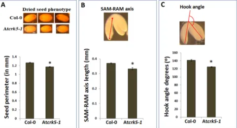
2.1. The Atcrk5-1 Mutant Exhibits a Considerable Delay in the Progression of the Phases of Embryogenesis We could recognize a notable size difference when comparing the wild typeArabidopsis thaliana (Col-0) and the Atcrk5-1mutant dry seeds (containing bent-cotyledon embryo stages) (Figure1A, pictures). We could determine significantly smaller seed sizes for the mutant when the dried seed contours were measured (Figure1A, diagram). After two days’ imbibition of dried seeds at 4◦C, we carefully pressed out the inverted stage embryos from the seeds by forceps, and measured the average length of the embryo axis from the shoot apical meristem (SAM) to the root apical meristem (RAM) in both of the genotypes. We found that the length of the axis was significantly shorter in Atcrk5-1 embryos as compared to the wild type Col-0 ones (Figure1B). Additionally, the bending angles of hypocotyl hooks of these bent-cotyledon-stage embryos were also measured. It is notable that the hypocotyl hook of the Atcrk5-1mutant embryos was already more opened at this stage when compared to the wild type ones (Figure1C; see Reference 61. for more details).
Int. J. Mol. Sci. 2019, 20, x FOR PEER REVIEW 4 of 24
2.1. The Atcrk5-1 Mutant Exhibits a Considerable Delay in the Progression of the Phases of Embryogenesis We could recognize a notable size difference when comparing the wild type Arabidopsis thaliana (Col-0) and the Atcrk5-1 mutant dry seeds (containing bent-cotyledon embryo stages) (Figure 1A, pictures). We could determine significantly smaller seed sizes for the mutant when the dried seed contours were measured (Figure 1A, diagram). After two days’ imbibition of dried seeds at 4 °C, we carefully pressed out the inverted stage embryos from the seeds by forceps, and measured the average length of the embryo axis from the shoot apical meristem (SAM) to the root apical meristem (RAM) in both of the genotypes. We found that the length of the axis was significantly shorter in Atcrk5-1 embryos as compared to the wild type Col-0 ones (Figure 1B). Additionally, the bending angles of hypocotyl hooks of these bent-cotyledon-stage embryos were also measured. It is notable that the hypocotyl hook of the Atcrk5-1 mutant embryos was already more opened at this stage when compared to the wild type ones (Figure 1C; see Reference 61. for more details).
Figure 1. Differences in seed and embryo sizes and embryo bent-cotyledon hook angles of wild type (Col-0) and mutant (Atcrk5-1) lines. (A) Pictures show dry seeds of the wild type (Col-0) and mutant (Atcrk5-1). The diagram shows the quantification of the seed size in wild type and mutant lines after two days’ imbibition of dried seeds at 4 °C. The seed contours were measured by ImageJ. The averages are from 100 independent seeds from the wild type and mutant, respectively. Standard errors (SE) are also shown. The mutant value indicated by asterisk is significantly lower compared to the wild type (Student’s t-test: p < 0.01, n = 100). (B) Embryo axis length measured from shoot apical meristem (SAM) until root apical meristem (RAM) was calculated in bent-cotyledon-stage embryos arised from the (A) experiment. Pictogram shows how the SAM-RAM axis was measured. All values are averages of at least 100 bent-cotyledon embryos. Standard errors (SE) are also shown. Asterisk depicts significant difference between the wild type and mutant embryos (Student’s t-test: p < 0.05, n = 100). (C) Differences in the hook angles of wild type and mutant embryos at the bent-cotyledon-stage (after two days’ imbibition of seeds at 4 °C). The averages and standard errors (SE) are shown. The mutant value is significantly different in comparison with the wild type (Student’s t-test: * p < 0.01, n = 75).
Pictogram shows the mode of measuring embryo hook angles (C). All experiment was repeated three times.
Thereafter, the individual developmental stages of the embryos were tested to find possible morphological differences between the wild type and mutant embryos. Microscopic images of embryos photographed by a CELL-R Olympus Microscope, are presented in Figure 2. The embryos were isolated from siliques1–11 (where S1–S11 represent silique position numbers from the youngest Figure 1.Differences in seed and embryo sizes and embryo bent-cotyledon hook angles of wild type (Col-0) and mutant (Atcrk5-1)lines. (A) Pictures show dry seeds of the wild type (Col-0) and mutant (Atcrk5-1). The diagram shows the quantification of the seed size in wild type and mutant lines after two days’ imbibition of dried seeds at 4◦C. The seed contours were measured by ImageJ. The averages are from 100 independent seeds from the wild type and mutant, respectively. Standard errors (SE) are also shown. The mutant value indicated by asterisk is significantly lower compared to the wild type (Student’s t-test: p<0.01,n=100). (B) Embryo axis length measured from shoot apical meristem (SAM) until root apical meristem (RAM) was calculated in bent-cotyledon-stage embryos arised from the (A) experiment. Pictogram shows how the SAM-RAM axis was measured. All values are averages of at least 100 bent-cotyledon embryos. Standard errors (SE) are also shown. Asterisk depicts significant difference between the wild type and mutant embryos (Student’s t-test:p<0.05,n= 100). (C) Differences in the hook angles of wild type and mutant embryos at the bent-cotyledon-stage (after two days’ imbibition of seeds at 4◦C). The averages and standard errors (SE) are shown. The mutant value is significantly different in comparison with the wild type (Student’s t-test: *p<0.01,n= 75). Pictogram shows the mode of measuring embryo hook angles (C). All experiment was repeated three times.
Thereafter, the individual developmental stages of the embryos were tested to find possible morphological differences between the wild type and mutant embryos. Microscopic images of embryos photographed by a CELL-R Olympus Microscope, are presented in Figure 2. The embryos were isolated from siliques1–11 (where S1–S11 represent silique position numbers from the youngest to oldest) of greenhouse plants of the same age. A sequential shift could be observed in the stages of Atcrk5-1embryo development from the globular/early heart embryo stages onward (from silique4; S4)
Int. J. Mol. Sci.2019,20, 6120 5 of 23
when compared to the wild type embryos (Figure2). Moreover, in the S10–S11 siliques, the Atcrk5-1 embryos considerably differed in morphology from the wild type ones (Figure2).
Int. J. Mol. Sci. 2019, 20, x FOR PEER REVIEW 5 of 24
to oldest) of greenhouse plants of the same age. A sequential shift could be observed in the stages of Atcrk5-1 embryo development from the globular/early heart embryo stages onward (from silique4;
S4) when compared to the wild type embryos (Figure 2). Moreover, in the S10–S11 siliques, the Atcrk5-1 embryos considerably differed in morphology from the wild type ones (Figure 2).
Figure 2. Embryogenesis of the wild type Arabidopsis thaliana Col-0 and the Atcrk5-1 mutant. Bright field microscopic images from a CELL-R Olympus Microscope. Embryo shapes were visualized after chloral hydrate treatment. S1–S11 = silique developmental stages where S1 represents the youngest and S11 the oldest siliques formed after pollination. As compared to wild type (A) embryos, there is a shift in Atcrk5-1 mutant (B) embryo development initiated from the globular/early heart embryo stages found in silique5 (S5). Red arrow represents the start and sequential direction of delay. Note that the wild type (Col-0) bent-cotyledon-stage embryos have 180° hook bending, while the mutant embryos at the same developmental stage (Atcrk5-1) have much less (around 130°) bending angle.
(Scale bars = 10 µm for S1–S7 and 100 µm for S9–S11).
Figure 3 indicates quantitative analysis of the progression of embryo development in wild type and Atcrk5-1 siliques. The distribution of sequential embryo developmental phases in the S1–S11 siliques is indicated in percentage by color coding the different stages. These quantitative data indicate that, in the wild type, the S4/S5 siliques contained basically globular, early heart and heart embryos, while in the same type of siliques of the mutant mainly had the eight-cell, globular, and, at a lesser extent, early heart embryos. The wild type S6 siliques mainly consisted of torpedo and early torpedo embryos, while the mutant S6 siliques were dominated by heart-stage embryos but still also contained eight-cell and globular type embryos. The S7 siliques of the wild type had mostly late torpedo and, at a lesser extent, upright embryos, while the mutant S7 siliques still mainly had embryos in the globular and heart (and a few percentage of early torpedo) embryo stages. In the S8/S9 siliques of the wild type, there were mainly torpedo, upright, and walking stick (app. 90° cotyledon bending) stage embryos. In contrast, the mutant S8/S9 siliques had still embryos in the early heart, heart, early torpedo, and torpedo stages with a few percentage of upright ones. The wild type S10/S11 siliques already contained principally walking stick and bent-cotyledon embryos, but the S10/S11 siliques from the mutant only contained upright and walking stick embryos (Figure 3).
Figure 2.Embryogenesis of the wild typeArabidopsis thalianaCol-0 and the Atcrk5-1mutant. Bright field microscopic images from a CELL-R Olympus Microscope. Embryo shapes were visualized after chloral hydrate treatment. S1–S11=silique developmental stages where S1 represents the youngest and S11 the oldest siliques formed after pollination. As compared to wild type (A) embryos, there is a shift in Atcrk5-1mutant (B) embryo development initiated from the globular/early heart embryo stages found in silique5 (S5). Red arrow represents the start and sequential direction of delay. Note that the wild type (Col-0) bent-cotyledon-stage embryos have 180◦hook bending, while the mutant embryos at the same developmental stage (Atcrk5-1) have much less (around 130◦) bending angle. (Scale bars=10 µm for S1–S7 and 100µm for S9–S11).
Figure3indicates quantitative analysis of the progression of embryo development in wild type and Atcrk5-1siliques. The distribution of sequential embryo developmental phases in the S1–S11 siliques is indicated in percentage by color coding the different stages. These quantitative data indicate that, in the wild type, the S4/S5 siliques contained basically globular, early heart and heart embryos, while in the same type of siliques of the mutant mainly had the eight-cell, globular, and, at a lesser extent, early heart embryos. The wild type S6 siliques mainly consisted of torpedo and early torpedo embryos, while the mutant S6 siliques were dominated by heart-stage embryos but still also contained eight-cell and globular type embryos. The S7 siliques of the wild type had mostly late torpedo and, at a lesser extent, upright embryos, while the mutant S7 siliques still mainly had embryos in the globular and heart (and a few percentage of early torpedo) embryo stages. In the S8/S9 siliques of the wild type, there were mainly torpedo, upright, and walking stick (app. 90◦cotyledon bending) stage embryos.
In contrast, the mutant S8/S9 siliques had still embryos in the early heart, heart, early torpedo, and torpedo stages with a few percentage of upright ones. The wild type S10/S11 siliques already contained principally walking stick and bent-cotyledon embryos, but the S10/S11 siliques from the mutant only contained upright and walking stick embryos (Figure3).
Therefore, embryos in the S11 siliques of the Atcrk5-1mutant resembled those in the S9 siliques of the wild type, which supported that, at the end of the investigations, the embryo development was delayed approx. by two phases in the mutant as compared to the wild type Col-0 (Figures2and3).
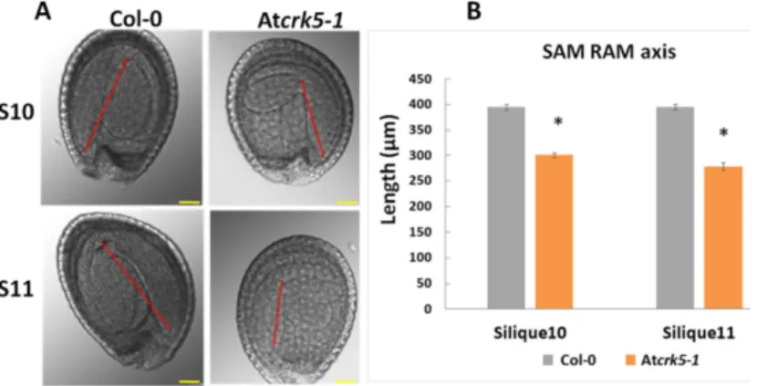
Furthermore, the hypocotyl hook of the embryos in the bent-cotyledon-stage of S10/S11 siliques was considerably less closed in the mutant than in the wild type (Figure4A). We found significant decrease in the axis length when we compared the SAM-RAM distance of the mutant embryos to that of the wild type Col-0 in the S10–S11 siliques, (Figure4B).
Int. J. Mol. Sci.2019,20, 6120 6 of 23
Int. J. Mol. Sci. 2019, 20, x FOR PEER REVIEW 6 of 24
Figure 3. Quantitative analysis of embryogenesis in wild type Arabidopsis thaliana (Col-0) and Atcrk5- 1 mutant in correlation to silique development. Distribution of the different embryo developmental stages from S1 until S11 depicted in percentage (%). A notable shift in Atcrk5-1 embryo development can be observed from the globular embryo stages in silique 4 (S4). Minimum 50 seeds were investigated for both genotypes for each embryo developmental stage of green-house-grown plants.
The experiments were repeated three times with the same results.
Therefore, embryos in the S11 siliques of the Atcrk5-1 mutant resembled those in the S9 siliques of the wild type, which supported that, at the end of the investigations, the embryo development was delayed approx. by two phases in the mutant as compared to the wild type Col-0 (Figures 2,3).
Furthermore, the hypocotyl hook of the embryos in the bent-cotyledon-stage of S10/S11 siliques was considerably less closed in the mutant than in the wild type (Figure 4A). We found significant decrease in the axis length when we compared the SAM-RAM distance of the mutant embryos to that of the wild type Col-0 in the S10–S11 siliques, (Figure 4B).
Figure 4. Comparison of hypocotyl bending and the SAM-RAM distance of bent-cotyledon-stage embryos. (A) Embryo length was measured from SAM to RAM in silique10 and silique11-derived embryos after preclearing in Hoyer’s solution. The SAM-RAM axis is indicated with red line. Note that the axes of the Atcrk5-1 embryos are shorter in comparison to those of the wild type. (B) Quantification of embryo axis length differences. Asterisks depict significant differences between the wild type and mutant embryos (Student’s t-test: * p < 0.05, n ≥ 50). Scale bars = 100 µm.
The relative expression of genes coding for the transcription factors LEC1, LEC2, FUS3, and ABI3 involved in embryo maturation and dormancy regulation processes were investigated to further Figure 3.Quantitative analysis of embryogenesis in wild typeArabidopsis thaliana(Col-0) and Atcrk5-1 mutant in correlation to silique development. Distribution of the different embryo developmental stages from S1 until S11 depicted in percentage (%). A notable shift in Atcrk5-1embryo development can be observed from the globular embryo stages in silique 4 (S4). Minimum 50 seeds were investigated for both genotypes for each embryo developmental stage of green-house-grown plants. The experiments were repeated three times with the same results.
Int. J. Mol. Sci. 2019, 20, x FOR PEER REVIEW 6 of 24
Figure 3. Quantitative analysis of embryogenesis in wild type Arabidopsis thaliana (Col-0) and Atcrk5- 1 mutant in correlation to silique development. Distribution of the different embryo developmental stages from S1 until S11 depicted in percentage (%). A notable shift in Atcrk5-1 embryo development can be observed from the globular embryo stages in silique 4 (S4). Minimum 50 seeds were investigated for both genotypes for each embryo developmental stage of green-house-grown plants.
The experiments were repeated three times with the same results.
Therefore, embryos in the S11 siliques of the Atcrk5-1 mutant resembled those in the S9 siliques of the wild type, which supported that, at the end of the investigations, the embryo development was delayed approx. by two phases in the mutant as compared to the wild type Col-0 (Figures 2,3).
Furthermore, the hypocotyl hook of the embryos in the bent-cotyledon-stage of S10/S11 siliques was considerably less closed in the mutant than in the wild type (Figure 4A). We found significant decrease in the axis length when we compared the SAM-RAM distance of the mutant embryos to that of the wild type Col-0 in the S10–S11 siliques, (Figure 4B).
Figure 4. Comparison of hypocotyl bending and the SAM-RAM distance of bent-cotyledon-stage embryos. (A) Embryo length was measured from SAM to RAM in silique10 and silique11-derived embryos after preclearing in Hoyer’s solution. The SAM-RAM axis is indicated with red line. Note that the axes of the Atcrk5-1 embryos are shorter in comparison to those of the wild type. (B) Quantification of embryo axis length differences. Asterisks depict significant differences between the wild type and mutant embryos (Student’s t-test: * p < 0.05, n ≥ 50). Scale bars = 100 µm.
The relative expression of genes coding for the transcription factors LEC1, LEC2, FUS3, and ABI3 involved in embryo maturation and dormancy regulation processes were investigated to further
Figure 4. Comparison of hypocotyl bending and the SAM-RAM distance of bent-cotyledon-stage embryos. (A) Embryo length was measured from SAM to RAM in silique10 and silique11-derived embryos after preclearing in Hoyer’s solution. The SAM-RAM axis is indicated with red line. Note that the axes of the Atcrk5-1embryos are shorter in comparison to those of the wild type. (B) Quantification of embryo axis length differences. Asterisks depict significant differences between the wild type and mutant embryos (Student’s t-test: *p<0.05,n≥50). Scale bars=100µm.
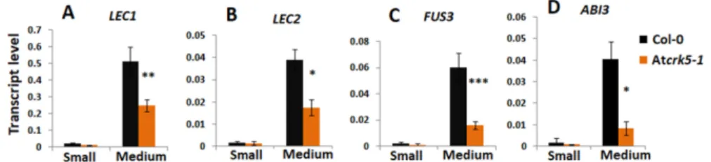
The relative expression of genes coding for the transcription factors LEC1, LEC2, FUS3, and ABI3 involved in embryo maturation and dormancy regulation processes were investigated to further support the delay in embryo development. Small (S1-S4) and medium-sized (S5-S7) siliques of the wild type (Col-0) and mutant (Atcrk5-1) plants were collected and analysed by qRT-PCR. The transcription factors LEC1, LEC2, and FUS3 act at late embryogenesis; therefore, we did not notice gene expression in small siliques. In the medium size siliques (from S5–S7), these genes were significantly less expressed in theAtcrk5-1mutant than in the control (Figure5A–C). The expression level of theABI3 transcription factor gene was also dropped at each silique stage in theAtcrk5-1mutant (Figure5D). The reduced transcription factor expressions in medium-sized siliques are in accordance with the delayed embryogenesis in the seeds of the Atcrk5-1mutant.
2.2. The Delayed Development of the Atcrk5-1 Embryos Is Linked to Their Decreased Gibberellin Synthesis and Level
The delay in Atcrk5-1embryo development started at the globular/early heart embryo stages found in silique4 (S4). The rapid expansion of wild type embryos takes place in this period (from S5 till S11) [3]. This was correlated with the elevated levels of bioactive gibberellins GA1and GA3
Int. J. Mol. Sci.2019,20, 6120 7 of 23
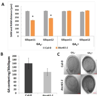
reaching their maximum at the walking stick embryo stage [3]. Bioactive gibberellins regulate late embryogenesis via modulating DELLA protein level in the embryos. GA deficiency and DELLA protein accumulation both led to abnormal embryos that are characterized by a shortened embryo axis and unbended cotyledons [3]. Based on these observations, the delayed morphogenesis of the Atcrk5-1 embryos might be explained by the reduced level of bioactive GAs. Therefore, a GA-antibody-based Elisa kit measured the total GA content of medium-sized siliques (S5–to–S7) collected from mutant and wild type plants. It was found that the medium-sized siliques of the Atcrk5-1mutant contained less GA than those of the wild type plants at the same age (Figure6A). We found that GA3treatment restored the SAM-RAM distance of the mutant embryos to the wild type level at S11–S12 stages when we treated greenhouse-grown wild type Arabidopsis and Atcrk5-1mutant plants by 20µM GA3from the early development of the inflorescence onward (Figure6B). Additionally, the hypocotyl bending phenotype of the Atcrk5-1mutant embryos found in S10–S12 (Figure4) was also restored to the wild type level by the exogenous GA3treatment (Figure6C).
Int. J. Mol. Sci. 2019, 20, x FOR PEER REVIEW 7 of 24
support the delay in embryo development. Small (S1-S4) and medium-sized (S5-S7) siliques of the wild type (Col-0) and mutant (Atcrk5-1) plants were collected and analysed by qRT-PCR. The transcription factors LEC1, LEC2, and FUS3 act at late embryogenesis; therefore, we did not notice gene expression in small siliques. In the medium size siliques (from S5–S7), these genes were significantly less expressed in the Atcrk5-1 mutant than in the control (Figure 5A–C). The expression level of the ABI3 transcription factor gene was also dropped at each silique stage in the Atcrk5-1 mutant (Figure 5D). The reduced transcription factor expressions in medium-sized siliques are in accordance with the delayed embryogenesis in the seeds of the Atcrk5-1 mutant.
Figure 5. Expression level of embryo maturation/dormancy related genes at different silique stages quantified by qRT-PCR. The relative transcript levels were determined in small siliques (S; S1–S4, 2DAP) and medium siliques (M, S5–S7, 3DAP) for (A) LEC1, (B) LEC2, (C) FUS3, and (D) ABI3.
Ubiquitin1 (UBQ-1) was used as an internal control. The values are means (+/−) SD of two independent biological repeats. Student’s t-test: * p < 0.05, ** p < 0.01, *** p < 0.001,. Primers used are listed in Table S1.
2.2. The Delayed Development of the Atcrk5-1 Embryos Is Linked to Their Decreased Gibberellin Synthesis and Level
The delay in Atcrk5-1 embryo development started at the globular/early heart embryo stages found in silique4 (S4). The rapid expansion of wild type embryos takes place in this period (from S5 till S11) [3]. This was correlated with the elevated levels of bioactive gibberellins GA1 and GA3
reaching their maximum at the walking stick embryo stage [3]. Bioactive gibberellins regulate late embryogenesis via modulating DELLA protein level in the embryos. GA deficiency and DELLA protein accumulation both led to abnormal embryos that are characterized by a shortened embryo axis and unbended cotyledons [3]. Based on these observations, the delayed morphogenesis of the Atcrk5-1 embryos might be explained by the reduced level of bioactive GAs. Therefore, a GA- antibody-based Elisa kit measured the total GA content of medium-sized siliques (S5–to–S7) collected from mutant and wild type plants. It was found that the medium-sized siliques of the Atcrk5-1 mutant contained less GA than those of the wild type plants at the same age (Figure 6A). We found that GA3
treatment restored the SAM-RAM distance of the mutant embryos to the wild type level at S11–S12 stages when we treated greenhouse-grown wild type Arabidopsis and Atcrk5-1 mutant plants by 20 µM GA3 from the early development of the inflorescence onward (Figure 6B). Additionally, the hypocotyl bending phenotype of the Atcrk5-1 mutant embryos found in S10–S12 (Figure 4) was also restored to the wild type level by the exogenous GA3 treatment (Figure 6C).
Figure 5.Expression level of embryo maturation/dormancy related genes at different silique stages quantified by qRT-PCR. The relative transcript levels were determined in small siliques (S; S1–S4, 2DAP) and medium siliques (M, S5–S7, 3DAP) for (A)LEC1, (B)LEC2, (C)FUS3, and (D)ABI3. Ubiquitin1 (UBQ-1)was used as an internal control. The values are means (+/−) SD of two independent biological repeats. Student’s t-test: *p<0.05, **p<0.01, ***p<0.001. Primers used are listed in Table S1.
We also examined the gene expression levels of the gibberellin 20-oxidases (AtGA20ox2and AtGA20ox3), which are highly expressed GA biosynthesis enzymes in dry and imbibed seeds [51]
and they are responsible for converting the inactive intermediates of GA biosynthesis into bioactive forms [3]. TheAtGA20ox2andAtGA20ox3genes have high expression during the expansion stage of embryogenesis (4–7 days after pollination, 4–7 DAP) [3]. The expression level of the gibberellin deactivation geneGA2ox4, which also has a high expression level in embryos at this stage (4–7 DAP; [3]), was also tested. Furthermore, the genes of DELLA proteins canonically known to participate in GA signaling during embryogenesis [3,32] were included into the gene expression study. Siliques of wild type (Col-0) and mutant (Atcrk5-1) plants were collected according to their developmental stages, like small (three DAP) and medium (five DAP) ones corresponding to embryo developmental stages S1–S4 and S5–S6–S7, respectively. The gene expression analysis was focused on the medium silique stage, where decreased GA content was observed in the Atcrk5-1mutant. When considering the genes coding for DELLA proteins, we found a slight gene expression level elevation inRGL1(Figure7A) andGAI (Figure7D) genes, while the expression levels of theRGL2,RGL3, andRGA(Figure7B,C,E) genes did not change in theAtcrk5-1as compared to that of the wild type. However, theGA20ox2/GA20ox3gene expression levels were decreased in the siliques of the Atcrk5-1mutant in comparison to the wild type (Figure7F,G). This indicates that the insufficient conversion of the inactive GA intermediates into their bioactive forms might cause the fall-offin the GA level at the medium silique stage (Figure6B). The expression level of theGA2ox4was also decreased (Figure7H) in the Atcrk5-1medium siliques, which might relate to the reduced negative feedback of GAs on their biosynthesis.
Int. J. Mol. Sci.2019,20, 6120 8 of 23
Int. J. Mol. Sci. 2019, 20, x FOR PEER REVIEW 8 of 24
Figure 6. Role of gibberellic acid in the delayed embryo development of Atcrk5-1. (A) Quantification of embryo axis length differences in siliques S11–S12. Embryo axis from SAM-to-RAM was measured after the treatment of developing inflorescences of wild type (Col-0) and Atcrk5-1 mutant plants without (GA −) or with 20 µM GA3 (GA +). The exogenous GA3 supply restored the length of the mutant embryos to the wild type level. (Student’s t-test:* p < 0.05, n = 30). The data presented are the means of two biological repeats. (B) Quantification of the total GA content in medium stage siliques (S5–S6–S7). 30-30 siliques were collected from wild type (Col-0) and mutant (Atcrk5-1) inflorescence.
The total GA content was determined using a GA-antibody-based Elisa kit. (C) Rescue of the delayed embryo bending phenotype in S11 of the Atcrk5-1 mutant by 20 µM GA3 treatment with its noticeable change from walking stick to bent cotyledon stage, while Col-0 remains in the bent cotyledon stage.
Representative images are shown from experiments repeated two times with 30-30 mutant and wild type embryos. Scale bars = 100 µm.
We also examined the gene expression levels of the gibberellin 20-oxidases (AtGA20ox2 and AtGA20ox3), which are highly expressed GA biosynthesis enzymes in dry and imbibed seeds [51] and they are responsible for converting the inactive intermediates of GA biosynthesis into bioactive forms [3]. The AtGA20ox2 and AtGA20ox3 genes have high expression during the expansion stage of embryogenesis (4–7 days after pollination, 4–7 DAP) [3]. The expression level of the gibberellin deactivation gene GA2ox4, which also has a high expression level in embryos at this stage (4–7 DAP;
[3]), was also tested. Furthermore, the genes of DELLA proteins canonically known to participate in GA signaling during embryogenesis [3,32] were included into the gene expression study. Siliques of wild type (Col-0) and mutant (Atcrk5-1) plants were collected according to their developmental stages, like small (three DAP) and medium (five DAP) ones corresponding to embryo developmental stages S1–S4 and S5–S6–S7, respectively. The gene expression analysis was focused on the medium silique stage, where decreased GA content was observed in the Atcrk5-1 mutant. When considering the genes coding for DELLA proteins, we found a slight gene expression level elevation in RGL1 (Figure 7A) and GAI (Figure 7D) genes, while the expression levels of the RGL2, RGL3, and RGA (Figure 7B,C,E) genes did not change in the Atcrk5-1 as compared to that of the wild type. However, the GA20ox2/GA20ox3 gene expression levels were decreased in the siliques of the Atcrk5-1 mutant in comparison to the wild type (Figure 7F/G). This indicates that the insufficient conversion of the inactive GA intermediates into their bioactive forms might cause the fall-off in the GA level at the medium silique stage (Figure 6B). The expression level of the GA2ox4 was also decreased (Figure 7H) Figure 6.Role of gibberellic acid in the delayed embryo development of Atcrk5-1. (A) Quantification of embryo axis length differences in siliques S11–S12. Embryo axis from SAM-to-RAM was measured after the treatment of developing inflorescences of wild type (Col-0) and Atcrk5-1mutant plants without (GA
−) or with 20µM GA3(GA+). The exogenous GA3supply restored the length of the mutant embryos to the wild type level. (Student’s t-test:*p<0.05,n=30). The data presented are the means of two biological repeats. (B) Quantification of the total GA content in medium stage siliques (S5–S6–S7). 30-30 siliques were collected from wild type (Col-0) and mutant (Atcrk5-1) inflorescence. The total GA content was determined using a GA-antibody-based Elisa kit. (C) Rescue of the delayed embryo bending phenotype in S11 of the Atcrk5-1mutant by 20µM GA3treatment with its noticeable change from walking stick to bent cotyledon stage, while Col-0 remains in the bent cotyledon stage. Representative images are shown from experiments repeated two times with 30-30 mutant and wild type embryos.
Scale bars=100µm.
Int. J. Mol. Sci. 2019, 20, x FOR PEER REVIEW 9 of 24
in the Atcrk5-1 medium siliques, which might relate to the reduced negative feedback of GAs on their biosynthesis.
Figure 7. Relative expression level of selected genes involved in Gibberellic Acid (GA) metabolism and signaling at different silique stages quantified by qRT-PCR. The relative transcript levels were determined in small siliques (S; S1–S4, 3DAP) and medium siliques (M, S5–S7, 5DAP) for the DELLA GA signaling genes (A) RGL1, (B) RGL2, (C) RGL3, (D) GA-INSENSITIVE (GAI), and (E) REPRESSOR OF ga1-3 (RGA), the gibberellin biosynthesis genes (F) GA20ox2 and (G) GA20ox3, and the gene of the GA catabolism enzyme (H) GA2ox4.Ubiquitin1 (UBQ-1) was used as an internal control. The values are means (+/−) SD of two independent biological repeats. . Student’s t-test: * p < 0.05, ** p < 0.01, *** p
< 0.001. Primers used are listed in the Table S1.
2.3. The Auxin Level Is Decreased in the Atcrk5-1 Mutant Embryos in Comparison with the Wild Type Ones.
It is known that there is crosstalk between auxin and GA during plant developmental regulation, including embryogenesis [52,60]. We pointed out a GA shortage in the Atcrk5-1 embryos at the globular/ heart/torpedo embryo stages (in medium siliques S5–S7, Figure 6B). Among others, GA is required for proper auxin transport in Arabidopsis [33]. Therefore, we tested the auxin distribution in the Atcrk5-1 mutant embryos during their development. The DR5::GFP marker (a synthetic promoter that responds to the presence of auxin linked to GFP gene [61]) was introduced into wild type (Figure 8A) and Atcrk5-1 plants (Figure 8B). Fluorescence microscopy was used to detect auxin- regulated GFP fluorescence in various embryo stages. We found a strong decrease in the DR5::GFP signal in the Atcrk5-1 embryos all along from the eight-cell stage until the walking stick stage of the embryos (Figure 8).
Figure 7. Relative expression level of selected genes involved in Gibberellic Acid (GA) metabolism and signaling at different silique stages quantified by qRT-PCR. The relative transcript levels were determined in small siliques (S; S1–S4, 3DAP) and medium siliques (M, S5–S7, 5DAP) for the DELLA GA signaling genes (A)RGL1, (B)RGL2, (C)RGL3, (D)GA-INSENSITIVE(GAI), and (E)REPRESSOR OF ga1-3(RGA), the gibberellin biosynthesis genes (F)GA20ox2and (G) GA20ox3, and the gene of the GA catabolism enzyme (H) GA2ox4.Ubiquitin1(UBQ-1) was used as an internal control. The values are means (+/−) SD of two independent biological repeats. Student’s t-test: *p<0.05, **p<0.01, ***p<
0.001. Primers used are listed in the Table S1.
Int. J. Mol. Sci.2019,20, 6120 9 of 23
2.3. The Auxin Level Is Decreased in the Atcrk5-1 Mutant Embryos in Comparison with the Wild Type Ones.
It is known that there is crosstalk between auxin and GA during plant developmental regulation, including embryogenesis [52,60]. We pointed out a GA shortage in the Atcrk5-1embryos at the globular/
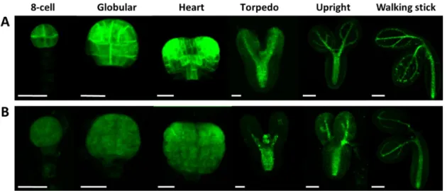
heart/torpedo embryo stages (in medium siliques S5–S7, Figure6B). Among others, GA is required for proper auxin transport in Arabidopsis [33]. Therefore, we tested the auxin distribution in the Atcrk5-1mutant embryos during their development. The DR5::GFP marker (a synthetic promoter that responds to the presence of auxin linked to GFP gene [61]) was introduced into wild type (Figure8A) and Atcrk5-1plants (Figure8B). Fluorescence microscopy was used to detect auxin-regulated GFP fluorescence in various embryo stages. We found a strong decrease in the DR5::GFP signal in the Atcrk5-1embryos all along from the eight-cell stage until the walking stick stage of the embryos (Figure8). Int. J. Mol. Sci. 2019, 20, x FOR PEER REVIEW 10 of 24
Figure 8. The auxin sensor DR5::GFP activity was recorded in wild type Arabidopsis and mutant Atcrk5-1 embryos at several developmental stages. Auxin distribution during the embryo development in wild type (A) and in the mutant (B). In both cases, 5-5 embryos were investigated from three independent experiments. Note the decreased auxin levels in each embryo developmental stage in the Atcrk5-1 mutant. Scale bars: eight-cell to torpedo = 15 µm; upright and walking stick = 100 µm.
2.4. Expression and Abundance of Auxin Efflux Proteins During Embryogenesis of Wild Type and Atcrk5-1 Mutant Plants
Auxin is known to determine cell division patterns and cell specification during all stages of embryogenesis, starting from the first division of the zygote and this is strongly dependent on PINs localization and activity [4,10,18,60]. The expression and abundance of the auxin efflux transporters PIN1, PIN3, PIN4, and PIN7 were investigated at various embryo developmental stages in order to investigate whether the delayed embryogenesis of the Atcrk5-1 mutant is associated with altered auxin transport pattern or efficiency. The PIN4 and PIN7 gene expressions were reduced in the small silique stage (S1–S4) of the Atcrk5-1 mutant (Figure 9A,B), while the PIN1 gene expression was down regulated in the mutant siliques during the small and medium stages inclusive the torpedo stage (S1–
S4 and S5–S7, Figure 9C).
Figure 9. Relative expression level of auxin efflux carrier PIN genes at different silique stages quantified by qRT-PCR. The relative transcript levels were determined in small siliques (S1–S4, 3DAP) and medium siliques (S5–S7, 5DAP) for (A) PIN4, (B) PIN7, (C) PIN1. Ubiquitin1 (UBQ-1) was used as an internal control. The values are means (+/−) SD of two independent biological repeats. Student’s t-test: * p < 0.05, ** p < 0.01, *** p < 0.001. Primers used are listed in the Table S1.
GFP-tagged PIN proteins were produced in wild type (Col-0) and mutant (Atcrk5-1) backgrounds to investigate their protein level and distribution. At early embryogenesis, PIN1 and PIN7 are required for normal embryo development. In the proembryo, PIN1 first localizes non-
Figure 8.The auxin sensor DR5::GFP activity was recorded in wild typeArabidopsisand mutant Atcrk5-1 embryos at several developmental stages. Auxin distribution during the embryo development in wild type (A) and in the mutant (B). In both cases, 5-5 embryos were investigated from three independent experiments. Note the decreased auxin levels in each embryo developmental stage in the Atcrk5-1 mutant. Scale bars: eight-cell to torpedo=15µm; upright and walking stick=100µm.
2.4. Expression and Abundance of Auxin Efflux Proteins During Embryogenesis of Wild Type and Atcrk5-1 Mutant Plants
Auxin is known to determine cell division patterns and cell specification during all stages of embryogenesis, starting from the first division of the zygote and this is strongly dependent on PINs localization and activity [4,10,18,60]. The expression and abundance of the auxin efflux transporters PIN1, PIN3, PIN4, and PIN7 were investigated at various embryo developmental stages in order to investigate whether the delayed embryogenesis of the Atcrk5-1mutant is associated with altered auxin transport pattern or efficiency. ThePIN4andPIN7gene expressions were reduced in the small silique stage (S1–S4) of theAtcrk5-1mutant (Figure9A,B), while thePIN1gene expression was down regulated in the mutant siliques during the small and medium stages inclusive the torpedo stage (S1–S4 and S5–S7, Figure9C).
GFP-tagged PIN proteins were produced in wild type (Col-0) and mutant (Atcrk5-1) backgrounds to investigate their protein level and distribution. At early embryogenesis, PIN1 and PIN7 are required for normal embryo development. In the proembryo, PIN1 first localizes non-polarly but, at the globular stage, it becomes localized to the basal membrane of provascular cells facing the hypophysis [10].
During early embryogenesis, PIN7 first localizes at the apical side of suspensor cells and at the 32-cell (globular embryo) stage PIN7 polarity changes and the protein becomes associated with the basal membrane of suspensor cells. PIN1 and PIN7 polarization changes correlate with the reversion of the auxin flow [4,10,12].
Int. J. Mol. Sci.2019,20, 6120 10 of 23
Int. J. Mol. Sci. 2019, 20, x FOR PEER REVIEW 10 of 24
Figure 8. The auxin sensor DR5::GFP activity was recorded in wild type Arabidopsis and mutant Atcrk5-1 embryos at several developmental stages. Auxin distribution during the embryo development in wild type (A) and in the mutant (B). In both cases, 5-5 embryos were investigated from three independent experiments. Note the decreased auxin levels in each embryo developmental stage in the Atcrk5-1 mutant. Scale bars: eight-cell to torpedo = 15 µm; upright and walking stick = 100 µm.
2.4. Expression and Abundance of Auxin Efflux Proteins During Embryogenesis of Wild Type and Atcrk5-1 Mutant Plants
Auxin is known to determine cell division patterns and cell specification during all stages of embryogenesis, starting from the first division of the zygote and this is strongly dependent on PINs localization and activity [4,10,18,60]. The expression and abundance of the auxin efflux transporters PIN1, PIN3, PIN4, and PIN7 were investigated at various embryo developmental stages in order to investigate whether the delayed embryogenesis of the Atcrk5-1 mutant is associated with altered auxin transport pattern or efficiency. The PIN4 and PIN7 gene expressions were reduced in the small silique stage (S1–S4) of the Atcrk5-1 mutant (Figure 9A,B), while the PIN1 gene expression was down regulated in the mutant siliques during the small and medium stages inclusive the torpedo stage (S1–
S4 and S5–S7, Figure 9C).
Figure 9. Relative expression level of auxin efflux carrier PIN genes at different silique stages quantified by qRT-PCR. The relative transcript levels were determined in small siliques (S1–S4, 3DAP) and medium siliques (S5–S7, 5DAP) for (A) PIN4, (B) PIN7, (C) PIN1. Ubiquitin1 (UBQ-1) was used as an internal control. The values are means (+/−) SD of two independent biological repeats. Student’s t-test: * p < 0.05, ** p < 0.01, *** p < 0.001. Primers used are listed in the Table S1.
GFP-tagged PIN proteins were produced in wild type (Col-0) and mutant (Atcrk5-1) backgrounds to investigate their protein level and distribution. At early embryogenesis, PIN1 and PIN7 are required for normal embryo development. In the proembryo, PIN1 first localizes non-
Figure 9.Relative expression level of auxin efflux carrierPINgenes at different silique stages quantified by qRT-PCR. The relative transcript levels were determined in small siliques (S1–S4, 3DAP) and medium siliques (S5–S7, 5DAP) for (A)PIN4, (B)PIN7, (C)PIN1.Ubiquitin1 (UBQ-1)was used as an internal control. The values are means (+/−) SD of two independent biological repeats. Student’s t-test: *p<
0.05, **p<0.01, ***p<0.001. Primers used are listed in the Table S1.
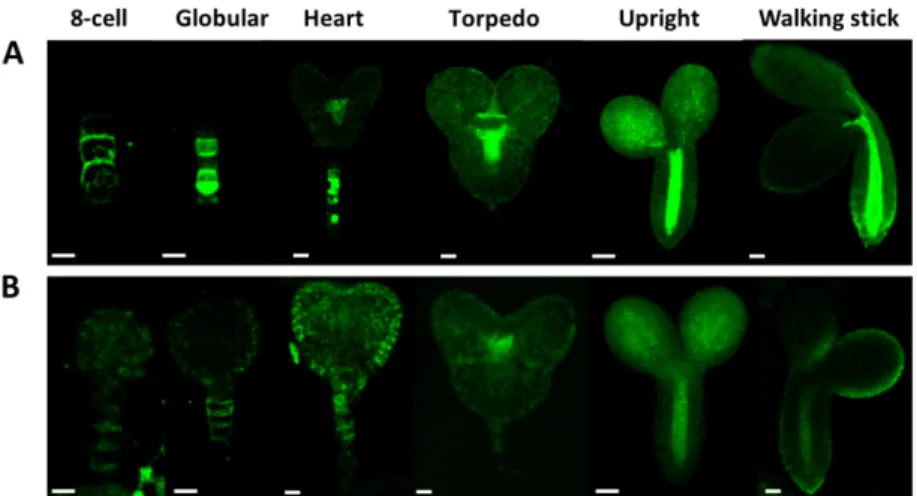
The pattern of PIN1/PIN7 distribution seems to be unaffected by the Atcrk5-1mutation (Figures10 and11). Figure10shows PIN1-GFP abundance in wild type (A) and Atcrk5-1(B) embryos from the eight-cell stage to the walking stick embryo stage. The PIN1-GFP signal intensity is lower in the Atcrk5-1mutant compared to the wild type in all embryogenesis stages, but especially in the early ones (up to the heart stage). Likewise, the PIN7-GFP signal intensity was drastically decreased in the Atcrk5-1embryos at each embryo stage (Figure11).
Int. J. Mol. Sci. 2019, 20, x FOR PEER REVIEW 11 of 24
polarly but, at the globular stage, it becomes localized to the basal membrane of provascular cells facing the hypophysis [10]. During early embryogenesis, PIN7 first localizes at the apical side of suspensor cells and at the 32-cell (globular embryo) stage PIN7 polarity changes and the protein becomes associated with the basal membrane of suspensor cells. PIN1 and PIN7 polarization changes correlate with the reversion of the auxin flow [4,10,12].
The pattern of PIN1/PIN7 distribution seems to be unaffected by the Atcrk5-1 mutation (Figures 10 and 11). Figure 10 shows PIN1-GFP abundance in wild type (A) and Atcrk5-1 (B) embryos from the eight-cell stage to the walking stick embryo stage. The PIN1-GFP signal intensity is lower in the Atcrk5-1 mutant compared to the wild type in all embryogenesis stages, but especially in the early ones (up to the heart stage). Likewise, the PIN7-GFP signal intensity was drastically decreased in the Atcrk5-1 embryos at each embryo stage (Figure 11).
Figure 10. Level and distribution of PIN1-GFP in wild type (A) and in Atcrk5-1 mutant (B) embryos during embryogenesis. Note that the PIN1-GFP signal is much less intense in the Atcrk5-1 mutant then in the control embryos. In both cases 5-5 embryos were investigated from three independent experiments. Scale bars: eight-cell to torpedo = 15 µm; upright; and, walking stick = 100 µm.
Figure 11. Comparison of the PIN7-GFP abundance and distribution in developing Arabidopsis embryos. PIN7-GFP intensity distribution during embryo development in wild type (A) and in the mutant (B). Note the decreased abundance of PIN7-GFP in each developmental stage of Atcrk5-1 Figure 10.Level and distribution of PIN1-GFP in wild type (A) and in Atcrk5-1mutant (B) embryos during embryogenesis. Note that the PIN1-GFP signal is much less intense in the Atcrk5-1mutant then in the control embryos. In both cases 5-5 embryos were investigated from three independent experiments. Scale bars: eight-cell to torpedo=15µm; upright; and, walking stick=100µm.
The abundance and distribution of the PIN3 and PIN4 proteins was also investigated. PIN3 is expressed at the basal pole of heart stage embryos during embryo development [10]. For PIN3-GFP, we did not find any abundance or localization alterations in the Atcrk5-1mutant when compared to the wild type during embryo development (Figure S1). The PIN4 protein first appears during early embryogenesis at the globular stage in the descendants of the hypophysial cell and in the provascular initials of root meristem [10]. We have found that the PIN4-GFP signal intensity was also decreased in the Atcrk5-1 embryos in comparison to the wild type, like in the cases of PIN1-GFP and PIN7-GFP (Figure S2).
2.5. Expression and Abundance of Auxin Influx (AUX1) Proteins During Embryogenesis of Wild Type and Atcrk5-1 Mutant Plants
Auxin influx is also required for proper embryogenesis. AUX1, LAX1, and LAX2 are required for both shoot and root pole formation of embryos, in cooperation with PIN efflux carriers [17,24].
Int. J. Mol. Sci.2019,20, 6120 11 of 23
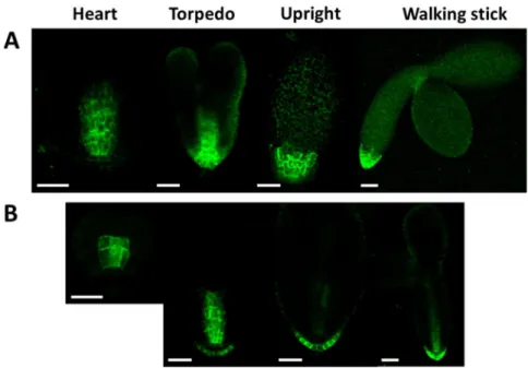
AUX/LAX protein expressions start from the heart stage in Arabidopsis embryos. We compared the localization pattern of the AUX1::YFP signal in the wild type and mutant (Atcrk5-1) embryos during different phases of embryo development (Figure12). We found that the AUX1::YFP expression level was decreased in the Atcrk5-1mutant in all of the embryo developmental stages in comparison to the wild type. Moreover, the distribution of the AUX1::YFP protein was more restricted at the root pole in the mutant background.
Int. J. Mol. Sci. 2019, 20, x FOR PEER REVIEW 11 of 24
polarly but, at the globular stage, it becomes localized to the basal membrane of provascular cells facing the hypophysis [10]. During early embryogenesis, PIN7 first localizes at the apical side of suspensor cells and at the 32-cell (globular embryo) stage PIN7 polarity changes and the protein becomes associated with the basal membrane of suspensor cells. PIN1 and PIN7 polarization changes correlate with the reversion of the auxin flow [4,10,12].
The pattern of PIN1/PIN7 distribution seems to be unaffected by the Atcrk5-1 mutation (Figures 10 and 11). Figure 10 shows PIN1-GFP abundance in wild type (A) and Atcrk5-1 (B) embryos from the eight-cell stage to the walking stick embryo stage. The PIN1-GFP signal intensity is lower in the Atcrk5-1 mutant compared to the wild type in all embryogenesis stages, but especially in the early ones (up to the heart stage). Likewise, the PIN7-GFP signal intensity was drastically decreased in the Atcrk5-1 embryos at each embryo stage (Figure 11).
Figure 10. Level and distribution of PIN1-GFP in wild type (A) and in Atcrk5-1 mutant (B) embryos during embryogenesis. Note that the PIN1-GFP signal is much less intense in the Atcrk5-1 mutant then in the control embryos. In both cases 5-5 embryos were investigated from three independent experiments. Scale bars: eight-cell to torpedo = 15 µm; upright; and, walking stick = 100 µm.
Figure 11. Comparison of the PIN7-GFP abundance and distribution in developing Arabidopsis embryos. PIN7-GFP intensity distribution during embryo development in wild type (A) and in the mutant (B). Note the decreased abundance of PIN7-GFP in each developmental stage of Atcrk5-1
Figure 11. Comparison of the PIN7-GFP abundance and distribution in developing Arabidopsis embryos. PIN7-GFP intensity distribution during embryo development in wild type (A) and in the mutant (B). Note the decreased abundance of PIN7-GFP in each developmental stage of Atcrk5-1 embryos. In both cases 5-5 embryos were investigated from three independent experiments. Scale bars:
eight-cell to torpedo=15µm; upright/walking stick=100µm.
2.6. AtCRK5 Can Phosphorylate the Auxin Efflux Proteins PIN1, PIN4 and PIN7 in vitro
The auxin efflux PIN1, PIN3, PIN4, and PIN7 proteins are all supposed to participate in auxin transportation during embryogenesis [17,20,60,62]. The localization and activation of the various PIN proteins are known to be affected by their phosphorylation status [15,63–65]. We have already published that AtCRK5 phosphorylates the PIN2 and PIN3 auxin efflux transporters and this may have a role in the gravitropic response of theArabidopsisroot upon gravistimulation and the hypocotyl hook bending during skotomorphogenesis [57,59]. The decreased abundance of PIN1-GFP, PIN4-GFP, and PIN7-GFP in Atcrk5-1mutant embryos might reflect a defect in PIN protein stability and/or localization. We speculated that all PIN proteins might be targets of phosphorylation by the AtCRK5 kinase based on the high sequence homology between different PIN hydrophilic loop regions. We cloned the hydrophilic loop domain regions of PIN1, PIN4, and PIN7 into a bacterial protein expression vector in order to support this hypothesis. After transformation and expression intoEscherichia coli cells, we purified the GST (Glutathione S-Transferase) tagged GST-PIN1HL and MBP (Maltose Binding Protein) tagged MBP-PIN4HL and MBP-PIN7HL. In the case of GST-PIN1HL, we performed an in vitro radioactive kinase assay while using the purified AtCRK5 protein kinase. Figure13shows the result of this phosphorylation assay.
Furthermore, a non-radioactive kinase assay was performed to determine whether the AtCRK5 kinase could phosphorylate the PIN4HL and/or the PIN7HL loop regions. The proteins of the kinase reaction were separated in a denaturing polyacrylamide gel and the GST-PIN1HL, MBP-PIN4HL, and MBP-PIN7HL protein bands were excised and sent for analysis by mass spectrometry after staining with Coomassie dye.
Int. J. Mol. Sci.2019,20, 6120 12 of 23
Int. J. Mol. Sci. 2019, 20, x FOR PEER REVIEW 12 of 24
embryos. In both cases 5-5 embryos were investigated from three independent experiments. Scale bars: eight-cell to torpedo = 15 µm; upright/walking stick= 100 µm.
The abundance and distribution of the PIN3 and PIN4 proteins was also investigated. PIN3 is expressed at the basal pole of heart stage embryos during embryo development [10]. For PIN3-GFP, we did not find any abundance or localization alterations in the Atcrk5-1 mutant when compared to the wild type during embryo development (Figure S1). The PIN4 protein first appears during early embryogenesis at the globular stage in the descendants of the hypophysial cell and in the provascular initials of root meristem [10]. We have found that the PIN4-GFP signal intensity was also decreased in the Atcrk5-1 embryos in comparison to the wild type, like in the cases of PIN1-GFP and PIN7-GFP (Figure S2).
2.5. Expression and Abundance of Auxin Influx (AUX1) Proteins During Embryogenesis of Wild Type and Atcrk5-1 Mutant Plants
Auxin influx is also required for proper embryogenesis. AUX1, LAX1, and LAX2 are required for both shoot and root pole formation of embryos, in cooperation with PIN efflux carriers [17,24].
AUX/LAX protein expressions start from the heart stage in Arabidopsis embryos. We compared the localization pattern of the AUX1::YFP signal in the wild type and mutant (Atcrk5-1) embryos during different phases of embryo development (Figure 12). We found that the AUX1::YFP expression level was decreased in the Atcrk5-1 mutant in all of the embryo developmental stages in comparison to the wild type. Moreover, the distribution of the AUX1::YFP protein was more restricted at the root pole in the mutant background.
Figure 12. Distribution of AUX1::YFP during embryo development in the wild type (A) and in the Atcrk5-1 mutant (B). In all embryo developmental stages there is lower level of AUX1-YFP in the Atcrk5-1 mutant. The localisation of the auxin influx protein (AUX-YFP) is also different in the mutant and the wild type backgrounds. In both cases 5-5 embryos were investigated from three independent experiments. Scale bars = 100 µm.
2.6. AtCRK5 Can Phosphorylate the Auxin Efflux Proteins PIN1, PIN4 and PIN7 in vitro
The auxin efflux PIN1, PIN3, PIN4, and PIN7 proteins are all supposed to participate in auxin transportation during embryogenesis [17,20,60,62]. The localization and activation of the various PIN proteins are known to be affected by their phosphorylation status [15,63–65]. We have already published that AtCRK5 phosphorylates the PIN2 and PIN3 auxin efflux transporters and this may have a role in the gravitropic response of the Arabidopsis root upon gravistimulation and the
Figure 12. Distribution of AUX1::YFP during embryo development in the wild type (A) and in the Atcrk5-1mutant (B).In all embryo developmental stages there is lower level of AUX1-YFP in the Atcrk5-1mutant. The localisation of the auxin influx protein (AUX-YFP) is also different in the mutant and the wild type backgrounds. In both cases 5-5 embryos were investigated from three independent experiments. Scale bars=100µm.
Int. J. Mol. Sci. 2019, 20, x FOR PEER REVIEW 13 of 24
hypocotyl hook bending during skotomorphogenesis [57,59]. The decreased abundance of PIN1-GFP, PIN4-GFP, and PIN7-GFP in Atcrk5-1 mutant embryos might reflect a defect in PIN protein stability and/or localization. We speculated that all PIN proteins might be targets of phosphorylation by the AtCRK5 kinase based on the high sequence homology between different PIN hydrophilic loop regions. We cloned the hydrophilic loop domain regions of PIN1, PIN4, and PIN7 into a bacterial protein expression vector in order to support this hypothesis. After transformation and expression into Escherichia coli cells, we purified the GST (Glutathione S-Transferase) tagged GST-PIN1HL and MBP (Maltose Binding Protein) tagged MBP-PIN4HL and MBP-PIN7HL. In the case of GST-PIN1HL, we performed an in vitro radioactive kinase assay while using the purified AtCRK5 protein kinase.
Figure 13 shows the result of this phosphorylation assay.
Figure 13. AtCRK5 phosphorylates the PIN1 hydrophilic loop in vitro. We performed in vitro radioactive phosphorylation assay with His6-AtCRK5 and two substrates: Myelin Basic Protein (MBP) as a positive control, and GST-PIN1-HL loop. White asterisks indicate the phosphorylation event on MBP (* asterisk) and GST-PIN1HLloop (** asterisk) proteins, respectively. We carried out glutathione S-Transferase (GST) column purification after the kinase reactions shown in the last three columns to remove the HIS6-CRK5 kinase because the His6-CRK5 and the GST-PIN1 protein sizes are nearly identical and we could not distinguish the phosphorylation signals.
Furthermore, a non-radioactive kinase assay was performed to determine whether the AtCRK5 kinase could phosphorylate the PIN4HL and/or the PIN7HL loop regions. The proteins of the kinase reaction were separated in a denaturing polyacrylamide gel and the GST-PIN1HL, MBP-PIN4HL, and MBP-PIN7HL protein bands were excised and sent for analysis by mass spectrometry after staining with Coomassie dye.
We could identify some potential phosphorylation sites for the PIN1, PIN4, and PIN7 hydrophilic loop. AtCRK5 protein kinase can phosphorylate the PIN1 protein at Ser-252 or Ser-253 sites. The phosphorylation site is at Ser-271 in the case of the PIN4 protein. In the case of PIN7 protein, two phosphopeptides were detected in the digest, the Ser-431 and the Ser-277/Ser-278, respectively (Figure S3).
3. Discussion
3.1. The AtCRK5 Protein Kinase Controls the Gibberellin Level Influencing Seed Size and Embryogenesis Ca2+ has a pivotal role as a secondary messenger in many abiotic and biotic stress responses in plants. Environmental cues trigger Ca2+ concentration alterations in plant cells that are perceived by calmodulin, several calcium-dependent protein kinases (CDPK superfamily members) and
Figure 13. AtCRK5 phosphorylates the PIN1 hydrophilic loop in vitro. We performed in vitro radioactive phosphorylation assay with His6-AtCRK5 and two substrates: Myelin Basic Protein (MBP) as a positive control, and GST-PIN1-HL loop. White asterisks indicate the phosphorylation event on MBP (* asterisk) and GST-PIN1HLloop (** asterisk) proteins, respectively. We carried out glutathione S-Transferase (GST) column purification after the kinase reactions shown in the last three columns to remove the HIS6-CRK5 kinase because the His6-CRK5 and the GST-PIN1 protein sizes are nearly identical and we could not distinguish the phosphorylation signals.
We could identify some potential phosphorylation sites for the PIN1, PIN4, and PIN7 hydrophilic loop. AtCRK5 protein kinase can phosphorylate the PIN1 protein at Ser-252 or Ser-253 sites. The phosphorylation site is at Ser-271 in the case of the PIN4 protein. In the case of PIN7 protein, two phosphopeptides were detected in the digest, the Ser-431 and the Ser-277/Ser-278, respectively (Figure S3).

![heart/torpedo embryo stages (in medium siliques S5–S7, Figure 6B). Among others, GA is required for proper auxin transport in Arabidopsis [33]](https://thumb-eu.123doks.com/thumbv2/9dokorg/1064165.70358/9.892.221.674.384.598/torpedo-embryo-stages-siliques-figure-required-transport-arabidopsis.webp)