Endoplasmic reticulum–retained podocin mutants are massively degraded by the proteasome
Received for publication, December 5, 2017, and in revised form, January 23, 2018 Published, Papers in Press, January 30, 2018, DOI 10.1074/jbc.RA117.001159
Maria-Carmen Serrano-Perez‡§, Frances C. Tilley‡§, Fabien Nevo‡§, Christelle Arrondel‡§, Selim Sbissa‡§, Gaëlle Martin‡§, Kalman Tory¶, Corinne Antignac‡§储, and Géraldine Mollet‡§1
From the‡Laboratory of Hereditary Kidney Diseases, Inserm UMR 1163, Imagine Institute, Paris 75015, France, the§Université Paris Descartes-Sorbonne Paris Cité, Imagine Institute, Paris 75015, France, the¶MTA-SE Lendület Nephrogenetic Laboratory, Hungarian Academy of Sciences and First Department of Pediatrics, Semmelweis University, Budapest 1083, Hungary, and the
储
Département de Génétique, Assistance Publique-Hôpitaux de Paris, Hôpital Necker-Enfants Malades, Paris 75015, FranceEdited by George N. DeMartino
Podocin is a key component of the slit diaphragm in the glo- merular filtration barrier, and mutations in the podocin-encod- ing geneNPHS2are a common cause of hereditary steroid-re- sistant nephrotic syndrome. A mutant allele encoding podocin with a p.R138Q amino acid substitution is the most frequent pathogenic variant in European and North American children, and the corresponding mutant protein is poorly expressed and retained in the endoplasmic reticulum bothin vitroandin vivo.
To better understand the defective trafficking and degradation of this mutant, we generated human podocyte cell lines stably expressing podocinwt or podocinR138Q. Although it has been proposed that podocin has a hairpin topology, we present evi- dence for podocinR138QN-glycosylation, suggesting that most of the protein has a transmembrane topology. We find thatN-gly- cosylated podocinR138Qhas a longer half-life than non-glycosy- lated podocinR138Q and that the latter is far more rapidly degraded than podocinwt. Consistent with its rapid degradation, podocinR138Q is exclusively degraded by the proteasome, whereas podocinwtis degraded by both the proteasomal and the lysosomal proteolytic machineries. In addition, we demonstrate an enhanced interaction of podocinR138Qwith calnexin as the mechanism of endoplasmic reticulum retention. Calnexin knockdown enriches the podocinR138Qnon-glycosylated frac- tion, whereas preventing exit from the calnexin cycle increases the glycosylated fraction. Altogether, we propose a model in which hairpin podocinR138Qis rapidly degraded by the protea- some, whereas transmembrane podocinR138Q degradation is delayed due to entry into the calnexin cycle.
Nephrotic syndrome is clinically characterized by protein- uria, edema, hypoalbuminemia, and hyperlipidemia, and is a
consequence of glomerular filtration barrier (GFB)2dysfunc- tion. The prognosis of steroid-resistant nephrotic syndrome (SRNS) is poor, with a high proportion of patients rapidly devel- oping end-stage renal disease, requiring dialysis or transplanta- tion (1, 2). The GFB is comprised principally of podocytes, spe- cialized epithelial cells that interdigitate at junctions known as slit diaphragms (SDs). Mutations in theNPHS2gene, encoding the slit diaphragm (SD) protein podocin, are the most frequent monogenic cause of SRNS in childhood (3, 4). Importantly, the missense mutationNPSH2: R138Q (p.R138Q) accounts for 20%
of all SRNS-causing alleles in Europe and North America (4), and is associated with an early-onset and rapidly progressing form of the disease (5). In accordance with the severe clinical phenotype, podocin p.R138Q (PodR138Q) is retained in the ER of podocytes and does not reach the SD, thus impairing correct functioning of the GFB (6, 7).
Podocin belongs to the stomatin and prohibitin homology domain (PHB) protein family and is specifically expressed in podocytes. It has been proposed that podocin acts as a molec- ular scaffold for other SD proteins in lipid raft membrane sub- domains. For example, podocin interacts with both nephrin and CD2AP through its carboxyl terminus, and participates in various signaling events at the SD (8 –10). Podocin has a pre- dicted hairpin-loop topology, with both the N and C termini facing the cytoplasm, and its hydrophobic domain anchored either to the inner leaflet of the plasma membrane (PM) or to the outer leaflet of the ER-membrane (7). However, an alterna- tive transmembrane topology has been described for stomatin and podocin, particularly when a conserved proline residue, critical for the kink of the hairpin topology, found at position 118 in podocin, is mutated, and has been linked toN-glycosy- lated forms of these proteins (11, 12). In addition, although the stability and degradation of podocin has been associated with a short internalization motif located in its C terminus and to an interaction with the ubiquitin ligase Ubr4 (13, 14), the mecha- This work was supported by European Union’s Seventh Framework Pro-
gramme Grant FP7/2007–2013/n°305608-EURenOmics (to C. Antignac), State funding from the Agence Nationale de la Recherche under “Inves- tissements d’avenir” program Grant ANR-10-IAHU-01 (to C. Antignac), and Association des Malades d’un Syndrome Néphrotique (AMSN) (Pro- gramme Ambition Recherche 2008 –2011 (to G. Mollet). The authors declare that they have no conflicts of interest with the contents of this article.
This article containsFigs. S1–S5.
1To whom correspondence should be addressed: Inserm UMR1163,Imagine Institute, 24 Boulevard du Montparnasse, 75015 Paris, France. Tel.: 33-1-42- 75-43-46; Fax: 33-1-42-75-42-25; E-mail:geraldine.mollet@inserm.fr.
2The abbreviations used are: GFB, glomerular filtration barrier; SRNS, steroid- resistant nephrotic syndrome; SD, slit diaphragm; PHB, prohibitin homo- logy domain; ER, endoplasmic reticulum; HA, human influenza hemagglu- tinin; WGA, wheat germ agglutinin; PNGase F, peptide:N-glycosidase F; Bz, bortezomib; QC, quality control; ERAD, endoplasmic reticulum–associated degradation; Cnx, calnexin; PM, plasma membrane; Cst, castanospermine;
Kif, kifunensine; dg, double-glycosylated; mg, mono-glycosylated; ng, non-glycosylated.
ARTICLE cro
4122 J. Biol. Chem.(2018) 293(11) 4122–4133
at SEMMELWEIS UNIV OF MEDICINE on June 14, 2018http://www.jbc.org/Downloaded from
nisms of PodR138Q degradation are still unexplored. In this study, we report the degradation pathways followed by PodR138Q, which might aid to establish new therapeutic strategies.
Results
PodocinR138Qis predominantly N-glycosylated
We generated two cell lines stably expressing two human influenza hemagglutinin (HA) tags at the N terminus of wild- type (WT) podocin (Podwt) and PodR138Qby lentiviral trans- duction of a human podocyte cell line. Although both cell lines expressed comparable levels of podocin mRNA (Fig. 1A), we found significantly higher levels of Podwtprotein in comparison to PodR138Q(Fig. 1B). We then confirmed by immunofluores- cence the expected subcellular localization at the PM for Podwt (co-localization with the wheat germ agglutinin, WGA) and at the ER for PodR138Q(a reticular pattern staining partially colo- calizing with calnexin) (Fig. 1C) (7). We demonstrated that nei- ther Podwtnor PodR138Qstable overexpression caused ER stress as evidenced by the lack of up-regulated levels of BiP protein (GRP78), an ER-chaperone widely used as an ER-stress indica-
tor (Fig. S1) (15–17). As positive controls of ER stress induction caused by misfolded mutant proteins, we generated human podocyte cell lines stably expressing V5-tagged nephrinwt (Nephwt) or nephrinS366R (NephS366R), because this latter mutant has been previously shown to induce ER stress (Fig.
S1A) (18).
Interestingly, we observed three specific bands for both Podwt and PodR138Q on podocin immunoblots. The fastest migrating band (lower band) was predominantly observed in Podwt protein extracts, whereas the slowest migrating band (upper band) was predominantly found in PodR138Qextracts (Fig. 1B). To determine whether the upper bands in the WT and mutant podocin triplets wereN-glycosylated forms, we either inhibitedN-glycosylation by treating cells with tunicamycin, or treated cell lysates with peptide:N-glycosidase F (PNGase F) to digestN-glycan groups. Upper and middle bands of the podocin triplets disappeared using both strategies (Fig. 1D) allowing us to define the upper, middle, and lower bands of the podocin triplets as double-glycosylated (dg), monoglycosylated (mg), and non-glycosylated (ng) podocin. To test whether the differ- ence in protein levels of Podwtand PodR138Qwas due to these Figure 1. Stably over-expressed podocinR138Qis largelyN-glycosylated, a feature shared only with other ER-retained podocin mutants.qRT-PCR (A), Western blot (B,D, andE), and immunofluorescence analysis (C) comparing podocin expression levels, protein band distribution, and subcellular localization, respectively, in 2HA-Podwtand 2HA-PodR138Qexpressing podocyte cell lines.B, ng, non-glycosylated podocin;mg, mono-glycosylated podocin;dg, double- glycosylated podocin. Quantification of total podocin (ng, mg and dg) from three independent experiments are shown as mean⫾S.D. *,p⬍0.05. HA monoclonal antibody was used to identify podocin and-actin served as loading control.C,polyclonal anti-podocin AP-P35 (Pod) and monoclonal calnexin AF18 (Cnx) were used as primary antibodies. Cell membrane and nuclei were labeled with WGA and Hoechst (H), respectively.Scale bar⫽30m.D,cells were treated overnight with 10g/ml of tunicamycin (Tm) to impairN-glycosylation (upper immunoblot). Alternatively, cell lysates were treated with PNGase F, an enzyme that digestsN-glycans from glycoproteins (lower immunoblot). Polyclonal AP-P35 or monoclonal anti-HA were used to immunoblot podocin.E, immunoblots of different podocin mutants known to possess different subcellular localizations.ER,endoplasmic reticulum–retained podocin mutants;V, vesicular podocin mutants;PM, plasma membrane localized podocin mutants.
Podocin
R138Qquality control and ERAD
at SEMMELWEIS UNIV OF MEDICINE on June 14, 2018http://www.jbc.org/Downloaded from
differential glycosylation patterns, we also created podocyte cell lines stably expressing HA-tagged Podwt and PodR138Q bearing mutations in the amino acid residues required for N-glycosylation, Asn199 and Asn355 (Podwt(N199Q,N355S) and PodR138Q(N199Q,N355S)). Immunoblots of protein extracts from these cells revealed that although the podocin triplet disappears with mutation of residues Asn199and Asn355(Fig.
S1B), glycosylation alone does not account for the differ- ences in protein levels of Podwt and PodR138Q, as there remains increased amounts of Podwt(N199Q,N355S)compared with PodR138Q(N199Q,N355S).
Our results showed that the mutant PodR138Qwas predomi- nantlyN-glycosylated, similarly to PodP118L(12), another ER- retained podocin mutant, suggesting that the majority of intra- cellular PodR138Q has a transmembrane topology (11, 12).
However, a small part of the Podwtpool was also glycosylated, consistent with the observations of WT podocin and stomatin by other authors (11, 12). We tested by immunoblot the protein expression pattern of a series of podocin mutants that present different subcellular localizations, confirming that only ER-re- tained mutants, such as PodR168C, were enriched inN-glycosy- lated forms (Fig. 1E).
PodocinR138Qhas a shorter half-life than Podwtand is rapidly degraded by the proteasome
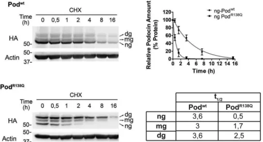
To investigate whether the difference in protein levels of Podwtand PodR138Qwas due to a higher degradation rate of the mutant protein, we determined the half-life (t1⁄2) of both pro- teins using a cycloheximide time course experiment, quantify- ing separately the glycosylated and non-glycosylated forms (Fig. 2). No differences were observed between thet1⁄2of the three Podwt forms, whereas a drastically reduced t1⁄2 was detected for ng-PodR138Qwhen compared with Podwt(7-fold).
Consistent with this finding, we also demonstrated that thet1⁄2
of the glycosylation mutant PodR138Q(N199Q,N355S) was also reduced around 7-fold compared with Podwt(N199Q,N355S)
(Fig. S2). Interestingly, ng-PodR138Q (i.e. hairpin-like to-
pology) appeared to be degraded faster than theN-glycosy- lated forms of PodR138Q(i.e.those with transmembrane to- pology), implying that the hairpin-like structure is more exposed to the intracellular degradative machinery (Fig. 2 andFig. S2).
Because ng-PodR138Qis degraded very quickly, we hypothe- sized that the proteasome was mediating its clearance. We therefore used the reversible proteasome inhibitor bortezomib (Bz) to perform dose-response and time course experiments (Fig. 3). We observed that ng-PodR138Qaccumulated in a dose- dependent manner, whereas levels of ng-Podwt increased significantly only at the highest Bz dose of 1M(Fig. 3A). Con- sistent with their longert1⁄2, both glycosylated Podwtand glyco- sylated PodR138Qwere not increased at any dose of Bz after 2 h (Fig. 3A). Thus, we next performed an overnight time course (16 h) using the lowest effective dose of Bz (0.1M) (Fig. 3B).
We found that only ng-PodR138Qaccumulated with time, and, most interestingly, the balance between ng- and dg-PodR138Q was inverted, thus after 16 h the proportions of each PodR138Q band resembled the proportions of each Podwtband in podocin immunoblots (Fig. 3B, colored graph). Levels of all forms of Podwtwere increased only at the longest time points. Similar findings were observed when we performed an overnight Bz time course on cells expressing the glycosylation mutants Podwt(N199Q,N355S) and PodR138Q(N199Q,N355S), with protein levels of the WT protein only significantly increased at the longest time point of 16 h, and a trend for levels of PodR138Q(N199Q,N355S)to accumulate in the presence of 0.1M Bz (Fig. S3A). The increased amount of Podwtand PodR138Q after an overnight treatment with 0.1MBz was confirmed by immunofluorescence analysis (Fig. 3C). As expected, a sus- tained exposure to Bz led to ER stress, as indicated by BiP induction (Fig. 3C), but to a higher extent in cells expressing PodR138Q(Fig. S3). Therefore, we concluded that the protea- some contributes to the degradation of both Podwt and PodR138Q, but more actively for ng-PodR138Q.
Figure 2. PodocinR138Qhas a short half-life.Immunoblot analysis of the time course of podocin degradation after inhibition of protein synthesis with cycloheximide (CHX) (25M) in podocyte cell lines. Densitometry data from three independent experiments are represented as mean⫾S.D. Half-lives were estimated by fitting a one-phase exponential decay curve to the data, as in the graph shown for ng-Podwtand ng-PodR138Q, and are summarized in the table.
Podocin
R138Qquality control and ERAD
at SEMMELWEIS UNIV OF MEDICINE on June 14, 2018http://www.jbc.org/Downloaded from
PodocinR138Qis not degraded by lysosomes
Because protein degradation proceeds via two major routes:
the proteasomal and the autophagic-lysosomal pathways, we investigated whether lysosomal degradation contributes to Podwtand PodR138Qproteolysis. We therefore treated the cells with ammonium chloride (NH4Cl), a weak base known to inhibit lysosomal proteases. NH4Cl treatment significantly increased levels of the ng-Podwtprotein fraction and levels of Podwt(N199Q,N355S), implying that the lysosomal machinery is involved in Podwtdegradation but not in PodR138Qproteoly- sis; indeed, we observed levels of both PodR138Q and PodR138Q(N199S,N355S)to fall after a 16-h NH4Cl treatment (Fig.
4AandFig. S4B). Then, by confocal microscopy, we confirmed the absence of PodR138Qin the late endosome/lysosome com- partment labeled with CD63 (Fig. 4B). Conversely, Podwtis pre-
dominantly present in this compartment, as already described (13). Taken together, our data suggest that Podwtis mainly degraded in lysosomes, in contrast to PodR138Q, which is exclu- sively degraded by the proteasome.
PodocinR138Qenhanced interaction with calnexin
MisfoldedN-glycosylated proteins located in the ER mem- brane are submitted to a strict quality control (QC) by the so- called calnexin (Cnx) cycle, being retained by the 90-kDa chap- erone Cnx until they reach their native conformation or are otherwise sent for ER-associated degradation (ERAD). We hypothesized that if PodR138Qis mostlyN-glycosylated and pos- sesses a transmembrane topology, interaction with Cnx may be a mechanism of PodR138QER retention, as has already been described for some nephrin mutants (18). Immunoblots of Figure 3. PodocinR138Qis degraded by the proteasome.Podocyte cell lines were treated with the proteasome inhibitor Bz or vehicle (DMSO) and processed for immunoblot (AandB) or immunofluorescence analysis of podocin (C).A,Bz dose-response (0.02 to 1M) after 2 h of treatment.B,time course of podocin after addition of 0.1MBz.AandB,podocin was identified using AP-P35 (Pod) primary antibody. BiP analysis was included to track ER stress (B). Quantification of ng bands is shown in thelower graphsinAand in themiddle graphsinB. Color graphs inBrepresent the percentage amount of each podocin band relative to total podocin. Quantitative results are shown as mean⫾S.D. (n⫽3).Asterisksrefer to non-stimulated cells (ns or 0 h). *,p⬍0.05 and **,p⬍0.01.C,podocin was detected by incubation with monoclonal HA primary antibody (ingreen). Hoechst nuclei labeling was included (inblue). All images were taken using the same confocal microscope settings to allow comparison of the intensity of fluorescence.Scale bar⫽40m.
Podocin
R138Qquality control and ERAD
at SEMMELWEIS UNIV OF MEDICINE on June 14, 2018http://www.jbc.org/Downloaded from
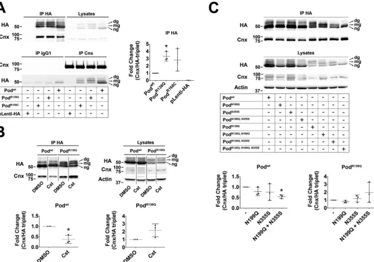
HEK293T cells transfected with HA-tagged WT podocin and the two ER-retained mutants, PodR138Q and PodR168C(7), showed that similarly to our findings from podocyte cell lines stably expressing Podwtand PodR138Qpodocin was present as three differentially glycosylated species (Fig. 5A). Co-immuno- precipitation studies revealed an interaction between Cnx and both WT and mutant podocin, with the mutant proteins inter- acting to a greater extent compared with the WT protein (Fig.
5A). That we found Cnx also interacted with Podwtwas not surprising, because HA immunoprecipitation enriched a cer- tain proportion ofN-glycosylated Podwt(Fig. 5A,upper panel).
Surprisingly, ng-podocin, WT, and mutants, also co-immuno- precipitated with Cnx, suggesting that hairpin podocin might interact with Cnx either indirectly, perhaps through oligomer- ization with the podocin transmembrane fraction via its N terminus (19), or directly, through membrane or cytosolic domains. To test whether impairing the interaction of PodR138Q with Cnx would allow its ER exit and promote membrane local- ization, we first treated the cells with castanospermine (Cst), a specific inhibitor of glucosidases I and II that prevents the sugar
trimming necessary for the recognition of the substrate protein N-glycan groups by the lectin domain of Cnx. Cst decreased Cnx interaction with Podwt, but not with PodR138Q(Fig. 5B), suggesting that PodR138Qinteraction with Cnx isN-glycan inde- pendent. Similar results were obtained when these experiments were performed using cells expressing glycosylation mutants Podwt(N199Q,N355S) and PodR138Q(N199Q,N355S) (Fig. 5C), sup- porting the idea that the interaction with Cnx is more lectin dependent for Podwtthan for PodR138Q. Finally, we found that introduction of p.N199Q and p.N355S substitutions into the PodR138Qmutant in podocytes did not prevent its ER retention, implying that suppression of theN-glycan-dependent interac- tion with Cnx is not sufficient to bring PodR138Qto the PM (Fig.
S4A), and supporting the idea that podocin is capable of lectin- dependent binding to Cnx.
Glycosylated podocinR138Qenters the calnexin cycle
Because we observed PodR138Q(N199Q,N355S) is still able to interact with Cnx, we next knocked-down Cnx in the two stable podocyte cell lines using siRNA (Fig. 6AandFig. S5A). Inter- Figure 4. PodocinR138Qis not degraded in the lysosomal compartment.A,time course immunoblot analysis of podocin content after the impairment of lysosomal degradation with NH4Cl (50 mM). The polyclonal antibody AP-P35 (Pod) was used to immunolabel podocin. Graphs show densitometry quantifica- tion of ng-podocin in three independent experiments. Data are normalized to-actin and then to non-treated cells (0 h).Asterisksrefer to 0 h. **,p⬍0.01.B, double immunofluorescence staining of podocin (AP-P35;green) and the lysosome/late endosome marker CD63 (cyan). Nuclei labeling by Hoechst is included (blue).Scale bar⫽10m.
Podocin
R138Qquality control and ERAD
at SEMMELWEIS UNIV OF MEDICINE on June 14, 2018http://www.jbc.org/Downloaded from
estingly, we observed that levels of N-glycosylated forms of PodR138Q were decreased upon Cnx knockdown, in contrast to levels of ng-PodR138Q, which were significantly increased (Fig. 6A,colored graph), suggesting that Cnx may play a role in stabilization of the transmembrane form of N-glycosylated PodR138Q, or indeed, that interaction with Cnx promotes glyco- sylation of PodR138Q. However, in support of the former state- ment, the total amount of PodR138Qwas slightly decreased after Cnx knockdown, suggesting that interaction with Cnx might delay PodR138Q degradation (Fig. 6A, lower graph). Next, we treated cells with kifunensine (Kif), a drug that inhibits the activity of␣-mannosidase I, and thus the mannose trimming that tags Cnx substrates for ERAD (Fig. 6B). Here, our results were precisely the opposite to those obtained upon Cnx knock- down, that is, levels of ng-PodR138Qwere significantly reduced, whereas levels of dg-PodR138Qwere increased (Fig. 6B,colored graph). Furthermore, the total amount of PodR138Qwas aug- mented after 4 h of treatment (Fig. 6B,lower graph), demon- strating that Kif treatment partially prevented PodR138Q degradation. These last data support the idea that onlyN-gly- cosylated, and thus transmembrane PodR138Q, enters the Cnx
cycle before being directed to ERAD. Finally, we observed by immunofluorescence that neither knockdown of Cnx nor blocking entry into the ERAD pathway with Kif were sufficient to target PodR138Qto the PM (data not shown). No significant changes on Podwtlevels were observed upon Cnx knockdown, but 4 h of Kif treatment did increase the levels of dg-Podwt, suggesting that glycosylated Podwtmay also enter the Cnx cycle (Fig. S5B).
Bortezomib partially re-addresses PodR138Qto the plasma membrane
While studying the proteasomal degradation of PodR138Q, we observed by immunofluorescence that a fraction of PodR138Q was localized, after Bz exposure, to thin filopodia PM protru- sions, similarly to Podwtin untreated cells, whereas PodR138Q was completely absent from these structures before treatment (Fig. 7A). Targeting of PodR138Qto filopodia, quantified as the percentage of WGA-positive filopodia that were also positive for podocin, revealed that Bz treatment increased plasma mem- brane targeting of PodR138Q, as soon as 2 h after Bz treatment, although levels did not reach those of Podwt(Fig. 7A,graph).
Figure 5. PodocinR138Qhas an enhanced interaction with calnexin.A–C,co-immunoprecipitation analyses of podocin and calnexin in HEK293T cells.A, co-immunoprecipitation of HA-tagged Podwt, PodR138Q, PodR168C, or an empty HA lentiviral vector (pLenti-HA) with Cnx.B,cells were treated with the glucosidase I and II inhibitor castanospermine (Cst; 500M, 16 h) before performing HA-immunoprecipitation to study the lectin-dependent interaction of Cnx with Podwtand PodR138Q.C,co-immunoprecipitation of Cnx with podocin in cells overexpressing HA-tagged Podwtand PodR138Qwith or without mutated N-glycosylation sites Asn199and Asn355.A–C,monoclonal anti-HA and anti-Cnx AF18 were used to identify podocin and Cnx, respectively.Graphsrepresent the densitometry quantification of Cnx when podocin is immunoprecipitated (IP HA). Data are normalized to total immunoprecipitated podocin (HA tripletin IP HA) and represent at least three independent experiments. *,p⬍0.05.
Podocin
R138Qquality control and ERAD
at SEMMELWEIS UNIV OF MEDICINE on June 14, 2018http://www.jbc.org/Downloaded from
We also observed a decreased interaction of PodR138Qwith Cnx in HEK293T cells treated with Bz, which may serve as an indi- cator of PodR138Q accumulation outside the ER (Fig. 7B).
Because we do not see PodR138Qat the PM (Fig. 7A) and the glycosylated forms of PodR138Qseem to be insensitive to Bz (Fig. 3), we hypothesize that only ng-PodR138Qis reaching the PM upon Bz treatment. Taking into account all our data we propose a scheme depicting a dynamic interchange between the hairpin and the transmembrane topology at the different steps of ER QC and subsequent ERAD (Fig. 7C). Briefly, hairpin PodR138Q is detected by the cytoplasmic QC machinery and rapidly degraded by the proteasome. In the meantime,N-gly- cosylated transmembrane PodR138Qenters the Cnx cycle to be eventually sent for ERAD. Because proteasomal degradation takes place in the cytosol, transmembrane PodR138Qwould lose itsN-glycan groups at some point between retrotranslocation and proteasomal degradation (20 –22), possibly adopting a hairpin topology when exposed to the cytosol. Bz treatment would then inhibit the degradation of ng-PodR138Q, giving it the chance to follow the secretory pathway and reach the PM.
Discussion
Despite p.R138Q being the most common podocin mutation causing SRNS in European and North American children (4), little is known about how this mutation affects podocin stability and degradation. In this study, we have found that human PodR138Q, when stably overexpressed in human podocyte cell lines, is clearly resolved as a triplet on immunoblots. The upper
bands of these triplets correspond to N-glycosylated forms, which is particularly intriguing because it implies that the C terminus of most PodR138Qis inside the ER lumen.N-Glycosyl- ation, and evidence of transmembrane topology, has been already described for stomatin, for the short isoform of podocin and for podocinP118L, another ER-retained podocin mutant (11, 12, 23). Whereas Pro118, an amino acid that is highly conserved throughout the stomatin family, is located within the hydro- phobic intramembrane region and is responsible for the kink of the hairpin topology (12), Arg138 and Arg168, which are also very well conserved, are located within the PHB domain, far away from this region. Nevertheless, we hypothesize that mis- sense mutations in the PHB region may also destabilize podocin hairpin topology, the folding of which is already intrinsically inefficient (11). Indeed, most of the missense mutations affect- ing the PHB domain typically result in ER-retention (7), and only podocin mutants that are known to be retained in the ER presentN-glycosylation levels comparable with PodR138Q. The switch in topology has important implications for the stability and the degradation of PodR138Q. We found the glycosylated PodR138Qforms to be more stable than ng-PodR138Q. The latter, with a hairpin structure, can be rapidly degraded by the protea- some. In contrast, we found evidence that the transmembrane isoform of PodR138Q enters the calnexin cycle, which may explain its longert1⁄2. Of course, it remains that glycosylation and deglycosylation events may be affecting the observed dif- ferences in stability of the different glycosylated podocin spe- Figure 6. PodocinR138Qenters the calnexin cycle.AandB, immunoblot analysis of podocin (HA) after Cnx knockdown (siCnx) (A) or impairment of Cnx cycle exit through the inhibition of␣-mannosidase I with kifunensine (Kif) (B) in HA WT and R138Q stably expressing podocyte cell lines.A,two siRNA oligonucleo- tides against Cnx were tested, alone (siCnx(1) andsiCnx(2)) or in combination (siCnx(1⫹2)). Luciferase siRNA (siLuc) served as control. Cnx protein amount was quantified to confirm Cnx knockdown (upper left graph).B,effect of kifunensine on podocin protein content.ns,non-stimulated cells.AandB,podocin was quantified as the relative percentage amount of each band within the triplet (color graph) or as total protein (the three bands altogether;lower graph). *,p⬍ 0.05 and **,p⬍0.01.
Podocin
R138Qquality control and ERAD
at SEMMELWEIS UNIV OF MEDICINE on June 14, 2018http://www.jbc.org/Downloaded from
cies, and undoubtedly these processes are also contributing to our results. That said, based on our experiments with Podwt(N199Q,N355S) and PodR138SQ(N199Q,N355S), which show these proteins behave in the same way as ng-Podwt and ng-PodR138Q, we believe that glycosylation of ng-podocin is not contributing to the observed increased stability of the glycosy- lated species, and in fact the longert1⁄2of mg- and dg-PodR138Q are due to decreased exposure to the intracellular degradative machinery. According to our results, transmembrane PodR138Q would be exclusively located in the ER as an intermediary of PodR138QQC, and is there stabilized by interaction with Cnx.
Indeed, in contrast to what has been shown for PodP118L(12), we did not find PodR138Qto be localized at the PM by immuno- fluorescence. This same model may apply for misfolded Podwt, because it also interacts with Cnx and its glycosylated forms are enriched after Kif treatment. Nevertheless, the increased levels of Podwtfollowing NH4Cl addition, together with the lack of response to Bz at short times, suggest that Podwtis mainly degraded in the endosome/lysosome compartment, a finding in accordance with results from other authors (13).
Cnx recognizes a complex code of glucose and mannose trimming in theN-glycan groups of its substrate proteins (21, Figure 7. Bortezomib partially re-addresses podocinR138Qto the plasma membrane.A,immunofluorescence analysis showing PodR138Q(green) in the presence of filopodia, through colocalization with the PM marker WGA (red), after a 2-h treatment with Bz (0.1M) or vehicle (DMSO). Podwtco-localization with WGA is included as a positive control. Hoechst nuclei labeling (H) is also shown (blue).Scale bar⫽10m. Image analysis of the WGA co-localization with podocin only at filopodia was obtained through the quantification of regions of interest (ROIs) that carefully delimit cell perimeters. Graph corresponds to the quantification of one representative experiment. One-way analysis of variance followed by Dunnett’s post-test was used as statistical analysis. **,p⬍0.01 and
***,p⬍0.001.B,co-immunoprecipitation (IP HA) of HA-podocin and Cnx after Bz addition (16 h at 0.1M). The graph represents the densitometry quantifi- cation of Cnx when podocin is immunoprecipitated. Data are normalized to total immunoprecipitated podocin (HA triplet) and represent to at least three independent experiments. ***,p⬍0.001.C,schematic summarizing the influence of different treatments on PodR138Qtopology and subcellular localization.
Redandgreen arrowsindicate a dynamic change to the transmembrane “wrong” or hairpin “right” topology, respectively.Numbers circled in yellow: (1) there is an imbalance of hairpin PodR138Qtoward a transmembrane topology, (2) decreasing PodR138Qinteraction with Cnx through siCnx transfection favors the hairpin topology, (3) stabilization of PodR138Qinteraction with Cnx, through inhibition of Cnx cycle exit with Kif, enhances podocin transmembrane topology, and (4) inhibition of PodR138Qproteasomal degradation with Bz increases the proportion of hairpin PodR138Qand is the only treatment that allows partial relocalization to the PM.EC, extracellular matrix;PM, plasma membrane;C,cytosol.
Podocin
R138Qquality control and ERAD
at SEMMELWEIS UNIV OF MEDICINE on June 14, 2018http://www.jbc.org/Downloaded from
24 –26). The fact that PodR138Qtopology and degradation is sensitive to mannose trimming implies that most probably there is an interaction with Cnx through itsN-glycan groups.
This result apparently contradicts the data obtained by the spe- cific inhibition of theN-glycan-dependent interaction of Cnx with PodR138Q, because the interaction with Cnx was not decreased. Nevertheless, there is an increasing amount of liter- ature suggesting alternative sites of interaction to the lectin domain of Cnx (i.e.the Cnx transmembrane domain), espe- cially in the case of mutant proteins (27–29). An interesting proposal is that there are at least two different types of interac- tion occurring sequentially or simultaneously; one carbohy- drate based, through theN-glycan groups, and one peptide based, outside the Cnx lectin domain (30 –32). This would explain why inhibiting lectin binding is insufficient to impair PodR138Q interaction with Cnx. In contrast, Cnx interaction with Podwt seemed to be more dependent on the N-glycan groups, even though Podwtis predominantly non-glycosylated.
However, we cannot rule out a second Cnx interaction site also for Podwt, because deletion ofN-glycan sites did not completely impair the interaction with Cnx and ng-Podwtalso co-immu- noprecipitated with Cnx. Finally, it is tempting to speculate that N-glycosylation and interaction with Cnx may be part of nor- mal podocin biosynthesis and initial folding, and not only a mechanism of podocin quality control. In fact, Cnx is part of the ribosome–translocon complex and its initial association to nas- cent glycoproteins is usually co-translational (33, 34).
The targeting of PodR138Q to the cell membrane using chemical chaperones as glycerol, trimethylamine-N-oxide, and DMSO has already been reported in transiently transfected human podocytes (35). Nevertheless, there is still no treatment available for SRNS patients carrying the p.R138Q mutation.
Here, we report the therapeutic potential of Bz (Velcade威), a reversible inhibitor of the 26S proteasome that has been approved and successfully used in multiple myeloma therapy (36, 37). PodR140Q, the mouse equivalent to PodR138Q, is poorly expressed in the glomeruli of the constitutiveNphs2R140Q/R140Q
knock-in mice (6), similarly to our findings in podocyte cell lines. By studying PodR138Qt1⁄2and the degradation pathways it follows, we have found that low intracellular levels of PodR138Q are due to rapid proteasomal degradation. Bz not only increases PodR138Q protein levels, but allows a significant amount of PodR138Qto reach the PM, at least at filopodia. This could be simply caused by an overflow from the ER, because 2 h of Bz treatment already quadruples PodR138Qprotein levels. Indeed, proteasomal inhibition has been found to inhibit the retro- translocation of most ERAD substrates, thus giving them the chance to remain in the secretory pathway (21, 38, 39). Alter- natively, because de-glycosylation by cytoplasmic peptide:N- glycanases (PNGases) is an additional step in glycoprotein ERAD (40), Bz would specifically rescue the ng-PodR138Q already targeted for proteasomal degradation. In support of this idea, a promising effect of Bz is that it enriches the ng-PodR138Q fraction corresponding to the potentially functional hairpin topology of PodR138Qat the SD. A successful therapy would need not only to bring PodR138Qto the PM, but also promote its hairpin topology. Interestingly, Bz has been reported to rescue other misfolded mutant proteins back to the PMin vitroandin
vivo(42). Additionally, second generation proteasomal inhibi- tors are currently being tested in clinical trials with less severe side effects than those of Bz, such as neurotoxicity or lym- phopenia (43). Altogether, although we lackin vivostudies, we propose that increasing ng-PodR138Qprotein levels through the inhibition of the proteasomal degradation may be a reasonable strategy to treat patients with p.R138Q mutation and possibly with other ER-retained podocin mutations.
Experimental procedures
Plasmids, cell culture, and establishment of lentiviral cell lines Human podocin-coding constructs were generated as described by Toryet al.(44). Human wildtypeNPHS1cDNA, encoding nephrin, was amplified from the construct described by Philippeet al.(6) and subcloned into NotI and SpeI sites of LentiORF pLEX-MCS (Open Biosystems). Thus, the encoded proteins consist of podocin with two hemagglutinin tags (2HA) fused to its N terminus, and nephrin with a V5 tag fused to its N terminus. Site-directed mutagenesis (QuikChange kit, Strat- agene) was used to generate the missense mutations used in this study: p.R138Q, p.R168C, p.N199Q, p.N355S for podocin, and p.S366R for nephrin. All constructs were verified by Sanger sequencing. A human immortalized podocyte cell line (AB8/
13), obtained by transfection of the temperature-sensitive mutant tsA58 of the SV40-T-antigen-encoding gene, was kindly provided by M. A. Saleem (45). Stable podocyte cell lines were obtained by transduction of the above cell line with lenti- viral vectors expressing either podocin (wildtype or mutants) or nephrin (wildtype or mutants) at a multiplicity of infection of 1, and subsequently selected by puromycin (2g/ml). Podocytes were cultured at 33 °C with 7% CO2in RPMI 1640 medium supplemented with 10% fetal bovine serum, insulin/transferrin/
selenium, glutamine, and penicillin/streptomycin (all from Life Technologies). At this growth-permissive temperature, podo- cytes are proliferating and undifferentiated, and do not express either endogenous podocin or nephrin. HEK293T cells were maintained in Dulbecco’s modified Eagle’s medium supple- mented with 10% fetal bovine serum, glutamine, and penicillin/
streptomycin (all from Life Technologies). Cell lines used in this study were tested mycoplasma-free.
Antibodies, enzymes, and chemical compounds
PNGase F was purchased from New England BioLabs. Tuni- camycin, cycloheximide, bortezomib, NH4Cl, castanosper- mine, and kifunensine were purchased from Sigma. The commercial antibodies used were as follows: mouse anti-HA (HA.11 clone 16B12, Covance), mouse anti-calnexin (Cnx) (clone AF18, Enzo Life Sciences), mouse anti-CD63 (clone H5C6, DSHB), and mouse anti-calnexin (clone C5C9, Cell Sig- naling Technology). Rabbit anti-podocin AP-P35 (Pod) was described previously (46). Alexa Fluor 555-conjugated wheat germ agglutinin (WGA555) (W32464, Life Technologies) was used to stain the plasma membrane. Rat IgG2a (isotype control clone 2H3, MBL) for immunoprecipitation control experi- ments was purchased from MBL. Secondary antibodies for immunoblotting were sheep anti-mouse and donkey anti-rab- bit HRP-conjugated antibodies (GE Healthcare UK). Secondary antibodies for immunofluorescence were donkey anti-rabbit
Podocin
R138Qquality control and ERAD
at SEMMELWEIS UNIV OF MEDICINE on June 14, 2018http://www.jbc.org/Downloaded from
and anti-mouse Alexa Fluor 488- and 647-conjugated antibod- ies (Life Sciences).
siRNA experiments
The following siRNAs were used to transfect podocyte cell lines stably expressing podocin to specifically knockdown the humanCNX (calnexin) gene: siRNA-1, 5⬘-AAGACGAUAC- CGAUGAUGAAA-3⬘and siRNA-2, 5⬘AAUGUGGUGGUGC- CUAUGUGA-3⬘. siRNA against the luciferase gene (Luc, 5⬘-GCCAUUCUAUCCUCUAGAGGAUG-3⬘) was used as a siRNA control. siRNAs were transfected using Lipofectamine威 RNAiMAX Reagent (Invitrogen) at a concentration of 20 nM
and the efficacy of siRNA was tested 3 days after transfection.
Quantitative real-time PCR
Total RNA was extracted from podocyte cell lines using the Qiagen extraction RNeasy威 kit (Qiagen) and treated with DNase I. Oneg of total RNA was reverse-transcribed using SuperScript II according to the manufacturer’s protocol (Invit- rogen). The relative expression levels of the studied mRNAs were determined by quantitative real-time PCR using Absolute SYBR Green ROX Mix (ABgene) with the following specific primers for human NPHS2: forward (929F) 5⬘-GGCTGA- AGCGCAAAGACAAG-3⬘ and reverse (988R) 5⬘-GCAGC- CTTTTCCGCTTCTG-3⬘. Human hypoxanthine phosphori- bosyltransferase (Hprt) was used as an internal standard. Data were analyzed with the 2⫺⌬⌬Ctmethod (47).
Immunofluorescence
Podocytes were cultured on type I collagen-coated coverslips and either transiently transfected using FuGENE威HD (Pro- mega) or treated with different drugs. When appropriate, cells were washed once with cold PBS, incubated with Alexa Fluor 555-conjugated WGA (1:600) for 15 min at 4 °C, and then fixed with ice-cold ethanol for 5 min. Fixed cells were blocked with 1⫻ PBS, 1% BSA for 30 min before incubation with mouse anti-HA primary antibodies (1:500) or rabbit anti-podocin AP-P35 (1:300) when combined with mouse anti-calnexin (1:500) or mouse anti-CD63 (1:200), followed by Alexa Fluor 488- or 647-conjugated secondary antibodies (1:200). Confocal images were captured using a⫻40 oil objective attached to a Leica SP8 confocal microscope. At least five random fields, with the only condition that filopodia were well visualized in the WGA channel, were considered for the quantification of the percentage area of WGA colocalization with podocin using ImageJ 1.48i software. Masks were carefully created to specifi- cally quantify the labeling of the filopodia. Confocal settings and Image J thresholds were kept the same.
Immunoblotting
Proteins from podocyte cell lines were extracted in lysis buffer containing 150 mMNaCl, 50 mMTris-HCl, pH 7, 0.5%
Triton X-100 with CompleteTM protease inhibitors (Roche Applied Science). Protein dosage was then performed using the BCA protein assay kit (Thermo Scientific). Fifty micrograms of protein were loaded on a 10% SDS-PAGE gel and transferred to a nitrocellulose membrane. Membranes were blocked in 5%
skimmed milk in 1⫻ Tris-buffered saline, 0.1% Tween 20
(TBST) for 1 h and incubated with primary antibodies at a 1:1,000 dilution. After washing, membranes were incubated with HRP-conjugated secondary antibodies diluted 1:10,000 in TBST, 5% milk for 1 h at room temperature. Signals were detected using ECL reagents (Amersham Biosciences) and acquired in a Fusion Fx7 darkroom (Vilber Lourmat). Densi- tometry quantification was performed using Bio-1D software.
Immunoprecipitation
HEK293T cells were transiently transfected with HA-tagged WT podocin and mutants using calcium phosphate. Forty- eight h post-transfection, cells were lysed in 150 mMNaCl, 25 mMTris-HCl, pH 8, 0.5% Triton X-100 with protease inhibi- tors, and HA-tagged podocin was immunoprecipitated using theMACSTMEpitope Tag Protein Isolation Kit (Miltenyi Bio- tec). Briefly, fresh lysates (1–1.5 mg of protein) were incubated either with mouse anti-calnexin antibodies, followed by a 30-min incubation with magnetic beads-coupled to protein A, or directly with magnetic beads coupled to a HA antibody. A rat IgG2a isotype control was included to discard unspecific interactions. Immunoprecipitated proteins were isolated usingMACS威Separation Columns in a magneticMACS separator and subsequently eluted with 1⫻Laemmli buffer.
Lysates and immunoprecipitated samples were subjected to immunoblot.
Statistical analyses
All immunoblots were normalized to-actin and then to the corresponding control group or the immunoprecipitated pro- tein in control conditions. Statistical analysis of at least three independent experiments was done using the one-sample two- tailedttest (41) or a one-way analysis of variance,pvalues: *, p⬍0.05; **,p⬍0.01; ***,p⬍0.001. GraphPad Prism 5 software was used to perform all statistical analyses (mean⫾S.D.).
Author contributions—M.-C. S.-P., F. C. T., C. Antignac, and G. Mol- let conceptualization; M.-C. S.-P., F. C. T., F. N., and G. Mollet formal analysis; M.-C. S.-P., F. C. T., K. T., and G. Mollet validation;
M.-C. S.-P., F. C. T., F. N., C. Arrondel, S. S., G. Martin, and K. T.
investigation; M.-C. S.-P., F. C. T., F. N., C. Arrondel, S. S., and G.
Martin methodology; M.-C. S.-P., F. C. T., C. Antignac, and G. Mollet writing-original draft; M.-C. S.-P., F. C. T., F. N., C. Arrondel, S. S., G.
Martin, K. T., C. Antignac, and G. Mollet writing-review and editing;
C. Antignac supervision; C. Antignac and G. Mollet project admin- istration; G. Mollet funding acquisition.
Acknowledgments—We thank Ana Maria Cuervo (Albert Einstein College, NY) for helpful discussion and advice; Gisèle Froment, Didier Nègre, and Caroline Costa from the Lentivectors Production Facility/
SFR BioSciences Gerland-Lyon Sud (UMS3444/US8) and the Confo- cal Platform at Imagine Institute.
References
1. Mekahli, D., Liutkus, A., Ranchin, B., Yu, A., Bessenay, L., Girardin, E., Van Damme-Lombaerts, R., Palcoux, J. B., Cachat, F., Lavocat, M. P., Bourdat- Michel, G., Nobili, F., and Cochat, P. (2009) Long-term outcome of idio- pathic steroid-resistant nephrotic syndrome: a multicenter study.Pediatr.
Nephrol.24,1525–1532CrossRef Medline
Podocin
R138Qquality control and ERAD
at SEMMELWEIS UNIV OF MEDICINE on June 14, 2018http://www.jbc.org/Downloaded from
2. Tune, B. M., and Mendoza, S. A. (1997) Treatment of the idiopathic ne- phrotic syndrome: regimens and outcomes in children and adults.J. Am.
Soc. Nephrol.8,824 – 832Medline
3. Boute, N., Gribouval, O., Roselli, S., Benessy, F., Lee, H., Fuchshuber, A., Dahan, K., Gubler, M. C., Niaudet, P., and Antignac, C. (2000) NPHS2, encoding the glomerular protein podocin, is mutated in autosomal reces- sive steroid-resistant nephrotic syndrome. Nat. Genet. 24, 349 –354 CrossRef Medline
4. Sadowski, C. E., Lovric, S., Ashraf, S., Pabst, W. L., Gee, H. Y., Kohl, S., Engelmann, S., Vega-Warner, V., Fang, H., Halbritter, J., Somers, M. J., Tan, W., Shril, S., Fessi, I., Lifton, R. P.,et al. (2015) A single-gene cause in 29.5 % of cases of steroid-resistant nephrotic syndrome.J. Am. Soc. Neph- rol.26,1279 –1289CrossRef Medline
5. Hinkes, B. G., Mucha, B., Vlangos, C. N., Gbadegesin, R., Liu, J., Has- selbacher, K., Hangan, D., Ozaltin, F., Zenker, M., Hildebrandt, F., and Arbeitsgemeinschaft für Paediatrische Nephrologie Study, G. (2007) Ne- phrotic syndrome in the first year of life: two thirds of cases are caused by mutations in 4 genes (NPHS1, NPHS2, WT1, and LAMB2).Pediatrics 119,e907–919CrossRef Medline
6. Philippe, A., Weber, S., Esquivel, E. L., Houbron, C., Hamard, G., Ratelade, J., Kriz, W., Schaefer, F., Gubler, M. C., and Antignac, C. (2008) A missense mutation in podocin leads to early and severe renal disease in mice.Kidney Int.73,1038 –1047CrossRef Medline
7. Roselli, S., Moutkine, I., Gribouval, O., Benmerah, A., and Antignac, C.
(2004) Plasma membrane targeting of podocin through the classical exocytic pathway: effect of NPHS2mutations.Traffic5,37– 44CrossRef Medline
8. Huber, T. B., Kottgen, M., Schilling, B., Walz, G., and Benzing, T. (2001) Interaction with podocin facilitates nephrin signaling.J. Biol. Chem.276, 41543– 41546CrossRef Medline
9. Huber, T. B., Schermer, B., Müller, R. U., Höhne, M., Bartram, M., Calixto, A., Hagmann, H., Reinhardt, C., Koos, F., Kunzelmann, K., Shirokova, E., Krautwurst, D., Harteneck, C., Simons, M., Pavenstädt, H.,et al. (2006) Podocin and MEC-2 bind cholesterol to regulate the activity of associated ion channels.Proc. Natl. Acad. Sci. U.S.A.103,17079 –17086CrossRef Medline
10. Schwarz, K., Simons, M., Reiser, J., Saleem, M. A., Faul, C., Kriz, W., Shaw, A. S., Holzman, L. B., and Mundel, P. (2001) Podocin, a raft-associated component of the glomerular slit diaphragm, interacts with CD2AP and nephrin.J. Clin. Invest.108,1621–1629CrossRef Medline
11. Kadurin, I., Huber, S., and Gründer, S. (2009) A single conserved proline residue determines the membrane topology of stomatin.Biochem. J.418, 587–594CrossRef Medline
12. Schurek, E. M., Völker, L. A., Tax, J., Lamkemeyer, T., Rinschen, M. M., Ungrue, D., Kratz, J. E., 3rd, Sirianant, L., Kunzelmann, K., Chalfie, M., Schermer, B., Benzing, T., and Höhne, M. (2014) A disease-causing mu- tation illuminates the protein membrane topology of the kidney-ex- pressed prohibitin homology (PHB) domain protein podocin. J. Biol.
Chem.289,11262–11271CrossRef Medline
13. Gödel, M., Ostendorf, B. N., Baumer, J., Weber, K., and Huber, T. B. (2013) A novel domain regulating degradation of the glomerular slit diaphragm protein podocin in cell culture systems.PLoS ONE8,e57078CrossRef Medline
14. Rinschen, M. M., Bharill, P., Wu, X., Kohli, P., Reinert, M. J., Kretz, O., Saez, I., Schermer, B., Höhne, M., Bartram, M. P., Aravamudhan, S., Brooks, B. R., Vilchez, D., Huber, T. B., Müller, R. U., Krüger, M., and Benzing, T. (2016) The ubiquitin ligase Ubr4 controls stability of podocin/
MEC-2 supercomplexes. Hum. Mol. Genet. 25, 1328 –1344CrossRef Medline
15. Li, W. W., Sistonen, L., Morimoto, R. I., and Lee, A. S. (1994) Stress induc- tion of the mammalian GRP78/BiP protein gene: in vivo genomic foot- printing and identification of p70CORE from human nuclear extract as a DNA-binding component specific to the stress regulatory element.Mol.
Cell. Biol.14,5533–5546CrossRef Medline
16. Wooden, S. K., Li, L. J., Navarro, D., Qadri, I., Pereira, L., and Lee, A. S.
(1991) Transactivation of the grp78 promoter by malfolded proteins, gly- cosylation block, and calcium ionophore is mediated through a proximal
region containing a CCAAT motif which interacts with CTF/NF-I.Mol.
Cell. Biol.11,5612–5623CrossRef Medline
17. Yoshida, H., Haze, K., Yanagi, H., Yura, T., and Mori, K. (1998) Identifica- tion of the cis-acting endoplasmic reticulum stress response element re- sponsible for transcriptional induction of mammalian glucose-regulated proteins. Involvement of basic leucine zipper transcription factors.J. Biol.
Chem.273,33741–33749CrossRef Medline
18. Drozdova, T., Papillon, J., and Cybulsky, A. V. (2013) Nephrin missense mutations: induction of endoplasmic reticulum stress and cell surface rescue by reduction in chaperone interactions.Physiol. Rep.1,e00086 Medline
19. Huber, T. B., Simons, M., Hartleben, B., Sernetz, L., Schmidts, M., Gund- lach, E., Saleem, M. A., Walz, G., and Benzing, T. (2003) Molecular basis of the functional podocin-nephrin complex: mutations in the NPHS2 gene disrupt nephrin targeting to lipid raft microdomains.Hum. Mol. Genet.
12,3397–3405CrossRef Medline
20. Hirsch, C., Blom, D., and Ploegh, H. L. (2003) A role for N-glycanase in the cytosolic turnover of glycoproteins.EMBO J.22,1036 –1046CrossRef Medline
21. Lederkremer, G. Z., and Glickman, M. H. (2005) A window of opportunity:
timing protein degradation by trimming of sugars and ubiquitins.Trends Biochem. Sci.30,297–303CrossRef Medline
22. Olzmann, J. A., Kopito, R. R., and Christianson, J. C. (2013) The mamma- lian endoplasmic reticulum-associated degradation system.Cold Spring Harb. Perspect. Biol.5,A013185CrossRef
23. Völker, L. A., Schurek, E. M., Rinschen, M. M., Tax, J., Schutte, B. A., Lamkemeyer, T., Ungrue, D., Schermer, B., Benzing, T., and Höhne, M.
(2013) Characterization of a short isoform of the kidney protein podocin in human kidney.BMC Nephrol.14,102CrossRef Medline
24. Frenkel, Z., Gregory, W., Kornfeld, S., and Lederkremer, G. Z. (2003) Endoplasmic reticulum-associated degradation of mammalian glycopro- teins involves sugar chain trimming to Man6 –5GlcNAc2.J. Biol. Chem.
278,34119 –34124CrossRef Medline
25. Hammond, C., Braakman, I., and Helenius, A. (1994) Role ofN-linked oligosaccharide recognition, glucose trimming, and calnexin in glycopro- tein folding and quality control.Proc. Natl. Acad. Sci. U.S.A.91,913–917 CrossRef Medline
26. Ware, F. E., Vassilakos, A., Peterson, P. A., Jackson, M. R., Lehrman, M. A., and Williams, D. B. (1995) The molecular chaperone calnexin binds Glc1Man9GlcNAc2 oligosaccharide as an initial step in recognizing un- folded glycoproteins.J. Biol. Chem.270,4697– 4704CrossRef Medline 27. Cannon, K. S., Hebert, D. N., and Helenius, A. (1996) Glycan-dependent
and -independent association of vesicular stomatitis virus G protein with calnexin.J. Biol. Chem.271,14280 –14284CrossRef Medline
28. Fontanini, A., Chies, R., Snapp, E. L., Ferrarini, M., Fabrizi, G. M., and Brancolini, C. (2005) Glycan-independent role of calnexin in the intracel- lular retention of Charcot-Marie-tooth 1A Gas3/PMP22 mutants.J. Biol.
Chem.280,2378 –2387CrossRef Medline
29. Swanton, E., High, S., and Woodman, P. (2003) Role of calnexin in the glycan-independent quality control of proteolipid protein.EMBO J.22, 2948 –2958CrossRef Medline
30. Arunachalam, B., and Cresswell, P. (1995) Molecular requirements for the interaction of class II major histocompatibility complex molecules and invariant chain with calnexin.J. Biol. Chem.270,2784 –2790CrossRef Medline
31. Lederkremer, G. Z. (2009) Glycoprotein folding, quality control and ER- associated degradation.Curr. Opin. Struct. Biol.19,515–523CrossRef Medline
32. Williams, D. B. (2006) Beyond lectins: the calnexin/calreticulin chaperone system of the endoplasmic reticulum.J. Cell Sci.119,615– 623CrossRef Medline
33. Chen, W., Helenius, J., Braakman, I., and Helenius, A. (1995) Cotransla- tional folding and calnexin binding during glycoprotein synthesis.Proc.
Natl. Acad. Sci. U.S.A.92,6229 – 6233CrossRef Medline
34. Lakkaraju, A. K., Abrami, L., Lemmin, T., Blaskovic, S., Kunz, B., Kihara, A., Dal Peraro, M., and van der Goot, F. G. (2012) Palmitoylated calnexin is a key component of the ribosome-translocon complex.EMBO J.31, 1823–1835CrossRef Medline
Podocin
R138Qquality control and ERAD
at SEMMELWEIS UNIV OF MEDICINE on June 14, 2018http://www.jbc.org/Downloaded from