DOI: 10.1556/066.2019.48.1.13
HIGH-THROUGHPUT MICROALGAE CULTIVATION WITH ADJUSTABLE LED-MODULE APPLYING DIFFERENT COLOURS
FOR NANNOCHLOROPSIS AND CHLORELLA MICROCULTURES
B. KISSa and Á. NÉMETHa*
aFermentation Pilot Plant Laboratory, Department of Applied Biotechnology and Food Science, Budapest University of Technology and Economics, H-1111 Budapest, Műegyetem rkp. 3. Hungary
(Received: 11 April 2018; accepted: 12 July 2018)
Utilization of algae includes both macroalgae for human consumption dating back to thousands of years, as well as the application of microalgae in health promoting dietary supplements. The autotrophic growth of microalgae is slow, but can be accelerated by optimizing their cultivation conditions. Effi ciency optimizations for time and economy should be performed in many parallel experiments. A new high-throughput microalgae cultivation method is presented here, applying 24-low-well microplate with varying illumination, in which the cell growth is followed via evaluation of scanned images. A strain of the genus Nannochloropsis and two Chlorella vulgaris species have been chosen as well described and frequently applied model organisms in order to test the recently developed cultivation system. In these scaled down experiments, the custom design lighting panel was tested by studying the effect of the colour of illumination on cell growth kinetics. RGB LEDs (i.e. light emitting diodes, red: 622 nm, green: 528 nm, and blue: 467 nm) were used individually or together providing red, green, blue, and white colours.
While the effect of light’s colour on algae growth was evaluated, also the new system was proven to be suitable for comparing maximal growth rates for different microalgae strains. While the tested two Chlorella isolates reached 1.2–1.4 g l–1 concentrations, the Nannochloropsis strain reached 1.4 g l–1 fi nal cell dry weight, and specifi c growth rates were observed between 0.58–0.62 day–1.
Keywords: Chlorella, Nannochloropsis, growth rate, LED, microscale, microplate, light conditions
As other photosynthesizing organisms, microalgae are capable of CO2 fi xation and the production of organic matter. Their advantage over plants is that they can photosynthesize much faster and with greater reproductive capacity due to their higher photoconversion effi ciency. In addition to these properties, their intracellular content can be used industrially from the feed/food industry through cosmetic industry to biofuel production. Therefore, examining the conditions affecting their intracellular content is very important in today’s technological development (MOHSENPOUR & WILLOUGHBY, 2013; FÓZER et al., 2017).
The materials produced by algae have a great variety. Among their major ingredients with industrial interest are chlorophyll, fatty acids, tocopherols, sterols, proteins, carbohydrates, vitamins, minerals, antioxidants, and pigments (CHACON-LEE & GONZÁLEZ- MARINO, 2010; BENNAMOUN et al., 2015; ARIEDE et al., 2017; FÓZER et al., 2017; SIDDIQUI &
* To whom correspondence should be addressed.
Phone: +36 1 463 2595; fax: +36 1 463 3855; e-mail: naron@f-labor.mkt.bme.hu
This is an open-access article distributed under the terms of the Creative Commons Attribution-NonCommercial 4.0 International License (https://creativecommons.org/licenses/by-nc/4.0/), which permits unrestricted use, distribu- tion, and reproduction in any medium for non-commercial purposes, provided the original author and source are credited, a link to the CC License is provided, and changes – if any – are indicated.
PRASAD, 2017). Many ingredients, like vitamin A, B1, and B2 or fatty acids are used as supplementation of essential nutrients (SIDDIQUI & PRASAD , 2017). Other components, such as phycobilin or phytosterol, are utilized as functional foods (CHACON-LEE & GONZÁLEZ-MARINO, 2010). Microalgae have some other high-value compounds like β-carotene (CHACON-LEE &
GONZÁLEZ-MARINO, 2010) and phycocyanin (BENNAMOUN et al., 2015) utilized for their antioxidant effect.
However, algae are often growing relatively slowly in comparison to other micro- organism. In order to improve their cultivation technology, the application of scale down is strongly recommended. While one of the smallest and most inexpensive means for investigation of microbial growth, a 96-well-microtiter plate (MTP), is suitable for algae cultivation, too, in an incubator shaker, its adjustable illumination is not yet solved. Therefore, we had started developing an illuminating system for MTPs with the application of LED chips as light source, since their size makes it possible to individually illuminate every well on an MTP. Furthermore, the electrical energy conversion of LEDs to light is one of the best among light sources, and thus LEDs have good potential in the development of economically feasible processes of algae production. Additionally, the emitted spectra of LED should fulfi l the requirements of the cultivated algae, while not wasting light by emitting unnecessary wavelengths (LEE & PALSSON, 1995; CARVALHO et al., 2011). By combination and dimming of different LED chips, LED panels can be adjusted easily to meet lighting requirements.
Like in case of all photoautotrophic cultures (including strains of Nannochloropsis and Chlorella), intensity, wavelength, and periodicity are the main factors of lighting infl uencing both biomass productivity and intracellular content (FU et al., 2013), which also determine the fi nal application potential in the industry (GOLDMAN, 1979). For growing algae cultures, light is the source of energy essential for photoautotrophic growth, so the successful cultivation of microalgae highly depends on the effective utilization of the light’s energy (SANDNES et al., 2005) and thus on the parameters of light.
Wavelength is probably the most important parameter, as the spectrum of light can infl uence the metabolic processes of algae. This is explained by the different energy content of different wavelengths that induce different biochemical processes in the cells. Algae strains can have various colour bodies with different photoabsorbent compounds like chlorophyll a, b, and c, which infl uence the light absorption spectrum of the incident light, meaning also that these bodies have an optimum (a sharper peak) characteristic wavelength.
While Nannochloropsis species only have chlorophyll a, Chlorella species have both chlorophyll a and b, resulting different light utilization patterns (LUBIAN et al., 2000; SHU et al., 2012).
The 400–700 nm range of light that can be used for photosynthesis is called photosynthetically active radiation (PAR).
The effect of wavelength on growth of algae has been examined in several studies. CHEN
and co-workers (2013) cultivated Nannochloropsis oceania in a 1 litre glass photobioreactor on modifi ed BG-11 medium. The photobioreactor was illuminated with different coloured LEDs (i.e., white, blue, red, or yellow) at a light intensity of ca. 150 μmol m–2 s–1 with wavelength set to 670 nm (red), 580 nm (blue), 475 nm (yellow), 430 to 450 nm, and 520 to 560 nm (white). They found that white light has the greatest improving effect on the fi nal concentration of microalgae biomass, reached on the 4th day of cultivation (2.1 g l–1), followed by yellow (2 g l–1), red (1.8 g l–1), and blue (1.6 g l–1) light. While white light containing the whole spectrum was found to result in the highest biomass concentration (CHEN et al., 2013), blue light induced the highest eicosapentaenoic acid (EPA) production of
the algae, which metabolite is primarily used in the treatment of cardiovascular diseases in human medicine (MAKI et al., 2014; BOROW et al., 2015; SCHWANKE et al., 2015).
DAS and co-workers (2011) studied the effect of light on phototrophic culture of Nannochloropsis sp. (isolated in Singapore). They found that illumination with different wavelengths changes the intracellular protein and carbohydrate compositions. When lit with blue light, cells contained 15% carbohydrate and 60% protein, whereas under red light, this ratio was 39:29. Nannochloropsis sp. achieved a specifi c growth rate of 0.64 and 0.66 day–1 in phototrophic and mixotrophic cultures with blue lighting.
TEO and co-workers (2014) also studied the effect of different light (blue, red, green, and white) on algae and showed that specifi c growth rates of Nannochloropsis species were similar under blue (1.64 day–1) and red (1.61 day–1) illumination. The higher growth rate was explained by the induction of more Rubisco enzymes (ribulose-bisphosphatase / carboxylase / oxygenase and carbonic anhydrase enzyme) in the cells caused by the blue light. The higher Rubisco enzyme activity is clearly associated with the enhanced growth rate and better storage of triglycerides. Red light is often proved to be harmful to algae cells (SHU et al., 2012), reducing the growth rate. However, the harmful effects of red light can be eliminated by blue illumination for short periods, which can regenerate the damaged enzymes.
BLAIR and co-workers (2014) presented the effect of wavelengths and growth medium composition on the growth of Chlorella vulgaris. They used light with different wavelengths (blue, white (clear), green, and red) in order to test their effect on algal growth. While cultivations under white (0.369 day–1), blue (0.235 day–1), and red (0.140 day–1) lights showed the highest growth rates on day 3, the green (0.137 day–1) light provided the highest growth rate on day 2.
In this study, we investigated the applicability of a new illumination system by comparing the growth of three microalgae strains belonging to the genera of Chlorella and Nannochloropsis. They were exposed to four different colours of illumination in order to determine the effect of light quality on biomass productivity and on specifi c growth rate, as well as study the operation of the new system.
1. Materials and methods
1.1. Strains and media
Experiments were carried out with three industrially relevant strains, widely studied and described (BLAIR et al., 2014; SAFIA et al., 2014), namely: a strain from the genus Nannochloropsis and two isolates of Chlorella vulgaris species (CvT, CvH), all belonging to green microalgae. They were cultivated on a modifi ed version of BG-11 medium widely used for blue and green algae culturing. Modifi ed BG-11 medium (CHO et al., 2015) contained 1.5 g l–1 NaNO3, 75 mg l–1 MgSO4∙7H2O, 40 mg l–1 K2HPO4∙7H2O, 36 mg l–1 CaCl2∙2H2O, 20 mg l–1 Na2CO3, 6 mg l–1 FeNH4SO4, 6 mg l–1 citric acid, 1 mg l–1 Na2MgEDTA, and 1 ml A5 trace solution per litre. The A5 solution contained 2.86 g l–1 H3BO3, 1.81 g l–1 MnCl2∙4H2O, 0.391 g l–1 NaMoO4∙2H2O, 0.222 g l–1 ZnSO4, 0.079 g l–1 CuSO4∙5H2O, and 0.05 g l–1 CoCl2∙6H2O. Deionized (Simplicity, EMD Millipore, Darmstadt, Germany) distilled water was used for preparing the medium. Before inoculation, volumes of this medium were sterilized by autoclaving at 121 °C for 20 min (3870ELV, Tuttnauer Europe, Breda, Netherlands) and used both for shaken fl ask precultivation and MTP experiments.
1.2. Cultivation
24-well microplate with white plastic walls and transparent bottom (Polystyrene 24 roundwell microplates from Porvair) was used in our experiments. The useful volume of each well was 1.5 ml.
The novel RGB LED panel was attached to the transparent bottom white walls hindered cross-lighting, thus each well could have different light conditions. We designed the panel in a way that each well on the microplate has its own RGB type LED chips: red 622 nm, green:
528 nm, blue: 467 nm. Jumpers on the panel allow switching on/off the corresponding LEDs and potentiometers (variable resistance) are to adjust LED intensity. While the length of lit and dark periods can also be adjusted, during the experiments presented in this paper we applied 8 h dark period after 16 h of light.
For isolation from the outside world but allowing effi cient gas exchange, we applied sandwich covers® that were kind gift of Enzyscreen (www. enzyscreen.com, the Netherlands), with its cloths replaced with a perforated silicone sheet.
The microplate and the corresponding illumination module were placed in an incubator shaker (Innova 40, New Brunswick Scientifi c, Enfi eld, CT, USA) and cultivated at 25 °C and 250 r.p.m. to obtain adequate mixing for gas exchange. Four different lights were tested:
white light (all three colours on), red, green, and blue.
1.3. Inoculation
We used for inoculum a pre-culture from a standing fl ask lit with natural light sources at ambient temperature. The dry weight of the pre-culture was set to 0.15–0.2 g l–1 for all three strains with appropriate amount of deionized sterile water.
1.4. Measuring methods
In order to detect cell growth in the wells of MTP, we regularly took the whole MTP out from the shaker, and after equalizing and re-homogenizing the culture volumes in a sterile box (i.e.
complemented up to the original volume of 1.5 ml, and shaken for a further 5 min), we put it on a scanner (LG workstation) for capturing photos (Fig. 1) with IrfanView v.4.37. software.
PixelTools software was used to read the greenscale value of the RGB photo at fi ve representative points indicated with red dots in Figure 1. With the application of a white cover, large cell concentration resulted deeper green colours (greenscale=0), and empty cells gave light green or even white (greenscale=255) pixels. For quantifi cation, we made a calibration curve as follows: a cell-suspension dilution series was prepared, and the optical density, cell dry weight, and greenscale values were determined for each step. Greenscale values were determined as described above, optical density was measured in triplicates at 560 nm with a spectrophotometer Pharmacia LKB Ultrospec Plus, Pharmacia Co., U.S.A. using distilled water as blank. Cell dry weight (CDW) was determined with fi ltration of 10 ml cell suspension through 0.22 μm pore size membrane followed by drying for 3 h at 105 °C.
1.5. Evaluation of results: kinetic auxiliary model
A generalized logistic equation (EDWARDS & WILKE, 1968) was fi tted to the measured data (CDW) with SigmaPlot (Systat Software, Inc. 14.0)(x-CDW, t-time):
x=xmax/1+exp(a×t0+b×t1+c×t2+d×t3) (Eq. 1)
dx/dt= –x×(1–x/xmax)×(b+2×c×t+3×d×t2) (Eq. 2)
μ=1/x×dx/dt (Eq. 3)
We determined the parameters (xmax, a, b, c, d) of each cultivation through non-linear regression, and fi rst growth rates (dx/dt) were calculated (Eq. 2), then specifi c growth rates were assessed (Eq. 3) to fi nd their maximum values. The effects of different illuminations on maximal specifi c growth rate were evaluated.
Statistical evaluations were done with MINITAB Release 14.
Fig. 1. The distribution of measured and averaged points in each well
2. Results and discussion
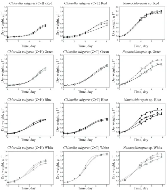
The main goal of this report is to present a simple, cheap, and reliable system for studying autotrophic cultivation of microalgae in small scale. To demonstrate its usefulness, we carried out experiments with three algae isolates under 4 different illumination conditions. Each experiment was repeated three times. Observed growth curves are presented in Figure 2, demonstrating excellent reproducibility especially for the two Chlorella isolates, since the repeated curves overlap each other.
Fig. 2. Parallel growth curves of Chlorella and Nannochloropsis sp.,
▼: red light, ○: green light, ●: blue light, : white light
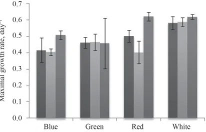
In order to further verify the usefulness of our microscale algae cultivation system, specifi c growth rates of the previous growth curves (three parallels) are presented in Figure 3. While out of the presented 12 μmax values in 9 cases low errors were observed (below 10%), the overall average of errors is 10.26%. Thus, the parallel experiments are really similar, resulting good reproducibility.
Fig. 3. Averages of maximal specifi c growth rates of the three isolates (Chlorella vulgaris strains – CvT and CvH and Nannochloropsis sp. – N.sp.) under different colours of light
: CvH; : CvT; : N.sp.
In Figure 4A, the experiments were grouped by the light conditions and by the isolates representing the average growth curves of the triplicates. Thus, one can observe the effect of illumination both on the basis of growth curves (Fig. 4A) and fi nal biomass concentration (xmax values) (Fig. 4B).
Fig. 4. Photosynthetic growth curves of Chlorella and Nannochloropsis spp. grown under different colours of light (A) and the observed fi nal biomass production (CDW) (B)
A): : Blue Logistic; : Green Logistic; : Red Logistic; : White Logistic;
: Blue; : Green; : Red; : White B): : Blue; : Green; : Red; : White
Cultivation of the two Chlorella strains resulted in similar patterns of biomass production, reaching similar values of fi nal maximum biomass concentration (xmax, g l–1) with the highest value at white colour illumination followed by green, blue, and red. At the same time, in case of Nannochloropsis, the effect of illumination was not so signifi cant. However, highest xmax was reached under red light, followed by green, white, and blue.
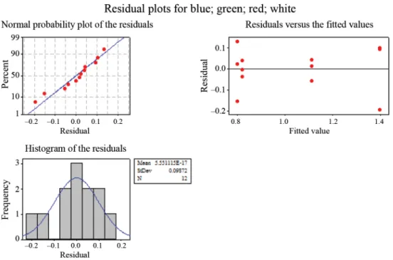
To further verify the goodness of the illuminated microplate cultivation system, we statistically compared via one-way ANOVA analysis the different illumination setups. These revealed, that different illuminations setups result in signifi cantly different growth curves at P<0.05 (Fig. 5). This confi rmed that the measured differences are not random originated, but related to the applied cultivation conditions (i.e. illumination).
Fig. 5. A representative statistical examinations of observed cell dry weight measurements of Chlorella vulgaris (CvT)
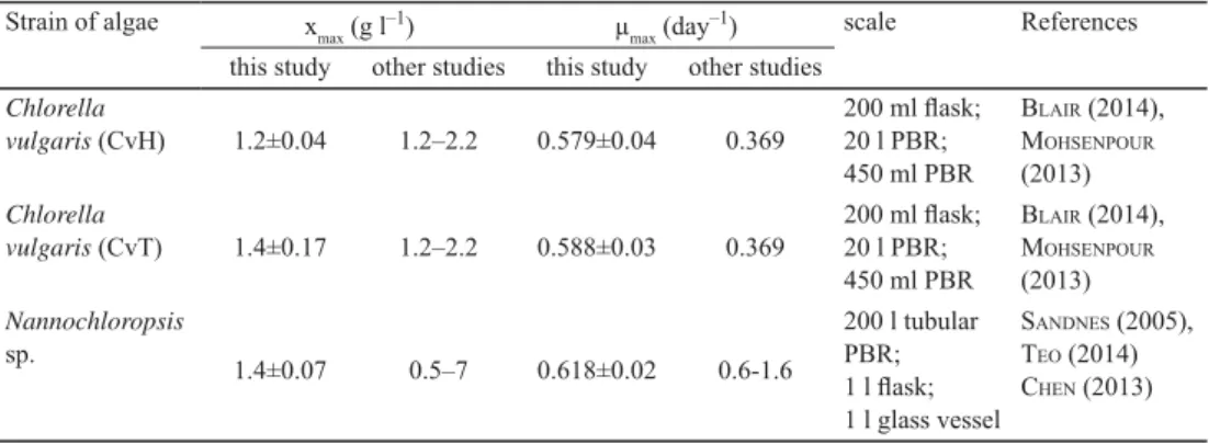
The obtained fi nal xmax concentrations as well as the observed maximal specifi c growth rates (μmax) are comparable with results of other authors applying even larger scale cultivation (Table 1).
Table 1. Maximal dry weight (xmax) and maximal specifi c growth rate (μmax) obtained in this study compared with results of other studies
Strain of algae xmax (g l–1) μmax (day–1) scale References
this study other studies this study other studies Chlorella
vulgaris (CvH) 1.2±0.04 1.2–2.2 0.579±0.04 0.369
200 ml fl ask;
20 l PBR;
450 ml PBR
BLAIR (2014), MOHSENPOUR
(2013) Chlorella
vulgaris (CvT) 1.4±0.17 1.2–2.2 0.588±0.03 0.369
200 ml fl ask;
20 l PBR;
450 ml PBR
BLAIR (2014), MOHSENPOUR
(2013) Nannochloropsis
sp. 1.4±0.07 0.5–7 0.618±0.02 0.6-1.6
200 l tubular PBR;
1 l fl ask;
1 l glass vessel
SANDNES (2005), TEO (2014) CHEN (2013)
PBR: packed bed reactor
3. Conclusions
Based on the presented experiments, it can be concluded that despite the small scale (1.5 ml), our unique lighting design made it possible to perform parallel microalgae cultivations in 24-well microplates. The goodness of the presented cultivation method (including cultivation in MTP, scanning the MTP, and evaluation of photos by greenscale) was verifi ed in three ways: 1) the parallel experiments (done in triplicates) had low errors (<10%); 2) the different setups resulted statistically signifi cant differences in fi nal CDW; 3) the observed maximum biomass concentrations, as well as maximal specifi c growth rates were comparable to results of other authors. Our results also showed that there is no difference among the two Chlorella isolates: both grow better when illuminated with white (mixed) colour. In Nannochloropsis cultures the effect of the applied illumination seemed to be less enhanced.
*
Authors are grateful to Enzyscreen for the microplate holder and Sandwich covers®, as well as to József Pekár for construction of the lighting module. Nannochloropsis sp. was a kind gift from KUKK Ltd., Chlorella vulgaris were kind gifts from Balaton Limnological Institute in 2010.
References
ARIEDE, M.B., CANDIDO, T.M., JACOME, A.L.M., VELASCO, M.V.R., DE CARVALHO, J.C.M. & BABY, A.R. (2017):
Cosmetic attributes of algae – A review. Algal Res., 25, 483–487.
BENNAMOUN, L., AFZAL, M.T. & LÉONARD, A. (2015): Drying of alga as a source of bioenergy feedstock and food supplement – A review. Renew. Sustain. Energy Rev., 50, 1203–1212.
BLAIR, M.F., KOKABIAN, B. & GUDE, V.G. (2014): Light and growth medium effect on Chlorella vulgaris biomass production. JECE, 2, 665–674.
BOROW, K.M., NELSON, J.R. & MASON, R.P. (2015): Biologic plausibility, cellular effects, and molecular mechanisms of eicosapentaenoic acid (EPA) in atherosclerosis. Atherosclerosis, 242, 357–366.
CARVALHO, A.P., SILVA, S.O., BAPTISTA, J.M. & MALCATA, F.X. (2011): Light requirements in microalgal photobioreactors: An overview of biophotonic aspects. Appl. Microbiol. Biot., 89, 1275–1288.
CHACON-LEE, T.L. & GONZÁLEZ-MARINO, G.E. (2010): Microalgae for “healthy” foods—Possibilities and challenges.
Comp. Rev. Food Sci. F., 9, 655–675.
CHEN, C.-Y., CHEN Y.-C., HUANG H-C., HUANG C.-C., LEE W.-L. & CHANG J.-S. (2013): Engineering strategies for enhancing the production of eicosapentaenoic acid (EPA) from an isolated microalga Nannochloropsis oceanica CY2. Bioresour. Technol., 147, 160–167.
CHO, H.U, KIM, Y.M., CHOI, Y.N., XU, X., SHIN, D.Y. & PARK, J.M. (2015): Effects of pH control and concentration on microbial oil production from Chlorella vulgaris cultivated in the effl uent of a low-cost organic waste fermentation system producing volatile fatty acids. Bioresour. Technol., 184, 245–250.
DAS, P., LEI, W., AZIZ, S.S. & OBBARD, J.P. (2011): Enhanced algae growth in both phototrophic and mixotrophic culture under blue light. Bioresour. Technol., 102, 3883–3887.
EDWARDS, W.H. & WILKE, C.R. (1968): Mathematical representation of batch culture data. Biotechnol. Bioeng., 10, 205–232.
FÓZER, D., VALENTINYI, N., RACZ, L. & MIZSEY, P. (2017): Evaluation of microalgae-based biorefi nery alternatives.
Clean Technol. Envir., 19(2), 501–515.
FU, W.Q., GUOMUNDSSON, O., PAGLIA, G., HERJOLFSSON, G., ANDRESSON, O.S., PALSSON, B.O. & BRYNJOLFSSON, S.
(2013): Enhancement of carotenoid biosynthesis in the green microalga Dunaliella salina with light-emitting diodes and adaptive laboratory evolution. Appl. Microbiol. Biot., 97, 2395–2403.
GOLDMAN, J.C. (1979): Temperature effects on steady-state growth, phosphorus uptake, and the chemical composition of a marine phytoplankter. Microb. Ecol., 5, 153–166.
LEE, C.G. & PALSSON, B.O. (1995): Light-emitting diode-based algal photobioreactor with external gas-exchange. J.
Ferment. Bioeng., 79, 257–263.
LUBIAN, L.M., MONTERO, O., MORENO-GARRIDO, I., HUERTAS, E., SOBRINO, C., VALL, M.G. & PAR, G. (2000):
Nannochloropsis (Eustigmatophyceae) as source of commercially valuable pigments. J. Appl. Phycol., 12, 249–255.
MAKI, K.C., YURKO-MAURO, K., DICKLIN, M.R., SCHILD, A.L. & GEOHAS, J.G. (2014): A new, microalgal DHA- and EPA-containing oil lowers triacylglycerols in adults with mild-to-moderate hypertriglyceridemia.
Prostaglandins, Leukotrienes and Essential Fatty Acids (PLEFA), 91, 141–148.
MOHSENPOUR, S.F. & WILLOUGHBY, N. (2013): Luminescent photobioreactor design for improved algal growth and photosynthetic pigment production through spectral conversion of light. Bioresour. Technol., 142, 147–153.
SAFIA, C., ZEBIBA, B., MERAHA, O., PONTALIERA, P.-Y. & VACA-GARCIA, C. (2014): Morphology, composition, production, processing and applications of Chlorella vulgaris: A review. Renew. Sustain. Energy Rev., 35, 265–278.
SANDNES, J.M., KÄLLQVIST, T., WENNER, D. & GISLEROD, H.R. (2005): Combined infl uence of light and temperature on growth rates of Nannochloropsis oceanica: Linking cellular responses to large-scale biomass production.
J. Appl. Phycol., 17, 515–525.
SCHWANKE, R.C., MARCON, R., BENTO, A.F. & CALIXTO, J.B. (2015): EPA- and DHA-derived resolvins’ actions in infl ammatory bowel disease. Eur. J. Pharmacol., 785, 156–164.
SHU, C.H., TSAI, C.H., LIAO, W.H., CHEN, K.Y. & HUANG, H.C. (2012): Effects of light quality on the accumulation of oil in a mixed culture of Chlorella sp. and S. cerevisiae. J. Chem. Technol. Biot., 87, 601–607.
SIDDIQUI, M.W. & PRASAD, K. (2017): Plant secondary metabolites. Vol 1., Biological and therapeutic signifi cance.
CRC Press and Apple Academic Press, New York, pp. 1–285.
TEO, C.L., ATTA, M., BUKHARI, A., TAISIR, M., YUSUF, A.M. & IDRIS, A. (2014): Enhancing growth and lipid production of marine microalgae for biodiesel production via the use of different LED wavelengths. Bioresour. Technol., 162, 38–44.